INTRODUCTION
Bleeding of esophageal varices is the main cause of morbi dity and mortality in children and adults with portal hyperten sion (PH) (13). Despite therapeutic advances, mortality due to acute episodes of upper gastrointestinal bleeding (UGIB) secondary to esophageal varices occurs in 5%19% of children with PH(1,3,5,10,12,22). Mortality rates of 19% have been reported within 35 days after episodes of bleeding varices among North American children with liver disease of several etiologies(5). Thus, it is essential to establish measures to prevent new UGIB episodes due to rupture of varices in these patients.
According to the Baveno V Consensus Workshop, which in volved adult patients with cirrhosis, treatment with betablockers in combination with band ligation is the most eficient method of secondary prophylaxis, although such results and recommen dations cannot be extrapolated to patients in the pediatric age group(1,2,3,4,5,10,12,13,14,22).
Studies involving secondary prophylaxis in children and adolescents are predominantly case series. According to current recommendations, endoscopic band ligation is the method of
Evaluation of endoscopic secondary prophylaxis
in children and adolescents with
esophageal varices
Júlio Rocha
PIMENTA
1, Alexandre Rodrigues
FERREIRA
1,2, Eleonora Druve Tavares
FAGUNDES
1,2,
Paulo Fernando Souto
BITTENCOURT
1, Alice Mendes
MOURA
3and Simone Diniz
CARVALHO
1Received 23/6/2016 Accepted 29/9/2016
ABSTRACT – Background – Bleeding of esophageal varices is the main cause of morbidity and mortality in children and adults with portal hypertension and there are few studies involving secondary prophylaxis in children and adolescents. Objective – To evaluate the eficacy of endoscopic secondary prophylaxis in prevention of upper gastrointestinal bleeding in children and adolescents with esophageal varices. Methods – This is a prospective analysis of 85 patients less than 18 years of age with or without cirrhosis, with portal hypertension. Participants underwent endoscopic secondary prophylaxis with sclerotherapy or band ligation. Eradication of varices, incidence of rebleeding, number of endoscopic sessions required for eradication, incidence of developing gastric fundus varices and portal hypertensive gastropathy were evaluated. Results – Band ligation was performed in 34 (40%) patients and sclerotherapy in 51 (60%) patients. Esophageal varices were eradicated in 81.2%, after a median of four endoscopic sessions. Varices relapsed in 38 (55.1%) patients. Thirtysix (42.3%) patients experienced rebleeding, and it was more prevalent in the group that received sclerotherapy. Gastric varices and portal hypertensive gastropathy developed in 38.7% and 57.9% of patients, respectively. Patients undergoing band ligation showed lower rebleeding rates (26.5% vs 52.9%) and fewer sessions required for eradication of esophageal varices (3.5 vs 5). Conclusion – Secondary prophylaxis was effective in eradicating esophageal varices and controlling new upper gastrointestinal bleeding episodes due to the rupture of esophageal varices. Band ligation seems that resulted in lower rebleeding rates and fewer sessions required to eradicate varices than did sclerotherapy.
HEADINGS – Esophageal and gastric varices. Portal hypertension. Ligation. Sclerotherapy. Child. Adolescent.
Declared conflict of interest of all authors: none Disclosure of funding: no funding received
1 Setor de Gastroenterologia Pediátrica do Hospital das Clínicas da Faculdade de Medicina da Universidade Federal de Minas Gerais (UFMG), Belo Horizonte, MG, Brasil; 2 Departamento de
Pediatria da UFMG, Belo Horizonte, MG, Brasil; 3 Faculdade de Medicina da UFMG, Belo Horizonte, MG, Brasil.
Correspondence: Júlio Rocha Pimenta. Rua Samuel Pereira, 178/02. Bairro Anchieta – CEP: 30310-550 – Belo Horizonte, MG, Brasil. E-mail: [email protected]
choice for children and adolescents, and betablocker therapy is not recommended(69,11,1418,20,2426). Both band ligation and sclerotherapy have high rates of variceal eradication, approximately 80%100%, and rebleeding rates of 030%(69,11,1417,24,26). Zargar et al. performed a randomized pediatric study comparing band ligation and sclero therapy in children, achieving better results in the band group(25). Secondary prophylaxis should always be used in children(16,20). However, additional studies are necessary to determine the best type of prevention. The present study aims to describe the results of endoscopic therapy as secondary prophylaxis in children and adolescents with UGIB due to esophageal varices followed at the Hospital das Clínicas, Universidade Federal de Minas Gerais (HCUFMG).
METHODS
Participants
Patients <18 years old with portal hypertension who had an UGIB episode due to rupture of esophageal varices and underwent secondary prophylaxis according to the protocol established by the service were included in the study. Exclusion criteria included non adherence to the protocol for secondary prophylaxis.
Protocol
Patients with UGIB secondary to esophageal varices, after managing the acute episode, were referred our service to undergo endoscopic secondary prophylaxis: band ligation is the procedure of choice for endoscopic secondary prophylaxis except in patients in whom the procedure is technically not possible, usually in small patients and children under two years. In such cases sclerotherapy is the procedure adopted as a form of secondary prophylaxis. Endoscopic prophylaxis was initiated two weeks after the UGIB episode. Upper digestive endoscopy (UDE) was performed at the Digestive Endoscopy Unit at the Instituto Alfa de Gastroen terologia of HCUFMG by three pediatric endoscopists who, for most procedures, were all present during the exam. The varices were classiied according to the Japanese classiication (Japanese Research Society for Portal Hypertension, 2a edition)(23): grade I (small caliber): small varicose veins, not tortuous; grade II (me dium caliber): slightly enlarged and tortuous varices, occupying less than a third of the esophageal lumen; grade III (large caliber): nodular varicose veins, similar to rosary beads, occupying more than a third of the esophageal lumen. In patients with varices of different sizes, the one with the largest caliber was used for classiication.
Gastric varices were classiied as esophagogastric varices ex tending to small curvature (GEV1S type), esophagogastric varices extending to the gastric fundus (GEV2S type), isolated gastric fundus varices (IGV1S) or gastric fundus and/or duodenum varices (IGV2S)(19).
The presence of red spots and portal hypertensive gastropathy were investigated in each endoscopic examination, and were classi ied as present or absent. Variable gastropathy was described as mild if there was a mosaic pattern of mild grade without any red spots, and described as severe when the mosaic pattern was superimposed by red spots or if any other red spots were present. Gastric antral vascular ectasia was reported when aggregates of red spots arranged in a linear pattern or diffuse lesions were found(18,21).
Band ligation was performed using the multiband ligator. The band began next to the gastroesophageal junction, moving crani ally with a distance of 5 cm. In each session, the varicose vein was tied using an elastic band, and all identiied varices were treated.
Sclerotherapy was performed in patients who technically was not possible to carry out the band ligation, with a transparent Telon injector (diameter 23), using a freehand technique. The injections were made both intravascularly and in the perivascular space, and the sclerosing agent used was 3% ethamolin. The injected amount ranged between 1 and 2 mL per varicose vein, with a maxi mum of 10 mL per session, according to the size of the vessels. All identiied varices underwent sclerotherapy.
Patients underwent endoscopy every three weeks until all varices were eradicated. After eradication, UDE was performed quarterly for the irst 6 months, then every six months and, if they remained
without varices, annually. UDE was performed acutely to manage any episodes of UGIB.
Clinical followup was carried out at the Pediatric Hepatology Clinic of HCUFMG. The diagnosis of cirrhosis and congenital hepatic ibrosis was based on clinical and histological evaluation. Diagnosis of extrahepatic portal vein obstruction was conirmed through Doppler ultrasonography of hepatic vessels. All patients underwent laboratory tests at the time of consultation to evaluate liver biochemistry (aspartate aminotransferase, alanine aminotrans ferase, alkaline phosphatase, gamma glutamyl transferase) and function (prothrombin activity, albumin), blood counts including platelet counts, and other exams when pertinent to the patient’s condition. Patients with cirrhosis were classiied according to the ChildPugh criteria at the beginning of secondary prophylaxis.
Term definitions (studied variables)
Eradication: when all visible varices had been thrombosed by sclerotherapy or were too thin for suction in band ligation, or when absent.
Rebleeding: occurrence of an UGIB episode by rupture of esophageal varices, after beginning prophylaxis, with clinical re percussions and in need of urgent UDE.
Early: rebleeding during prophylaxis and before eradication (not associated with complications of the endoscopy procedure).
Late: rebleeding after eradication.
Relapse: reappearance of varices needing endoscopic treatment in a patient who had already had all varices eradicated.
Appearance of portal hypertensive gastropathy: gastropathy emergence in a patient who did not have it at the irst UDE prior to prophylaxis.
Appearance of gastric varices: emergence of gastric fundus varices (GEV2, IGV1, IGV2) in a patient who did not have them at the irst UDE prior to prophylaxis.
Statistical analysis and ethical aspects
The database was developed and analyzed using the SPSS 17 program. Continuous variables with normal distribution were evaluated using Student’s t test and expressed as mean and standard deviation (SD). Continuous variables without normal distribution were expressed through median and interquartile range (IR) (25% 75%) and compared using the nonparametric KruskalWallis test. The comparison of the distribution of dichotomous variables was analyzed through the chisquare test, with Yates correction or Fisher’s exact test, twotailed, if necessary. The probability of signiicance was considered signiicant when less than 0.05 (P<0.05). This study has been approved by the Ethics Research Committee of UFMG.
RESULTS
Patients characteristics
The median age at UGIB was 6.8 years (IR 25%75%: 1.9 8.8) among the patients with cirrhosis, and in patients with cir rhosis secondary to biliary atresia the median was 2.9 years (IR 25%75%: 1.64.6) and in those with cirrhosis by autoimmune hepatitis 8.2 years (IR 25%75%: 6.110.5) (P=0.26). In the non cirrhosis group, the median was 5.4 years (IR 25%75%: 2.68.6) (P=0.626). In patients with EHPVO and CHF, the median age was 4.7 (IR 25%75%: 2.28.0) and 7.5 years (IR 25%75%: 6.5 10.7), respectively.
In those with cirrhosis, the most frequent cause was biliary atresia in 14 (37.8%) patients, followed by cryptogenic cirrhosis in 8 (21.6%), primary sclerosing cholangitis in 5 (13.5%) and au toimmune hepatitis in 5 (13.5%). Other causes included alpha1 antitrypsin deiciency in 3 (8.1%), BuddChiari syndrome in 1 (2.7%) and choledochal cyst in 1 (2.7%).
The UDE performed at the beginning of secondary prophy laxis showed esophageal varices of small caliber in 12 (14.1%) patients, medium caliber in 37 (43.5%) patients and large caliber in 36 (42.4%) patients. In 46 (54.1%) patients there were signals suggestive of bleeding (red spots). Gastric varices were observed in 54 (63.5%) patients and portal hypertensive gastropathy in 28 (32.9%) patients.
Evaluation of secondary prophylaxis
Analyzing the whole group, eradication of esophageal varices was achieved in 69 (81.2%) patients, requiring a median of four endoscopic sessions for eradication (IR 25%75%: 26). Varices relapsed in 38 (44.7%) patients. The esophageal varices were eradicated in 70.3% of those in the cirrhosis group and in 89.6% of patients without cirrhosis (Table 2 and 3).
TABLE 2. Evaluation of secondary prophylaxis comparing patients with and without cirrhosis (n = 85)
Cirrhosis n = 37
Non cirrhosis n = 48
Total
n = 85 P value
Eradication of varices 26 (70.3%) 43 (89.6%) 69 (81.2%) 0.047
Number of endoscopic sessions for eradication
Median (IR 25%-75%) 4 (2-5.8) 5 (3-6) 4 (2-6) 0.251
Relapse of esophageal varices 14 (53.8%) 24 (55.8%) 38(55.1%) 0.369
Rebleeding Early Late
15 (40.5%) 13
2
21 (43.8%) 15
6
36 (42.3%) 28
8
0.939 0.498
Presence of gastropathy at the beginning of secondary prophylaxis 15 (40.5%) 13 (27.1%) 28 (32.9%) 0.281
Appearance of gastropathy during secondary prophylaxis 10 (45.5%) 23 (65.7%) 33 (57.9%) 0.217
Presence of gastric varices at the beginning of secondary prophylaxis 25 (67.6%) 29 (60.4%) 54 (63.5%) 0.651
Appearance of gastric varices during secondary prophylaxis 7(58.3%) 5 (26.3%) 12 (38.7%) 0.966
Death 9(24.3%) 1 (2.1%) 10 (11.8%) 0.004
Follow up Median (years) IR 25%-75%
5.9 3.1-8.9
7.5 5.1-12.5
6.6
3.8-10 0.072
IR: interquartile range
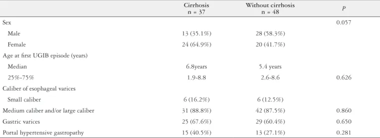
TABLE 1. Clinical and endoscopic characteristics of patients with and without cirrhosis at the beginning of secondary prophylaxis
Cirrhosis n = 37
Without cirrhosis
n = 48 P
Sex 0.057
Male 13 (35.1%) 28 (58.3%)
Female 24 (64.9%) 20 (41.7%)
Age at irst UGIB episode (years)
Median 6.8years 5.4 years
25%-75% 1.9-8.8 2.6-8.6 0.626
Caliber of esophageal varices
Small caliber 6 (16.2%) 6 (12.5%)
Medium caliber and/or large caliber 31 (88.8%) 42 (87.5%) 0.860
Gastric varices 25 (67.6%) 29 (60.4%) 0.650
Of the 36 (42.4%) patients with rebleeding, 28 (77.8%) oc curred before completion of the endoscopic sessions for secondary prophylaxis (early bleeding) and 8 (22.2%) after the varices were eradicated (late bleeding). The median time between the eradication and rebleeding was 1.4 years (IR 25%75%: 0.72.9). There were no bleeding episodes between management of the acute bleeding and the beginning of secondary prophylaxis.
Gastric fundus varices appeared in 12 (38.7%) patients among the 31 who did not have them at the beginning of secondary prophylaxis, and portal hypertensive gastropathy appeared in 33 (62.3%) patients of the 53 that did not have it at the beginning of prophylaxis.
Ten deaths occurred during the study, nine of them in the cirrhosis group (24.3%). Six patients died due to complications secondary to UGIB and three from complications after liver trans plant. Esophageal stenosis was observed due to the endoscopic procedure in four (4.7%) patients: three had sclerotherapy and one had band ligation. All were treated with dilation, which reversed the stenosis. It was not assessed dysphagia after endoscopic procedures.
The results of secondary prophylaxis for the patients with portal hypertension are described in Tables 2 and 3.
Evaluation of endoscopic prophylaxis with relation to the method used
For secondary prophylaxis, 51 (60%) patients underwent scle rotherapy and 34 (40%) underwent band ligation. In those who underwent sclerotherapy, varices were eradicated in 41 (80.4%) patients in a median of ive endoscopic sessions. Varices recurred in 25 (60.9%) patients, and in 11 (44%) of them, the recurrent varices were eradicated during further endoscopic procedures. Rebleeding causing clinical consequences occurred in 27 (52.9%) patients, 23 (85.2%) instances of which occurred between endoscopic sessions and 4 (14.8%) after varices were eradicated.
In those who received band ligation, varices were eradicated in 28 (82.4%) patients after a median of 3.5 endoscopic sessions. Varices recurred in 13 (46.4%) of them, and were subsequently
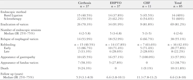
TABLE 3. Results of secondary prophylaxis for patients with portal hypertertion (n = 85)
Cirrhosis n = 37
EHPVO n = 37
CHF n = 11
Total n = 85
Endoscopic method Band Ligation Sclerotherapy
15 (40.5%) 22 (59.5%)
14 (37.8%) 23 (62.2%)
5 (45.5%) 6 (54.6%)
34 (40%) 51 (60%)
Eradication of varices 26 (70.3%) 34 (91.9%) 9 (81.8%) 69 (81.2%)
Number of endoscopic sessions
Median (IR 25%-75%) 4 (2-5.8) 5 (3-6.8) 5 (3-5) 4 (2-6)
Relapse of esophageal varices 14 (53.9%) 18 (52.9%) 6 (66.7%) 38 (55.1%)
Rebleeding Early Late
n = 15 (40.5%) 13 (86.7%)
2 (13.3%)
n = 14 (37.8%) 10 (71.4%)
4 (28.6%)
n = 7 (63.6%) 5 (71.4%) 2 (28.6%)
n = 36 (42.4%) 28 (77.8%)
8 (22.2%)
Appearance of gastropathy 10 (45.5%) 16 (57.1%) 7 (100.0%) 33 (57.9%)
Appearance of fundus varices 7 (58.3%) 5 (27.8%) 0 12 (38.7%)
Death 9 (24.3%) 0 1 (9.1%) 10 (11.8%)
Follow up (years)
Median (IR 25%-75%) 5.9 (3.1-8.9) 6.6 (3.8-10.1) 11.3 (7.8-13.2) 6.6 (3.8-10)
EHPVO: extrahepatic portal vein obstruction; IR: interquartile range.
TABLE 4. Comparison between band ligation and sclerotherapy
Band ligation n = 34
Sclerotherapy n = 51 P
Sex Male Female
14 (41.2%) 20 (58.8%)
27 (52.9%) 24 (47.1%)
0.399
Age at diagnosis (years) Median
IR 25%-75%
5.9 2.5-8.8
2.4 1.1-5.5
0.005
Number of sessions for eradication
Median IR 25%-75%
3.5 2-5
5.0 3-8
0.006
Relapse of esophageal
varices 13 (46.4%) 25 (61.0%) 0.344
Time to relapse Median (month) IR 25%-75%
12.98 8.9-22.2
16.0 9.3-26.1
0.051
Rebleeding Early Late
9 (26.5%) 5 4
27(52.9%) 23
4
0.028 0.164
Appearance of gastropathy
19 13 (68.4%)
38 20 (52.6%)
0.393
Appearance of fundus varices
14 5 (35.7%)
17 7 (41.2%)
0.952
IR: interquartile range.
DISCUSSION
Secondary prophylaxis aims to prevent new UGIB episodes, and it is already well established that both adults and children should be treated(4,20). Data of studies done with adults cannot be extrapolated to the pediatric age group since, in adults, the main cause of PH is liver cirrhosis, while in pediatric patients, half of cases are due to EHPVO, whose clinical evolution is different from that of cirrhosis and in whom the hepatocellular function is preserved(1,10). Furthermore, comorbidities are common in adults and can increase morbidity and mortality in this age group. Another factor that differs among groups is the hemodynamic response to bleeding or drugs(1,10,18,20). This study aims to contribute our experience to that described in the literature of endoscopic secondary prophylaxis in children and adolescent who had UGIB secondary to esophageal varices(69,11,14,1518,20,2426).
Regarding the approach for secondary prophylaxis in adults with cirrhosis, nonselective beta blockers associated with band ligation should be used(4). Sclerotherapy, despite effectively eradicat ing varices, is used less because of higher complication rates than those with band ligation(4,20). In children, according to opinion of pediatric experts on the Baveno V consensus committee, secondary prophylaxis should be performed with endoscopic therapy, and rubber band ligation has been the preferred method. Drug therapy with beta blockers is not recommended, as studies proving its utility in children have yet to be performed(20).
Analyzing the whole evaluated group, eradication of esophageal varices was achieved in 81.2%, which is within the reported 80% 100% range for endoscopic therapy in pediatric patients for both band ligation and sclerotherapy(69,11,1417,24,26).
Several studies demonstrated the effectiveness of endoscopic sclerotherapy for preventing new UGIB episodes in children with PH(8,14,15,24,26). Poddar et al. followed 207 children with EHPVO, and varices were eradicated in 95% of them, after a mean of 4.5 endoscopic sessions(14). Itha et al. described 163 children with EHPVO provided secondary prophylaxis with sclerotherapy, in whom esophageal varices were eradicated in 80% after a mean of 7.6 endoscopic sessions(8). In the present study, sclerotherapy eradicated esophageal varices in 80% of patients, after a median of ive sessions, similar to reported data for pediatric patients.
Relapse of esophageal varices is reported in the pediatric literature with a frequency of 10%40% of cases after the use of sclerotherapy as secondary prophylaxis(8,14,15,24,26). However, a higher rate of relapse of esophageal varices (61%) was observed in the present study. The rebleeding rate observed (52.9%) in this study was also higher than has been described in the literature (012%) (8,14,15,24,26). This difference may relect the fact that the group treated
with sclerotherapy had a lower median age lower than that of the studies mentioned, and in this group of patients it was not possible to perform band ligation because the ligature device could not pass by the cricopharyngeus. The lower age may predispose to a higher frequency of rebleeding.
Similar to what has already been observed in other stud ies(8,14), both portal hypertensive gastropathy and gastric fundus varices arose frequently after eradication of esophageal varices with sclerotherapy. The present indings are consistent with those of Itha et al., who followed 163 children with EHPVO who were given secondary prophylaxis with sclerotherapy(8), and Poddar et al., who followed 274 children with EHPVO also given secondary prophylaxis with sclerotherapy(14).
Portal hypertensive gastropathy and gastric fundus varices may arise after secondary prophylaxis because endoscopic therapy does not alter the blood pressure in the portal system. Thus the eradi cation of esophageal varices may lead to redistribution of blood low to other portal system sites, explaining the increased incidence of gastric varices and portal hypertensive gastropathy. However, UGIB due to bleeding of these sites is more dificult to approach endoscopically. These patients may be the ones who most beneit from drug therapy, since propranolol reduces blood pressure in the entire portal system. However, further studies are necessary to conirm this hypothesis.
In 2002, Mckiernan et al. irst described the use of a multiband ligator in children(11). They eradicated esophageal varices after a median of two sessions with a success rate of 92.8%. On the other hand, Karrer et al.(9) and Fox et al.(6) needed a mean of four sessions to eradicate varices, similar to the present study, in which band ligation eradicated varices in 82% of patients after a median of 3.5 endoscopic sessions. In the literature, relapse of varices after band ligation in children is highly variable, between 9% and 75%(6,7,9,11,16,17), and our indings it within that range. Early and late rebleeding rates were similar to those published, approximately 7%27%(6,7,9,11,16,17). The rates at which PH gastropathy and gastric varices developed were also high, at 68.4% and 35.7% respectively.
In adults, the superiority of band ligation relative to sclero therapy in secondary prophylaxis of esophageal varices is well established(4). Zargar et al. compared 25 children treated with band ligation to 24 children treated with sclerotherapy(25). Band ligation required fewer endoscopic sessions to eradicate varices, had lower rates of early rebleeding (4% vs 25%: P=0.049) and had fewer major complications (esophageal ulcer, stenosis and pneumonia) than sclerotherapy (4% vs 25%: P=0.049). The authors concluded that band ligation has signiicant advantages over sclerotherapy in terms of effectiveness and safety, and it should be the irst choice for to eradicate varices(25).
Both methods were equally effective at eradicating esophageal varices, but with a statistically signiicant difference in the number of sessions required, where band ligation achieved early eradication, which is in agreement with the results of Zagar et al.(25). Another difference observed was the higher early rebleeding rate in the scle rotherapy group, as demonstrated in other studies(4,25). However, this comparison has limitations, as the study was not randomized, which also was the case with most pediatric studies(69,11,1417,24,26). The group treated with sclerotherapy also had a lower median age than the group treated with band ligation. The lower median age in the sclerotherapy group relects the greater dificulty in performing band ligation for younger patients, which limits the comparison. This difference also can be related to the technique required for sclerotherapy, which might require a higher frequency of perivas cular injections and fewer intravascular injections, necessitating more sessions for eradication and higher rebleeding rates during secondary prophylaxis.
Endoscopic secondary prophylaxis is effective in controlling new episodes of UGIB due to rupture of esophageal varices in patients both with and without cirrhosis, regardless of the endoscopic tech nique used. However, portal hypertensive gastropathy and fundic varices clearly arise after eradication of esophageal varices. High rates of relapse of esophageal varices and of rebleeding were observed, but these events were wellcontrolled with new additional endoscopic treatment. Thus, band ligation and sclerotherapy are acceptable methods for secondary prophylaxis in childhood, although higher rebleeding rates were observed in the sclerotherapy group. However,
this fact that should be interpreted with caution, since it arises from a nonrandomized study. Further randomized studies with more subjects are required to make a reliable conclusion on the subject.
Authors’ contributions
Pimenta JR: implementation of research, writing and statisti cal analysis. Ferreira AR: implementation of research, writing and statistical analysis. Bittencourt PFS: search execution. Fagundes EDT: search execution. Moura AM: data collection. Carvalho SD: search execution.
REFERENCES
1. Bhasin DK, Malhi NJS. Variceal bleeding and portal hypertension: much to learn, much to explore. Endoscopy. 2002;34:11928.
2. Bosch J, Abraldes JG, Groszmann R. Current management of portal hyperten sion. J Hepatol. 2003;38:S54S68.
3. D’Amico G, Pagliaro L, Bosh J. The treatment of portal hypertension: a me taanalytic review. Hepatology. 1995;22:33254.
4. De Franchis R. Revising consensus in portal hypertension: Report of the Baveno V consensus workshop on methodology of diagnosis and therapy in portal hypertension. J Hepatol. 2010;53:7628.
5. Duché M, Ducot B, Ackermann O et al. Experience with endoscopic management of highrisk gastroesophageal varices, with and without bleeding, in children with biliary atresia. Gastroenterology. 2013;145:8017.
6. Fox VL, CarrLocke DL, Connors PJ, et al. Endoscopic ligation of esophageal varices in children. J Pediatr Gastroenterol Nutr. 1995;20:2028.
7. Hall R, Lilly JR, Stiegmann GV. Endoscopic esophageal varix ligation: technique and preliminary results in children. J Pediatr Surg. 1988;23:12223.
8. Itha S, Yachha SK. Endoscopic outcome beyond esophageal variceal eradication in children with extrahepatic portal venous obstruction. J Pediatr Gastroenterol Nutr. 2006;42:196200.
9. Karrer FM, Holland RM, Michael JA, et al. Portal vein Thrombosis: treatment of variceal hemorrhage by endoscopic ligation. J Pediatr Surg. 1994;8:114951. 10. Lykavieris P, Gauthier F, Hadchouel P, et al. Risk of gastrointestinal bleeding
during adolescence and early adulthood in children with portal vein obstruction. J Pediatr. 2000;136:8058.
11. McKiernan PJ, Beath SV, Davison SM. A prospective study of endoscopic esophageal variceal ligation using a multiband ligator. J Pediatr Gastroentrol Nutr. 2002;34:20711.
12. McKiernan PJ. Treatment of variceal bleeding. Gastroenterol Endoc Clin North Am. 2001;11:789812.
13. Molleston JP. Variceal bleeding in children. J Pediatr Gastroenterol Nutr. 2003;37:53845.
14. Poddar U, Thapa BM, Singh K. Frequency of gastropathy and gastric varices in children with extrahepatic portal venous obstruction treated with sclerotherapy. J Gastroenterol Hepatol. 2004;19:125356.
15. Poddar U, Thapa BR, Singh K. Endoscopic sclerotherapy in children: experience with 257 cases of extrahepatic portal venous obstruction. Gastrointest Endosc. 2003;57:6836.
16. Pokharna RK, Kumar S, Khatri PC, et al. Endoscopic variceal ligation using multiband ligator. Indian Pediatr. 2005;42:1314.
17. Price MR, Sartorelli KH, Karrer FM, et al. Management of esophageal varices in children by endoscopic variceal ligation. J Pediatr Surg. 1996;31: 105659.
18. Santos, J.M., Ferreira AR, Fagundes EDT, et al. Endoscopic and pharmacolog ical secondary prophylaxis in children and adolescents with esophageal varices. J Pediatr Gastroenterol Nutr. 2013;56:938.
19. Sarin SK, Kumar A. Gastric varices: proile, classiication and management. Am J Gastroenterol. 1989;84:12449.
20. Shneider BL, Bosch J, de Franchis R, et al. Portal hypertension in children: expert pediatric opinion on the report of the Baveno V Consensus Workshop on Methodology of Diagnosis and Therapy in Portal Hypertension. Pediatr Transplant. 2012;16:42637.
21. Spina GP, Arcidiacono R, Bocsh J, et al. Gastric endoscopic features in portal hypertension: inal report of a consensus conference, Milan, Italy, September, 1992. J Hepatol. 1994;21:4617.
22. Stringer MD, Howard ER, Mowat AP. Endoscopic sclerotherapy in the man agement of esophageal varices in 61 children with biliary atresia. J Pediatr Surg. 1989;24:43842.
23. Tajiri T, Yoshida H, Obara K et al, General rules for recording endoscopic indings of esophagogastric varices. Dig Endosc. 2010;22:p. 19.
24. Yachha SK, Sharma BC, Kumar M, et al. Endoscopic sclerotherapy for esopha geal varices in children with extrahepatic portal venous obstruction: a followup study. J Pediatr Gastroenterol Nutr. 1997;24:4952.
25. Zargar SA, Javid G, Khan BA, et al. Endoscopic ligation compared with sclero therapy for bleeding esophageal varices in children with extrahepatic portal venous obstruction. Hepatology 2002;36:66672.
26. Zargar SA, Yattoo GN, Javid G, et al. Fifteenyear follow up of endoscopic injection sclerotherapy in children with extrahepatic portal venous obstruction. J Gastroenterol Hepatol. 2004;19:13945.
Pimenta JR, Ferreira AR, Fagundes EDT, Bittencourt PFS, Moura AM, Carvalho SD. Avaliação da proilaxia secundária endoscópica em crianças e adolescentes com varizes de esôfago. Arq Gastroenterol. 2017,54(1):216.
RESUMO – Contexto – Os episódios de sangramento das varizes esofágicas são a principal causa de morbidade e mortalidade em crianças e adultos com hipertensão porta e poucos são os estudos envolvendo a proilaxia secundária em crianças e adolescentes. Objetivo – Avaliar a eicácia da proilaxia endoscópica secundária na prevenção de hemorragia digestiva alta em crianças e adolescentes com varizes de esôfago. Métodos – Estudo prospectivo com 85 pacientes menores de 18 anos com hipertensão porta, cirróticos e não cirróticos. A proilaxia secundária endoscópica foi realizada através de ligadura elástica ou escleroterapia. Foram avaliadas erradicação de varizes, incidência de ressangramento, número de sessões endoscópicas necessárias para a erradicação, incidência de surgimento de varizes gástricas e da gastropatia da hipertensão porta. Resultados – Ligadura elástica foi realizada em 34 (40%) pacientes e escleroterapia em 51 (60%). As varizes de esôfago foram erradicadas em 81,2% após mediana de quatro sessões endoscópicas. Foi observada recidiva de varizes de esôfago em 38 (55,1%) pacientes. Ressangramento por ruptura de varizes de esôfago ocorreu em 36 (42,3%) pacientes e foi mais prevalente no grupo submetido à escleroterapia. O surgimento de varizes gástricas e gastropatia da hipertensão porta ocorreram em 38,7% e 57,9% respectivamente. Os pacientes submetidos à ligadura elástica apresentaram taxas menores de ressangramento (26,5% vs 52,9%) e número menor de sessões necessárias para erradicação das varizes de esôfago (3,5 vs 5). Conclusão – A proilaxia secundária endoscópica mostrouse eicaz para erradicação de varizes de esôfago e evitar novos episódios de hemorragia digestiva alta secundária à ruptura de varizes de esôfago. A ligadura elástica endoscópica provavelmente apresenta menores taxas de ressangramento e número menor de sessões necessárias para erradicação das varizes de esôfago, quando comparada à escleroterapia.