INTRODUCTION
Esophageal cancer is a rapidly progressive disease with poor
survival rates. Most of these tumors are either adenocarcinoma
or squamous cell carcinoma histologic types. Less frequent cancer
subtypes, such as melanoma, lymphomas or neuroendocrine tumors
are uncommon and poorly explored
(34).
Neuroendocrine esophageal neoplasms are rare and most of
studies are case reports. The classiication and nomenclature are
not well established and knowledge acquired in neuroendocrine
neoplasms of lungs and certain gastrointestinal sites, such as
pancreas, usually guide esophageal neuroendocrine management.
Neuroendocrine neoplasm subtypes are grouped based upon
shared neuroendocrine features. The subtypes comprise small cell
carcinoma, large cell neuroendocrine carcinoma, typical carcinoid,
and atypical carcinoid
(140).
This article is a descriptive report of clinical and pathological
features of cases of neuroendocrine esophageal tumors in our
institute, along 8 years of experience, and a review of the literature.
METHODS
All esophageal cancer patients’ charts of our institution were
reviewed, between 2008 and 2016. Cases of neuroendocrine
neo-Primary neuroendocrine neoplasm
of the esophagus – Report of 14 cases from
a single institute and review of the literature
Francisco
TUSTUMI
1, Flavio Roberto
TAKEDA
2, Rodrigo Hideki
UEMA
3,
Guilherme Luiz Stelko
PEREIRA
2, Rubens Antonio Aissar
SALLUM
1and Ivan
CECCONELLO
1Received 9/7/2016 Accepted 22/8/2016
ABSTRACT – Background –
Most prevalent esophageal neoplasm is squamous cell carcinoma and adenocarcinoma. Other tumors are uncommon and
poorly studied. Primary neuroendocrine esophageal neoplasm is a rare carcinoma and most of its therapy management is based on lung neuroendocrine
studies. Neuroendocrine tumors can be clustered in the following subtypes: high grade (small cell carcinoma or large cell carcinoma) and low grade
(carcinoids).
Objective
– The present study aims to assess clinical and pathological neuroendocrine esophageal tumors in a single oncologic center.
Methods
– A retrospective analysis of patients and review of the literatures was performed.
Results
– Fourteen patients were identiied as neuroendocrine
tumors, 11 male and 3 female patients. Mean age was 67.3 years old. Ten patients were classiied as small cell, 3 as large cell and 1 as carcinoid. Four
patients presented squamous cell carcinoma simultaneously and 1 also presented adenocarcinoma. Main sites of metastasis were liver, peritoneum,
lung and bones. Most patients died before 2 years of follow-up. Patient with longer survival died at 35 months after diagnosis.
Conclusion
–
Neuro-endocrine esophageal tumors are rare; affect mainly men in their sixties or seventies. High grade tumors can be mixed to other subtypes neoplasms,
such as adenocarcinoma and squamous cell carcinoma. Most of these patients have poor overall survival rates.
HEADINGS –
Esophageal neoplasms. Carcinoid tumor. Neuroendocrine carcinoma.
Declared conflict of interest of all authors: none Disclosure of funding: no funding received
1 Cirurgia do Aparelho Digestivo, Faculdade de Medicina, Hospital das Clínicas, USP, SP, Brasil; 2 Instituto do Câncer, Faculdade de Medicina, Hospital das Clínicas, USP, SP, Brasil; 3 Faculdade
de Medicina, Hospital das Clínicas, Universidade de São Paulo, SP, Brasil.
Correspondence: Francisco Tustumi. Faculdade de Medicina, do Hospital das Clínicas, da Universidade de São Paulo. Rua Teodoro Sampaio, 632, ap. 71 – CEP: 05406-000 – São Paulo, SP, Brasil. E-mail: [email protected]
plasm were identiied and clinical and pathological features of these
patients were assessed.
For review of the literature, database search was performed
in MEDLINE, with search algorithm: (“neuroendocrine” [All
Fields] OR “carcinoid tumor” [MeSH Terms] OR “carcinoid” [All
Fields] OR “small cell carcinoma” [MeSH Terms] OR “large cell
carcinoma”) AND (“oesophagus” [All Fields] OR “esophagus” [All
Fields] OR esophageal [All Fields]). Period searched up to 2016, with
no idiom restriction. Review studies and no full-text studies were
excluded. When more than one publication of a single trial existed,
only the publication with the most complete data was included.
RESULTS
A total of 1,574 of esophageal cancers were reviewed.
Four-teen (0.89%) cases of neuroendocrine neoplasm were selected, 3
female and 11 male patients (Table 1). Mean age was 67.3 (range
47 to 80) years old. Table 2 shows immunohistochemical panel of
neuroendocrine neoplasm.
Three were classified as large cell esophageal carcinoma
(LCEC), 10 as small cell carcinoma (SCEC), and 1 as carcinoid.
Distal and middle esophagus were more often affected.
Main symptoms were dysphagia (14∕14) and weight loss (mean
11.7 ± Std Dev 6.3 kg). Duration of symptoms prior to diagnosis
was 6.2 ± Std Dev 3.3 months.
Associated squamous cell carcinoma could be seen in 4∕14 cases
and associated adenocarcinoma (adenoneuroendocrine) in 1∕14.
Immunohistochemical panel can be seen in Table 2.
Of these patients, 10∕14 had previous history of high amount
of alcohol intake and 12∕14 were tobacco smokers.
Most cases were diagnosed at late stages (III and IV),
ac-cordingly AJCC 7th Edition
(122).
Metastasis sites were lungs, liver,
adrenal, peritoneal and bones.
Cause of death was pneumonia in five cases, urinary tract
infection in one case, and sepsis of unknown origin in one case.
The five remaining patients, cause of death was not clearly
established.
Curative intent surgery was performed in 4∕14 patients, of which
one is still alive at 26 months of follow-up.
TABLE 1.
Main characteristics of the 14 patients diagnosed with neuroendocrine tumor
Patient Age
Sex
KPS ECOG
Stage
Histologic
subtype
Associated
neoplasm
Site
Survival
(months)
Esophagec-tomy
Chemothe-
rapy
Radiothe-raphy
1
79
M
50
4
Unstaged
SCCE
No
Lower
2
No
No
No
2
65
M
90
1
IIIA
SCCE
No
Middle
Alive at 26
months
Yes
No
No
3
60
M
80
1
IIIB
LCCE
No
Middle and
lower
13
No
IP
30 cGy
4
65
M
30
4
IA
Carcinoid
No
Lower
7
No
No
No
5
75
F
90
1
IIA
LCCE
No
Lower
13
Yes
IP, PC
45 cGy
6
51
M
100
0
IIIC
SCCE
No
Upper and
middle
Lost to follow-up
at 4 months
No
EP
No
7
58
M
80
2
IV
SCCE
SCC
Middle
2
No
No
No
8
64
M
100
0
IIIA
SCCE
SCC
Middle and
lower
35
Yes
IP, PT
45 cGy
9
47
M
100
0
IV
SCCE
No
Lower
10
No
IP, PT
50.4 cGy
10
78
F
70
1
IV
SCCE
SCC
Lower
13
No
IP, PT
45 cGy
11
75
M
80
1
IIIC
SCCE
No
Upper and
middle
12
No
IP
45 cGy
12
80
F
50
3
IV
SCCE
SCC
Middle
6
No
No
No
13
76
M
60
3
IV
SCCE
No
Middle
5
No
IP
No
14
69
M
90
1
IIIB
LCCE
EA
Lower
18
Yes
(neoadjuvant), IP
XP
No
SCEC: small cell esophageal cancer; LCEC: large cell esophageal cancer; KPS: Karnofsky Performance Status; ECOG: Eastern Coorporative Oncology Group; SCC: squamous cell carcinoma; EA: esophageal adenocarcinoma; PC: Paclitaxel and Carboplatin; PT: Cisplatin and Paclitaxel; IP: Irinotecan and Cisplatin; EP: Etoposide and Cisplatin; XP: Capecitabine and Cisplatin.
TABLE 2.
Immunohistochemical panel of patients with esophageal neuroendocrine neoplasm
Patient
Grade of cellular
differentiation
Syp
CgA
Ki-67(%)
Ber-EP4
Ck7
AE-1/AE-3 35BH11
P63
CD 56
TTF-1
Vimentin
1
Poorly
+
NP
>90
NP
NP
NP
NP
NP
+
NP
NP
2
Poorly
NP
-
80-90
NP
NP
NP
+
-
+
+
NP
3
Poorly
NP
+
90
+
NP
-
NP
-
NP
NP
NP
4
Well
+
+
<1
NP
NP
NP
NP
NP
NP
NP
NP
5
Poorly
+
+
>90
NP
+
NP
NP
-
NP
NP
NP
6
Poorly
+
+
90
NP
NP
NP
NP
NP
NP
+
NP
7
Poorly
+
+
NP
+
NP
NP
NP
+
NP
NP
NP
8
Poorly
-
+
50
NP
+
+
NP
+
+
+
+
9
Poorly
+
+
80-90
NP
NP
NP
+
-
+e
NP
NP
10
Poorly
+
+
NP
NP
-
+
+
-
+
+
NP
11
Poorly
NP
-
NP
-
NP
+
+
-
+
NP
NP
12
Poorly
+
+
90
NP
NP
+
NP
+
+
NP
NP
13
Poorly
NP
NP
NP
NP
NP
+
+
+
+
+
+
14
Poorly
+
+
70
-
+
NP
NP
-
NP
NP
NP
(+): positive; (-): negative; Syp: Synaptophysin; CgA: Chromogranine A; Ck: Cytokeratin; NP: not performed.
Most cases had low survival rates (see Figure 2). Patient who
lived longer died at 35 months of pneumonia. Patient “2” is still
alive without disease up to this paper publication. Patients “6” lost
to follow-up, with disease. The remaining patients died with disease.
studies data concerning carcinoid tumors and LCEC prevalence.
Most of the studies approaching LCEC were performed in West
countries.
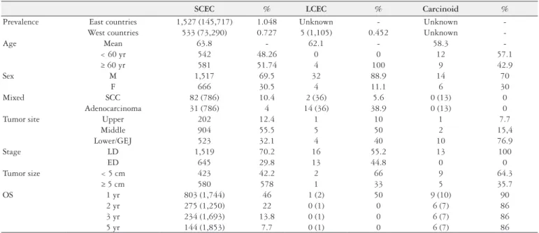
Neoplasms were staged as limited disease (LD) or extend
dis-ease (ED). LD is deined as disdis-ease conined to the esophagus and
adjacent organs with or without regional lymph node involvement
while ED is deined for neoplasm with distant spread
(59). Main
features and differences concerning neuroendocrine subtypes are
presented in Table 3.
DISCUSSION
Neuroendocrine esophageal neoplasms are exceedingly rare,
and hence there are few large sample clinical studies approaching
this issue. Consequently, most of the knowledge is based on
neu-roendocrine lung neoplasms.
Neuroendocrine lung tumors are classiied as low grade
(car-cinoids) or as high grade (SCEC and LCEC)
(120).
SCEC have nuclear appearance, with inely granular chromatin;
lack of predominant nucleoli; nuclear fragility; fusiform cells; scant
cytoplasm and indistinct cells borders; high mitotic rate; and large
area of necrosis
(120). (Figure 3).
FIGURE 2.
Kaplan-Meir curve for survival of patients with esophageal
neuroendocrine neoplasm
Patient diagnosed with carcinoid tumor presented myeloibrosis
during follow-up, leading to pancytopenia and early death.
Review of the literature
For review of the literature, a total number of articles by database
search were 1007. After excluding duplicates and screening by title
and abstract, 154 articles remained
(1-33, 35-79, 81-115, 117-119, 121, 123-139, 141-162).
A cumulative sample size of 2,957 patients was evaluated, in
20 different countries. Of this patients, 2,899 were SCEC (77% in
East countries; 23% in West countries); 35 were LCEC (6% in East
countries; 94% in West countries); and 23 were carcinoid (54% in
East countries; 43% in West countries).
Among all esophageal malignances, prevalence of SCEC was
1.05% in East countries and 0.72% in West countries. There are few
TABLE 3.
Main features and differences concerning neuroendocrine subtypes
SCEC
%
LCEC
%
Carcinoid
%
Prevalence
East countries
1,527 (145,717)
1.048
Unknown
-
Unknown
-
West countries
533 (73,290)
0.727
5 (1,105)
0.452
Unknown
-Age
Mean
63.8
-
62.1
-
58.3
-
< 60 yr
542
48.26
0
0
12
57.1
≥ 60 yr
581
51.74
4
100
9
42.9
Sex
M
1,517
69.5
32
88.9
14
70
F
666
30.5
4
11.1
6
30
Mixed
SCC
82 (786)
10.4
2 (36)
5.6
0 (13)
0
Adenocarcinoma
31 (786)
4
14 (36)
38.9
0 (13)
0
Tumor site
Upper
202
12.4
1
10
1
7.7
Middle
904
55.5
5
50
2
15,4
Lower/GEJ
523
32.1
4
40
10
76.9
Stage
LD
1,519
70.2
16
55.2
13
100
ED
645
29.8
13
44.8
0
0
Tumor size
< 5 cm
423
42.2
2
66
9
64.3
≥ 5 cm
580
578
1
33
5
35.7
OS
1 yr
803 (1,744)
46
1 (2)
50
9 (10)
90
2 yr
275 (1,250)
22
0 (1)
0
6 (7)
86
3 yr
234 (1,693)
13.8
0 (1)
0
6 (7)
86
5 yr
144 (1,853)
7.7
0 (1)
0
6 (7)
86
OS: overall survival; GEJ: gastroesophageal junction; SCEC: small cell esophageal cancer; LCEC: large cell esophageal cancer.
LCEC presents non-small cell nuclear features (vesicular
clumpy chromatin, predominant nucleoli), abundant cytoplasm,
and large cell size
(120). (Figure 4).
a systemic disease and, just like in lung cancer, chemotherapy is
the mainstay of therapy
(17). Additional therapy (surgery or
radio-therapy) should be considered, but randomized controlled trials
still unavailable
(80, 116).
Our data suggests a much good prognosis for low grade
neu-roendocrine tumors, with high overall survival rate. For limited
disease (LD) carcinoid, surgical intervention is the treatment of
choice
(18).
Although low incidence of esophageal neuroendocrine tumors,
our results give a better picture of the behavior of this rare
condi-tion. The present study shows this disease affects mainly men in
sixties or seventies. Middle and lower esophageal thirds are most
fre-quently affected. Nevertheless, future multicenter efforts are needed
for randomized clinical trials evaluating therapeutic guidelines.
ACKNOWLEDGMENT
We would like to state our acknowledgment for Rafaela Brito
Bezerra Pinheiro efforts on pathology analysis and microscopy
pictures.
Authors’ contributions
Tustumi F: study elaboration and data collect. Takeda FR:
study elaboration. Uema RH: data collect. Pereira GLS:
onco-logic review. Sallum RAA: orientation of the study. Cecconello I:
orientation of the study.
FIGURE 4.
Large cell esophageal carcinoma (LCEC). Solid nests of large
and intermediate cells, with eosinophilic cytoplasm, vesicular nuclei and
prominent nucleoli, focal necrosis and high mitotic rate (a) (HE, 200x).
Chromogranin positive (400x) (b).
Carcinoids typical morphology includes coarsely granular “salt
and pepper” chromatin, overall uniformity, prominent vascularity, lack
of prominent nucleoli, low mitosis rate. Usually no necrosis is seen
(120).
Esophageal high grade tumors tend to be aggressive. Usually,
patients are diagnosed lately, with widespread disease, and with
poor prognosis. Currently, clinical treatment strategies of high
grade cancers neuroendocrine neoplasms are very limited and
full of contradiction. High grade neoplasms are often regarded as
REFERENCES
1. Adachi K, Tateno Y, Okada H. An esophageal collision tumor. Clin Gastroenterol Hepatol. 2014;12:17-8.
2. Akagi I, Miyashita M, Makino H, Nomura T, Shimanuki K, Satake S, Hagiwara N, Uchida E. Small cell carcinoma of the esophagus: a case report. J Nippon Med Sch. 2014;81:97-100.
3. Ando T, Hosokawa A, Yamawaki H, Hasumoto Y, Kajiura S, Itaya Y, et al. Esophageal small-cell carcinoma with syndrome of inappropriate secretion of antidiuretic hormone. Intern Med. 2011;50:1099-103.
4. Atsumi K, Shioyama Y, Nomoto S, Ohga S, Toba T, Sasaki T, et al. Chemoradia-tion for small cell esophageal carcinoma: report of 11 cases from multi-instituChemoradia-tion experience. J Radiat Res. 2010;51:15-20.
5. Asayama M, Fuse N, Yoshino T, Yano T, Tahara M, Doi T, et al. Amrubicin for the treatment of neuroendocrine carcinoma of the gastrointestinal tract: a retro-spective analysis of ive cases. Cancer Chemother Pharmacol. 2011;68:1325-30. 6. Atsumi K, Shioyama Y, Nomoto S, Ohga S, Toba T, Sasaki T, et al. Chemoradia-tion for small cell esophageal carcinoma: report of 11 cases from multi-instituChemoradia-tion experience. J Radiat Res. 2010;51:15-20.
Tustumi F, Takeda FR, Uema RH, Pereira GLS, Sallum RAA, Cecconello I. Neoplasia neuroendócrina primária de esôfago – Relato de 14 casos e revisão
de literatura. Arq Gastroenterol. 2017,54(1):4-10.
RESUMO – Contexto –
As neoplasias esofágicas mais prevalentes são o adenocarcinoma e o carcinoma espinocelular. Outros subtipos histológicos
são incomuns e pouco estudados. Neoplasia neuroendócrina esofágica é uma patologia rara e seu manejo atualmente se baseia nos conhecimentos
prévios de tumores neuroendócrinos de pulmão. Tumores neuroendócrinos podem ser divididos nas seguintes formas: alto grau (pequenas células ou
grandes célula) e baixo grau (carcinoides).
Objetivo
– Avaliar clínica e patologicamente os tumores de esôfago em um centro oncológico referenciado.
Métodos –
Foi realizada análise retrospectiva e revisão da literatura de neoplasias neuroendócrinas de esôfago.
Resultados –
Foram identiicados 14
pacientes com tumores neuroendócrino, sendo 11 homens, 3 mulheres. Idade média foi de 67,3 anos de idade. Desses pacientes, 10 foram classiicados
como pequenas células, 3 como grandes células e 1 como carcinoide. Foram encontrados quatro casos de tumor misto neuroendócrino e carcinoma
espinocelular, e um caso de tumor misto adenoneuroendócrino. Principal sítio de metástases foi fígado, peritônio, pulmão e ossos. A maioria dos
pa-cientes foi a óbito em até 2 anos de seguimento. Paciente com sobrevida mais longa foi a óbito após 35 meses do diagnóstico.
Conclusão
– Neoplasias
neuroendócrinas de esôfago são raras, afetam principalmente o sexo masculino na 7ª ou 8ª década de vida. A maioria dos pacientes com tumores de
alto grau tem sobrevida curta.
DESCRITORES –
Neoplasias esofágicas. Tumor carcinoide. Carcinoma neuroendócrino.
7. Babich JP, Klein J, Khullar P, Friedel DM. Endoscopic mucosal resection of a mid-esophageal carcinoid with EUS guidance. Endoscopy. 2010;42(Suppl 2):E302-3. 8. Basu S, Nair N. (18)FDG PET in primary oat cell carcinoma of the esophagus.
Indian J Cancer. 2005;42:60-2.
9. Bennouna J, Bardet E, Deguiral P, Douillard JY. Small cell carcinoma of the esophagus: analysis of 10 cases and review of the published data. Am J Clin Oncol. 2000;23:455-9.
10. Bibeau F, Chateau MC, Guiu M, Assenat E, Azria D, Lavaill R, et al. Small cell carcinoma with concomitant adenocarcinoma arising in a Barrett’s oesophagus: report of a case with a favourable behaviour. Virchows Arch. 2008;452:103-7. 11. Briggs JC, Ibrahim NB. Oat cell carcinomas of the oesophagus: a
clinico-patho-logical study of 23 cases. Histopathology. 1983;7:261-77.
12. Brown RS Jr, Peppercorn MA, Farraye FA, Glotzer DJ, Antonioli DA, Taylor G. Carcinoid tumor of the esophagus. J Clin Gastroenterol. 1994;19:338-9. 13. Caldwell CB, Bains MS, Burt M. Unusual malignant neoplasms of the
14. Cary NR, Barron DJ, McGoldrick JP, Wells FC. Combined oesophageal adeno-carcinoma and carcinoid in Barrett’s oesophagitis: potential role of enterochro-mafin-like cells in oesophageal malignancy. Thorax. 1993;48:404-5.
15. Casas F, Ferrer F, Farrús B, Casals J, Biete A. Primary small cell carcinoma of the esophagus: a review of the literature with emphasis on therapy and prognosis. Cancer. 1997;80:1366-72.
16. Chatni SS, Ravindran HK, Narayanan A, Balakrishnan V. Small cell carcinoma of the esophagus. Saudi J Gastroenterol. 2008;14:149-50.
17. Chen SB, Yang JS, Yang WP, Weng HR, Li H, Liu DT, Chen YP. Treatment and prognosis of limited disease primary small cell carcinoma of esophagus. Dis Esophagus. 2011;24:114-9.
18. Chen KC, Chang YL, Pan CT, Lee YC. Esophageal atypical carcinoid tumor with tracheal invasion. J Thorac Cardiovasc Surg. 2007;134:524-5.
19. Chen WW, Wang F, Chen S, Wang L, Ren C, Luo HY, et al. Detailed analysis of prognostic factors in primary esophageal small cell carcinoma. Ann Thorac Surg. 2014;97:1975-81.
20. Chin K, Baba S, Hosaka H, Ishiyama A, Mizunuma N, Shinozaki E, et al. Irino-tecan plus cisplatin for therapy of small-cell carcinoma of the esophagus: report of 12 cases from single institution experience. Jpn J Clin Oncol. 2008;38:426-31. 21. Chino O, Makuuchi H, Ozawa S, Shimada H, Nishi T, Kise Y, et al. Small Cell
Type of Esophageal Neuroendocrine Carcinoma Resembling a Submucosal Tumor. Tokai J Exp Clin Med. 2015;40:36-9.
22. Chong FK, Graham JH, Madoff IM. Mucin-producing carcinoid (“composite tumor”) of upper third of esophagus: a variant of carcinoid tumor. Cancer. 1979;44:1853-9.
23. Chow V, Law S, Lam KY, Luk JM, Wong J. Telomerase activity in small cell esophageal carcinoma. Dis Esophagus. 2001;14:139-42.
24. Chuah SK, Hu TH, Kuo CM, Chiu KW, Kuo CH, Wu KL, et al. Upper gas-trointestinal carcinoid tumors incidentally found by endoscopic examinations. World J Gastroenterol. 2005;11:7028-32.
25. Clark T, Lee MJ, Munk PL. Primary small-cell carcinoma of the oesophagus with spontaneous oesophageal perforation following chemotherapy. Australas Radiol. 1996;40:250-3.
26. Cook MG, Eusebi V, Betts CM. Oat-cell carcinoma of the oesophagus: a recently recognized entity. J Clin Pathol. 1976;29:1068-73.
27. Cox PM, Vazir MH, Petty RK, Law S, Dhillon AP. Cerebellar cortical degener-ation in associdegener-ation with small-cell carcinoma of the oesophagus. Neuropathol Appl Neurobiol. 1989;15:175-83.
28. Craig SR, Carey FA, Walker WS, Cameron EW. Primary small-cell cancer of the esophagus. J Thorac Cardiovasc Surg. 1995;109:284-8.
29. Deepak P, Devi R, Pillai H. Esophageal small cell carcinoma with synchronous renal cell carcinoma: a case report with review of the literature. Case Rep Gas-troenterol. 2011;5:196-200.
30. del Valle Ruiz SR, Torres Bermúdez JR, Montoya Tabares MJ, Merino Montes J, Sánchez de la Villa G. [Extrapulmonary small cell carcinoma located in the oesophagus. Cir Esp. 2010;88:47-8.
31. Doherty MA, McIntyre M, Arnott SJ. Oat cell carcinoma of esophagus: a report of six British patients with a review of the literature. Int J Radiat Oncol Biol Phys. 1984;10:147-52.
32. Eccles DM, Allan SG, Sang CT, Cornbleet MA. Small cell carcinoma of the oesophagus: report of three cases and review of published cases. Thorax. 1989;44:749-50.
33. Einspanier GR, Caleel RT, Milford AF 3rd. Carcinoid tumors of the esophagus: report of a case. J Am Osteopath Assoc. 1987;87:500-3.
34. Enzinger PC, Mayer. R. Esophageal Cancer. N Engl J Med. 2003;349:2241-52. 35. Feng JF, Huang Y, Zhao Q, Chen QX. Clinical signiicance of preoperative
neutrophil lymphocyte ratio versus platelet lymphocyte ratio in patients with small cell carcinoma of the esophagus. Scientiic World Journal. 2013:504365. 36. Feng W, Harada H, Zhang P, Mitsuya K, Zheng X, Yasui H, et al. Metastatic brain
tumors from small-cell esophageal cancer: clinical characteristics and outcome. J Cancer Res Ther. 2014;10:256-8.
37. Fenlon HM, O’Keane C, Carney DN, Breatnach E. Imaging of small cell carci-noma of the oesophagus. Clin Radiol. 1995;50:634-8.
38. Fukuchi M, Sakurai S, Tsukagoshi R, Naitoh H, Kuwano H. A case of large cell neuroendocrine carcinoma of the esophagogastric junction. Esophagus. 2015;12:295-9.
39. Funakoshi S, Hashiguchi A, Teramoto K, Miyata N, Kurita S, Adachi M, et al. Second-line chemotherapy for refractory small cell neuroendocrine carcinoma of the esophagus that relapsed after complete remission with irinotecan plus cisplatin therapy: Case report and review of the literature. Oncol Lett. 2013;5:117-22.
40. Gao R, Zhang Y, Wen XP, Fu J, Zhang GJ. Chemotherapy with cisplatin or carboplatin in combination with etoposide for small-cell esophageal cancer: a systemic analysis of case series. Dis Esophagus. 2014;27:764-9.
41. Gollard R, Ellis C, VanderHarten C. Small cell/neuroendocrine tumors of the esophagus: presentation of two cases and review of the literature. Tumori. 2010;96:780-3.
42. González LM, Sanz-Esponera J, Saez C, Alvarez T, Sierra E, Sanz-Ortega J. Case report: esophageal collision tumor (oat cell carcinoma and adenocarcinoma) in Barrett’s esophagus: immunohistochemical, electron microscopy and LOH analysis. Histol Histopathol. 2003;18:1-5.
43. Goscinski MA, Xu R, Zhou F, Wang J, Yang H, Huang R, et al. Nuclear, cyto-plasmic, and stromal expression of ZEB1 in squamous and small cell carcinoma of the esophagus. APMIS. 2015;123:1040-7.
44. Goto T, Watabe H, Kawakami T, Watanabe M, Nakano K, Takai A, et al. [A case of small cell carcinoma of the esophagus with remarkable response to chemotherapy with CPT-11 and CDDP]. Nihon Shokakibyo Gakkai Zasshi. 2007;104:1204-11.
45. Gumprich T, Johnen G, Hummel T, Jaworska M, Schmitz I, Müller KM. [A rare case of a neuroendocrine carcinoma of the esophagus. Intermediate between a wel-differentiated neuroendocrine carcinoma and a low-differentiated neuroen-docrine carcinoma]. Pathologe. 2004;25:229-34.
46. Gupta NM, Goenka MK, Atri A, Vaiphei K. Carcinoid tumour of the oesoph-agus: a rare oesophageal cancer. Eur J Surg. 1996;162:841-4.
47. Heyes FL, Ayres J, Matthews HR. Oat cell carcinoma of esophagus. Int J Radiat Oncol Biol Phys. 1985;11:1573.
48. Hoang MP, Hobbs CM, Sobin LH, Albores-Saavedra J. Carcinoid tumor of the esophagus: a clinicopathologic study of four cases. Am J Surg Pathol. 2002;26:517-22.
49. Horai T, Kobayshi A, Tateishi R, Wada A, Taniguchi H, Taniguchi K, et al. A cytologic study on small cell carcinoma of the esophagus. Cancer. 1978;41:1890-6. 50. Hosseini S, Salek R, Nasrolahi H, Mohammadianpanah M, Judi M. Small Cell
Carcinoma of the Esophagus: Clinicopathological Features and Outcome of 22 Cases.Iran Red Crescent Med J. 2015;17:e20353.
51. Hou X, Wei JC, Wu JX, Wang X, Fu JH, Lin P, Yang HX. Multidisciplinary modalities achieve encouraging long-term survival in resectable limited-disease esophageal small cell carcinoma. PLoS One. 2013;8:e69259.
52. Hsu PK, Li AF, Hsu WH. Simultaneous thoracotomy and laparotomy for small cell carcinoma of oesophagogastric junction. ANZ J Surg. 2008;78:723-4. 53. Huang Q, Wu H, Nie L, Shi J, Lebenthal A, Chen J, et al. Primary
high-grade neuroendocrine carcinoma of the esophagus: a clinicopathologic and immunohistochemical study of 42 resection cases. Am J Surg Pathol. 2013;37: 467-83.
54. Huang YL, Chou SH, Chai CY, Hsu JS. Small cell neuroendocrine carcinoma of the esophagus. Kaohsiung J Med Sci. 2015;31:108-9.
55. Hudson E, Powell J, Mukherjee S, Crosby TD, Brewster AE, Maughan TS, et al. Small cell oesophageal carcinoma: an institutional experience and review of the literature. Br J Cancer. 2007;96:708-11.
56. Huncharek M, Muscat J. Small cell carcinoma of the esophagus. The Massa-chusetts General Hospital experience, 1978 to 1993. Chest. 1995;107:179-81. 57. Imai T, Sannohe Y, Okano H. Oat cell carcinoma (apudoma) of the esophagus:
a case report. Cancer. 1978;41:358-64.
58. Isoyama Y, Shioyama Y, Nomoto S, Ohga S, Nonoshita T, Onishi K, et al. Car-boplatin and etoposide combined with radiotherapy for limited-stage small-cell esophageal carcinoma: three cases and review of the literature. Jpn J Radiol. 2010;28:181-7.
59. Lv J, Linag J, Wang J, Wang L, He J, Xiao Z, Yin W. Primary Small Cell Carci-noma of the Esophagus. J Thorac Oncol. 2008;3:1460-5.
60. Johnson FE, Clawson MC, Bashiti HM, Silverberg AB, Broun GO Jr. Small cell undifferentiated carcinoma of the esophagus. Case report with hormonal studies. Cancer. 1984;53:1746-51.
61. Kadhim MM, Jespersen ML, Pilegaard HK, Nordsmark M, Villadsen GE. Mixed Adenoneuroendocrine Carcinoma Is a Rare but Important Tumour Found in the Oesophagus. Case Rep Gastrointest Med. 2016;2016:9542687.
62. Kanamoto A, Nakanishi Y, Ochiai A, Shimoda T, Yamaguchi H, Tachimori Y, et al. A case of small polypoid esophageal carcinoma with multidirectional differentiation, including neuroendocrine, squamous, ciliated glandular, and sarcomatous components. Arch Pathol Lab Med. 2000;124:1685-7.
63. Kanno K, Hikichi T, Saito K, Watanabe K, Takagi T, Shibukawa G, et al. A case of esophageal small cell carcinoma associated with hypercalcemia causing severe acute pancreatitis. Fukushima J Med Sci. 2007;53:51-60.
64. Kitajima T, Kaida S, Lee S, Haruta S, Shinohara H, Ueno M, et al. Mixed ad-eno(neuro)endocrine carcinoma arising from the ectopic gastric mucosa of the upper thoracic esophagus. World J Surg Oncol. 2013;11:218.
66. Krishnatreya M, Kataki AC, Sharma JD, Borthakur BB, Kalita M. Epidemiology of primary small cell carcinoma of the esophagus: A retrospective study. South Asian J Cancer. 2014;3:231-2.
67. Ku GY, Minsky BD, Rusch VW, Bains M, Kelsen DP, Ilson DH. Small-cell car-cinoma of the esophagus and gastroesophageal junction: review of the Memorial Sloan-Kettering experience. Ann Oncol. 2008;19:533-7.
68. Kukar M, Groman A, Malhotra U, Warren GW, Bogner P, Nwogu CE, et al. Small cell carcinoma of the esophagus: a SEER database analysis. Ann Surg Oncol. 2013;20:4239-44.
69. Kuo CH, Hsieh CC, Chan ML, Li AF, Huang MH, Hsu WH, Hsu HS. Small cell carcinoma of the esophagus: a report of 16 cases from a single institution and literature review. Ann Thorac Surg. 2011;91:373-8.
70. Kuriry H, Swied AM. Large-Cell Neuroendocrine Carcinoma of the Esophagus: A Case from Saudi Arabia. Case Rep Gastroenterol. 2015;9:327-34.
71. Lam KY, Law S, Tung PH, Wong J. Esophageal small cell carcinomas: clinico-pathologic parameters, p53 overexpression, proliferation marker, and their impact on pathogenesis. Arch Pathol Lab Med. 2000;124:228-33.
72. Law SY, Fok M, Lam KY, Loke SL, Ma LT, Wong J. Small cell carcinoma of the esophagus. Cancer. 1994;73:2894-9.
73. Lee YT, Chiu PW, Choi PC, Sung JJ. Esophageal small-cell cancer mimicking stromal tumor. Endoscopy. 2006;38(Suppl 2):E21-2.
74. Lee CG, Lim YJ, Park SJ, Jang BI, Choi SR, Kim JK, et al. Neuroendocrine tumor study group. The clinical features and treatment modality of esophageal neuroendocrine tumors: a multicenter study in Korea. BMC Cancer. 2014;14:569. 75. Li J, Chen X, Shen Y, Hou Y, Zhang S, Wang H, et al. A rare collision tumor
of squamous carcinoma and small cell carcinoma in esophagus involved with separate lymph nodes: a case report. J Thorac Dis. 2013;5:E203-6.
76. Lichtenstein S, Albert NE, Muchnik A, Abraham M. Small cell carcinoma: an unusual location in a young healthy female. J Gastrointestin Liver Dis. 2011;20:427-30.
77. Lim CS, Park SJ, Park MI, Moon W, Kim HH, Lee JS, et al. Successful endo-scopic mucosal resection of a low esophageal carcinoid tumor. Clin Endosc. 2013;46:576-8.
78. Lü JM, Liang J, Wang JW, He J, Xiao ZF, Zhang HX, et al. [Clinical analysis of 126 patients with primary small cell carcinoma of the esophagus]. Zhonghua Zhong Liu Za Zhi. 2009;31:121-5.
79. Lu J, Xue LY, Lu N, Zou SM, Liu XY, Wen P. Supericial primary small cell car-cinoma of the esophagus: clinicophatological and immunohistocemical analysis of 15 cases. Dis Esophagus. 2010;23:153-9.
80. Lu XJ, Luo JD, Ling Y, Kong YZ, Feng LL, Zhou J, Wang F. Management of Small Cell Carcinoma of Esophagus in China. J Gastrointest Surg. 2013;17:1181-87.
81. Macaigne G, Boivin JF, Chayette C, Chaieb S, Deplus R. [Small-cell carcinoma of the esophagus: presentation of a case and review of the literature]. Gastroenterol Clin Biol. 1999;23:988-90.
82. Madroszyk A, Egreteau J, Martin L, Queneau PE, Bosset JF, Merrouche Y. Small-cell carcinoma of the esophagus: report of three cases and review of the literature with emphasis on therapy. Ann Oncol. 2001;12:1321-5.
83. Maheswari GK, Baboo HA, Patel MH, Gopal U, Wadhwa MK. Primary malig-nant carcinoid tumor of the esophagus. Turk J Cancer. 2000;30:40-3. 84. Makino H, Tajiri T, Onda M, Sasajima K, Miyashita M, Nomura T, et al.
Effec-tiveness of preoperative chemotherapy using carboplatin (CBDCA) and surgery against an esophageal mall cell carcinoma. Dis Esophagus. 2002;15:237-41. 85. Martin R, Natesha RK, Hsu HK, Thompson I, Hoover EL. Synchronous small
cell and squamous cell carcinoma of the esophagus: case report and review of embryogenesis. J Tenn Med Assoc. 1988;81:288-90.
86. Maru DM, Khurana H, Rashid A, Correa AM, Anandasabapathy S, Krishnan S, et al. Retrospective study of clinicopathologic features and prognosis of high-grade neuroendocrine carcinoma of the esophagus. Am J Surg Pathol. 2008;32:1404-11. 87. Fattahi Masoum SH, Sharifi N, Taraz Jamshidi S, Sharifian A, Rezaee R.
Primary Small Cell Carcinoma of the Esophagus (PSCEC) Associated with Paraneoplastic Sweating Syndrome: A Case Report and Literature Review. Iran J Otorhinolaryngol. 2015;27:463-7.
88. Matsuda K, Sakurada K, Kokubo Y, Sato S, Nakazato Y, Kayama T. Esophageal neuroendocrine tumor metastatic to the pineal region. Brain Tumor Pathol. 2014;31:192-7.
89. Matsunaga M, Miwa K, Noguchi T, Sasaki Y. Small cell carcinoma of gastro-oe-sophageal junction with remarkable response to chemo-radiotherapy. BMJ Case Rep. 2012;2012.
90. Matsuoka M, Boku N, Yoshino T, Hironaka S, Onozawa Y, Fukutomi A, et al. Small cell carcinoma of the esophagus responding to fourth-line chemotherapy with weekly paclitaxel. Int J Clin Oncol. 2005;10:429-32.
91. Matsusaka T, Watanabe H, Enjoji M. Anaplastic carcinoma of the esophagus. Re-port of three cases and their histogenetic consideration. Cancer. 1976;37:1352-8.
92. McCullen M, Vyas SK, Winwood PJ, Loehry CA, Parham DM, Hamblin T. Long-term survival associated with metastatic small cell carcinoma of the esophagus treated by chemotherapy, autologous bone marrow transplantation, and adjuvant radiation therapy. Cancer. 1994;73:1-4.
93. Medgyesy CD, Wolff RA, Putnam JB Jr, Ajani JA. Small cell carcinoma of the esophagus: the University of Texas M. D. Anderson Cancer Center experience and literature review. Cancer. 2000;88:262-7.
94. Mimori K, Mori M, Kuwano H, Sugimachi K. Hyperthermochemoradiotherapy is effective for small cell carcinoma of the esophagus. J Surg Oncol. 1995;59:63-6. 95. Mitani M, Kuwabara Y, Shinoda N, Sato A, Fujii Y. Long-term survivors after the resection of limited esophageal small cell carcinoma. Dis Esophagus. 2000;13:259-61.
96. Mori M, Matsukuma A, Adachi Y, Miyagahara T, Matsuda H, Kuwano H, et al. Small cell carcinoma of the esophagus. Cancer. 1989;63:564-73.
97. Muguruma K, Ohira M, Tanaka H, Kubo N, Yamashita Y, Sawada T, et al. Long-term survival of advanced small cell carcinoma of the esophagus after resection: a case report. Anticancer Res. 2013;33:595-600.
98. Mulder LD, Gardiner GA, Weeks DA. Primary small cell carcinoma of the esophagus: case presentation and review of the literature. Gastrointest Radiol. 1991;16:5-10.
99. Nakajima Y, Zenda S, Minashi K, Yano T, Tahara M, Doi T, et al. Non-surgical approach to small cell carcinoma of the esophagus: does this rare disease have the same tumor behavior as SCLC? Int J Clin Oncol. 2012;17:610-5.
100. Nawroz IM. Malignant carcinoid tumour of oesophagus. Histopathology. 1987;11:879-80.
101. Nayal B, Vasudevan G, Rao AC, Kudva R, Valliathan M, Mathew M, Rao L. Primary Small Cell Carcinoma of The Esophagus - An Eight Year Retrospective Study. J Clin Diagn Res. 2015;9:EC04-6.
102. Nemoto K, Zhao HJ, Goto T, Ogawa Y, Takai Y, Matsushita H, et al. Radia-tion therapy for limited-stage small-cell esophageal cancer. Am J Clin Oncol. 2002;25:404-7.
103. Nevarez A, Saftoiu A, Bhutani MS. Primary small cell carcinoma of esophagus: clinic-pathological features and therapeutic options. Current Health Sciences Journal. 2011;37:31-4.
104. Nichols GL, Kelsen DP. Small cell carcinoma of the esophagus. The Memorial Hospital experience 1970 to 1987. Cancer. 1989;64:1531-3.
105. Tokunaga N, Itaba S, Nakamura K, Yamada M, Okamoto R, Aso A, et al. [A case of lymph node metastasis from esophageal small-cell-type endocrine cell carcinoma diagnosed by endoscopic ultrasound-guided ine-needle aspiration]. Nihon Shokakibyo Gakkai Zasshi. 2012;109:1360-6.
106. Nishimaki T, Suzuki T, Fukuda T, Aizawa K, Tanaka O, Muto T. Primary small cell carcinoma of the esophagus with ectopic gastrin production. Report of a case and review of the literature. Dig Dis Sci. 1993;38:767-71.
107. Noguchi T, Takeno S, Kato T, Wada S, Noguchi T, Uchida Y, et al. Small cell carcinoma of the esophagus; clinicopathological and immunohistochemical analysis of six cases. Dis Esophagus. 2003;16:252-8.
108. Ohmura Y, Takiyama W, Mandai K, Doi T, Nishikawa Y. Small cell carcinoma of the esophagus: a case report. Jpn J Clin Oncol. 1997;27:95-100.
109. Okuma HS, Iwasa S, Shoji H, Takashima A, Okita N, Honma Y, et al. Irinotecan plus cisplatin in patients with extensive-disease poorly differentiated neuroendo-crine carcinoma of the esophagus. Anticancer Res. 2014;34:5037-41. 110. Okumura T, Shimada Y, Omura T, Hirano K, Nagata T, Tsukada K. MicroRNA
proiles to predict postoperative prognosis in patients with small cell carcinoma of the esophagus. Anticancer Res. 2015;35:719-27.
111. Liñán Padilla A, Milla Sabas A, Abad Zamora JM, Vázquez Medina A, Ibáñez F, Alcántara F, Hernández de la Torre JM. [Oat-cell carcinoma of the esophagus pre-sentation of two cases and literature review]. Rev Esp Enferm Dig. 2007;99:415-9. 112. Pantvaidya GH, Pramesh CS, Deshpande MS, Jambhekar NA, Sharma S, Desh-pande RK. Small cell carcinoma of the esophagus: the Tata Memorial Hospital experience. Ann Thorac Surg. 2002;74:1924-7.
113. Partensky C, Chayvialle JA, Berger F, Souquet JC, Moulinier B. Five-year survival after transhiatal resection of esophageal carcinoid tumor with a lymph node metastasis. Cancer. 1993;72:2320-2.
114. Poynton AR, Walsh TN, Kelly A, Harney M, Stuart R, Daly PA, Hennessy TP. Small cell carcinoma of the oesophagus. Eur J Surg Oncol. 1997;23:509-12. 115. Purdy S, Gaffney EF. Ultrastructural and immunocytochemical deinition of component neoplasms in an unusual gastro-oesophageal collision tumour. Histopathology. 1986;10:525-34.
116. Raja S, Rice TW, Rajeswaran J, Zhong J, Blackstone EH. Esophageal Small-Cell Cancer: Study of a Rare Disease. Dis Esophagus. 2013;26:690-5.
117. Rankin R, Nirodi NS, Browne MK. Carcinoid tumour of the oesophagus: report of a case. Scott Med J. 1980;25:245-9.
119. Reid HA, Richardson WW, Corrin B. Oat cell carcinoma of the esophagus. Cancer. 1980;45:2342-7.
120. Rekhtman N. Neuroendocrine Tumors of the Lung. An Update. Arch Pathol Lab Med. 2010;134:1628-38.
121. Reyes CV, Chejfec G, Jao W, Gould VE. Neuroendocrine carcinomas of the esophagus. Ultrastruct Pathol. 1980;1:367-76.
122. Rice TW, Blackstone EH, Rusch VW. 7th Edition of AJCC Cancer Staging Man-ual: Esophagus and Esophagogastric Junction. Ann Surg Oncol. 2010;17:1721-4. 123. Rosenthal SN, Lemkin JA. Multiple small cell carcinomas of the esophagus.
Cancer. 1983;51:1944-6.
124. Sabanathan S, Graham GP, Salama FD. Primary oat cell carcinoma of the oesophagus. Thorax. 1986;41:318-21.
125. Sahai P, Baghmar S, Nath D, Arora S, Bhasker S, Gogia A, Sikka K, Kumar R, Chander S. Extrapulmonary Small Cell Carcinoma - a Case Series of Oropha-ryngea and Esophageal Primary Sites Treated with Chemo-Radiotherapy. Asian Pac J Cancer Prev. 2015;16:7025-9.
126. Sahnane N, Furlan D, Monti M, Romualdi C, Vanoli A, Vicari E, et al. Microsatel-lite unstable gastrointestinal neuroendocrine carcinomas: a new clinicopathologic entity. Endocr Relat Cancer. 2015;22:35-45.
127. Sato T, Mukai M, Ando N, Tashiro Y, Iri H, Abe O, Watanabe Y. Small cell car-cinoma (non-oat cell type) of the esophagus concomitant with invasive squamous cell carcinoma and carcinoma in situ. A case report. Cancer. 1986;57:328-32. 128. Saw EC, Yu GS, Wagner G, Heng Y. Synchronous primary neuroendocrine
carcinoma and adenocarcinoma in Barrett’s esophagus. J Clin Gastroenterol. 1997;24:116-9.
129. Schuerle T, Aoun E, Farah K. Small cell carcinoma of the oesophagus: a rare cause of dysphagia. BMJ Case Rep. 2013. doi: 10.1136/bcr-2013-200468. 130. Shimoda T, Koizumi W, Tanabe S, Higuchi K, Sasaki T, Nakayama N, et al.
Small-cell carcinoma of the esophagus associated with a paraneoplastic neu-rological syndrome: a case report documenting a complete response. Jpn J Clin Oncol. 2006;36:109-12.
131. Shinohara Y, Takeno S, Takahashi Y, Moroga T, Yamashita S, Kawahara K. Successful chemoradiotherapy for small-cell carcinoma of the esophagus in an octogenarian Japanese woman: report of the oldest case and review of long-term survival cases. Ann Thorac Cardiovasc Surg. 2014;20:237-42.
132. Shirafuji T, Kanda F, Sekiguchi K, Higuchi M, Yokozaki H, Tanaka K, Takahashi H, Toda T. Anti-Hu-associated paraneoplastic encephalomyelitis with esophageal small cell carcinoma. Intern Med. 2012;51:2423-7.
133. Siegal A, Swartz A, Aronheim M. Malignant carcinoid tumour of the oesophagus. Thorax. 1991;46:76.
134. Song Y, Wang W, Tao G, Zhu W, Zhou X, Pan P. Survival beneit of radiotherapy to patients with small cell esophagus carcinoma - An analysis of Surveillance Epidemiology and End Results (SEER) data. Oncotarget. 2015. doi:10.18632/ oncotarget.6764.
135. Sun KL, He J, Cheng GY, Chai LX. Management of primary small cell carcinoma of the esophagus. Chin Med J (Engl). 2007;120:355-8.
136. Tanabe G, Kajisa T, Shimazu H, Yoshida A. Effective chemotherapy for small cell carcinoma of the esophagus. Cancer. 1987;60:2613-6.
137. Tanaka T, Matono S, Nagano T, Nishimura K, Murata K, Yamana H, et al. Surgical management for small cell carcinoma of the esophagus. Dis Esophagus. 2010;23:502-5.
138. Terada T. Small cell neuroendocrine carcinoma of the esophagus: report of 6 cases with immunohistochemical and molecular genetic analysis of KIT and PDGFRA. Int J Clin Exp Pathol. 2013;6:485-91.
139. Tetreault SA, Kossman C, Mason J. Syngeneic bone marrow transplantation for small cell carcinoma of the esophagus. Bone Marrow Transplant. 1999;24:813-4. 140. Travis, WD, Brambilla, E, Muller-Hermelink, HK, Harris, CC. The concept of
pulmonary neuroendocrine tumours. In: Pathology & Genetics: Tumours of the Lung, Pleura, Thymus, and Heart. IARC Press, Lyon 2004; p.19.
141. Veits L, Lang-Schwarz C, Volkholz H, Falkeis C, Vieth M, Schulz H. Mixed adenoneuroendocrine carcinoma (MANEC) of the esophagogastric junction predominantly consisting of poorly differentiated neuroendocrine carcinoma. Endoscopy. 2013;45(Suppl 2)UCTN:E16-7.
142. Vera-Mémdez FJ, Barón JM, Hallal H, García-Solano J, Martínez-Albadalejo M. [Primary undifferentiated small cell carcinoma of the esophagus: case report and literature review]. Gastroenterol Hepatol. 2001;24:410-1.
143. Vos B, Rozema T, Miller RC, Hendlisz A, Van Laethem JL, Khanir K, Weber DC, El Nakadi I, Van Houtte P. Small cell carcinoma of the esophagus: a multicenter Rare Cancer Network study. Dis Esophagus. 2011;24:258-64.
144. Wakahama O, Nishikawa S, Miyashita K, Kudou T, Nagasaka A, Higuchi A. Carcinoid tumor of the esophagus: report of a case. Gastroenterol Endosc. 2000;42:1058-62.
145. Walker SJ, Steel A, Cullen MH, Matthews HR. Treatment of oesophageal small cell carcinoma by combined chemotherapy and surgical resection: report of two cases and review of published cases. Thorax. 1989;44:751-2.
146. Wilson CI, Summerall J, Willis I, Lubin J, Inchausti BC. Esophageal collision tumor (Large cell neuroendocrine carcinoma and papillary carcinoma) arising in a Barrett esophagus. Arch Pathol Lab Med. 2000;124:411-5.
147. Wu Z, Ma JY, Yang JJ, Zhao YF, Zhang SF. Primary small cell carcinoma of esophagus: report of 9 cases and review of literature. World J Gastroenterol. 2004;10:3680-2.
148. Xie MR, Xu SB, Sun XH, Ke L, Mei XY, Liu CQ, Ma DC. Role of surgery in the management and prognosis of limited-stage small cell carcinoma of the esophagus. Dis Esophagus. 2015;28:476-82.
149. Yachida S, Matsushita K, Usuki H, Wanibuchi H, Maeba T, Maeta H. Long-term survival after resection for small cell carcinoma of the esophagus. Ann Thorac Surg. 2001;72:596-7.
150. Yagi M, Abe Y, Sasaki Y, Nomura E, Sato T, Iwano D, et al. Esophageal carcinoid tumor treated by endoscopic resection. Dig Endosc. 2015;27:527-30.
151. Yamamoto J, Ohshima K, Ikeda S, Iwashita A, Kikuchi M. Primary esophageal small cell carcinoma with concomitant invasive squamous cell carcinoma or carcinoma in situ. Hum Pathol. 2003;34:1108-15.
152. Yamamoto S, Tsuda H, Sakano T, Aikoh S, Tamai S, Matsubara O. Esophageal pleomorphic giant cell carcinoma combined with small cell carcinoma. Pathol Int. 2007;57:523-8.
153. Yan H, Wang R, Jiang S, Zhu K, Feng R, Xu X, Meng X. NSE can predict the sensitivity to deinitive chemoradiotherapy of small cell carcinoma of esophagus. Med Oncol. 2014;31:796.
154. Yang L, Sun X, Zou Y, Meng X. Small cell type neuroendocrine carcinoma colliding with squamous cell carcinoma at esophagus. Int J Clin Exp Pathol. 2014;7:1792-5.
155. Yau KK, Siu WT, Wong DC, Chau CH, Li AC, Law BK, Li MK. Non-operative management of small cell carcinoma of esophagus. Dis Esophagus. 2007;20: 487-90.
156. Yazıcı O, Ozdemir NY, Sendur MA, Aksoy S, Zengin N. Current approaches for prophylactic cranial irradiation in extrapulmonary small cell carcinoma. Curr Med Res Opin. 2014;30:1327-36.
157. Yekeler E, Koca T, Vural S. A rare cause of the cough: primary small cell carci-noma of esophagus-case report. Case Rep Med. 2012;2012:870783.
158. Yun JP, Zhang MF, Hou JH, Tian QH, Fu J, Liang XM, Wu QL, Rong TH. Primary small cell carcinoma of the esophagus: clinicopathological and immu-nohistochemical features of 21 cases. BMC Cancer. 2007;7:38.
159. Zhan XK, Sun YK, Zhang W, Wang JW. [Clinical analysis of 81 cases with primary small cell carcinoma of the esophagus]. Zhonghua Zhong Liu Za Zhi. 2008;30:926-9.
160. Zhang Z, Xiao H, Xie F, Zhang H, Chen C, Xiao H, et al. High-incidence of PTEN mutations in Chinese patients with primary small cell carcinoma of the esophagus. BMC Cancer. 2014;14:19.
161. Zhou Q, Zou BW, Xu Y, Xue JX, Meng MB, Liu FJ, et al. DNA repair gene poly-morphisms and clinical outcome of patients with primary small cell carcinoma of the esophagus. Tumour Biol. 2015;36:1539-48.