AR
TIGO ORIGINAL / ORIGINAL AR
TICLE
INTRODUCTION
Following the vertical axis from above to below, there are three anatomically less resistant areas in the laryngopharyngeal segment, where the diverticular formations may be found. The uppermost area is bila terally less resistant, where the foramen allowing passage of the internal branch of the inferior laryngeal nerve is located. The next area is located on the poste-rior surface of the laryngopharyngeal wall, between the oblique and transverse fascicle of the cricopharyngeal muscle. This area, irst described by Killian, is the site where the posterior laryngopharyngeal diverticula can be found (Zenker’s diverticula). The last is located on the anterior insertion of the cricopharyngeal muscle over the cricoid cartilage, there is a lateral depression on each side, where the rarer Killian-Jamiesen laryngo-pharyngeal diverticula may occur(10, 12, 13, 19, 29).
The characteristics, consequences and ease of
LATERAL LARYNGOPHARYNGEAL
DIVERTICULA:
a videofluoroscopic study of
laryngopharyngeal wall in wind
instrumentalists
Milton Melciades Barbosa
COSTA
and Fátima Lago
ALVITE
ABSTRACT – Context - This paper analyze healthy musicians who play wind instruments. Objective - To identify possible diverticular for-mations on the laryngopharyngeal wall produced by pharyngeal overpressure during the use of these instruments. Methods - Through a videoluoroscopic method, 22 professional musicians had their pharynx analyzed in frontal face and proile, by swallowing 20 mL of barium sulfate solution and blowing against resistance. Results - All the volunteers showed lateral laryngopharyngeal diverticula (3 unilateral and 19 bilateral) with areas ranging from 0.7 to 6 cm2. Trumpet and clarinet players showed larger diverticula, on both
the right and left sides. Any important complaints were noted spontaneously or after questions. In the barium-swallow analyses, the 41 diverticula previously identiied in the blowing tests were not seen or appeared to be smaller, because of the free lux passage from the pharynx to the esophagus. Despite the existence of the other, less resistant areas on the laryngopharyngeal segment, no other protrusions could be found in this group of wind instrumentalists. Conclusions - The lateral laryngopharyngeal diverticula that occur in blow instrumentalists is distinct of diverticula produced by laryngopharyngeal overpressure determined by abnormally high resistance to lux passage from pharynx to esophagus. In musicians is the persistent and continuous pharyngeal overpressure induced by the resistance of the instrument’s mouthpiece will strongly distend the anatomically less resistant areas of the pharynx, producing a large protrusion. Laryngopharyngeal overpressure without abnormal resistance to lux passage explain the way blow instrumentalists protrusions did not appear as full sacs in a barium-swallow test, despite their larger dimensions. As inal conclusion the musician-acquired diverticula must be considered as an “occupational overuse syndrome”.
HEADINGS –Zenker diverticulum. Fluoroscopy. Occupational disorders. Cumulative trauma disorders. Music.
This study was performed at the Laboratory of Digestive Motility and Videofluoroscopy of Biomedical Science Institute of the Federal University of Rio de Janeiro, Brazil. Correspondence: Dr. Milton M. B. Costa - Laboratório de Motilidade Digestiva e Imagem - Bloco F1 - Sala 8, Instituto de Ciências Biomédicas, Universidade Federal do Rio de Janeiro - 21941-590 - Rio de Janeiro, Brazil. E-mail: [email protected]
imaging of Zenker’s diverticula leads us to believe that they are the commonest type of pharyngeal di-verticula(17). They are considered to be 4 times more frequent than Killian-Jamiesen diverticula(19). How-ever, the lateral laryngopharyngeal protrusions are at least 9 times more frequent than the Zenker type(6).
Lateral laryngopharyngeal diverticula have been va-riously considered to be rare(17, 23, 31) or common(10, 20); as a pathological entity(1, 10, 11, 22, 25, 27, 28); and also as a variation of the normal morphology(2). They have been described as a pouch or bulge with a narrow or wide neck(10), and also as true(10) and as false diverticula(6). They have been termed a well-known entity(2, 10), or also as a subject that remains open to new research(6).
A lateral laryngopharyngeal diverticulum is formed by the pharyngeal internal layers passing through a hole develo-ped in the thyrohyoid membrane, via a mechanism similar to that observed in the protrusions that occur in the lower alimentary tract(4, 6, 8).
The anatomically less resistant laryngopharyngeal areas are adequate to sustain the physiological pressure of swal-lowing and phonation. Nevertheless, when subjected to continuous and atypical overpressure regimens, they can be a site of out-pou ching. Physiologically, during swallowing, the pressure in the pharynx will ind the pharyngoesophageal transition open, allowing passage for food under pressure to the esophagus, and protecting the less resistant areas on the laryngopharynx wall. In a group of human subjects with 33 diagnosed lateral laryngopharyngeal diverticula, only 4 sub-jects showed no degree of dysphagia to impede the pharyngeal clearance(6).
Usually a lateral laryngopharyngeal diverticulum is a small protrusion(6, 10). Larger lateral diverticula are normally described as case reports, especially in players of wind instru-ments. The constant overpressure on the pharynx is conside-red to be the main factor responsible for the development of these diverticula(12, 15, 26).
The aims of this study were to identify, in a group of long-time wind instrumentalists, who are considered healthy individuals, possible morphological injury to the pharyngeal wall, related to the continuous use of their instruments.
METHODS
This study was carried out in full agreement with the ethi-cal guidelines proposed by the World Mediethi-cal Association (WMA Helsinki Declaration, Finland, 1995, supplemented by the 52nd WMA General Assembly, Edinburgh, United Kingdom, 2000, with amendments in Washington, 2002 and Tokyo, 2004). The protocol was previously approved by the Ethics Committee for Scientiic Investigation of the Federal University of Rio de Janeiro, RJ, Brazil (CEP - No. 240/07) in agreement with Resolution 196/96 of the Brazi-lian Ministry of Health. All volunteers gave their informed consent to participate.
The pharynxes of 22 healthy male volunteers (who presented no spontaneous complaints), professional wind instrumentalists, with ages between 27 and 56 years (mean 35 years) were evaluated by a videoluoroscopic method, on the left lateral and antero-posterior planes.
Before the videoluoroscopic examination, all the volun-teers were questioned about their general health and specii-cally about hoarseness, raucousness, dysphonia, phonatory
weariness, cervical pain, dificulty in swallowing (dysphagia), choking, or pain in association with deglutition (odinophagia). All of them were requested to report any other complaints.
The musicians were seated on a special chair, to enable them to change their position with respect to the x-ray emis-sion tube, thus varying the radiological incidence without having to physically move their bodies. A calibration system allowed the quantiication of dimensional variations in the object, by means of the metric scale attached to the chair. The calibration grid pattern (with known measurements) is registered in an analogous frontal plane to the plane occupied by the study object(7). All the measurements were made from frontal images (antero-posterior incidence).
All the musicians were submitted to a videoluoroscopic study for morphological and functional analysis of the pha-ryngeal dynamics during swallowing of 20 mL of contrast medium (barium sulfate solution diluted 50% with distilled water). Each volunteer was observed with at least three dis-tinct gulps in each swallow test. After the contrast-medium series, the musicians were instructed to blow against two ingers, seeking to produce an equal or larger resistance to that necessary to use their own instruments. The blowing against resistance was also performed in each videoluoro-scopic incidence, with intermittent effort and rest.
The videoluoroscopic examinations were carried out with a Philips BV 22 C-arm (Philips; The Netherlands) with a 100 kV, 20 mA, Philips LR24424 intensiier (Philips; The Netherlands). The television system is black and white, based on the NTSC standard, comprising a black and white 20” Philips monitor and a black and white CCD Sony Mythos B/W (Sony; USA) camera (0.1 lux; f = 3.6 mm; 400-line resolution) coupled with the image intensiier.
The images generated by the radiological equipment are captured from a video output (BNC) of the black and white monitor, and are recorded simultaneously on an analog VHS Panasonic NV-MV 40 video recorder (Panasonic; Brazil) and on a digital Philips DVD recorder model DVDR 3455H (Philips; USA), with the image control being displayed on the Panasonic CT-1383VY 13” color monitor screen (Pana-sonic; Mexico).
All protrusions identiied were measured in the antero-pos terior plane, using institutional software able to measure areas based on a previous calibration (Videomed Version 1-16.9.2002-alpha)(5). Each area was measured at least twice. Each result is the mean of the measured areas. The statisti-cal analysis used the Mann-Whitney test with a signiicance level of P<0.05 (GraphPad Prism version 5.00, Graphpad software, 2003, La Jolla, California, USA).
RESULTS
TABLE 1. Volunteers grouped by wind instrument frequencies
Ident. Age Instrument Years of use Hours per day Complaint
Side and area (cm2)
Barium solution air Left right left right
RXSS 28 trumpet 09 06 no 0.1 0.4 1.7 2.2
GNF 36 trumpet 27 06 no 0.0 0.3 2.7 6.0
MAPF 32 trumpet 10 02 no 0.4 0.3 1.3 2.3
CMM 33 trumpet 11 04 no 0.6 0.6 3.4 5.0
CASJ 29 trumpet 17 05 no 0.0 0.0 1.4 4.0
HLF 36 trumpet 20 05 no 0.0 0.0 3.0 2.7
M1 32.3 15.66 4.6 0.18 0.27 2.25 3.7
ARG 53 saxophone 30 03 no 0.2 0.0 0.9 0.7
CZ 27 saxophone 12 06 no 1.4 0.3 2.0 1.7
RP 40 saxophone 25 06 no 0.3 0.0 0.9 0.0
RMF 30 saxophone 3 04 no 1.1 0.0 2.6 1.6
WRB 37 saxophone 08 02 no 0.0 0.3 0.0 0.7
M2 37.4 15.6 4.2 0.6 0.12 1.28 0.94
CAC 34 clarinet 22 03 no 0.0 1.0 1.3 2.0
ASS 31 clarinet 13 06 no 0.0 3.4 3.5 6.0
FO 36 clarinet 17 06 no 0.2 0.5 2.5 3.2
FAF 35 clarinet 19 04 no 0.4 0.2 0.8 1.5
JLSO 40 clarinet 28 05 no 0.1 0.4 0.9 2.5
M3 35.2 19.8 4.8 0.14 1.1 1.8 3.04
MSN 32 lute 20 04 no 0,0 0,0 1,5 0.0
ERS 34 lute 25 04 no 0,3 0,7 0,9 1,1
M4 33 22.5 04 0,15 0,35 1,2 0,55
LF 46 oboe 28 04 no 0.4 0.3 3.4 1,4
LPS 38 saxhorn 21 04 no 0.0 0.0 2.5 3.1
MZ 40 bassoon 22 03 no 0.0 0.0 1.5 2.6
RGC 34 trombone 15 03 no 0.4 0.4 1.4 0.8
M5 39.5 21.5 3.5 0.25 0.41 2.2 1.97
MF 35.4 19 4.2 0.3 0.5 1.75 2.0
M= Mean, MF= Final Mean. The table presents the measurements of areas (cm2) of the lateral pharyngeal protrusion, obtained by a videoluoroscopic study using a barium solution and air as
the contrast media
All 22 musicians are professionals, who have played their wind instruments for a mean period of 19 years (from 3 to 30 years). They reported playing their instruments from 2 to 6 h per day, for a mean of 4.2 h. Trumpet (6), saxophone (5), and clarinet (5) were the most frequent instruments played by the volunteers. Other, less frequently played instruments were the lute, oboe, saxhorn, bassoon, and trombone. All the musicians blew against two closed ingers apposed on the lips, which allowed us to identify, in all of them (100%), a unilateral (3 volunteers) or bilateral (19 volunteers) air protrusion; these protrusions varied in size from 0.7 to 6 cm2. The dimensions of the diverticula observed with air as the contrast medium varied with respect to the size and side, and showed no relationship to the length of time that the musi-cian had used the instrument. Eight diverticula showed areas from 0.7 to 0.9 cm2 (5 on the left side and 3 on the right); 12 showed areas between 1 and 1.9 cm2 (6 on the right side and 6 on the left); and of 15 musicians with bilateral diverticula, 21 ranged from 2 cm2 to 6 cm2 (6 on both sides, 6 on the right, and 3 on the left). There was no correlation between the length of time that the musician had played the instrument
and the area of the diverticula. Also, neither side showed a statistically signiicant prevalence (Figure 1).
The trumpet and clarinet players showed the largest mean areas on the right and left sides (trumpet right side 3.7 cm2 and left side 2.25 cm2; clarinet right side 3.04 cm2 and left side 1.8 cm2).
The three groups of trumpet, clarinet, and saxophone players were large enough to allow statistical analysis. The trumpet and clarinet produced the same level of injury on the left (P = 0.4633) and on the right side (P = 0.4642). The trumpet and clarinet produced more overload than the saxo-phone, with statistical signiicance on the right side of the pharyngeal wall (trumpet x saxophone P = 0.0043, clarinet x saxophone P = 0.0317), and without statistical signiicance on the left side (trumpet x saxophone P = 0.2222, clarinet x saxophone P = 0.6723). There were no statistical differences in the lateral laryngopharyngeal right and left side resistance to overpressure imposed by the trumpet (P = 0.0996), clarinet (P = 0.2492) or saxophone (P = 0.4620).
In the barium-swallow observation, ive volunteers did not show their laryngopharyngeal diverticula (four of them had been identiied with the air test as bilateral). In seven other volunteers, unilateral protrusions were identiied (ive of them showed bilateral diverticula with the air test). Bilateral diverticula were identiied in 10 of the 19 subjects identiied with air distension. All the 27 protrusions identiied with barium were under-scaled. Twenty of them with dimensions less them 0.5 cm2 were only identiied after air identiication in frame-by-frame analyses. Six, also under-scaled, ranged from 0.6 cm2 to 1.4 cm2. One volunteer, a clarinet player, showed a unilateral right-side diverticulum with the barium solution (3.4 cm2). However, with air distension this right diverticulum measured 6.00 cm2 and the left side showed a diverticulum with a diameter of 3.5 cm2. The other 14 air-identiied diverticula were not seen by the barium study (Figures 2, 3 and 4).
The volunteers did not present any spontaneous com-plaints. Speciic questions about hoarseness, raucousness, dysphonia, phonatory weariness, cervical pain, dificulty in swallowing (dysphagia), choking, or pain in association with deglutition (odinophagia) received negative responses, and also none of these symptoms were observed. Nevertheless, cervical lateral pressure, during instrument use, was admitted
FIGURE 2. (A) Frame from a barium solution swallow, showing a large unilateral diverticulum (arrow) and (B) the same volunteer, a clarinet player, blows against resistance, showing large bilateral laryngopharyngeal diverticula (arrow)
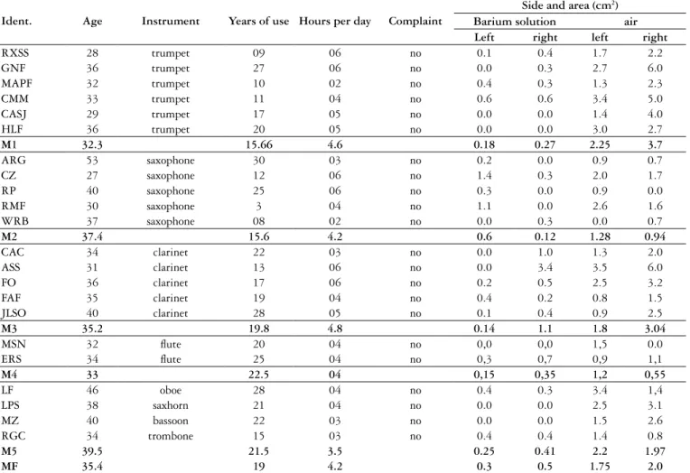
FIGURE 3. From a barium solution swallow: frame (A) shows the lateral contour of the laryngopharynx with a discrete irregularity, probably not ap-preciated because there is no residual volume of contrast after the swallow. (B, C and D) show a sequence, obtained by blowing against resistance, by the same trumpet player shown in (A). These images show large diverticula on both sides, where the left diverticulum (1) is smaller than the right (2). In frames (C and D), for didactical interest, we artiicially accentuated the lateral and inferior contour of the laryngopharynx to show the total air pharyngeal distension accentuating the pyriform recess (indicated by arrows in C). In (D) we estimated with a circle (arrow) the projection of the limit between the diverticula on both sides and the laryngopharynx
as “normal” discrete discomfort. This was admitted especially by the three volunteers in whom intermittent protrusions were seen at the cervical external surface in association with blowing against resistance (Figure 5).
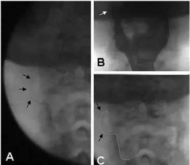
FIGURE 5. A man, without cervical extension, showing external cervical projections of the lateral pharyngeal diverticula (arrows) evidenced by blowing against resistance
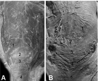
FIGURE 6 (A). Right and superior anatomically less resistant lateral laryngopharyngeal triangular formation, for passage of the laryngeal su-perior nerve (1). The superior margin (2) great horn of the hyoid bone; the inferior margin (3) is formed by the thyropharyngeal muscle, and the vertical margin (4) is formed by the thyrohyoid muscle; 4- esophagus. (B) a sagittal section, where the arrow indicates the trans-illuminated thin area on the top of the pyriform recess. In (C) a lateral external view showing an irregular thin area, where the arrow indicates the internal branch of the superior laryngeal nerve entrance; 5- esophagus
No other protrusions could be found in this group of wind instrumentalists despite the existence of the other, less resis-tant areas on the laryngopharyngeal wall (Figures 6 and 7).
DISCUSSION
In a group of 22 wind instrumentalists examined by means of a videoluoroscopic study, we could observe 41 pro-trusions on the less resistant lateral laryngopharyngeal area by air distension, and these protrusions ranged in size from 0.7 to 6 cm2. In the same group, under a videoluoroscopic barium-swallow study, the lateral diverticula that were pre-viously observed in the air-distension study did not appear, or appeared as a small protrusion. With air distension, the 41 diverticula were clearly identiied, and with barium as the contrast medium, only 7 protrusions larger than 0.6 cm were clearly identiied. Nineteen others were identiied from frame-by-frame analyses, and showed under-scaled areas less than 0.5 cm2 and only after their identiication with air distension. Two different mechanisms can cause lateral laryngopha-ryngeal diverticula. They can be produced by phalaryngopha-ryngeal intra-luminal overpressure without abnormal resistance to lux passage from the pharynx to the esophagus (in wind instrumentalists), or by overpressure produced by abnor-mally high resistance to lux passage from the pharynx to the esophagus (in dysphagia). These two mechanisms
plain the differences between the deinition that consider the diverticular formations as small protrusions that appear as full sacs in a barium-swallow test(6, 10, 13) and our results for the musician subjects examined in this study, in which the protrusions did not appear as full sacs in a barium-swallow test, despite their larger dimensions.
Physiologically, during swallowing, the high pressure into the pharynx inds the pharyngoesophageal transition open, giving food under pressure free passage to the esophagus and indirectly protecting the less resistant areas on the upper sides of the lateral laryngopharyngeal wall.
Pharyngeal protrusions are frequently associated with dysphagia. In a previous study(6) that examined 33 lateral laryngopharyngeal diverticula, dysphagia was not identiied in only four cases. The dysphagic process usually causes pha-ryngeal overpressure secondary to a high resistance to lux passage from the pharynx to the esophagus, because of the lower eficiency of the mechanism responsible for opening the pharyngoesophageal transition.
The anatomically less resistant area located above the narrow region, which conigures the pharyngeal esophageal transition, is a loor where the lateral laryngopharyngeal protrusions emerge(4, 6, 8). These anatomically less resistant areas provide suficient elasticity to sustain a physiological and certainly, some discrete overpressure.
Nevertheless, a continuous overpressure regimen, secon-dary to a lux resistance produced by dysphagia, can gener-ate a small retractile lgener-ateral protrusion that can be seen in a barium-swallow study, as a transient sac with a large neck or without a neck. This sac, which has some residual elas-ticity, is best deined as a pouch. The presence of somewhat larger protrusions leads to the deinition of diverticula as protrusions that appear as full barium sacs, variable in size, connected to the persistent sac by a proportionately short neck(6, 10, 13).
In wind instrumentalists, the protrusions are produced by intra-luminal pharyngeal overpressure, and do not involve dificulty in swallowing. The persistent and continuous pha-ryngeal overpressure induced by the resistance of the instru-ment’s mouthpiece will strongly distend the anatomically less resistant areas of the pharynx, producing a large protrusion that is best deined as an acquired diverticulum. Neverthe-less, these diverticula either did not appear, or appeared as a discrete protrusion when studied with the barium solution, because there is no abnormal resistance to lux passage from the pharynx to the esophagus during the swallowing process.
Acquired laryngopharyngeal diverticula are a conse-quence of overpressure. Under continuous overpressure, a permanent protrusion, with or without a neck, can be pro-duced. This permanent large sac, with or without contrast retention, must be considered as a diverticulum. Therefore, to analyze the pharyngeal wall it is necessary to complement the barium-swallow study with a test of pharyngeal disten-sion against resistance.
Despite the existence of the other, less resistant areas on the laryngopharyngeal segment, no other protrusions could be found in this group of wind instrumentalists. The
video-luoroscopic lateral proile study showed a large pharyngeal distension against the closed pharyngoesophageal transition, when the musicians blew against resistance, allowing us to presume that the other, less resistant areas were exempt from the overpressure effect produced by wind instruments. We believe that this exemption from overpressure results from the inferior position of the two other, less resistant areas.
The anatomically weaker area in the lateral laryngopha-ryngeal wall has characteristics of individual weakness, which is accentuated with aging(6). All the wind instrumentalists analyzed showed acquired diverticula, and the dimensions had no relationship to the age of the subject or the period of time of instrument use, indicating some degree of individua-lity in the weakness characteristics.
Blowing against resistance, as is necessary to play a wind instrument, clearly produces overload and damage in the ana-tomically weaker area in the laryngopharynx. Nevertheless, the kind of instrument seems to be more important than the length of time that it has been played. There were several examples in the group as a whole and also within the groups of users of the same instruments, where larger diverticula were found in musi-cians who had played their instruments for shorter periods. In the groups of trumpet, saxophone and clarinet players where the number of musicians allowed a comparison, the length of time that the instrument had been played and the size of the acquired diverticula showed no correlation.
There was no statistically signiicant difference in the lateral laryngopharyngeal right and left side resistance to overpressure imposed by the trumpet, clarinet or saxophone. The trumpet and clarinet players showed similar degrees of injury on the left and right sides. However, trumpet and clarinet overload produced more injury than the saxophone on the right and left sides, with statistically signiicant da-mage on the right side. These results lead us to believe that the overload is the factor responsible for the lateral pharyn-golaryngeal injury, and that there is no real difference in the resistance of the two sides.
Without a clear dimension reference, absence of symp-toms was previously considered to be common in cases of pharyngeal lateral diverticula(6, 9, 10). In agreement with these previous observations, no spontaneous complaints were expressed by the 22 subjects all of them wind instru-mentalists with large diverticula. Nevertheless, there are descriptions of complaints associated with lateral laryn-gopharyngeal diverticula, such as dysphagia(10), cervical aching and odynophagia(15), dysphagia and hoarseness(11), dysphagia, suffocation, and cervical discomfort(9), aspiration after swallowing (16, 28), laryngeal superior nerve neuralgia(3), cervical mass(24), halitosis, and the sensation of a foreign body in the throat(18). For this reason, all these complaints were directly mentioned to each volunteer, and all of them denied any such complaints.
were the cause, we certainly would have found at least one of these complaints in this group of wind instrumentalists, not their complete absence.
In three volunteers, we observed at the cervical external surface, an intermittent protrusion associated with blowing against resistance. These external cervical projections, which are associated with large diverticula, must be the cervical mass that had been mentioned previously(24). These persons also mentioned the perception of cervical pressure, described as discomfort, associated with instrument use, as observed previously(9).
Acquired lateral laryngopharyngeal diverticula were identiied in 100% of the musicians studied. Therefore, these diverticula should be considered as an “occupational overuse syndrome” (OOS) or, as previously described for other lesions that are produced by repetitive actions, as a “cumulative trauma disorder” (CTD) or “repetitive strain injury” (RSI). Under these several designations, this syndrome is responsible
for causing injury by repetitive tasks, as observed here in the wind instrumentalists. The absence of clear discomfort or other evident symptoms does not negate the possibility that the repetitive overload produced by a wind instrument is responsible for this condition(14, 21, 30).
CONCLUSION
Acquired lateral laryngopharyngeal diverticula are a consequence of pharyngeal overpressure. The absence of full barium sacs upon lateral laryngopharyngeal barium-swallow examination of the diverticula of the musicians indicates a lack of dificulty in the lux passage from the pharynx to the esophagus. The acquired diverticula produced by use of a wind instrument must be considered as an OOS. The lack of symptoms does not exclude the possibility that the repetitive tasks are the main factor responsible for the development of these diverticula.
Costa MMB, Alvite FL. Divertículo faríngeo-lateral: estudo videoluoroscópico da laringofaringe em instrumentistas de sopro. Arq Gastroenterol. 2012;49(2):99-106.
RESUMO – Contexto - São apresentados os resultados de um estudo em proissionais sadios, instrumentistas de sopro. Objetivo - Identiicar possíveis formações diverticulares produzidas nas paredes da laringofaringe pela alta pressão que a distende durante o uso desses instrumentos. Métodos - Uti-lizando o método videoluoroscópico examinou-se a faringe de 22 músicos proissionais nas incidências frontais e peril com deglutição de 20 mL de solução de sulfato de bário e após, soprando contra resistência. Resultados - Em todos os voluntários detectou-se a presença de divertículo faríngeo-lateral (3 unilaterais e 19 bilaterais) com áreas de projeção variando de 0,7 a 6 cm2. Trompetistas e clarinetistas apresentaram divertículos grandes e
bilaterais. Nenhuma queixa importante foi referida espontaneamente ou mesmo após questionamento. No estudo baritado da deglutição ou não se detectaram ou se detectaram com pequena dimensão, devido à livre passagem do luxo de contraste para o esôfago, os 41 divertículos identiicados quando os músicos sopraram contra resistência. Embora existam na laringofaringe outras áreas anatomicamente menos resistentes, nenhuma delas apresentou protrusão no grupo de instrumentistas de sopro estudado. Conclusões - A formação diverticular lateral que ocorre nos instrumentistas de sopro é distinta daquela que ocorre por aumento da pressão laringofaríngea determinada por resistência anormalmente elevada à livre passagem do luxo para o esôfago. As formações diverticulares laterais nos instrumentistas devem-se ao prolongado e repetitivo supranormal aumento da pressão laringofaríngea produzida pelo sopro contra resistência. A ausência, durante a deglutição, de resistência aumentada ao luxo do meio de contraste em passagem para o esôfago explica o fato de os divertículos de maior dimensão, identiicados pela distensão por ar, não apresentarem retenção da solução de sulfato de bário em seu interior durante a deglutição. Pode-se ainda concluir, com base na presença em 100% dos casos, que os divertículos faríngeo-laterais observados nos instrumentistas de sopro constituem-se em síndrome de sobrecarga funcional.
REFERENCES
1. Atkinson L. Pharyngeal diverticula with particular reference to lateral protrusions of various types. Arch Middx Hosp. 1952;2:245-54.
2. Bachman AL, Seaman WB, Macken KL. Lateral pharyngeal diverticula. Radio-logy. 1968;91:774-82.
3. Bagatzounis A, Geyer G. Lateral pharyngeal diverticulum as a cause of superior laryngeal nerve neuralgia. Laryngorhinootologie. 1994;73:219-21.
4. Cardenal L. Diccionario terminológico de ciencias médicas. 6. ed. Barcelona: Salvat; 1958. p.1304.
5. Costa MMB, Moreno MPR. Videomed. Software without patent. Rio de Janeiro: Center for Electronic Computation, Universidade Federal do Rio de Janeiro. Brazil: NCE/UFRJ; Version 1-16.9.2002-alpha.
6. Costa MM, Koch HA. Lateral laryngopharyngeal diverticulum: anatomical and videoluoroscopic study. Eur Radiol. 2005;15:1319-25.
7. Costa MMB, Canevaro LV, Koch HA, DeBonis R. Videoluoroscopy chair for the study of swallowing and related disorders. Radiol Bras. 2009;42:179-84. 8. Crawford JM. The gastrointestinal tract. In: Cotran RS, Kumar V, Collins T,
editors. Robbins pathologic basis of disease. 6th ed. Philadelphia: WB Saunders; 1999. p.755-842.
9. Curtis DJ, Cruess DF, Crain M, Sivit C, Winters C Jr, Dachman AH. Lateral pharyngeal outpouchings: a comparison of dysphagia and asymptomatic patients. Dysphagia. 1988;2:156-61.
10. Ekberg O, Nylander G. Lateral diverticula from the pharyngo-esophageal junction area. Radiology. 1983;146:117-22.
11. Ettman IK, Ramey DR 3rd. Lateral pharyngeal diverticulum: unusual cause of dysphagia and hoarseness. Am J Gastroenterol. 1967;47:490-7.
12. Flores-Franco RA, Limas-Frescas NE. The overused airway: lessons from a young trumpet player. Med Probl Perform Art. 2010;25:35.
13. Fowler WG. Lateral pharyngeal diverticula. Ann Surg. 1962;155:161-5. 14. Fry HJ. Overuse syndrome of the upper limb in musicians. Med J Aust.
1986;144:182-5.
15. Hankins WD. Traumatic hernia of the lateral pharyngeal walls. Radiology. 1944;42:499.
16. Huang PC, Scher RL. Endoscopic management of lateral pharyngeal pouch. Ann Otol Rhinol Laryngol. 1999;108:408-10.
17. Kaufman SA. Lateral pharyngeal diverticula. Am J Roentgenol Radium Ther Nucl Med. 1956;75:238-41.
18. Lee SW, Lee JY. Lateral pharyngeal diverticulum. Otolaryngol Head Neck Surg. 2010;143:309-10.
19. Lerut T, Coosemans W, Decaluwé H, Decker G, De Leyn P, Nafteux P, van Raemdonck D. Zenker’s diverticulum. Multimidia manual of cardio-thoracic surgery. (February 24, 2009). doi:10.1510/mmcts.2007.002881.
20. Liston SL. Lateral pharyngeal diverticula. Otolaryngol Head Neck Surg. 1985;93:582-5.
21. McKeag D. Overuse injuries. The concept in 1992. Prim Care. 1991;18:851-65. 22. McMyn JK. Lateral pharyngeal diverticula. J Fac Radiologists Lond. 1957;8:421-3. 23. Pace-Balzan A, Habashi SM, Nassar WY. View from within: radiology in focus
lateral pharyngeal diverticulum. J Laryngol Otol. 1991;105:793-5.
24. Pinto JA, Marquis VB, de Godoy LB, Magri EN, Brunoro MV. Bilateral hypo-pharyngeal diverticulum. Otolaryngol Head Neck Surg. 2009;141:144-5. 25. Ramsey GH, Watson JS, Gramiak R, Weinberg SA. Cineluorographic analysis
of the mechanism of swallowing. Radiology. 1955;64:498-518.
26. Rommelfanger KW. Lateral pharyngeal pouches (Author’s transl). Laryngol Rhinol Otol (Stuttq). 1980;59:710-4.
27. Rubesin SE, Jessurum J, Robertson D, Jones B, Bosma JF, Donner MW. Lines of the pharynx. Radiographics. 1987;7:217-37.
28. Rubesin SE. The pharynx. Structural disorders. Radiol Clin North Am. 1994;32:1083-101.
29. Rubesin SE, Levine M S.Killian-Jamieson diverticula: radiographic indings in 16 Patients. AJR Am J Roentgenol. 2001;177:85-9.
30. van Tulder M, Malmivaara A, Koes B. Repetitive strain injury. Lancet. 2007;369:1815-22.
31. Weller MD, Porter MJ, Rowlands J. An audit of pharyngeal pouch surgery using endoscopic stapling. The patient’s viewpoint. Eur Arch Otorhinolaryngol. 2004;261:331-3.