AR
TIGO ORIGINAL / ORIGINAL AR
TICLE
INTRODUCTION
Eosinophilic esophagitis (EoE) is a chronic, immune/antigen-mediated(5, 13) inlammatory esopha-geal disorder(24, 25) with a rapidly increasing preva-lence(6, 17). Recent epidemiologic data indicate that EoE is now the second leading cause of chronic esophagitis, behind gastroesophageal relux disease(20, 26) and a frequent cause of dysphagia(19).Its prevalence in children(6) and adults(20) almost reaches levels comparable to Crohn’s disease. EoE affects all age groups with a peak between the age of 20 and 50(4). EoE is clinico-pathologically characterized by esophageal symptoms in combination with a dense esophageal eosinophilia, both being refractory to proton pump inhibitors. Notably, eosinophilic in-lammation is absent in the stomach, small intestine and colon(9).
EoE’s leading symptom in adolescents and adults is dysphagia for solids with the imminent risk of prolonged food impactions(1, 2, 4, 21). Furthermore, pa-tients frequently complain of retrosternal pain that is unrelated to swallowing activity(9).
All studies agree that 80%-90% of patients have endoscopic features, most of them nonspeciic(25, 27) and none of them are pathognomonic of EoE(5, 13). Sometimes the endoscopic patterns do not explain food impaction episodes, and in these cases a mano-metric study could be reasonable.
EOSINOPHILIC ESOPHAGITIS:
manometric and pHmetric findings
Monica Maria Cardoso
MONNERAT
and Eponina Maria de Oliveira
LEMME
ABSTRACT – Context - Eosinophilic esophagitis is an entity characterized by an esophageal inlammatory iniltrate of eosinophils, manifested by dysphagia, intermittent food impactions and symptoms similar to gastroesophageal relux disease (GERD), that predominantly affects young adults. There may be association of eosinophilic esophagitis with GERD, and motor abnormalities have been described. Objective - The main objectives of this study are to describe the indings at esophageal manometry and pH monitoring in patients with eosinophilic esophagitis. Methods - Cross-sectional study of 20 patients with a diagnosis of eosinophilic esophagitis, submitted to esophageal manometry and 24h pH monitoring. Were analysed the manometric changes and the presence of abnormal relux on pH monitoring. Results - Twenty patients (15 men, 5 women) had a mean age of 29 years. Motility disorders were found in 25% (5/20) patients with ineffective esophageal motility being the most common inding. pH monitoring revealed abnormal relux on 25%, without any relationship with manometric indings. Conclusions - Manometric abnormalities were observed in 25% of patients and abnormal relux on pH monitoring also in 25%. This study showed no relationship between abnormal relux and the presence of manometric changes.
HEADINGS – Eosinophilic esophagitis. Esophageal motility disorders. Gastroesophageal relux.
Esophagus Unit, Gastroenterology Division, Clementino Fraga Filho University Hospital, Federal University of Rio de Janeiro, RJ, Brazil.
Correspondence: Prof. Eponina M. Oliveira Lemme - Travessa Santa Terezinha, 14 – Tijuca - 20271-070 - Rio de Janeiro, RJ, Brazil. E-mail: [email protected] Lucendo et al.(14) described manometric abnor-malities in 26/30 (86,6%) adult patients with EoE, and observed that the motor disorders exclusively affected the part of the esophagus comprising smooth muscle. When the diagnosis of gastroesophageal relux disease (GERD) vs EoE in not apparent despite en-doscopy and biopsy, intraesophageal pH monitoring may be of use in excluding pathologic relux as either the primary or a concomitant cause for esophageal eosinophilia, as recommended by the Consensus guidelines(5, 13).
Some reports suggest that the interaction between GERD and EoE can be complex, and that the notion of establishing a clear distinction between the two disorders may be too simplistic. There are at least four situations in which GERD might be associated with esophageal eosinophils: (1) GERD causes esophageal injury that results in a mild eosinophilic iniltration; (2) GERD and EoE coexist but are unrelated; (3) EoE contributes to or causes GERD; or (4) GERD contributes to or causes EoE(14).
The aim of this study was to evaluate the etio-logy of dysphagia and the presence of relux in EoE, analyzing pHmetric and manometric disorders and if there was a correlation between them.
METHODS
and 24-hour pH monitoring in patients with eosinophilic esophagitis (EoE). The protocol of this research was approved by the Commission of Ethics in Research of Clementino Fraga Filho Hospital University, Federal University of Rio de Ja-neiro, RJ, Brazil. Cases were 20 symptomatic patients, recruited from the Gastroenterologic Division at Hospital University, based on previous clinical documentation of esophageal symp-toms suggestive of EoE and current endoscopic biopsies with histopathology conirming EoE as deined by the Consensus guidelines(5, 13) and had at least 15 eosinophils/high power ield. From 20 patients, 15 were man and 5 women, the average age was 29 years, with higher prevalence (60%) between 15 and 30 years. The predominant symptoms were the recurrent food impaction (55%), heartburn (35%), dysphagia for solids (25%) and thoracic pain (15%), with average duration 34.6 months (3 months to 10 years). Every patient had been submitted to upper digestive endoscopy. All of them had endoscopic ab-normalities, alone or combined. Noticeable loss of vascular pattern was found in all patients, with whitish plaques and longitudinal furrows. Patients received information about esophageal manometry and 24-hour pH monitoring exams and their possible side effects and complications. All subjects gave written informed consent.
Manometric study
Esophageal manometry (EM) was carried out at The Esophagus Unit of the University Hospital, using perfu-sion computerized equipment (ALACER, Brazil) and an 8-channel, 4,5 mm diameter polyvinyl catheter, with the patient in supine position and after the 6-hour fast. After administering topical anesthesia using lidocaine, the ma-nometry tube was inserted trough the anesthetized nostril. A baseline gastric waveform was identiied on the monitor screen to conirm placement of the four catheter sensing ports in the stomach after which an end-expiratory LES pressure in mm Hg was obtained by the station pull trough method. The mean of the highest values recorded from each of the four ports was considered the LES pressure. Relax-ation of the sphincter was evaluated after at least 10 wet swallows. After LES measurements, one of distal channels of the manometry catheter was positioned 3 cm above the LES upper border.
The esophageal body motility was measured by recording the response to at least 10 wet swallows of 3-5 mL volume. The manometry catheter was subsequently slowly withdrawn until the proximal hole of the catheter showed a good pres-sure waveform of the upper esophageal sphincter (UES) and relaxation of the UES pressure was monitored during the wet swallow. The catheter was further withdrawn until the second port recorded the UES pressure and after wet swallows the pharyngeal contraction and relaxation of the UES were recorded.
For diagnosing of motility abnormalities was used the In-ternational Classiication of Esophageal Motor Disorders(22) adapting where relevant, to the normal values employees in the Esophagus Unit from study(12) in healthy volunteers (Table 1).
24-hour pH ambulatory monitoring
Twenty-four-hour intraesophageal pH studies were per-formed with portable digital recorders (MKIII, AL2 Alacer and Sigma SMP 2128, MG), an antimony catheter and an outer electrode. This test was performed after a 6-hour fast, introducing the catheter trough the nostril and placed 5 centimeters proximal to the LES (localized by manometric study) after calibrating the catheter for pH 1.07 and 7.01 using reference solutions.
Patients continued their normal daily activities and were allowed a normal diet, with no citric fruits or soft drinks. Proton pump inhibitors (PPI) were discontinued at least 7 days prior to the examination and prokinetics agents 24 hours before the test.
It was considered reflux episode when the pH of the esophagus to fall to less than 4 for at least 15 seconds. For the interpretation of the results they hired the percentage (%) of total time pH<4 and in the upright position and supine position; normal values as % total time < 4.5%, % upright time < 7.0% and % supine time < 2.5%(8).
RESULTS
Esophageal manometry
Esophageal manometry was normal in 75% of patients. Only ive patients (25%) presented manometric disturbances. The study of the upper esophageal sphincter was normal in all patients, including rest pressure, relaxation and pharyn-goesophageal coordination. Motor disorders affect only the smooth muscles of esophagus. The LES was normal in 18 patients (90%), hypertensive (38.2 mm Hg) with normal re-laxation in 1 patient (5%) and excessive hypotensive (4.6 mm Hg) in another. The study of the esophageal body showed ineffective esophageal motility in 3 patients (15%). Esopha-geal manometry results are presented in Table 2.
TABLE 1. Esophageal motility abnormalities (Spechler, Castell(22)) Motility abnormalities Manometricindings
Ineffective esophageal motility
Number of low amplitude peristaltic contractions (< 30 mm Hg) and/or nontransmitted contractions > 20% of total number of wet swallows used for esophageal body study
Hypotensive LES LES pressure < 10 mm Hg
Hypertensive LES LES pressure > 32 mm Hg(12)
LES = lower esophageal sphincter
TABLE 2. Esophageal manometry results
Esophagealmanometryresults n = 20 (%patients))
LES hypertensive 1 (5%)
LES hypotensive 1 (5%)
Ineffective esophageal motility 3 (15%)
Normal esophageal manometry 15 (75%)
Total 20 (100%)
24-hour pH monitoring
PH monitoring was normal in 15/20 patients (75%) and revealed abnormal relux in 5/20 patients (25%), being 3 pa-tients with supine relux and 2 with bipositional relux. One patient presented bronchospasm coincident with relux (index of symptoms positive). Table 3 shows the indings described. The ive patients with abnormal relux on pH monitoring received double dose PPI during 8 weeks and then the esopha-geal biopsies were repeated. There was persistence of the endoscopic indings of EoE and the esophageal eosinophilia.
Primary disorders were found in 12 patients: 2 with achalasia, diffuse esophageal spasm in 7 and nutcracker esophagus in 3 patients. LES was normal in 100 patients, hypotensive in 12, hypertensive in 3 and incomplete relaxation was found in 2 achalasia patients. Unspeciic motor abnormalities (tertiary contractions, low-amplitude and ineffective esophageal mo-tility were reported in 35 patients (34.3%) and contractions of high amplitude in 11. Therefore, manometric disturbances were found in 41% of patients.
In our study, esophageal manometry was normal in 15 patients (75%). Esophageal manometry was abnormal in 5 patients in this study (25%), being the most frequent change ineffective esophageal motility, identiied in 3 patients. We also found a patient with hypotensive LES (4.5 mm Hg) and another with hypertensive LES (38.2 mm Hg). The up-per esophageal sphincter was normal in all patients. In our study, only one patient who presented manometric change had abnormal relux. Motility disorders were present in 40% (2/5) of patients with dysphagia for solids and 36% (4/11) of patients with recurrent food impaction.
Normal pH monitoring was found in patient who pre-sented hypotensive lower esophageal sphincter.
According to Korsapati et al.(10) esophageal manometry measures only the circular muscle function of the esophagus, and not all motility disorders are discovered by this method. Martin et al.(15) reported normal LES in 9 patients (81%), hypotensive in 2 (18%), and ineffective esophageal motility was identiied in 5 patients (45%), a percentage greater than the one described by other authors.
Lucendo et al.(14) described manometric abnormalities in 26/30 (86,6%) adult patients with EoE, and observed that the motor disorders exclusively affected the part of the esophagus comprising smooth muscle. The manometric study of the upper esophageal sphincter was normal in all patients, including resting pressure, dynamic behavior, and pharyngoesophageal motor coordination. The LES was hypotensive in 12/30 (40%), a hypoperistaltic motor pattern was found in 17/30 patients (56.7%), and a hyperkinetic pat-tern in 9 patients (30%).
Hejazi et al.(7) inferred that dysphagia in EoE can be attributed at least in part to motility disorders that can improves with treatment. Some authors suggest that there are different phases in the development of motor abnormali-ties, similarly to what happens in other disorders affecting esophageal function such as GERD and achalasia. Initially the motility is normal, then it would raise spastic hyper-contractility and diffuse esophageal spasm that evolutes to simultaneous low-am plitude contractions.
Rothenberg et al.(21) analysed the results of nine studies on esophageal pH monitoring in EoE, and abnormal results were found in 18% of 100 patients. In the publication of Martin et al.(15) abnormal relux on pH monitoring was found in two patients (18%).
GERD is a common condition and may affect 10% to 20% of healthy adults(3), while the EoE can affect up to 1% of a population(20). So, GERD and EoE coexist but are unrelated(16, 23).
TABLE 3. pH monitoring results
pH monitoring results n = 20 (% patients)
Normal pH monitoring 15 (75%)
Abnormal bipositional 2 (10%)
Abnormal supine 3 (15%)
Correlation between pH monitoring and manometry findings
There was no correlation between manometry and pH monitoring indings.
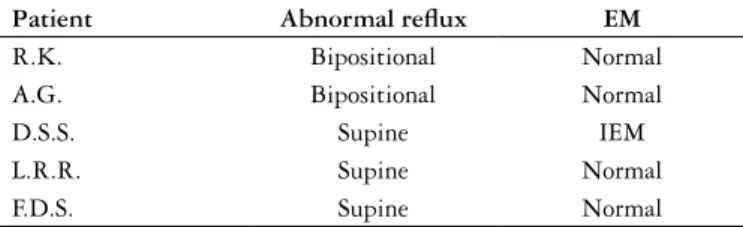
From ive patients with abnormal relux only one pre-sented abnormal manometry ineffective esophageal motility and from ive patients with abnormal manometry only one had abnormal relux (Tables 4 and 5).
TABLE 4. Abnormal pH monitoring x esophageal manometry
Patient Abnormal relux EM
R.K. Bipositional Normal
A.G. Bipositional Normal
D.S.S. Supine IEM
L.R.R. Supine Normal
F.D.S. Supine Normal
EM = esophageal manometry IEM = ineffective esophageal motility
TABLE 5. Abnormal esophageal manometry x pH monitoring
Patient AbnormalEM pH monitoring
E.R.M. Hypertensive LES Normal
A.E. Excessive hypotensive LES pressure Normal
D.S.S. IEM Abnormal supine
R.A. IEM Normal
J.R.S.L. IEM Normal
EM = esophageal manometry LES = lower esophageal sphincter IEM = ineffective esophageal motility
DISCUSSION
In our study, the presence of abnormal reflux in five patients (25%), attach to overlap between EoE and GERD. The ive patients with abnormal relux on pH monitoring received double dosage PPI during 8 weeks and then the esophageal biopsies were repeated. There was not remission of dysphagia, neither the endoscopic indings of EoE and the esophageal eosinophilia. This way, GERD was discarded as a cause of esophageal eosinophilia, as this continued after PPI treatment.
In 2007, Consensus guidelines(5), one of the diagnostic criteria for EoE is that there would be no improvement with esophageal eosinophilia with acid suppression. However, in a high percentage adult patients with eosinophilic iniltration in the esophagus present coexisting GERD(16). This ratiies that the partial response to PPI does not exclude EoE diagnosis, and that pH monitoring and esophageal manometry should be performed in patients with atypical symptoms or when there is no diagnostic deinition. It is important to emphasize that EoE can coexist with GERD; however, treatment with PPI allows control of acid relux but does not improves the dysphagia.
Another possible association between EoE and GERD is that eosinophils secret potent agents that can affect esopha-geal smooth muscle and nerve function. Cytotoxic effects of eosinophils may render esophageal epithelium more susceptible to relux injury(23).
We ind in our study 5/20 (25%) patients who presented manometric abnormalities (hypertensive LES in 1 patient, excessive hypotensive LES in other and IEM in 3 patients). Only 1 patient with ineffective esophageal motility had ab-normal relux on pH (supine position).
IEM was the most frequent manometric abnormality in this study, as in other publications about EoE(7, 15, 18). IEM has been associated with relux in both the supine and upright position, prolonged esophageal clearance, and delayed of bolus transport(11).
Five patients (25%) showed pathological relux on pH monitoring (two patients with bipositional reflux and three patients with supine relux). One of these patients presented IEM on manometry. The presence of abnormal relux in the other patients would be related to overlap between EoE and GERD. Our study may have some limitations for the analysis pHmetric and manometric features arising from the small number of patients with EoE, but there was no correlation between pHmetric and manometric indings.
In conclusion, manometric abnormalities were observed in 25% of patients and abnormal relux on pH monitoring also in 25%. Ineffective esophageal motility was the most frequent disorder in this study. There was no relationship between abnormal relux and the presence of manometric changes in these patients.
Monnerat MMC, Lemme EMO. Esofagite eosinofílica: achados manométricos e pHmétricos. Arq Gastroenterol. 2012;49(2):113-7.
RESUMO – Contexto - A esofagite eosinofílica é uma doença inlamatória crônica, caracterizada por iniltrado eosinofílico no esôfago e se manifesta por disfagia, impactações alimentares e sintomas similares aos da doença do reluxo gastroesofágico (DRGE), com maior incidência em adultos jovens. Pode haver associação da esofagite eosinofílica com a DRGE, e anormalidades motoras têm sido descritas. Objetivo - Os principais objetivos deste estudo são descrever as alterações manométricas e a presença de reluxo anormal à pHmetria esofágica em pacientes com esofagite eosinofílica. Métodos - Estudo transversal de 20 pacientes com diagnóstico de esofagite eosinofílica, submetidos a esofagomanometria e pHmetria esofagiana de 24 h. Foram analisadas as alterações manométricas e a presença de reluxo anormal à pHmetria. Resultados - Vinte pacientes (15 homens, 5 mulheres) com média de idade de 29 anos. Distúrbios da motilidade esofagiana foram encontrados em 25% dos pacientes, com predomínio da motilidade esofagiana ineicaz. A pHmetria revelou reluxo anormal também em 25%, sem relação entre os achados manométricos e pHmétricos. Conclusões - Anormalidades manométricas foram encontradas em 25% dos pacientes e reluxo anormal à pHmetria também em 25%. Neste estudo, não houve relação entre reluxo anormal e a presença de alterações à esofagomanometria.
REFERENCES
1. Arora AS, Yamazaki K. Eosinophilic esophagitis: asthma of the esophagus? Clin Gastroenterol Hepatol. 2004;2:523-30.
2. Attwood SE, Smyrk TC, Demeester TR, Jones JB. Esophageal eosinophilia with dysphagia: a distinct clinicopathologic syndrome. Dig Dis Sci. 1993;38:109-16. 3. Dent J, El Serag-HB, Wallander MA, Johansson S. Epidemiology of
gastroesopha-geal relux relux disease: a systematic review. Gut. 2005;54:710-7.
4. Desai TK, Stecevic V, Chang CH, Goldstein NS, Badizadegan K, Furuta GT. Association of eosinophilic inlammation with esophageal food impaction in adults. Gastrointest Endosc. 2005;61:795-801.
5. Furuta GT, Liacouras C, Collins MH, Gupta SK, Justinich C, Putnam PE, Bonis P, Hassall E, Straumann A, Rothemberg ME. Eosinophilic esophagitis in children and adults: a systematic review and consensus recommendations for diagnosis and treatment. Gastroenterology. 2007;133:1342-63.
6. Gupta SK, Fitzgerald JF, Kondratyuk T, HogenEsch H. Cytokine expression in normal and inlamed esophageal mucosa: a study into the pathogenesis of allergic eosinophilic esophagitis. J Pediatr Gastroenterol Nutr. 2006;42:22-6.
7. Hejazi RA, Reddymasu SC, Sostarich S, McCallum RW. Disturbances of esopha-geal motility in eosinophilic esophagitis: a case series. Dysphagia. 2010;25:231-7. 8. Johnson LF, DeMeester TR. Twenty-four hour pH monitoring of the distal
esophagus. A quantitative measure of gastroesophageal relux. Am J Gastroen-terol. 1974;62:325-32.
9. Kapel RC, Miller JK, Torres C, Aksoy S, Lash R, Katzka DA. Eosinophilic esophagitis: a prevalent disease in the United States that affects all age groups. Gastroenterology. 2008;134:1316-21.
10. Korsapati H, Babaei A, Bhargava V, Dohil R, Quin A, Mittal RK. Dysfunction of the longitudinal muscles of the esophagus in eosinophilic esophagitis. Gut. 2009;58:1056-62.
11. Lemme EM, Abrahão Jr, LJ, Manhães Y, Schechter R, Carvalho BB, Alvariz A. Ineffective esophageal motility in gastroesophageal erosive relux disease and in nonerosive relux disease. Are They Different? J Clin Gastroenterol. 2005;39:224-7. 12. Lemme EMO, Domingues GR, Silva LFD, Firman CG, Pantoja JAS. Esophageal
manometry: preliminary values in health adult volunteers. Gastroenterol Endosc Dig. 2001;24:29-35.
13. Liacouras CA, Furuta GT, Hirano I, Atkins D, Attwood SE, Bonis PA, Burks AW, Chehade M, Collins MH, Dellon ES, Dohil R, Falk GW, Gonsalves N, Gupta SK, Katzka DA, Lucendo AJ, Markowitz JE, Noel RJ, Odze RD, Putnam PE, Richter JE, Romero Y, Ruchelli E, Sampson HA, Schoepfer A, Shaheen NJ, Sicherer SH, Spechler S, Spergel JM, Straumann A, Wershil BK, Rothenberg ME, Aceves SS. Eosinophilic esophagitis: updated consensus recommendations for children and adults. J Allergy Clin Immunol. 2011;128:3-20.e6
14. Lucendo AJ, Pascual-Turrión JM, Navarro M, Comas C, Castillo P, Letrán A, Caballero MT, Larrauri J. Endoscopic, bioptic, and manometric indings in eo-sinophilic esophagitis before and after steroid therapy: a case series. Endoscopy. 2007;39:765-71.
15. Martin LM, Vaquero CS, Prudencio SS, Perona JC, Gisbert JP, Otero, RM. Eo-sinophilic esophagitis in adult – clinical, endoscopic, pH-metric, and manometric indings. Rev Esp Enferm Dig. 2008;100:476-80.
16. Molina Infante J, Fernando-Qoqiauri L, Mateos-Rodriguez JM, Pérez-Gallardo B, Prieto-Bermejo AB. Overlap of relux and eosinophilic esophagitis in two patients requiring different therapies: a review of the literature. World J Gastroenterol. 2008;14:1463-6.
17. Noel RJ, Putnam PE, Rothenberg ME. Eosinophilic esophagitis. N Engl J Med. 2004;351:940-1.
18. NurKo S, Rosen R. Esophageal dysmotility in patients who have eosinophilic Esophagitis.. Gastrointest Clin N Am. 2008;18:73-89.
19. Portmann S, Heer P, Bussmann Ch. Epidemiology of eosinophilic esophagitis: data from a community-based longitudinal study [abstract]. Gastroenterology. 2007;132:A-609.
20. Ronkainen J, Talley NJ, Aro P, Storskrubb T, Johansson SE, Lind T, Bolling-Sternevald E, Vieth M, Stolte M, Walker MM, Agréus L. Prevalence of esophageal eosinophils and eosinophilic esophagitis in adults: the population-based Kalixanda study. Gut. 2007;56:615-20.
21. Rothenberg ME, Mishra A, Collins MH, Putnam PE. Pathogenesis and clinical features of eosinophilic esophagitis. J Allergy Clin Immunol. 2001;108:891-4. 22. Spechler SJ, Castell DO. Classiication of oesophageal motility abnormalities.
Gut. 2001;49:145-51.
23. Spechler SJ, Genta R, Souza R. Thougts on the complex relationship between gastroesophageal relux relux disease and eosinophilic esophagitis. Am J Gas-troenterol. 2007;102:1301-4.
24. Straumann A, Bauer M, Fischer B, Blaser K, Simon HU. Idiopathic eosinophilic esophagitis is associated with a Th2-type allergic inlammatory response. J Allergy Clin Immunol. 2001;108:954-61.
25. Straumann A, Spichtin HP, Grize L, Bucher KA, Beglinger C, Simon H-U. Natural history of primary eosinophilic esophagitis: a follow-up of 30 adult patients for up to 11.5 years. Gastroenterology. 2003;125:1660-9.
26. Straumann A, Simon HU. Eosinophilic esophagitis: escalating epidemiology? J Allergy Clin Immunol. 2005;115:418-9.
27. Yan BM, Schaffer EA. Eosinophilic esophagitis: a newly established cause of dysphagia. World J Gastroenterol. 2006;12:2328-34.