Diego de Oliveira Hartmann
Dissertation presented to obtain the Ph.D degree in Biochemistry
Instituto de Tecnologia Química e Biológica António Xavier | Universidade Nova de LisboaOeiras,
December, 2014
Exploring ionic liquids
′
unique stimuli to
Diego de Oliveira Hartmann
Dissertation presented to obtain the Ph.D degree in Biochemistry
Instituto de Tecnologia Química e Biológica António Xavier | Universidade Nova de Lisboa
Oeiras, December, 2014
elucidate uncharacterised cellular and molecular
ii
Title: Exploring ionic liquids′ unique stimuli to elucidate uncharacterised cellular
and molecular mechanisms in filamentous fungi
Dissertation presented to obtain the Ph.D. degree in Biochemistry
Author: Diego de Oliveira Hartmann
Applied and Environmental Mycology Laboratory Instituto de Tecnologia Química e Biológica Universidade Nova de Lisboa
Av. da República
Estação Agronómica Nacional 2780-157 Oeiras
iii
I declare that the work presented in this thesis, except where otherwise stated, is based on my own research. It was supervised by Doctor Cristina Silva Pereira and
Doctor Ricardo O. Louro. The work was mainly performed in Instituto de
Tecnologia Química e Biológica, Universidade Nova de Lisboa, between May 2010
and December 2014.
I am grateful for the financial support provided by Fundação para a Ciência e
Tecnologia (grant reference SFRH/BD/66396/2009). The work was partially
v
vii
Acknowledgements ... xi
Summary ... xiii
Sumário ... xv
Thesis publications ... xix
List of acronyms ... xxi
CHAPTER I ... 1
Introduction 1.1.Fungi ... 3
1.2.The fungal cell envelope ... 13
1.2.1. Plasma membrane ... 13
1.2.2. Cell wall ... 19
1.3.Ionic liquids ... 26
1.4.Objectives ... 33
1.5. References ... 34
CHAPTER II ... 55
Unravelling the mechanism of toxicity of alkyltributylphosphonium chlorides in Aspergillus nidulans conidia 2.1. Abstract ... 58
2.2. Introduction ... 59
2.3. Materials and Methods ... 61
2.3.1. Chemicals ... 61
2.3.2. Ionic liquids ... 61
2.3.3. Fungal strain ... 61
2.3.4. Toxicity tests ... 62
2.3.5. Membrane and cell wall integrity assays ... 62
2.3.6. Scanning electron microscopy ... 64
2.3.7. Biodegradability assessment ... 64
2.4. Results and Discussion ... 65
2.5. Conclusions ... 75
2.6. Acknowledgements ... 76
2.7. References ... 77
CHAPTER III ... 85
viii
3.3. Materials and Methods ... 92
3.3.1. Chemicals ... 92
3.3.2. Ionic liquids ... 92
3.3.3. Fungal strain ... 92
3.3.4. Experimental conditions ... 92
3.3.5. Total RNA extraction and cDNA synthesis ... 93
3.3.6. Oligonucleotides design ... 94
3.3.7. Quantitative real-time PCR analysis ... 95
3.3.8. Statistical analysis ... 95
3.3.9. Microscopic analyses ... 95
3.4. Results and Discussion ... 97
3.4.1. Plasma membrane biosynthetic genes ... 97
3.4.2. Cell wall biosynthetic genes ... 101
3.4.3. Cell wall integrity pathway ... 105
3.5. Conclusions ... 108
3.6. Acknowledgements ... 110
3.7. References ... 111
CHAPTER IV ... 119
Plasma membrane permeabilisation as a mechanism of toxicity of ionic liquids: a matter of charge 4.1. Abstract... 122
4.2. Introduction ... 123
4.3. Materials and Methods ... 126
4.3.1. Chemicals ... 126
4.3.2. Ionic liquids ... 126
4.3.3. Fungal strain ... 126
4.3.4. Toxicity tests ... 127
4.3.5. Plasma membrane permeabilisation assay ... 127
4.3.6. Biodegradability assessment ... 128
4.3.7. Experimental conditions for gene expression analysis ... 129
4.3.8. Oligonucleotides design ... 130
4.3.9. Total RNA extraction and cDNA synthesis ... 130
4.3.10. Quantitative real-time PCR analysis ... 131
4.3.11. Statistical analysis ... 131
4.3.12. Molecular dynamics simulation ... 131
4.4. Results and Discussion ... 133
ix
4.4.3. Imidazolium-based ionic liquids, but not cholinium alkanoates,
permeabilise the fungal plasma membrane ... 137
4.4.4. Plasma membrane permeabilisation depends on the nature of the ionic liquids ions and of the membrane itself ... 143
4.4.5. Elongation of an alkyl chain in the cholinium cation induces plasma membrane permeabilisation ... 145
4.5. Conclusions ... 150
4.6. Acknowledgements ... 152
4.7. References ... 153
CHAPTER V ... 161
Ionic liquids stimuli can provide insights on the roles of sphingolipids intermediates as signalling molecules in Aspergillus nidulans 5.1. Abstract ... 164
5.2. Introduction ... 165
5.3. Materials and Methods ... 168
5.3.1. Chemicals ... 168
5.3.2. Ionic liquids ... 168
5.3.3. Fungal strain ... 168
5.3.4. Experimental conditions ... 168
5.3.5. Oligonucleotides design ... 169
5.3.6. Total RNA extraction and cDNA synthesis ... 171
5.3.7. Quantitative real-time PCR analysis ... 171
5.3.8. Statistical analysis ... 171
5.3.9. Extraction of sphingoid bases ... 172
5.3.10. High performance liquid chromatography ... 172
5.4. Results and Discussion ... 174
5.5. Conclusions ... 188
5.6. Acknowledgements ... 190
5.7. References ... 191
CHAPTER VI ... 197
Final discussion 6.1. Final discussion ... 199
xi
The completion of this thesis was only possible with the help of many people, to whom I wish to acknowledge.
First of all, I wish to thank my supervisors Cristina Silva Pereira and Ricardo O. Louro for making this work possible. Cristina, thank you not only for your continuous effort, dedication and enthusiasm, but for always believing in me and in my ideas. Nothing would be possible without your support.
Thanks to all former and present members of the Applied and Environmental Mycology and the Inorganic Biochemistry and NMR groups from for their continuous support. Especial thanks to all of those with whom I worked more closely, mainly Paula, Isabel, Tiago, Carlos and Cristina Leitão. A very sincere thank you! to Marija, who started as my mentor on ionic liquids, became my peer and, most especially, my friend.
I also thank all the collaborators who made the work of this thesis possible,
especially Gabriela Adamová and Prof. Kenneth R. Seddon from The Queen’s
University Ionic Liquid Laboratories; Prof. Luis Paulo N. Rebelo, from ITQB; Filipa Siopa and Prof. Carlos A. M. Afonso, Faculdade de Farmacia, Universidade de Lisboa; and Karina Shimizu and Prof. José N. Canongia Lopes, from Centro de Química Estrutural, Instituto Superior Técnico.
I would also like to thanks all my friends that have been with me during these last years, helping me and supporting me in many different ways. Pépe, Jane, Jojo, Bee, Amarula, City, Tiago, Micha, Number Three, thanks for all the coffees, drinks, talks and laughter. Zé, thank you for making the final steps so much better,
and for making me look at it as a new beginning. If it wasn’t for you all I am sure I
wouldn’t get so far. Thank you!
xiii
The emergence of new fungal pathogens, either of plants or animals, and the increasing number of reported cases of resistant human pathogenic strains to the available antifungal drugs reinforces the need for better understanding the biology of filamentous fungi. Conventional drugs target components of the fungal membrane or cell wall, therefore identifying novel intracellular targets, yet unique to fungi, is a global priority. Previous studies revealed that ionic liquids, a class of tuneable organic salts, are able to induce distinct metabolic alterations in filamentous fungi. These compounds, despite being often regarded as green solvents, mainly due to their lack of vapour pressure and non-flammability, can display certain toxicity, and their potential use in industrial scale reinforces the need for their toxicological
assessment. Ionic liquids′ distinct effects on filamentous fungi inspired the present
thesis, which focusses in better understanding the mechanisms of toxicity of these compounds and the response of filamentous fungi to their chemical stress. More importantly, it aims at investigating the unique chemical stimuli of ionic liquids as potential tools to unravel unknown cellular and molecular mechanisms in filamentous fungi.
Using the model filamentous strain Aspergillus nidulans, the mechanisms of
toxicity of four distinct families of ionic liquids were investigated. In a systematic study that employed predominantly fluorescence microscopy and gene expression
analyses (qRT-PCR), I was able to prove that alkyltributylphosphonium chlorides
are able to permeabilise the fungal plasma membrane in a manner dependent on the
length of the cation′s alkyl substituent (Chapters II and III). The same effect could
be observed for two other distinct families of ionic liquids, 1-alkyl-3-methylimidazolium chlorides and alkyl-(2-hydroxyethyl)-dimethylammonium bromides (Chapter IV). However, plasma membrane permeabilisation does not depend only on the length of the alkyl chain, but also on the charge of the ion carrying such chain. Cholinium alkanoates, for instance, although becoming
increasingly more toxic with the elongation of the anion′s alkyl chain, do not cause
xiv
the overall negative charge of the fungal cell wall seem to play a crucial role in the mechanisms of toxicity of ionic liquids. This hypothesis was further investigated by molecular dynamics simulations with representative ionic liquids of each family and artificial membranes (Chapter IV).
In addition to membrane permeabilisation, some ionic liquids were also able to damage the fungal cell wall and induce a stress response known as cell wall integrity pathway, as observed by the up-regulation of cell wall biosynthetic genes (Chapters III and IV). This signalling pathway is better understood in yeast, whereas
little knowledge is available for filamentous fungi, including Aspergillus nidulans.
Data suggest that ionic liquids are not only able to cause cell wall damage but also activate alternative stress response pathways, as already proposed to exist in this model fungus. Remarkably, ionic liquids also activate the sphingolipids biosynthetic pathway and induce the accumulation of intermediates such as the sphingoid bases dihydrosphingosine and phytosphingosine, as well as other unknown species. Each family of ionic liquids is able to induce a distinctive response, highlighting the uniqueness of their chemical stimuli (Chapter V). Intermediates in the sphingolipids biosynthetic pathway have already been reported to be important signalling elements in eukaryotes, in several biological processes, from cell cycle regulation to programed cell death. In yeast, some of these intermediates are involved in the cell wall stress response, but little information about the puzzling roles of these compounds in filamentous fungi is available to date.
The work presented in this thesis furthers the knowledge on ionic liquids
toxicological risk and constitutes the basis for exploring ionic liquids′ unique
xv
O surgimento de novos fungos patogénicos, tanto de plantas como de animais, e o crescente número de casos reportados de estirpes patogénicas resistentes às drogas antifúngicas disponíveis reforça a necessidade de maiores conhecimentos sobre a biologia dos fungos filamentosos. As drogas convencionais têm como alvo a membrana plasmática ou a parede celular fúngicas e, portanto, novos alvos intracelulares, únicos aos fungos, é uma prioridade global. Estudos preliminares revelam que líquidos iónicos, uma classe de sais orgánicos tuneáveis, são capazes de induzir, em fungos filamentosos, alterações metabólicas distintas. Estes compostos,
apesar de serem frequentemente considerados solventes “verdes”, principalmente
pela sua falta de pressão de vapor e não-flamabilidade, podem apresentar considerável toxicidade, e seu uso potencial em escala industrial reitera a necessidade de sua avaliação toxicológica. Os efeitos distintos dos líquidos iónicos em fungos filamentosos inspiram a presente tese, que visa melhor compreender os mecanismos de toxicidade destes compostos e a resposta dos fungos filamentosos ao seu stress químico. Ainda mais importante, visa investigar o estímulo químico único dos líquidos iónicos como potenciais ferramentas para elucidar mecanismos celulares e moleculares desconhecidos em fungos filamentosos.
Usando o fungo filamentoso modelo Aspergillus nidulans, os mecanismos
de toxicidade de quatro famílias distintas de líquidos iónicos foram investigados. Em um estudo sistemático que empregou principalmente microscopoia de
fluorescência e análise da expressão génica (qRT-PCR), fui capaz de provar que
xvi
química da membrana plasmática, que apresenta uma distribuição heterogénia de cargas em sua superfície, e a carga negativa da parede celular desempenham um papel fundamental nos mecanismos de toxicidade dos líquidos iónicos. Esta hipótese foi também suportada por estudos de simulação de dinámica molecular de líquidos iónicos representativos de cada família e membranas artificiais (Capítulo IV).
Adicionalmente à permeabilização da membrana plasmática, alguns líquídos iónicos são também capazes de causar dano à parede celular dos fungos e induzir uma resposta ao stress conhecida como via de da integridade da parede celular., como pode ser observado pela regulação positiva de genes da biosíntese da parede celular (Capítulos III e IV). Esta via de sinalização é melhor conhecida em levedura, enquanto pouco sabe-se em fungos filamentosos, incluindo Aspergillus nidulans. Os dados sugerem que os líquidos iónicos não só são capazes de causar dano à parede celular, mas também de ativar vias de resposta ao stress alternativas, como já sugerido de existir neste fungo modelo. É digno de nota que estes líquidos iónicos também ativam a via de biosíntese de esfingolípidos e induzem a acumulação de intermediários como diidroesfingosina e fitoesfingosina, bem como de outras espécies desconhecidas. Cada família de líquidos iónicos é capaz de induzir respostas distintas, dando ênfase à natureza única do seu estímulo químico (Capítulo V). Intermediários na via de biosíntese de esfingolípidos já foram descritos por serem importantes elementos sinalizadores em eucariotos, em vários processos biológicos, desde regulação do ciclo celular à morte celular programada. Em leveduras, alguns destes intermediários estão envolvidos na resposta ao stress da parede cellular, mas pouca informação sobre os intrigantes papéis destes compostos em fungos filamentosos está disponível atualmente.
xix
Pereira C. Unravelling the mechanism of toxicity of alkyltributylphosphonium
chlorides in Aspergillus nidulans conidia. New Journal of Chemistry, 2012, 36,
56-63. DOI:10.1039/C1NJ20470J
Hartmann DO and Silva Pereira C. A molecular analysis of the toxicity of
alkyltributylphosphonium chlorides in Aspergillus nidulans. New Journal of
Chemistry, 2013, 37, 1569-1577. DOI: 10.1039/C3NJ00167A
Hartmann DO, Siopa F, Shimizu K, Leitão MC, Canongia Lopes JN, Afonso
CAM, Silva Pereira C. Plasma membrane permeabilisation as a mechanism of
toxicity of ionic liquids: a matter of charge. manuscript in preparation.
Additional publications
Martins I, Hartmann DO, Alves PC, Planchon S, Renaut J, Leitão MC,
Rebelo LP, Silva Pereira C. Proteomic alterations induced by ionic liquids in
Aspergillus nidulans and Neurospora crassa. Journal of Proteomics, 2013, 94, 262-278, DOI: 10.1016/j.jprot.2013.09.015
Martins I, Hartmann DO, Alves PC, Martins C, Garcia H, Leclercq CC,
Ferreira R, He J, Renaut J, Becker JD, Silva Pereira C. Elucidating how the
saprophytic fungus Aspergillus nidulans uses the plant polyester suberin as carbon
source. BMC Genomics, 2014, 15, 613, DOI:10.1186/1471-2164-15-613
Martins TM, Hartmann DO, Planchon S, Martins I, Renaut J, Silva Pereira C.
The old 3-oxoadipate pathway revisited: new insights in the catabolism of aromatics
in the saprophytic fungus Aspergillus nidulans. Fungal Genetics and Biology,
xxi
APIs Active Pharmaceutical Ingredients
aw water activity
cDNA complementary DNA
CFW Calcofluor White
CWI Cell Wall Integrity pathway
DG18 Dichloran Glycerol Agar Base
DIC
Differential Interference Contrast
DMSO Dimethylsulfoxide
DNA Deoxyribonucleic acid
FEG-SEM
Field Emission Gun-Scanning Electron Microscope
FGSC The Fungal Genetics Stock Center
GPI Glycophosphatidylinositol
HeLa Immortal cervical cancer cell line derived from cells taken from
Henrietta Lacks on 1951
HMG-CoA 3-hydroxy-3-methylglutaryl-coenzyme A
HPLC High Performance Liquid Chromatography
log
10(
k
0)
1-octanol/water partition coefficient
logD Distribution coefficient
logP Partition coefficient
MAP Mitogen-activated protein
MIC Minimal Inhibitory Concentration
MFC Minimal Fungicidal Concentration
NMR Nuclear Magnetic Resonance spectroscopy
xxii
qRT-PCR quantitative Real-Time PCR
RNA Ribonucleic acid
ROS Reactive Oxygen Species
SDS Sodium dodecylsulphate
UPLC Ultra Performance Liquid Chromatography
Introduction
1.1.Fungi ... 3
1.2.The fungal cell envelope ... 13
1.2.1. Plasma membrane ... 13 1.2.2. Cell wall ... 19
1.3.Ionic liquids ... 26
1.4.Objectives ... 33
3
1.1. Fungi
The following pages provide general information on fungal biology and the diversity and classification of this group of organisms. Relevant aspects of the fungal cell, growth and development will be addressed. Special focus will be given to Ascomycota fungi, mainly for the members of the genus Aspergillus, including the model filamentous fungus Aspergillus nidulans.
Fungi constitute a group of eukaryotic organisms, thus possessing cellular features
common to e.g. animals and plants, such as enveloped nuclei, chromatin, intragenic
regions (introns) in their DNA, 80S ribosomes, cytoplasmic organelles, such as mitochondria and vacuoles, cytoskeleton, among others [1-3]. Nutritionally, they are heterotrophs (chemo-organotrophs), needing fixed organic compounds as a source of energy and carbon for cellular synthesis. The presence of a cell wall external to the plasma membrane prevents acquisition of nutrients by phagocytosis; thus, fungi need to absorb soluble nutrients, usually attained by the secretion of enzymes that degrade polymeric materials and release simpler molecules that can then pass through the cell boundaries [2, 4]. Fungi are ubiquitous organisms, and can be found in virtually all environments, being able to utilize a great variety of food sources, and survive under a wide range of temperatures and moisture degrees [3]. Most fungi are saprophytes (obtain nutrients from dead organic matter), but many can be pathogens, parasites or even display a symbiotic life-style with other organisms [2, 4].
Besides their strong role in natural ecosystems, e.g. in the decay of organic
matter, carbon recyclers, mutualistic relations with plants (mycorrhyzae) [5, 6] or as pathogens of plants and animals [7, 8], fungi are also important for human health
and economy [3, 4]. For millennia fungi have been used in human alimentation, e.g.
as a direct food source, in food processing and in brewery [4]. However, several fungal species can cause severe plant pathologies that lead to enormous economic losses in agriculture [9]. Fungal mycotoxins can also present a great risk for crop
production and human health (e.g. ergot alkaloids, trichothecenes, aflatoxins) [10,
4
per annum, more than 2 million invasive fungal infections, over 85 million mucocutaneous candidiasis infections and 1.7 thousand millions of superficial nail and skin infections are reported [12]. Emerging fungal infections are often associated to crossover fungal pathogens able to infect plants and humans [8, 12], hence able to establish infection by overcoming the host-specific barriers and grow at the elevated body temperature. Fungi constitute a high risk to immunocompromised individuals, such as HIV/AIDS, cancer, transplant and diabetes patients [13], which represent a significant percentage of the nowadays world population. Moreover, mortality rates of invasive fungal infections often exceed 50% [12, 13]. In contrast to their negative aspects for human health as significant (opportunistic) pathogens, many fungal strains are able to produce important pharmacological compounds, the classic example being the discovery of
penicillin from Penicillium rubens in 1928 [14, 15]. Many other biologically active
agents are derived from fungi, such as the antimicrobials cephalosporin and griseofulvin, the cholesterol-lowering agent lovastatin, the immunosuppressant cyclosporin, the plant growth hormones gibberellins, and food additives (carotenoids) [16]. Their high catabolic capacity and ability to produce extracellular enzymes have also been explored in many fermentation processes and
biotechnological applications, e.g production of organic acids, metabolites, food
fermentations [11]. Finally, in a different context, many fungal species are used as models for better understanding biological processes common to other eukaryotes;
for instance, the yeast Saccharomyces cerevisiae was the first eukaryotic genome to
be released [17] and is currently one of the most intensively studied model organisms in molecular and cell biology [18].
The diversity of fungal species is enormous, being estimated by
Hawksworth in 1991 to be more than 1.5 million [19]. This number is controversial, as many studies suggest much different estimations, from 500,000 to 9.9 million [20, 21]. In a recent study applying an algorithm that the number of higher taxa across
all life, Mora et al. predicted that the number of fungal species rounds 611,000
5
least one order of magnitude greater than the number of currently known species, roughly 80,000 to 100,000 [22, 23].
These known fungal species are currently organised within the kingdom
Fungi, but for long they have been associated with plants, being first included in the
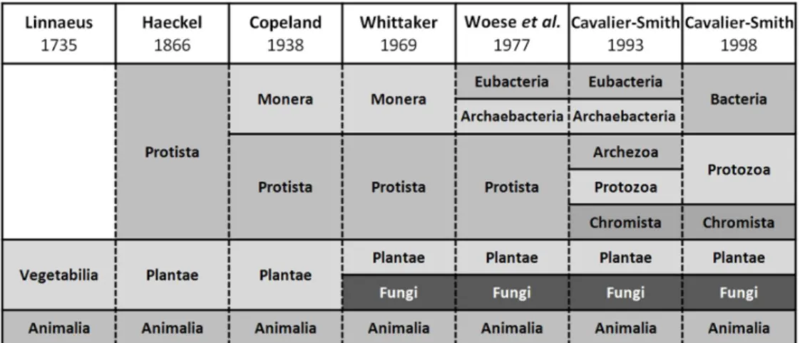
kingdom Vegetabilia, by Linnaeus in 1735 (Fig. 1.1) [24]. This persisted through
the creating of the three- and four-kingdom systems of Haeckel, in 1866 [25], and Copeland, in 1928 [26], respectively. Only with the creation of the five-kingdom system by Whittaker, in 1969 [27], fungi were classified in their own kingdom, a concept kept in classification systems proposed afterwards [28, 29]. In the six-kingdom system proposed by Cavalier-Smith, in 1998 , only “true fungi” are
considered to belong the kingdom Fungi, being oomycetes and slime molds placed
in the kingdoms Chromista and Protozoa, respectively [30, 31] (Fig. 1.1). Their
independence from plants is now strongly supported by molecular evidence that prove that plants constitute an independent evolutionary lineage, while fungi are more closely related to animals, constituting a monophyletic group [32]. They both share the common feature that their flagellate cells propel themselves with a single
posterior flagellum (opisthokonts), as opposed to plants and other eukaryotes, in
which these cells propel themselves with one or more anterior flagella [33].
Fig. 1.1. Schematic representation of the presence of the kingdom Fungi along the different
classification systems proposed by Linnaeus, Haeckel, Copeland, Whittaker, Woese et al.
6
The kingdom Fungi traditionally comprised four phyla of “true fungi”,
namely Chytridiomycota, Zygomycota, Ascomycota and Basidiomycota [2, 4]. This
classification has now been revised and, in the most recent internationally agreed
phylogenetic classification proposed by Hibbett et al. [34] (Fig. 1.2.), the former
Chytridiomycota and Zygomycota were considered as basal fungal lineages and are
subdivided in several phyla and subphyla. Ascomycota and Basidiomycota, the
largest phyla within fungi, are grouped together in the subkindom Dikarya.
Interestingly, Microsporidia a group of unicellular parasites of animals and protists
with highly reduced mitochondria were included in the kingdom Fungi. Overall, the
classification proposed by Hibbett et al. accepts, within the kingdom Fungi, one
subkingdom, seven phyla and ten subphyla [34].
Fig. 1.2. Phylogeny and classification of the kingdom Fungi according to Hibbett et al. [34].
Branch lengths do not intend to be proportional to genetic distances.
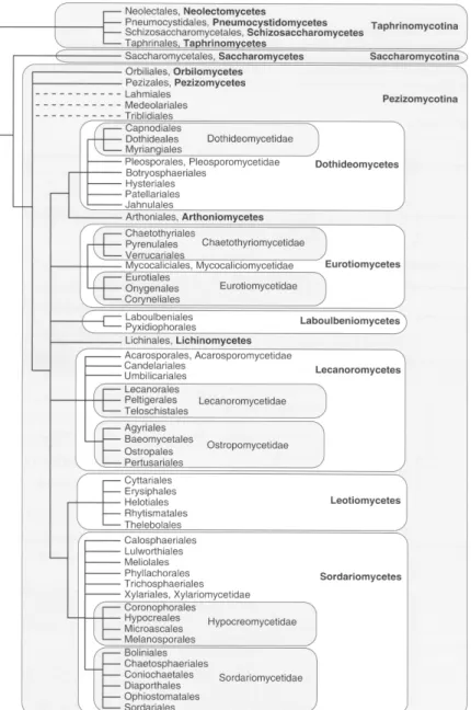
The phylum Ascomycota constitutes the most diverse group of fungi,
counting with ca. 60,000 known species and the largest number of clades of fungi
with septate hyphae. Three main subphyla are proposed in this classification:
7
Fig. 1.3. Phylogeny and classification of the phylum Ascomycota according to Hibbett et al.
[34]. Branch lengths do not intend to be proportional to genetic distances.
The basal group Taphrinomycotina comprises, among others, plant parasites,
the human pathogen Pneumocystis jirovecii [35] and the fission yeast
Schizosaccharomyces pombe, a significant model organism in cell biology of
eukaryotes [36, 37]. The most remarkable members of the subphylum
8
species of the genus Candida, which constitute important human pathogens [38].
The filamentous Ascomycota are contained in the subphylum Pezizomycotina that
comprises a wide diversity of fungal species and life-styles. Remarkably, included
in this group are e.g. Penicillium rubens (Eurotiomycetes, Eurotiales,
Trichocomaceae) from which the antibiotic penicillin was discovered [14, 15],
Fusarium spp. (Sordariomycetes, Hypocreales, Nectriaceae), important pathogens
of plants [39], Neurospora crassa (Sordariomycetes, Sordariales, Sordariaceae)
and Aspergillus nidulans (Eurotiomycetes, Eurotiales, Trichocomaceae) the last two considered model filamentous fungi in cell biology studies [40-42].
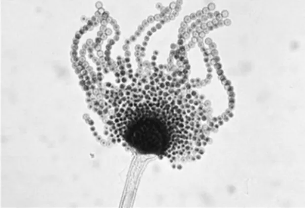
The genus Aspergillus has around 250 known species [43], with significant
roles in natural ecosystems and in human economy, including important plant and human pathogens, producers of mycotoxins, enzymes, organic acids and secondary metabolites [44]. The name of the genus comes from the asexual reproductive structures of these fungi, that resemble a device used to sprinkle holy water by the
Roman Catholic clergy, the asperges [11] (Fig. 1.4). Representative members of
this genus are A. fumigatus, A. niger, A. oryzae, A. terreus and A. nidulans. Their
importance justifies that, at the present, the genomes of several species are completely sequenced and available [40, 45-48]. Throughout this thesis the focus of
discussion will be the biology of the filamentous fungus A. nidulans and, whenever
appropriate, of the model yeast S. cerevisiae or other relevant, related species.
Fig. 1.4. Asexual reproductive structure (conidiophore) of Aspergillus flavus. Its shape
9
The fungal cell has the same basic constitution as other eukaryotic cells [4].
The genetic material is retained in a nuclear envelope, perforated by numerous pore complexes. The nucleoplasm is ribosome-free and is packed with the chromatin, and might contain one or two nucleoli [1]. Fungi also contain several cytoplasmic organelles, such as peroxisomes, vacuoles, endoplasmic reticulum, Golgi apparatus and mitochondria, although the latter differ from other eukaryotic cells for being more elongated, larger and for having their cristae arranged as lamellae parallel to the longitudinal axis [2, 4] (Fig. 1.5A). They also possess and endomembrane system able to divide the cell into functional and structural compartments for the synthesis, breakdown and transport of material into, within and out of the cell [49]. A cytoskeleton, composed of three types protein filaments (microtubules, microfilaments and intermediate filaments) is responsible for providing structure and organization to the cytoplasm, while also conferring shape to the cell [50].
A variety of cell shapes are present in the kingdom Fungi, when considering
the diversity of species, life-styles and complex life cycles these organisms can assume. Considering their vegetative growth, one could roughly distinguish two main cell morphologies, the yeast form, spherical, ellipsoidal or cylindrical; and the filamentous form, chains of cylindrical cells that form hyphae (Fig. 1.5) or pseudohyphae [51]. Some fungi can assume both morphologies in its life-cycle, and are usually called dimorphic.
Highly polarised, filamentousgrowth of hyphae [52] is possible due to the
10
filamentous fungi in colonising many types of substrates, taking advantage of the nutritional sources available.
Fig.1.5. A) Transmission electron micrograph of the near median section through hyphal tip
11
Vegetative growth can stop and the fungi can enter into cycles of asexual or sexual development, in each it produces spores for propagation or resistance in adverse environmental and nutritional conditions (Fig. 1.6). Filamentous
Ascomycota, herein exemplified by A. nidulans, undergo a complex process of
asexual development. It is usually triggered by many environmental conditions,
such as exposition to air, light or temperature [54, 58]. It begins with the formation of specialized aerial hyphae, the stalk, which extends vertically and can differentiate into a conidiophore. After polarised growth, the stalk undergoes isotropic growth at the tip, forming a vesicle. A series of specialised cells bud from the vesicle, forming a first layer of metulae. Each metula then originates two phialides that will then produce by mitoses the asexual spores, called conidia. Long chains of conidia can be produced per each phialide, and a single conidiophore can produce up to 100,000 conidia, that can spread in the environment carried up by the air [54, 58]. Typically,
A. nidulans form conidia in solid substrates, but it can also be observed on submerged cultures, usually under stress or upon nutrient limitations [58].
Under harsh environmental conditions, such as nutrient starvation, some
fungi can undergo sexual development, producing spores that are extremely
resistant. Sexual spores are produced through meiosis, which guaranties higher genetic variability compared to the parental cells, another strategy to overcome
environmental adversities [4]. In the case of Aspergillus species, not all have a
known sexual stage; about one third can reproduce sexually, such as A. nidulans,
A. fumigatus, A. flavus, A. parasiticus and A. nomius [54, 59-65]. These species can be classified as heterothallic, in case they need compatible mating partners, or homothallic, when this is not required. Ascomycota produce their sexual spores inside specialized cell called asci, from where the name of the phylum is derive [4].
12
entire population of dikaryotic cells that will produce a single fruiting body containing the ascospores-forming asci. These are formed by the fusion of the nuclei (karyogamy), which is followed by meiosis and subsequent mitosis, generating eight haploid nuclei that will be separated by membranes, forming eight ascospores. A final mitotic event occurs, generating the mature, binucleate
ascospores [54]. In Aspergillus nidulans, asci and ascospores are produced in a
closed fruiting body, called cleistothecia, which can reach 125-200 µm in diameter [54]. Spore dispersal occurs after mechanical rupture of the cleistothecia. Other types of fruiting bodies can also be produced by filamentous fungi, such as
apothecia, perithecia and pseudothecia, in Ascomycota, and basidiocarps, such as the
commonly named mushrooms, in Basidiomycota [2, 4].
Fig.1.6. Schematic representation of Aspergillus nidulans life cycle.
After being generated by the reproductive structures, both conidia and ascospores enter a state of dormancy, period in which they are relatively resistant to
various stress conditions, e.g. UV radiation, drought, high temperatures [54]. Once
the spores are dispersed and reach an appropriate substrate with adequate water and
nutritional sources, dormancy is broken and germination occurs. Emergence of a
13
1.2. The fungal cell envelope
The following pages give more detailed information on the structure and biosynthesis of the fungal plasma membrane and cell wall, relevant for the understanding of the work discussed in the following chapters. The knowledge herein presented is mainly based on the model yeast Saccharomyces cerevisiae; nevertheless, aspects of the biology of the filamentous fungus Aspergillus nidulans or other related species will highlighted.
1.2.1. Plasma membrane
The fungal plasma membrane, as all biological membranes, is composed of a thin film of lipids and proteins [1]. The lipids are arranged as a double layer, and serve as a relatively impermeable barrier for most water-soluble molecules [4]. The most abundant lipids in the plasma membrane are phospholipids, which usually contain a glycerol unit with two fatty acids, a phosphate group, and a simple organic molecule such as choline, inositol, serine or ethanolamine. Their shape and amphifilic nature,
i.e. polar head groups and long hydrophobic tails, allow the spontaneous formation
of bilayers in aqueous environments, where they tend to aggregate in ways that the hydrophobic tails face the interior and the hydrophilic head groups are exposed to the aqueous exterior [1].
Fatty acids are the basic elements of complex lipids, being incorporated
into e.g. phospholipids and sphingolipids [67]. The de novo synthesis of fatty acids
that occurs in the cytoplasm (it can also occur in the mitochondria [68]) begins with the formation of malonyl-CoA from acetyl-CoA, derived from citrate or acetate, in a reaction catalysed by an acetyl-CoA-carboxylase [69]. The fatty acid synthase
complex utilizes malonyl-CoA as a two carbon building block for the de novo
synthesis of saturated fatty acids. Aspergillus nidulans has two genes coding for the
subunits of the fatty acid synthase complex, namely fasA and fasB [70]. Further
elongation steps can occur in the endoplasmic reticulum by the addition of two carbons from malonyl-CoA [67], to generate very long chain fatty acids, that can be
utilised e.g. in the synthesis of sphingolipids [71]. The desaturation and
14
Together, this complex biosynthetic pathway allows a high diversity of molecule types. Importantly, the level of fatty acid saturation in the phospholipids will determine the membrane fluidity [1]. Membranes enriched in unsaturated fatty acids are more fluid than those with a high number of saturated fatty acid moieties [72].
Fatty acids are one of the substrates for the production of phosphatidic acid,
a central metabolite in the synthesis of phospholipids [67]. It can be used in the
biosynthetic pathways that will lead to the production of the most abundant
phospholipids in fungi: phosphatidylcholine, phosphatidylethanolamine,
phosphatidylinositol and phosphatidylserine [73]. This diversity of phospholipids provides an important feature to the plasma membrane: the lipid bilayer is
asymmetrical, i.e. the composition of phospholipids regarding their head groups
varies from the inner to the outer monolayer [1]. Since these head groups have different net charges, significant difference between the overall charges of each bilayer surface is observed.
Additionally to phospholipids, sterols and sphingolipids also play
fundamental roles in the structure of the fungal plasma membrane.[67] Ergosterol
is the major sterol present in the plasma membrane of a great number of fungal
species,[67, 74, 75] including Aspergillus nidulans.[76] It is very similar, in
structure and function, to the mammalian cholesterol, and it also accounts for the fluidity of the plasma membrane [4]. Its synthesis involves a complex pathway that,
in yeast, comprises almost thirty enzymes in the yeast S. cerevisiae (Fig. 1.7) [67].
15
enzymes in the pathway [83], generating sterol intermediates (e.g. lanosterol,
zymosterol, fecosterol and episterol) and, ultimately, ergosterol [67]. Despite the current knowledge in ergosterol biosynthesis in yeast, this pathway can present significant differences depending on the fungal species; for instance, alternative
branches and sterol intermediates have been identified in A. fumigatus [74].
Fig.1.7. Schematic representation of ergosterol biosynthesis in Saccharomyces cerevisiae.
Adapted from [67].
Sphingolipids constitute another class of plasma membrane lipids that
16
residues, thus being classified as complex sphingolipids [84]. The sphingolipid
biosynthetic pathway is characterised in greater detail for the yeast S. cerevisiae
(Fig. 1.8); however, knowledge of this pathway in filamentous fungi such as A.
nidulans is rather more limited (Fig. 1.9).
Fig.1.8. Schematic representation of sphingolipids biosynthesis in Saccharomyces
cerevisiae. Adapted from [67].
17
fatty acid to yield dihydroceramide and phytoceramide, respectively, a reaction catalysed by ceramide synthases [90, 91]. Addition of an inositolphosphate group renders the first complex sphingolipid, inositolphosphoryl ceramide [86, 92]. Mannose-inositolphosphoryl ceramide can be formed by the mannosylation of inositolphosphoryl ceramide [93], and addition of a second inositol phosphate group to the former yields mannosediinositolphosphoryl ceramide [94], the most complex product of this pathway. The long chain bases dihydrosphingosine and phytosphingosine can also enter an alternative route to generate phosphorylated sphingoid bases [71]. Several intermediates in the sphingolipids biosynthetic pathway can play important functions in the cell, other than being structural components of the membrane, as discussed ahead in this thesis (see Section 1.3).
Fig.1.9. Schematic representation of sphingolipids biosynthesis in Aspergillus nidulans.
Adapted from [89].
18
they have been implicated in protein sorting, secretion, endocytosis, and cell
polarity. In the filamentous fungus A. nidulans, these domains are present in
septation sites and particularly at the tip of the hyphae, where they likely play a vital role in the establishment of polarity and directionality of hyphal growth [91, 96].
Besides playing an important structural function in cells, sphingolipids have already been described to play numerous roles as signaling molecules in eukaryotes [97-100]. Sphingolipids intermediates, especially sphingoid bases and their phosphate species, and ceramides involved in the regulation of cell growth,
differentiation, and programmed cell death. In S. cerevisiae, sphingoid bases are
also involved as a second messenger in the stress response to cell wall damage [71]. The fungal plasma membrane is one of the main targets of the available
array of antifungal drugs. Ergosterol can only be found in fungi and, due to its
essential role in maintaining membrane integrity and cell viability, its biosynthetic pathway has been considered a major target in antifungal drug development [101, 102]. Azoles, such as miconazole, ketoconazole, fluconazole, itraconazole and
voriconazole, inhibit the 14α-demethylase, which leads to depletion of ergosterol
and accumulation of sterol precursors (e.g. lanosterol) [102, 103]. Despite being the
19
especially at later steps in the pathway, justifies the current interest in exploring them for the development of new therapeutic agents. Many natural compounds can inhibit sphingolipids biosynthesis in fungi. Myoricin and fumonisins, for example, are inhibitors of the early steps in sphingolipids biosynthesis, while aureobasidins inhibit later steps in the pathway [101].
1.2.2. Cell wall
The fungal cell wall accounts for a great percentage of the cell dry weight (15-30%), and is responsible for allowing cell morphogenesis and maintaining its shape, counteracting the turgor pressure and protecting the plasma membrane against mechanical damage [106, 107]. Seen by electron microscopy, the cell wall presents an electron-lucid inner layer that represents the skeletal layer composed mainly of
polysaccharides, predominantly chitin and 1,3-β-glucans. Aspergillus nidulans also
contains amorphous 1,3-α-glucans embedded within 1,3-β-glucan and chitin fibrils,
absent in the yeast cell wall [106, 108]. They form long, poorly branched 1,3-β
-glucans form an elastic network that is continuously stretched under normal osmotic conditions. These glucans contain nonreducing ends that may serve as acceptor sites
for chitin and 1,6-β-glucans, overall serving as a support for the outer electron-dense
layer of mannoproteins [106] (Fig. 1.10).
Fig.1.10. Representation of the cell wall of filamentous fungi, showing its main components:
20
The enzymes involved in the synthesis of chitin, 1,3-α-glucans and 1,3-β
-glucans are known to be localised at the plasma membrane; the glycan chains are extended in the cytosolic side of the membrane, while being extruded to the periplasmic region, where they become branched and are linked to the existing polysaccharide structure. The cell wall proteins follow the secretory pathway and, once outside the cytoplasm, are integrated to the cell wall. The majority of the enzymes and reactions that occur extracellularly are not known, being the intracellular cell wall biosynthesis better understood [106, 108].
Chitin is a polymer of linear chains of 1,4-β-N-acetylglucosamine residues,
and is synthesised and deposited in the cell surface by a group of enzymes called chitin synthases [10, 107]. The first and rate-limiting step in chitin synthesis is catalysed by glutamine-fructose-6-phosphate transaminase, an enzyme that produces glucosamine 6-phosphate from glutamine and fructose 6-phosphate [110]. Three
more steps yield N-acetylglucosamine 6-phosphate, N-acetylglucosamine
1-phosphate and, finally, UDP-N-acetylglucosamine, the substrate of chitin synthases.
Fungal chitin synthases can be classified into seven classes, according to their amino
acid similarities [111]. Saccharomyces cerevisiae only has three genes coding for
chitin synthases [106], while filamentous fungi can have many more (e.g. A. oryzae,
with eleven genes) which might reflect the greater content of this polymer in the cell
walls of filamentous fungi when compared to yeasts [107]. Aspergillus nidulans has
chitin synthases from the seven classes, and a total of eight genes coding for this type of enzymes [108, 111]. Despite the great number of chitin synthase genes in fungi, they are usually not redundant, and can play distinct roles throughout the organism’s life cycle. For instance, in yeast, Chs1 acts as a repair enzyme during cell separation; Chs2 is responsible for the synthesis of chitin at the septum; and Chs3 functions as the main source of the polymer in the mother cell, septum and
during cell wall stress. In A. nidulans, ChsA is expressed specifically during the
21
probably facilitate interaction of the synthases with the actin cytoskeleton and their correct localisation at the hyphal tip, being involved in cell wall synthesis at this site.
With few exceptions, all filamentous and Ascomycota have genes coding for chitin
synthases with myosin-motor like domains, reinforcing the importance of these type synthases in the polarised growth of hyphae [107].
Glucans are the more abundant polysaccharides in the cell wall [107]. The
main one, 1,3-β-glucan, form long, poorly branched chains, covalently linked to
chitin and other cell wall components. 1,3/1,4-β-glucans and 1,6-β-glucans are also
present, playing minor roles in linking polysaccharides and cell wall proteins to the
main 1,3-β-glucans chains [106]. While the yeast S. cerevisiae has more than one
gene involved in 1,3-β-glucan biosynthesis, filamentous Ascomycota have only one
(fksA in A. nidulans) [107, 108, 116]. The glucan synthase complex catalyses the
addition of UDP-glucose units, creating a linear chain of increasing length. The main source of UDP-glucose is the pentose phosphate pathway, but alternative pathways can be used to generate this substrate [106]. Although lacking in yeast,
most filamentous Ascomycota contain also 1,3-α-glucans in their cell walls. Their
genomes contain one or more genes coding for 1,3-α-glucan synthases, e.g. one in
the rice pathogen Magnaporthe griseae, two in N. crassa, three in A. fumigatus and
five in A. niger [107]. Aspergillus nidulans has two genes coding for 1,3-α-glucan
synthases, agsA and agsB [108, 117, 118]. The synthesis of other important glucans,
such as 1,6-β-glucans, responsible for cross-linking of chitin and cell wall proteins
to the main 1,3-β-glucan chain, is not completely understood, neither in yeast nor
filamentous fungi [106, 108].
Cell wall proteins constitute up to 10% of the fungal cell wall biomass
[119], and most of their known functions include cell wall synthesis, integrity or remodelling or can display other functions, such as hydrophobins, adhesion, ROS detoxification and iron acquisition [106-108]. The majority of the cell wall proteins
are linked to the 1,3-β-glucan skeleton by a glycosylphosphatidylinositol (GPI)
anchor covalently linked to 1,6-β-glucans and usually extending their effector
22
found in the skeletal inner layer of the cell wall and are thought to be directly linked
to the 1,3-β-glucan chains through an ester linkage between a hydroxyl group from
1,3-β-glucan and a deaminated glutamine residue from internal repeats in their
peptide sequence. Other non-covalently linked proteins can also be found associated
to the cell wall by e.g glycan-binding motifs, ionic interactions or disulphide bonds
with other cell wall proteins. There are over 20 cell wall proteins in S. cerevisiae;
however information on their abundance and role in A. nidulans is rather limited
[106, 108, 119].
Both yeasts and filamentous fungi possess genes coding for chitinases and glucanases that are able to cleave and, in some cases, re-form the bonds within and between the preformed cell wall polysaccharides [120]. These activities are crucial for the re-modelling of the cell wall, especially during growth and morphogenesis.
As a unique component of the fungal cell, the cell wall has also been explored as an important target for antifungal drugs. Echinocandins, such as
caspofungin, micafungin and anidulafungin, are noncompetitive inhibitors of 1,3-β
-glucan synthases, preventing the incorporation of glucose monomers to the -glucan chain [121]. They emerged as promising new therapies alternative to the traditional drugs that target ergosterol and its biosynthesis. However, new reported cases of resistance to echinocandins are emerging [122], highlighting the need of new classes of antifungal therapies [123]. Polyoxins and nikkomycins, on the other hand, are
competitive inhibitors of chitin synthases. Defective synthesis of either 1,3-β
-glucans or chitin leads to weakened cell walls and, consequently, fungal cell lysis [101, 124].
Upon damage to the cell wall, either chemical or physical, fungi respond by activating several genes involved in its biosynthesis, creating conditions that allow them to re-establish its integrity. This is regulated by a signalling cascade better
understood in S. cerevisiae, called cell wall integrity (CWI) pathway [125, 126]
(Fig.1.11). This pathway is also known to be present in a variety of filamentous
fungi, such as A. nidulans [126] and A. fumigatus [127], but many knowledge gaps
23
stress the cell wall, such as calcofluor white (chitin antagonist), Congo red (1,3-β
-glucan-binding dye) and cell wall lytic enzymes, or by antifungal agents that perturb the cell wall synthesis and composition. However, it can also be activated during growth, at regions of the highly polarised growth, where the cell undergoes the greatest wall stress, by hypo-osmotic shock and oxidative and heat stress [125]. Overall, these different stimuli culminate in plasma membrane stretch, considered the physical stimulus that ultimately activates the CWI pathway. This is possible due to the presence of sensors in the cell surface that detect and transmit alterations in the plasma membrane tension.
Fig.1.11. Schematic representation of the cell wall integrity pathway of Saccharomyces
24
Five sensors are described for yeast, Wsc1-3, Mid2 and Mtl1 [125]. These sensors are transmembrane proteins, imbedded in the plasma membrane and bound to the cell wall glucans. The information of cell wall stress is received by the surface sensors and transmitted to a small G-protein (Rho1) through the GEFs Rom1-2. Rho1 then activates a protein kinase in the pathway (Pck1) that is then responsible for the phosphorylation and activation of a mitogen-activated protein (MAP) kinase cascade. These MAP kinase cascade is thought to amplify the low signal initiated at the cell surface [125]. The downstream effector of this cascade, Mpk1 in yeast, is located mainly in the nucleus and is responsible for the activation
of the transcription factors (e.g. Rlm1, Swi4-Swi6 complex) that will ultimately
induce the transcription of the cell wall related genes and also of the gene coding for the kinase, in a positive-feedback that is thought to intensify the signal. The CWI pathway is not as linear as described above, and its composing elements can perform other functions upon cell wall stress conditions. For example, Rho1 induces the
activity of the 1,3-β-glucan synthase and influences actin dynamics through
regulation of the formins (actin-binding proteins) Bni1 and Bnr1, while Pck1
recruits chitin synthases to the cell surface and seems to induce N-glycosylation of
cell wall proteins in the endoplasmic reticulum lumen. Interfaces of the central CWI pathway with other signalling pathways is also observed, including calcium signalling, sphingolipid and phosphoinositide metabolisms and Tor protein kinases [125].
Many of the genes involved in the CWI pathway in yeast have homologs in filamentous fungi. However, most likely due to great differences in cell wall composition and growth, the CWI pathway of filamentous fungi is not entirely
identical to that of S. cerevisiae, and need to be further investigated. Up to know,
for example, only two membrane sensors have been described and characterized for
A. nidulans, WscA and WscB, and a third protein, homolog to the yeast Mid2, is also thought to play a role in the CWI pathway of this filamentous fungus [126, 128, 129]. Other elements of the pathway, such as the G-protein RhoA, the protein kinase PkcA, the MAP kinases BckA, MkkA and MpkA, and the transcription factor
25
signalling cascade in filamentous fungi is much different from that of the yeast
[126]. While in S. cerevisiae the majority of the genes involved in cell wall
biosynthesis is regulated by the CWI pathway, in A. nidulans the homolog pathway
seem to be essential only for the regulation of the genes involved in 1,3-α-glucan
synthesis and partially for transcription of the first step in chitin synthesis upon cell wall stress [125, 126, 128]. All other cell wall related genes are thought to be
regulated by an alternative, yet uncharacterised, CWI pathway in A. nidulans,
reinforcing the need for further studies in the characterisation and regulation of cell wall integrity pathways in filamentous fungi [130-133].
Fig.1.12. Schematic representation of the cell wall integrity pathway of Aspergillus nidulans.
26
1.3. Ionic liquids
The following paragraphs provide only general information on ionic liquids and some of the work performed on the understanding of their toxicity, and present examples of interesting biological applications of these compounds. Emphasis is given whenever possible to studies focussing eukaryotic organisms which are more relevant for the present thesis.
Ionic liquids have been described as molten salts that have, by definition, a melting point below 100 °C [134]. They are entirely ionic in nature, and usually consist of a large asymmetrical organic cation associated with a polyatomic anion that may be either organic or inorganic. In the design of ionic liquids, the asymmetry of the cation or anion is preferred over symmetrical species, since the former do not tend to pack so easily, therefore rendering salts with a lower melting point [134]. Structural modifications can be made either to the anion, to the cation, or in substituents on these two species. Therefore, millions of formulations become possible and, by altering their cationic or anionic components, their properties can be tailored [134,
135]. Their physical and chemical properties (e.g. melting point, viscosity,
solubility or hydrophobicity and chemical polarity or hydrogen bonding ability, respectively) can be tuned to best suit a specific process. The tuneability of such properties rendered ionic liquids as an excellent choice as alternative organic solvents in many chemical processes. They already found application in many reactions [136], as solvents [136, 137] and catalysts [138], and show great potential in electrochemistry [139, 140].
27
of compounds is, therefore, a priority, and should be taken in consideration for the conscious design of novel ionic liquids [145].
The current interest in ionic liquids toxicity began in the early 2000’s, with works like those performed by Pernak and co-workers, which developed new cationic surfactants with benzimidazolium or pyridinium cores and evaluated their antimicrobial properties, defined by their minimal inhibitory and lethal concentrations against bacterial and yeast strains relevant for human health [146]. The evaluation of the antimicrobial activity of
1-alkoxymethyl-(3-nicotionylaminomethyl)benzimidazolium,
1-alkoxymethyl-3-(1-benzimidazol-methylamino)pyridinium,
1-alkoxymethyl-3-[1-(benzotriazol-1-yl)methyl-amino]pyridinium chlorides and N,N’-bis[3-(1-alkoxymethyl)pyridinium
chloride]methylenediamine revealed that their toxicity increased with the elongation of the alkoxy chain, that varied between 2 to 12 carbon atoms. This constituted one of the first systematic studies on the toxicity of ionic liquids, and served afterwards as basis for evaluating the toxicity of many newly synthesised imidazolium- and pyridinium-based compounds, including 1-alkoxymethylcarbamoylpyridinium chlorides [147], methylimidazolium chlorides and bromides;
1-alkyl-3-hydroxyethyl-2-methylimidazolium chlorides [148] ,
1-alkoxymethyl-3-methylimidazolium chlorides, tetrafluoroborates and hexafluorophosphates [149], 1,3-dialkoxymethylimidazolium chlorides [150], 1-alkyl- and alkoxy-methylimidazolium lactates [151], alkoxymethyl-3-hydroxypyridinium and 1-alkoxymethyl-3-dimethylaminopyridinium chlorides [152]. The data revealed a clear trend towards a stronger toxic effect of ionic liquids with the increase in the length of the alkyl or the alkoxy side chain. The effect of the anions on the observed toxicities seemed to be secondary to the effect of the cations, but their broad diversity did not allow a conclusive analysis.
These initial studies inspired an escalating number of investigations of the toxic effects of several ionic liquids, expanding the research for other biological models. For example, the toxic effects of ionic liquids with 1-alkyl-3-methylimidazolium as the cation were analysed against leukemia and glioma rat cell
28
[155] and the toxicity of pyridinium-based ionic liquids has been analysed using the
freshwater snail Physa acuta [156], the zebra mussel Dreissena polymorpha [157],
and the green algae Pseudokirchneriella. subcapitata [158], focussing on the effect
of the alkyl side chain on the pyridinium cation. The effects of imidazolium- and
pyridinium-based ionic liquids in the bioluminescence of the bacterium Vibrio
fischeri was analysed [159]. Bacterial bioluminescence is directly linked to cellular respiration, so decrease in luminescence is indicative of toxicity. A correlation between toxicity and the 1-octanol/water partition coefficient of the cation was proposed, suggestive of higher lipophilicity with the increase in the length of the alkyl side chain and, therefore, greater interaction with the cell surfaces.
Other cationic head groups were also subject to investigations on their toxic effects, such as the aromatic quinolinium-based cations. Despite rarely studied, these ionic liquids showed great antimicrobial properties and cytotoxicity potential that increased with the elongation of the substituted alkyl chain [144, 160]. The effects of ionic liquids with nitrogen-containing alicyclic cations, namely
pyrrolidinium, piperidinium and morpholinium, were also investigated, e.g. their
aquatic toxicity against the zebrafish (Danio rerio) [161], and their cytotoxicity
against mammal cell lines [162, 163]. It was observed that increasing the number of
carbon atoms in the alicyclic ring generally increases toxicity, e.g. the piperidinium
cation (six-member rings) was more toxic than the pyrrolidinium cation (five-member rings) [163]. The morpholinium ionic liquids, due to the incorporation of an oxygen atom in the ring, were the least toxic of these alicyclic cations. Furthermore, the non-aromatic head groups appeared to be generally less toxic than their aromatic equivalents [162, 163]. Their lower toxicity, relative to the corresponding aromatic rings, was generally evident in the bioassays with models,
such as V. fischeri, the algae Scenedesmus vacuolatus and the aquatic plant Lemna
minor [164]. . As observed for imidazolium- and pyridinium-based ionic liquids,
29
Quaternary ammonium salts have already been known and studied for a considerable time, and were widely explored in numerous applications, such as disinfectants, surfactants, antistatic agents and catalysts [166]. Their properties depend on the chain length and functional groups, and on the anion [167]. Pernak and co-authors were the firsts to consider their antimicrobial activity against relevant strains, which was, as expected, governed by the length of the alkoxy and alkyl side chains [168, 169]. The toxicity of ammonium ionic liquids against other biological
models were also tested, including the bioluminescent bacterium V. fischeri [170],
the fresh water crustacean Daphnia magna and the algae P. subcapitata [171, 172],
zebrafish [172], and cell lines [165]. The toxicity trends previously described for
other cationic head groups were also observed, i.e. the highest toxicity was observed
for those carrying the longest alkyl chains and could be well correlated with the cation lipophilicity.
Some of the most interesting groups of quaternary ammonium-based ionic liquids are those containing the cholinium cation (2-hydroxyethyl-trimethylammonium cation). Its combination with benign anions constituted a major advance in the design of biocompatible ionic liquids [173-180]. The low toxicity of many cholinium ionic liquids have been demonstrated, including those
carrying as anions, e.g. saccharinate and acesulfamate [181], dimethylphosphate
[182], phosphate-based anions [183], lactates [184], and alkanoates [185]. The last
were analysed for the first time against filamentous fungi of the genus Penicillum,
and included a range of linear alkanoate anions and two structural isomers [185]. Their toxicity increased with the chain length of the anion, being the branched isomers less toxic than the corresponding linear ones with the equal number of carbon atoms. These cholinium alkanoates displayed, also, high biodegradability potential and great solvent ability [186].
30
assessment, still seldom investigated. Some halides, e.g. with the
tetrabutylphosphonium cation, showed levels of toxicity against the freshwater snail
P. acuta that were comparable to imidazolium-based ionic liquids carrying the same
chain length [156]. The antimicrobial properties of a series of alkyltrihexylphosphonium halides demonstrated a significant role of the structure of the cation in their toxicity, since the antimicrobial activity of the phosphonium chlorides decreased for the longest alkyl chains (8 to 14 carbon atoms) [188]. The
apparently high toxicity of tetraalkylphosphonium halides against V. fischeri and
D. magna [170, 171] and P. subcapitata [171, 189] was also demonstrated, as well
as their cytotoxicity against human and rat cell lines [165, 190]. The lack of systematisation in these studies, however, did not allow a conclusive rationalisation, a general problem when evaluating the toxicity of the highly diverse groups of ionic liquids [145].
The better knowledge of ionic liquids toxicity is crucial to identify their mechanisms of action and further advance towards their conscious design. Investigations on ionic liquids mechanisms of toxicity are, however, rather limited. The hydrophobic interaction between ionic liquids and biological membranes has been proposed as a main mechanism of toxicity of ionic liquids, largely supported by the observation that their toxic effects increase with the elongation of the side chains. This non-specific toxicity, called baseline toxicity or narcosis [191], appear correlated with the 1-octanol/water partition coefficients, suggestive of an increase in either the cation or anion lipophilicity with the increase in the number of carbons of the side chains [164], leading to greater probability of interaction with biological
membranes. Liposomes, i.e. phospholipid vesicles, were used to assess the effects
31
Investigating ionic liquids mechanisms of toxicity is not only necessary for a complete understanding of their environmental impact but is also necessary for the development of new green, environmentally friendly processes. As an example, the biocompatible and biodegradable cholinium alkanoates have been observed to have a great solvent ability towards the plant polymer suberin [185, 186]. Cholinium hexanoate was successfully employed in the extraction of suberin from cork, partially preserving the native structure of this polymer, which allowed, for the first time, the formation of suberin films with antibacterial properties [186, 196-198]. The deeper knowledge on ionic liquids toxicity can also can open doors for exploring their use in new biological applications. A good example of their early applications was the innovative use of 1-alkoxymethyl-3-methylimidazolium tetrafluoroborate in embalming and tissue preservation [199] and as a wood preservative [200], exploring their antimicrobial activity as a replacement for the highly volatile and toxic formalin.
The potential use of ionic liquids as drugs for important etiological agents is also under current investigation. For example, imidazolium-based ionic liquids have
been explored for their potential in combating Plasmodium falciparum [201] and
Trypanosoma cruzi [202], the causative agents of malaria and Chagas disease, respectively. Another good example is choline-geranate that has been proposed as a coadjutant in the treatment of wound infections, due to its antimicrobial activity, minimal toxicity to epithelial cells as well as skin, and effective permeation enhancement for drug delivery [203].

![Table 2.1. Minimal inhibitory and fungicidal concentrations (MIC and MFC, respectively) of the alkyltributylphosphonium chlorides, [P 4 4 4 n ]Cl, defined for Aspergillus nidulans](https://thumb-eu.123doks.com/thumbv2/123dok_br/15764175.640103/91.774.262.474.398.676/minimal-inhibitory-fungicidal-concentrations-respectively-alkyltributylphosphonium-chlorides-aspergillus.webp)
![Fig. 2.2. A) Minimal inhibitory and fungicidal concentrations (MIC and MFC, respectively) of the alkyltributylphosphonium chlorides, [P 4 4 4 n ]Cl, where n = 1, 3 - 8, 10, 12 or 14, defined for Aspergillus nidulans](https://thumb-eu.123doks.com/thumbv2/123dok_br/15764175.640103/93.774.126.610.185.818/minimal-inhibitory-fungicidal-concentrations-respectively-alkyltributylphosphonium-chlorides-aspergillus.webp)
![Fig. 2.3. Membrane integrity assay. Conidia were treated with 100 mM of alkyltributylphosphonium chlorides, [P 4 4 4 n ]Cl (n = 1, 4, 8 or 12), during one hour and stained with propidium iodide, PI](https://thumb-eu.123doks.com/thumbv2/123dok_br/15764175.640103/94.774.213.612.106.860/membrane-integrity-conidia-treated-alkyltributylphosphonium-chlorides-stained-propidium.webp)
![Table 2.2. Percentage of membrane-damaged conidia after one hour of incubation with alkyltributylphosphonium chlorides, [P 4 4 4 n ]Cl (n = 1, 4, 8 or 12), obtained as (number of propidium iodide-stained conidia / total number of co](https://thumb-eu.123doks.com/thumbv2/123dok_br/15764175.640103/95.774.81.654.580.734/percentage-membrane-damaged-incubation-alkyltributylphosphonium-chlorides-obtained-propidium.webp)
![Fig. 2.4. Cell wall damage assay. Conidia were treated with 100 mM of alkyltributylphosphonium chlorides, [P 4 4 4 n ]Cl (n = 1 or 12), during one, two or four hours and stained with Calcofluor White M2R (CFW)](https://thumb-eu.123doks.com/thumbv2/123dok_br/15764175.640103/97.774.81.652.259.869/damage-conidia-treated-alkyltributylphosphonium-chlorides-stained-calcofluor-white.webp)
![Fig. 2.5. Scanning Electron Microscopy (SEM) analysis of conidia treated with 100 mM of alkyltributylphosphonium chlorides, [P 4 4 4 n ]Cl (n = 1, 8 or 12), during two hours of incubation](https://thumb-eu.123doks.com/thumbv2/123dok_br/15764175.640103/99.774.104.629.103.884/scanning-electron-microscopy-analysis-conidia-alkyltributylphosphonium-chlorides-incubation.webp)
