Methods to preserve potentially toxigenic fungi
Lucas Costa Guimarães
1, Ana Paula Fernandes
2, Sara Maria Chalfoun
3,
Luís Roberto Batista
21
Instituto de Ciências da Saúde, Universidade Paulista, Brasília, DF, Brazil. 2
Departamento de Ciência dos Alimentos, Universidade Federal de Lavras, Lavras, MG, Brazil. 3
Empresa de Pesquisas Agropecuária de Minas Gerais, Lavras, MG, Brazil.
Submitted: June 12, 2012; Approved: September 09, 2013.
Abstract
Microorganisms are a source of many high-value compounds which are useful to every living being, such as humans, plants and animals. Since the process of isolating and improving a microorganism can be lengthy and expensive, preserving the obtained characteristic is of paramount importance, so the process does not need to be repeated. Fungi are eukaryotic, achlorophyllous, heterotrophic organ-isms, usually filamentous, absorb their food, can be either macro or microscopic, propagate them-selves by means of spores and store glycogen as a source of storage. Fungi, while infesting food, may produce toxic substances such as mycotoxins. The great genetic diversity of the Kingdom Fungi ren-ders the preservation of fungal cultures for many years relevant. Several international reference my-cological culture collections are maintained in many countries. The methodologies that are most fit for preserving microorganisms for extended periods are based on lowering the metabolism until it reaches a stage of artificial dormancy . The goal of this study was to analyze three methods for poten-tially toxigenic fungal conservation (Castellani’s, continuous subculture and lyophilization) and to identify the best among them.
Key words:mycotoxins, toxigenic fungi, methods for fungal conservation.
Introduction
Microorganisms are sources of many high value com-pounds that are useful to all living beings. Some of the most important products that use microorganisms in their pro-duction are vitamins, antibiotics, alcohol, enzymes, bio-surfactants, medicines etc. (Cameotra, 2007).
The international community considers the XXI cen-tury the Biotechnology era, where, mycelial fungi are con-sidered large biotechnology producers. In recent decades, these organisms have been biologically employed for the obtaining a series of active substances that are used in agri-culture, the food industry and especially in medicine. (Feo-filovaet al., 2009).
The isolation and improvement of a microorganism are long and expensive processes, thus it is essential to pre-serve the characteristic obtained so as not to need to repeat those procedures once again. The choice of a preservation technique for specific microorganism depends on the
char-acteristics of the method, maintenance costs, importance of the collection and equipment availability, among other fac-tors.
The preservation of fungal cultures is an essential ele-ment of systematics and biodiversity studies, because the fungi are a widely diverse group and for that various culti-vation and preserculti-vation methods are necessary to guarantee the viability and morphological, physiological and genetic integrity of the cultures over time. The cost and conve-nience of each method, however, should also be consid-ered. (Nakasoneet al., 2004).
Knowing how to preserve culture is to have simple and efficient techniques for such, it is of the most conspicu-ous importance in any laboratory where research activities are developed. (Romeiro, 1996).
The importance of culture preservation arises from the need to have the organism or specimen available at any time, for experimental ends, either for routine works or to Brazilian Journal of Microbiology 45, 1, 43-47 (2014) Copyright © 2014, Sociedade Brasileira de Microbiologia
ISSN 1678-4405 www.sbmicrobiologia.org.br
Send correspondence to L.C. Guimarães. Instituto de Ciências da Saúde, Universidade Paulista, Campus Brasília, Brasília, DF, Brazil. E-mail: [email protected].
meet requests from other researchers, for didactic ends, for comparative studies, etc. (Samson,et al., 2004).
Seeking to obtain the best method of microorganism preservation, the present work proposes to apply three pres-ervation methods of potentially toxigenic fungi isolated from foods and to identify the best among them.
Methodology
Collection of food samples
The samples were obtained from the retail market of the city of Lavras - MG, including unprocessed foods with deterioration signs (pear, potato, grape, Brazil nut, peanut, wheat) and foods still processed within the expiration pe-riod (canned corn, linseed, raisins and peanuts) as observed in Table 1. The samples obtained were taken to the EPAMIG/URESM Laboratory at the Federal University of Lavras where the microbiological analyses were con-ducted.
Microbiological analysis
The analyses of the samples were carried out through direct plating, which consisted of removal of fragments of the injured area of the food or the sample in an integral manner and aseptically transferring them to Petri dishes with PDA medium (Potato Dextrose Agar).
After plating, the dishes were maintained in BOD with temperature of 25 °C and a photoperiod of 12 h, for 5 days. After that period, the microorganisms were puri-fied, characterized, identified and stored.
Characterization
The characterization was conducted through macro-scopic and micromacro-scopic characteristics. The macromacro-scopic characteristics appraised were coloration and colony diam-eter, sclerotia presence or absence and coloration, color-ation of the underside of the colony in different media, among others.
Among the microscopic characteristics studied were the length of the conidiophores, forms and size of the conidia, texture of the conidia and conidiophores.
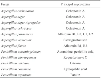
The identification of the species of the gerera Aspergillus was according to Klich (Klich, 2002) and Christensen (Christensen, 1982); the genusPenicillium ac-cording to Pitt (Pitt, 1988), and Samsonet al.(Samson,et al., 2004), where we only selected the fungi with toxigenic potential (Table 2).
Preservation methods
Lyophilization
The samples of the fungi were frozen in duplicate at -80 °C in an ultra-freezer, after the freezing the samples were taken to the Liotop, I model L101 lyophilizer, where the initial temperature was -50±2 °C. With the temperature stabilized, the vacuum was initiated with a pressure around 650mHg, after 48 h the lyophilization concluded with pres-sure around 150mHg.
Every two months the lyophilized fragments were isolated and put in 2% Malt Agar (MA) medium for the viability test, and the fungi revealed as toxigenic were also placed in specific medium for the toxigenic poten-tial test.
Sterile distilled water
The inoculation of glass flasks containing sterilized distilled water with a small portion of the culture medium (approximately 5 mm x 10 mm) with the fungus to pre-serve, was conducted. The flasks employed were the same used for the antibiotic, with a 6 mL capacity, filled with 4 mL of distilled water, sealed with their own rubber stopper and autoclaved at 121° under 1 atm for 30 min. After the autoclaving the transference of the fungi to the flasks with
Table 1- Fungi obtained and origin.
Fungi Origin
Aspergillus carbonarius Coffee
Aspergillus niger Grape
Aspergillus niger Agregados Coffee
Aspergillus ochraceus Potato
Aspergillus parasiticus Brazil-nut
Aspergillus versicolor Coffee
Aspergillus flavus Brazil-nut
Penicillium aurantiogriseum Coffee
Penicillium chrysogenum Peanut
Penicillium citrinum Wheat
Penicillium commune Mycology collection EPAMIG/CTSM
Penicillium expansum Coffee
Table 2- Fungi and mycotoxins potentially produced.
Fungi Principal mycotoxins
Aspergillus carbonarius Ochratoxin A
Aspergillus niger Ochratoxin A
Aspergillus niger Agregados Ochratoxin A
Aspergillus ochraceus Ochratoxin A
Aspergillus parasiticus Aflatoxin B1, B2, G1, G2
Aspergillus versicolor Esterigmatocistin
Aspergillus flavus Aflatoxin B1, B2
Penicillium aurantiogriseum Auranthine, penicillic acid
Penicillium chrysogenum Roquefortine c C
Penicillium citrinum Citrinin
Penicillium commune Cyclopaldic acid
Penicillium expansum Patulin
Source: Illustrated Manual on Identification of Some Seed-borne
water in an aseptic chamber was carried out in duplicate. The flask stopper was removed and pieces of culture me-dium containing fungal mycelium were transferred into the flasks.
Every two months the fragments were isolated and placed in 2% MA medium for the viability test, and the fungi revealed as toxigenic were also placed in a medium specific for the toxigenic potential test.
Continuous transference
The transference of the fungi was carried out in dupli-cate. The fungi were placed in petri plates with 2% MA me-dium sealed with parafilm, the cultures were stored in a in refrigerator at a temperature from 4 to 8 °C according to the literature, and every two months the fungi were transfered to other plates with 2% MA medium for the viability test, and those toxigenic were also placed in a medium specific for the toxigenic potential test.
Results
Fungal isolate viability test
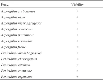
In Table 3 the fungal isolates can be evaluated at time 0, that is, before they were submitted to the preservation tests. They were found viable and pure.
Through the data shown in Table 4 the results of the viability tests of the isolates can be seen at times 1, 2, 3 and 4 corresponding to 2, 4, 6 and 8 months of preservation by the continuous subculture, Castellani and lyophilization methods, being represented by the + sign as viableand by the – sign as unviable.
According to the results obtained and represented by Table 4 one can observe that the isolates presented viability under the three preservation methods.
In relation to the methods used in the present work, the Castellani method is indicated as the most advanta-geous among the three methods used, therefore besides
having a high viability rate and preserving the characteris-tics of the isolates.
It was also observed that there weren’t any changes to the macroscopic and morphological characteristics of the tested fungi.
Discussion
The fungi, unlike most of the bacteria, are beings with a slow growth rate in culture media. On the other hand, many times there is contamination by bacteria or other fungi, which harms the preservation of the colonies. The existent techniques for the mycology collection mainte-nance are difficult, costly and frequently inefficient. The development of new ways of fungal preservation of for pro-longed periods becomes necessary. (Samsonet al., 2004).
In a study using 26 fungi strains preserved by the Castellani method for 2 years the viability of 100% of the strains was observed, with no alterations in the macro-scopic and morphological characteristics, demonstrating the efficiency of that method in the preservation of fungal culture viability of (e Bueno Gallardo, 1998).
Evaluating the results obtained we observed that they are in agreement with the affirmative of Cavalcanti (Feo-filovaet al., 2009). who obtained high viability in a study on the preservation by the Castellani, continuous subcul-ture and lyophilization methods, also affirming the Castel-lani method as viable in the preservation of dimorphic fungi and the lyophilization method is shown more efficient in the preservation of yeasts, presenting of viability rate of 100% in the conducted study.
Previous studies comparing the viability of fungal cultures preserved by the Castellani and lyophilization methods suggest that the Castellani method (distilled wa-ter) is more advantageous to maintain, in laboratory, differ-ent gerera and species of fungi, even comparing the two methods(e Qiangqiang Jiajun, 1998) in another study com-paring the viability of seventy eight isolates after twelve years, using the lyophilization and Castellani methods in the their preservation obtaining 89.7% of the isolates as via-ble preserved by the Castellani method and 87.2% of the isolates preserved by the lyophilization method. (Romeiro, 1996).
A study, where the use of the Castellani method and continuous subculture was compared in the preservation of hundred and eleven strains of different species of microor-ganisms during a seven-year the period, resulted in (71.2%) viability in the strains preserved by the Castellani method and (77.5%) viability in the strains preserved through con-tinuous subculture.(Samsonet al., 2004).
Conclusion
The lyophilization methods, Castellani and continu-ous subculture tested in this work were shown efficient in the preservation of the 12 isolates during the 4 times
Toxigenic fungi 45
Table 3- Represents the viability of potentially toxigenic fungal isolates at time 0.
Fungi Viability
Aspergillus carbonarius +
Aspergillus níger +
Aspergillus niger Agregados +
Aspergillus ochraceus +
Aspergillus parasiticus +
Aspergillus versicolor +
Aspergillus flavus +
Penicillium aurantiogriseum +
Penicillium chrysogenum +
Penicillium citrinum +
Penicillium commune +
Guimarães
et
al.
Fungi Time 1 2 months Repetition Lyophilization Castellani Continuous subculture Time 2 4 months Lyophilization Castellani Continuous subculture
A. carbonarius 1 and 2 + + + + + +
A. Níger 1 and 2 + + + + + +
A. niger Agregados 1 and 2 + + + + + +
A. ochraceus 1 and 2 + + + + + +
A. parasiticus 1 and 2 + + + + + +
A. versicolor 1 and 2 + + + + + +
A. flavus 1 and 2 + + + + + +
P. aurantiogriseum 1 and 2 + + + + + +
P. chrysogenum 1 and 2 + + + + + +
P. citrinum 1 and 2 + + + + + +
P. commune 1 and 2 + + + + + +
P. expansum 1 and 2 + + + + + +
Fungi Time 3 6 months Repetition Lyophilization Castellani Continuous subculture Time 4 8 months Lyophilization Castellani Continuous subculture
A. carbonarius 1 and 2 + + + + + +
A. niger 1 and 2 + + + + + +
A. niger Agregados 1 and 2 + + + + + +
A. ochraceus 1 and 2 + + + + + +
A. parasiticus 1 and 2 + + + + + +
A. versicolor 1 and 2 + + + + + +
A. flavus 1 and 2 + + + + + +
P. aurantiogriseum 1 and 2 + + + + + +
P. chrysogenum 1 and 2 + + + + + +
P. citrinum 1 and 2 + + + + + +
P. commune 1 and 2 + + + + + +
(8 months), however it is important to emphasize that the tested times correspond to a period of short duration, which suggests the need of more lengthy studies.
In relation to the methods used in the present work, the Castellani method is indicated as the most advantageous among the three methods used, therefore besides having a high viability rate and preserving the characteristics of the isolates, it is considered a simple method, one of low cost and that does not need electric power, and thus not being af-fected by any circumstances due to lack of electricity.
References
Bueno L, Gallardo R (1998) Preservación de hongos filamentosos en agua destilada estéril.Rev Iberoam Micol 15:166-168. Cameotra SS (2007) Preservation of microorganisms as deposits
for patent application. Biochem Biophys Res Commun 353:849-850.
Christensen CM (1982) TheAspergillus ochraceusgroup: twe new species from western soils and a synoption. Mycol 74:210-225.
Feofilova EP, Kuznetsova LS, Sergeeva YE, Galanina LA (2009) Species composition of food-spoiling mycelial fungi. Microbiol 78:112-116.
Klich MA (2002)Identification of common Aspergillus species.
Centraalbureau voor Schimmelcultures, Utrecht.
Nakasone KK, Peterson AW, Jong S (2004) Preservation and dis-tribution of fungal cultures.In:Mueller, G. M.; Bills, G. F.; Foster, M. S.Biodiversity of fungi, inventory and monitoring methods. Elsevier, San Diego, p. 37-47.
Pitt JI (1988) A laboratory guide to common Penicillium species. 2. ed. CSIRO Food, Australia.
Qiangqiang Z, Jiajun W, LI L (1998) Storage of fungi using sterile distilled water orlyophilization: comparison after 12 years. Mycoses 41:255-257.
Romeiro RS (1996) Preservação de culturas de bactérias fitopato-gênicas.Universidade Federal de Viçosa, Viçosa, MG. Samson RA, Houbraken AMP, Kuijpers AFA, Frank JM, Frisvad
JC (2004) New ochratoxin A or sclerotium producing spe-cies in Aspergillus section Nigri. Stud Mycol 50:23-43.
All the content of the journal, except where otherwise noted, is licensed under a Creative Commons License CC BY-NC.