Cate cho lamine -induce d vaso co nstrictio n
is se nsitive to carbo nic anhydrase I
activatio n
1Romanian Medical Academy, Center for Research and Medical Assistance,
Simleu Silvaniei, Salaj, Romania
2Faculty of Medicine, O radea, Romania 3Klinik Wilkenberg, Wilkenberg, Germany
4Medical Care Unit, Staten Island, New York, NY, USA
I. Puscas1, M. Coltau1,
L. Gilau2, R. Pasca1,
G. Domuta1, M. Baican3
and A. Hecht4
Abstract
We studied the relationship between alpha- and beta-adrenergic ago-nists and the activity of carbonic anhydrase I and II in erythrocyte, clinical and vessel studies. Kinetic studies were performed. Adrener-gic agonists increased erythrocyte carbonic anhydrase as follows: adrenaline by 75%, noradrenaline by 68%, isoprenaline by 55%, and orciprenaline by 62%. The kinetic data indicated a non-competitive mechanism of action. In clinical studies carbonic anhydrase I from erythrocytes increased by 87% after noradrenaline administration, by 71% after orciprenaline and by 82% after isoprenaline. The increase in carbonic anhydrase I paralleled the increase in blood pressure. Similar results were obtained in vessel studies on piglet vascular smooth muscle. We believe that adrenergic agonists may have a dual mechan-ism of action: the first one consists of a catecholamine action on its receptor with the formation of a stimulus-receptor complex. The second mechanism proposed completes the first one. By this second component of the mechanism, the same stimulus directly acts on the carbonic anhydrase I isozyme (that might be functionally coupled with adrenergic receptors), so that its activation ensures an adequate pH for stimulus-receptor coupling for signal transduction into the cell, result-ing in vasoconstriction.
Co rre spo nde nce I. Puscas
Center for Research and Medical Assistance 37 Dunarii Street 4775 Simleu Silvaniei, Salaj Romania
Fax: + 40-60-67-8320 E-mail: ccam@ netcompsj.ro
Received August 11, 1999 Accepted December 12, 2000
Ke y wo rds
·Adrenergic agonists ·Carbonic anhydrase ·Arterial blood pressure
Intro ductio n
Catecholamines released by the sympa-thetic nervous system and adrenal medulla are involved in regulating a host of physi-ological functions, particularly the integra-tion of responses to a range of stresses (1). Norepinephrine is the major neurotransmit-ter in the peripheral sympathetic nervous system, whereas epinephrine is the primary hormone secreted by the adrenal medulla in mammals (2).
Important factors in the response of any
cell or organ to sympathomimetic amines are the density and proportion of alpha- and beta-adrenergic receptors (3,4). Studies on DNA cloning have demonstrated the exist-ence of at least nine types of adrenergic receptors (5). Other studies have proved that binding of agonists to these receptors occurs when the catecholamine molecule is in the protonated state (6), all of these receptors being coupled with G proteins.
reac-tion of CO2, having a main role in the
mainte-nance of acid-basic equilibrium (7).
CA CA
H2O + CO2 Û H2CO3 Û H+ + HCO3
-Eight isozymes have been described so far, located in the membranes, cytoplasm and mitochondria of all organs. CA I is present both in erythrocytes and in vascular walls and its physiologic role has been in-completely elucidated (8). CA II is to be found both in erythrocytes and in the cyto-plasm. By its presence in the parietal cells of the gastric mucosa, it has a central role in HCl production, while in the kidney CA II is involved in the maintenance of urinary pH along with CA IV.
Regarding the physiological role of CA isozymes, our studies have shown that CA I is involved in the vascular changes (9) while CA II and CA IV are isozymes involved in the secretory processes (10).
The same studies showed that CA I and CA II are activated by nonsteroidal anti-inflammatory drugs (11) and vasodilating prostaglandins and diuretic agents (12,13) inhibit CA while vasoconstrictive prosta-glandins activate the enzyme (12). These studies have also demonstrated the involve-ment of carbonic anhydrase in the regulation of vascular and secretory processes in the organism (14).
Our previous work has shown that alpha-and beta-adrenergic agonists activated puri-fied and red cell CA while adrenergic antago-nists inhibited CA and reduced the activating effect of agonists on this enzyme (15,16).
In the present investigation we studied the relationship between alpha- and beta-adrenergic agonists and CA activity in vaso-constriction mechanism.
Mate rial and Me tho ds
Mate rial
Purified human erythrocyte CA I and
CA II, adrenaline, noradrenaline, isoprena-line, orciprenaisoprena-line, HEPES buffer,
p-nitrophenol, and Na2SO4 were obtained
from Sigma Chemical Co. (Deisenhofen, Germany), orciprenaline (Alupent) was purchased from Boehringer (Ingelheim, Germany), and noradrenaline and isopre-naline (vials) were obtained from Sicomed (Bucharest, Romania).
Expe rim e ntal de signs
Erythrocyte studies. We studied the ef-fects of adrenaline, noradrenaline, isoprena-line and orciprenaisoprena-line on CA I and CA II purified from human erythrocytes. Kinetic determinations were performed at concen-trations between 10 nM and 100 µM.
Kinetic studies were carried out in order to identify the mechanism of action of adre-nergic agonists on CA. Maximum reaction
rate (Vmax) and the Michaelis constant (Km)
were determined.
Clinical studies. The study was conducted according to the Declaration of Helsinki as modified by the 21st World Medical Assem-bly, Venice, Italy, 1983 and later by the 41st World Medical Assembly, Hong Kong, 1989. All patients gave informed consent for a protocol approved by the Ethics Committee of the Center for Research and Medical As-sistance in Simleu Silvaniei.
ortho-static hypotension, or had evidence from the screening tests of underlying illness or sig-nificant laboratory or electrocardiogram ab-normalities. In the acute experiment, group I
patients (N = 12) received noradrenaline iv
at the dose of 4 mg/1000 ml isotonic solution (4 µg/min over 30-min periods), group II patients (N = 14) received orciprenaline
(Alupent) iv at the dose of 0.5 mg, and group
III patients (N = 16) received isoprenaline
(Isoprenalin) sc at the dose of 0.2 mg.
Red cell CA I and CA II activity and arterial blood pressure values were deter-mined before and 30 min after drug adminis-tration and the blood count, routine chemis-try, urinalysis and electrocardiogram were repeated.
Vessel studies. In the animal experiments, 20 piglets weighing 25-30 kg were housed in air-conditioned quarters and had free access to tap water and standard food. Animals were divided into 4 groups of 5 piglets each and treated as follows in the acute
experi-ment: group 1 - noradrenaline, iv doses of 2
µg/min for 30 min; group 2 - orciprenaline
(Alupent), iv doses of 0.25 mg; group 3
-isoprenaline (Isoprenalin), sc doses of 0.1
mg, and group 4 (control group) - placebo. Arterial blood pressure was determined 30 min after drug administration and all piglets were sacrificed for isolation of vas-cular smooth muscle CA I. CA I activity was determined and compared to that obtained for the control group.
Expe rim e ntal pro ce dure
Differentiation of red cell CA I from CA II activity was performed by the nicotinate test (17), which relies on selective inhibition of CA I activity.
Vascular smooth muscle CA I was iso-lated from the small mesenteric arteries of the animals according to the technique of Lonnerholm et al. (8).
CA I and CA II activity was assessed using the stopped-flow method (18), which
consists of measuring the enzymatic activity
of CO2 hydration and is based on a
colori-metric method which measures changing pH. The time needed for the pH of the reagent mixture to decrease from its initial value of 7.5 to its final value of 6.5 was measured. The reaction was monitored spectrophoto-metrically at 400 nm using a rapid kinetic Hi-Tech SF-51MX spectrophotometer (Hi-Tech Scientific Ltd., Salisbury, England) equipped with a mixing unit and a system of two syringes which supply the reagents. The signal transmitted by the photomultiplier from the mixing chamber is received and visual-ized by a computer equipped with a math-ematical coprocessor and the kinetic soft-ware package RKBIN IS1.
We used p-nitrophenol (0.2 mM) as color indicator and HEPES (20 mM) as buffer.
Na2SO4 (0.1 M) was used to keep a constant
ionic strength. The CO2 solution at a
concen-tration of 15 mM (as substrate) was obtained
by bubbling twice-distilled water with CO2 to
saturation. All reagents were maintained at pH
7.5 and at room temperature (22-25oC).
Carbonic anhydrase activity was obtained by the formula:
A = T0 - T [enzyme units/ml]
T
where T0 represents the uncatalyzed reaction
time, and T the catalyzed reaction time (in the presence of CA). Activity is reported as enzyme units (EU) per ml.
In the CO2 hydration reaction catalyzed
by CA one enzyme unit will cause the pH to
drop from 7.5 to 6.5 per minute, at 25o
Statistical analysis
When repeated measure ANOVA showed significant differences between groups, the Newman-Keuls multiple comparison test was performed to determine which groups dif-fered significantly. Probabilities of P<0.05 were considered significant.
Re sults
Erythro cyte studie s
Adrenaline, noradrenaline, isoprenaline
and orciprenaline increased CA I and CA II activity in a dose-dependent manner. The effect started at 10 nM and reached a peak at 100 µM (Table 1).
Adrenaline increased CA I activity from 0.425 ± 0.01 to 0.743 ± 0.02 EU/ml (75%) (P<0.001), and CA II activity from 1.00 ± 0.01 to 1.472 ± 0.02 EU/ml (47%) (P<0.001). Noradrenaline increased CA I activity from 0.425 ± 0.01 to 0.714 ± 0.01 EU/ml (68%) (P<0.001), and CA II activity from 1.00 ± 0.01 to 1.436 ± 0.02 EU/ml (43%) (P<0.001).
Isoprenaline increased CA I activity from 0.425 ± 0.01 to 0.658 ± 0.02 EU/ml (55%) (P<0.001), and CA II activity from 1.00 ± 0.01 to 1.342 ± 0.01 EU/ml (34%) (P<0.001). Orciprenaline increased CA I activity from 0.425 ± 0.01 to 0.688 ± 0.02 EU/ml (62%) (P<0.001), and CA II activity from 1.00 ± 0.01 to 1.488 ± 0.01 EU/ml (49%) (P<0.001).
The kinetic data processed according to the Michaelis-Menten equation showed a non-competitive mechanism of action with
an increase in Vmax and a constant Km.The
kinetic results show that adrenergic agonists were bound to the active site of CA I in a position different from that of the enzyme
substrate, CO2 (Table 2).
Clinical studie s
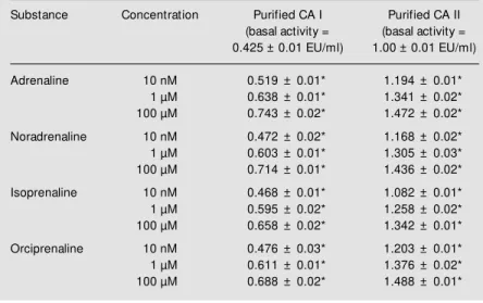
In group 1, noradrenaline increased red cell CA I activity from 0.268 ± 0.026 to 0.501 ± 0.042 EU/ml (87%) (P<0.001) and CA II activity from 1.081 ± 0.116 to 1.598 ± 0.134 EU/ml (48%) (P<0.05) (Fig-ure 1). Arterial blood press(Fig-ure rose from 130 ± 10 to 175 ± 15 mmHg (P<0.05) (Figure 2). In group 2, orciprenaline increased red cell CA I activity from 0.251 ± 0.030 to 0.429 ± 0.027 EU/ml (71%) (P<0.001) and CA II activity from 1.135 ± 0.110 to 1.288 ± 0.194 EU/ml (27%) (P<0.05) (Fig-ure 1). Arterial blood press(Fig-ure rose from 120 ± 10 to 155 ± 5 mmHg (P<0.05) (Figure 2). Table 1 - Effect of adrenergic agonists on isozyme I and II of carbonic anhydrase (CA).
The table show s the increase of the activity of CA isozymes induced by therapeutic agents such as adrenaline, noradrenaline, isoprenaline and orciprenaline. Values are reported as means ± SEM (N = 5 assessments) * P<0.05 compared w ith basal activity for each isozyme (New man-Keuls multiple comparison test).
Substance Concentration Purified CA I Purified CA II
(basal activity = (basal activity = 0.425 ± 0.01 EU/ml) 1.00 ± 0.01 EU/ml)
Adrenaline 10 nM 0.519 ± 0.01* 1.194 ± 0.01*
1 µM 0.638 ± 0.01* 1.341 ± 0.02*
100 µM 0.743 ± 0.02* 1.472 ± 0.02*
Noradrenaline 10 nM 0.472 ± 0.02* 1.168 ± 0.02*
1 µM 0.603 ± 0.01* 1.305 ± 0.03*
100 µM 0.714 ± 0.01* 1.436 ± 0.02*
Isoprenaline 10 nM 0.468 ± 0.01* 1.082 ± 0.01*
1 µM 0.595 ± 0.02* 1.258 ± 0.02*
100 µM 0.658 ± 0.02* 1.342 ± 0.01*
Orciprenaline 10 nM 0.476 ± 0.03* 1.203 ± 0.01*
1 µM 0.611 ± 0.01* 1.376 ± 0.02*
100 µM 0.688 ± 0.02* 1.488 ± 0.01*
Table 2 - Kinetic data for interaction betw een alpha- and beta-adrenergic agonists and purified carbonic anhydrase (CA) I.
CA I concentration = 3.68 x 10 nM , pH 7.5, T = 25oC. Data are reported as means ± SD
(N = 5 assessments). * P<0.05 compared to purified CA I (New man-Keuls multiple comparison test).
System Vmax (mM s-1) Km (mM )
CA I 1.332 ± 0.01 8.99 ± 0.2
CA I + adrenaline (100 µM ) 1.796 ± 0.02* 8.87 ± 0.1
CA I + noradrenaline (100 µM ) 1.733 ± 0.01* 8.91 ± 0.1
CA I + isoprenaline (100 µM ) 1.710 ± 0.02* 8.95 ± 0.2
In group 3, isoprenaline increased red cell CA I activity from 0.247 ± 0.015 to 0.450 ± 0.020 EU/ml (82%) (P<0.001) and CA II activity from 0.983 ± 0.105 to 1.474 ± 0.208 EU/ml (50%) (P<0.05) (Fig-ure 1). Arterial blood press(Fig-ure rose from 120 ± 10 to 160 ± 5 mmHg (P<0.05) (Figure 2).
Ve sse l studie s
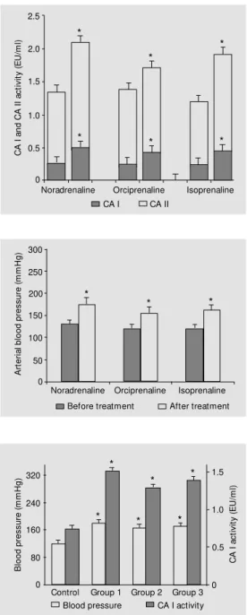
In animals, after the acute experiment the activity of vascular smooth muscle CA I was 0.812 ± 0.062 EU/ml and arterial blood pres-sure was 120 ± 10 mmHg in the control group. In all adrenergic-treated groups, vas-cular smooth muscle CA I activity and arte-rial blood pressure increased significantly (P<0.05) compared to controls, as follows: group 1 - CA I was 1.696 ± 0.124 EU/ml and arterial blood pressure was 180 ± 15 mmHg; group 2 - CA I was 1.408 ± 0.136 EU/ml and arterial blood pressure was 165 ± 10 mmHg; group 3 - CA I was 1.519 ± 0.143 EU/ml and arterial blood pressure was 170 ± 10 mmHg (Figure 3).
D iscussio n
Our group has studied the relationship between adrenergic agonists and CA (15,16), showing that CA was activated by a direct mechanism. The erythrocyte studies proved that alpha- and beta-adrenergic agonists are direct and strong CA I activators which have less effect on CA II.
Clinical and vessel studies have shown
that adrenergic agonists are powerful CA I activators both in erythrocytes and in vascu-lar smooth muscles. In humans, noradrena-line, orciprenaline and isoprenaline increased arterial blood pressure in volunteer subjects, in parallel with an increase of erythrocyte CA I activity. Parallelism between the in-crease in arterial blood pressure and erythro-cyte CA I activation was observed in all groups. The most potent activating effect on CA was induced by noradrenaline which
Figure 1 - Effect of noradrena-line (4 µg/min, over 30 min, iv), orciprenaline (0.5 mg, iv) and isoprenaline (0.2 mg, sc) on red blood cell CA I and CA II activity. Values are reported as means ± SEM ; N = 12-16 pat ient s. * P<0.05 compared w ith values before treatment (paired t-test).
Figure 2 - Effect of noradrena-line (4 µg/min, over 30 min, iv), orciprenaline (0.5 mg, iv) and isoprenaline (0.2 mg, sc) on ar-terial blood pressure. Values are reported as means ± SEM ; N = 12-16 patients. * P< 0.05 com-pared w ith values before treat-ment (paired t-test).
Figure 3 - Increase of vascular smooth muscle CA I activity and of arterial blood pressure values in piglets after treatment w ith noradrenaline (group 1), orci-prenaline (group 2) and iso-prenaline (group 3) compared to control. Values are reported as means ± SEM ; N = 5 piglets. * P<0.05 compared w ith control (paired t-test).
A rt e ri a l b lo o d p re s s u re ( m m H g ) 300
Before treatment After treatment Noradrenaline Orciprenaline Isoprenaline 250 200 150 100 50 0 C A I a n d C A I I a c ti v it y ( E U /m l) 2.5 Noradrenaline * 2.0 1.5 1.0 0.5 0 Orciprenaline Isoprenaline
CA I CA II
* * * * * B lo o d p re s s u re ( m m H g ) 320 C A I a c ti v it y ( E U /m l) 1.5 1.0 0.5 0
Blood pressure CA I activity Control Group 1 Group 2 Group 3 240
160
80
0
also produced the main increase in blood pressure. None of these patients presented any major side effects during the experi-ments.
In animals, administration of adrenergic agonists significantly increased CA activity, mainly of CA I in arteriolar smooth muscle as compared to controls, in parallel with an increase in arterial blood pressure.
These results agree with previous studies by our group which showed that CA I is involved in the modulation of vascular
cesses in the organism. In our conception, the pH increase induced by CA I inhibition might influence the binding of hypotensive stimuli to their specific receptors, followed by signal transduction to the cytoplasm of smooth muscle cells with subsequent vaso-dilating effects (9,14). Similarly, the reduc-tion in pH induced by vascular smooth muscle CA I activation with hypertensive agents may influence the membrane specific recep-tor and signal transduction in the vascular smooth muscle cytosol, with subsequent va-soconstrictive effects (14).
Regarding the role of pH changes in hy-pertension, our results support recent studies which have shown that primary hyperten-sion may be associated with perturbations of acid-base status or intracellular pH, respec-tively (19,20). Furthermore, the same stud-ies demonstrated a decrease of intracellular pH in hypertensive animal models as com-pared to normotensive animals (19,21). An evaluation of steady-state intracellular pH in erythrocytes using a nuclear magnetic reso-nance technique indicated that intracellular pH is reduced in erythrocytes from untreated patients with essential hypertension com-pared to treated patients and normotensive controls (22).
Other studies have reported that the blood pressure-lowering effects of calcium chan-nel blockade were inversely related to intra-cellular pH, i.e., the lower the initial pH, the greater the antihypertensive effect. Further-more, nifedipine consistently elevated intra-cellular pH values (23).
An enhanced activity of the H+
-Na+
anti-porter (as a major mechanism of cell defense against cellular acidification) has been re-ported in lymphocytes (24) as well as in the renal brush border membrane of spontane-ously hypertensive rats (25) and in hyperten-sive rats (26). Other authors have shown enhanced responsiveness of the renal
proxi-mal Na+
-H+
antiporter of hypertensive rats to stimulation with some hormones (27). It has
been assumed that an overactivity of this antiporter is not a primary process but rather reflects intracellular acidosis in sion, as studied in spontaneously hyperten-sive rat models (20,21).
Catecholamine-induced CA I activation has suggested a concept concerning the in-volvement of pH changes (induced by acti-vation) in vasoconstrictive processes due to adrenergic agonists. In keeping with this concept, catecholamines have a dual mech-anism of action: the first consists of adrener-gic agonists acting on their specific recep-tors with subsequent formation of a stimu-lus-receptor complex, followed by the cou-pling of G proteins and information trans-mission within the cell. The second mechan-ism suggests that catecholamines directly act on CA I (an isozyme that might be func-tionally coupled with adrenergic receptors), which by its activation accompanied by a fall in pH may be favorable to catechol-amine-binding to its specific receptor and by G proteins to facilitate information transmis-sion into the cell.
Our results suggest that CA I modulates vascular tone by means of pH changes (14). This role for pH in the vascular bed is sup-ported by our data as follows: a) the already known role of CA I in acid-base balance; b) the effect of vasoconstrictive substances that activate erythrocyte CA I and vascular smooth muscle CA I by a direct mechanism of ac-tion, and c) the effect of vasodilatory sub-stances along with drugs used in the treat-ment of hypertension which inhibit CA I by a direct mechanism of action both in erythro-cytes and in vascular smooth muscle.
Re fe re nce s
1. Hoffman BB & Lefkow itz RJ (1996). Cat-echolamines, sympathomimetic drugs, and adrenergic receptor antagonists. In: M olinoff PB & Ruddon RW (Editors), The Pharmacological Basis of Therapeutics. 9th edn. M cGraw -Hill, New York, 199-248. 2. Allw ood M J, Cobbold AF & Ginsberg J (1963). Peripheral vascular effects of nor-adrenaline, isopropylnornor-adrenaline, and dopamine. British M edical Bulletin, 19: 132-136.
3. Davey M (1986). Alpha adrenoceptors - an overview . Journal of M olecular and Cellu-lar Cardiology, 18 (Suppl 5): 1-15. 4. Brodde OE (1988). The functional
impor-tance of beta1 and beta2 adrenoceptors in
the human heart. American Journal of Car-diology, 62: 24C-29C.
5. Kobilka BK (1987).Cloning, sequencing and expression of the gene coding for the human platelet alpha1-adrenergic recep-tor. Science,238: 650-656.
6. Strader CD, Candelore M R, Hill WS, Sigal IS & Dixon RAF (1989). Identification of tw o serine residues involved in agonist activation of the ß-adrenergic receptor. Journal of Biological Chem istry, 264: 13572-13578.
7. M aren TH (1967). Carbonic anhydrase: chem ist ry, physiology and inhibit ion. Physiological Review s, 47: 595-781. 8. Lonnerholm G, Selking O & Wistrand PJ
(1985). Amount and distribution of car-bonic anhydrases I and II in the gas-trointestinal tract. Gastroenterology, 86: 1151-1161.
9. Puscas I, Vlaicu R & Lazar A (1997). Isosorbide nitrates, nitroglycerine and so-dium nitroprusside induce vasodilation concomitantly w ith inhibition of carbonic anhydrase I in erythrocytes. American Journal of Hypertension, 10: 124-128. 10. Puscas I, Coltau M , Baican M & Domuta G
(1999). A new concept regarding the mechanism of action of omeprazole. In-ternational Journal of Clinical Pharmacolo-gyand Therapeutics, 37: 286-293. 11. Puscas I, Coltau M & Pasca R (1996).
Nonsteroidal anti-inflammatory drugs ac-tivate carbonic anhydrase by a direct mechanism of action. Journal of Pharma-cology and Experimental Therapeutics, 277: 1146-1148.
12. Puscas I & Coltau M (1995). Prostaglan-dins having vasodilating effects inhibit car-bonic anhydrase w hile leukotriens B4 and
C4 increase carbonic anhydrase activity.
International Journal of Clinical Pharma-cologyand Therapeutics, 32: 176-181. 13. Puscas I, Coltau M , Baican M , Domuta G
& Hecht A (1999). Vasodilatory effect of diuretics is dependent on inhibition of vas-cular smooth muscle carbonic anhydrase by a direct mechanism of action. Drugs under Experimental and Clinical Research, XXV: 271-279.
14. Puscas I & Supuran CT (1995). Carbonic anhydrase, a modulator of the physiologi-cal and pathologiphysiologi-cal processes in the or-ganism: The theory of pH. The 4th Inter-national Conference on the Carbonic An-hydrases. Oxford, England, July 26-30, 1995, 7.6.
15. Puscas I, Buzas G & M oldovan A (1985). Effect of beta-adrenergic agonists and an-tagonists on carbonic anhydrase. Revue Roumaine de Biochemie, 22: 157-163. 16. Puscas I, Pasca R, Lazoc L & Kun C (1994).
Effects of alpha- and beta-adrenergic ago-nists and antagoago-nists on carbonic anhy-drase. In: Puscas I (Editor), Carbonic An-hydrase and M odulation of Physiologic and Pathologic Processes in the Organ-ism. Helicon Publisher House, Timisoara, Romania, 260-268.
17. Puscas I, Coltau M & Domuta G (1999). Rapid method for differentiation of car-bonic anhydrase I from carcar-bonic anhy-drase II activity. Analytical Letters, 32: 915-924.
18. Khalifah RG (1971). The carbon dioxide hydration activity of carbonic anhydrase: stop-flow kinetic studies on the native human isozymes B and C. Journal of Bio-logical Chemistry, 246: 2561-2573. 19. Batlle DC, Saleh A & Rombola G (1990).
Reduced intracellular pH in lymphocytes from the spontaneously hypertensive rat. Hypertension, 15: 97-103.
20. Batlle D, Sharma AM , Alsheikha M W, Sobrero M , Saleh A & Gutterm an C (1993). Renal acid excretion and intracel-lular pH in salt-sensitive genetic hyperten-sion. Journal of Clinical Investigation, 91: 2178-2184.
21. Saleh A & Batlle DC (1990). Kinetic prop-erties of the Na+/H+ antiporter of
lympho-cytes from the spontaneously hyperten-sive rat: role of intracellular pH. Journal of Clinical Investigation, 85: 1734-1739. 22. Resnick LM , Gupt a RK, Sosa RE,
Barbagallo M , M ann S, M arion R & Laragh JH (1987). Intracellular pH in human and experimental hypertension. Proceedings of the National Academy of Sciences, USA, 84: 7663-7667.
23. Resnick LM , Gupta RK, DiFabio B & Laragh JH (1994). Intracellular ionic con-sequences of dietary salt loading in es-sential hypertension. Relation to blood pressure and effects of calcium channel blockade. Journal of Clinical Investigation, 94: 1269-1277.
24. Feig PU, D’Occhio M A & Boylan JW (1987). Lymphocyte membrane sodium-proton exchange in spontaneously hyper-tensive rats. Hypertension, 9: 282-288. 25. M orduchow icz GA, Sheikh Hamad D, Jo
OD, Nord EP, Lee DB & Yanagaw a N (1989). Increased Na+/H+ antiport activity
in the renal brush border membrane of SHR. Kidney International, 36: 576-581. 26. Parenti P, Hanozet GM & Bianchi G
(1986). Sodium and glucose transport across renal brush border membranes of M ilan hypertensive rats. Hypertension, 8: 932-939.
27. Gesek FA & Schoolw erth AC (1991). Hor-mone responses of proximal Na+-H+