APPLICATION
OF AN ENZYME-LINKED
IMMUNOSORBENT
ASSAY (ELISA)
METHOD
TO THE DIAGNOSIS
OF
HUMAN
HYDATIDOSIS
Jorge A. Guisantes, 2 Manuel
F. Rubio,s and Ramdn Diaz4
An investigation was conducted to evaluate the usefulness of a special ELISA method for diagnosing human hydatidosis. The results suggest the test may prove useful in seroepidemiologic studies and that it can make a valuable contn’-
bution to diagnosis of pulmonary hydatidosis.
Introduction
For some time immunologic methods such as indirect hemagglutination, latex aggluti- nation, electrosyneresis or counterimmuno- electrophoresis, double diffusion in gel, im- munoelectrophoresis, and indirect immuno- fluorescence have been widely used in diag- nosing human hydatidosis. More recently, immunoenzymatic (ELBA) methods have been introduced into immunologic diagnosis. These methods were used first to detect intra- cellular antigens and antibodies in tissue sec- tions, but this use was soon broadened to in- clude detection of circulating antigens and an- tibodies, and in this manner the methods were rapidly and effectively incorporated into the procedures for immunodiagnosis of infectious and parasitic diseases.
At present, the ELISA methods’ sensitivity is believed similar to that of radioimmuno- assay; but they are cheaper and simpler to use than radioimmunoassay, and they permit complete quantification of the antigens and antibodies involved. They have therefore been readily accepted, and the number of research
‘Also published in Spanish in the Bolefin de la Oficina Sa- nilaria Pmmnm~ana 90(2):160-168, 1981.
%ection Chief, Department of Microbiology and Para- sitoloev, Universitv Clinic, School of Medicine, Univer- sity of Navarra, Pamplona, Spain.
3Resident, Department of Microbiology and Parasit- ology, University Clinic, University of Navarra.
*Chief, Department of Microbiology and Parasitology, University Clinic, University of Navarra.
workers using them for diagnosis of infectious and parasitic diseases is increasing steadily.
Underlying the successive emergence of dif- ferent immunologic methods for diagnosing hydatidosis has been the desire to achieve sev- eral aims-’ Including reduced performance time, more economical use of reagents (es-
pecially antigen), better reproducibility, greater sensitivity and specificity, and appli- cability to seroepidemiologic studies.
It appears that the ELISA methods serve all of these purposes. Certainly with regard to diagnosis of hydatidosis the initial results have appeared promising (1,Z). Therefore, in view of such findings and the good results obtained in diagnosing other parasitic diseases, we felt it would be of interest to evaluate a new in- direct ELISA method for diagnosing human hydatidosis. Accordingly, we subjected sera from hydatidosis cases and control sera to this ELISA test, the latex agglutination test, and the immunoelectrophoresis test commonly employed as the diagnostic reference method.
Materials and Methods
Antigen for the three tests was obtained from the hydatid fluid of fertile hepatic cysts in sheep. The fluid was dialyzed, lyophilized, and standardized by immunoelectrophoretic analysis (3), a procedure demonstrating the presence of antigen 5 (see Capron et al.-4) together with other antigenic fractions.
Guisantes et al. l DIAGNOSIS OF HUMAN HYDATIDOSIS 261
and specificity, 172 human sera were em- ployed. These included:
1) 76 preoperative sera from patients with surgi- cally confirmed hydatidosis (41 with hepatic hyda- tidosis, 23 with pulmonary hydatidosis, and 12 with multiple hydatidosis or hydatidosis at other loca- tions).
2) 96 control sera consisting of: (a) 30 sera from healthy donors;
(b) 30 sera from patients with various nonin- fectious and nonparasitic diseases-including dis- eases such as cirrhosis of the liver, hepatic and pul- monary neoplasms, and collagenopathies-that are capable of producing cross-reactions in serologic tests for hydatidosis.
(c) 36 sera from patients with various infec- tious and parasitic diseases-including amebiasis, ascariasis, brucellosis, distomatosis caused by Fus-
ciola hepatica, giardiasis, leprosy, malaria, oxyuria- sis, teniasis, toxoplasmosis, trichomoniasis, tuber- culosis, and typhoid fever.
All the sera were stored for varying lengths of time at -2O’C until used.
ELISA Test
The ELISA test employed goat sera anti- human-immunoglobulins conjugated with radish peroxidase from the Cappel Laborato- ries in Cochranville, Pennsylvania, U.S.A. The optimum dilution of the conjugate, de- termined by prior titrations, proved to be 1:800 in phosphate-buffered saline (pH 7.2) with 4 per cent bovine seroalbumin and 0.05 per cent Tween 20.
The technique used was the indirect meth- od described initially by Engvall and Perl- mann (51, employing the micro-technique proposed by Ruitenberg et al. (s), with some modifications designed to adapt it to the anti- gen-antibody system under study.
Phosphate-buffered saline (pH 7.2) with 0.02 per cent sodium azide.was used for pre- paring the antigen solution. The optimum an- tigen concentration (5 pg dry weight per ml) was determined by prior titrations.
Polystyrene microtiter plates with flat bot- tom wells (Microtiter Brand, Dynatech Com- panies) were used in the test. Each well was
sensitized with 0.1 ml of the antigen solution and incubated for 3 hours at 37%. The sensi- tized wells were then washed three times with distilled water containing 0.05 per cent Tween 20 before sera were added to them.
The sera were inactivated for 30 minutes at 56’C and were then diluted with phosphate- buffered saline (pH 7.2) containing 0.5 per cent bovine seroalbumin (Sigma Chemical Co., Missouri, U.S.A.). The initial dilution was 1: 10 and double dilutions ranging up to
1:20,480 were made in successive wells (see photograph).
The plates thus prepared were incubated for 1 hour at 37°C. They were then emptied and washed three times with distilled water containing Tween 20. Next, 0.1 ml of the peroxidase conjugate was placed in each well and the plates were again incubated for 1 hour at 37%. Following a further washing, 0.1 ml of substrate -a solution of 5-aminosalicylic acid (5AS) and H202-was placed in each well. The substrate was prepared by dissolv- ing 80 mg of 5AS in 100 ml of warm distilled water and adjusting the pH to 6.0 with NaOH (1N). Immediately before using the substrate, 0.05 per cent Hz02 was added to the 5AS solution at a rate of 1 ml for every 9 ml of 5AS solution.
The plates were then maintained at room temperature for 1 hour, after which the re- action was stopped by adding 0.025 ml of NaOH (1N) to each well. The results were then read visually; the final dilution that showed a color other than the color of negative sera was taken to be the highest titer yielding a positive response. Wells containing both posi- tively and negatively responding sera are shown in the accompanying photograph.
Latex Agglutination Test (LA)
10 20 40 80 160 320 640 1280 2560 5l20 lO240 20480
An ELISA microtitration plate with positive and negative sera. The wells in rows 1, 3, and 4 contain sera from hydatidosis patients, while those in the other rows
contain sera from healthy subjects or from disease patients with cirrhosis of the liver or collagenopathy.
tine buffer (pH 8.2). This optimum concen- tration was determined by prior titrations with sera from a group of known hydatidosis cases and healthy donors.
Immunoelectrophoresis Test (IEP)
The IEP method of Capron et al. (a), as described by Guisantes et al. (S), was used for this test. Those sera that gave rise to diag- nostic arc 5, as described by Capron et al. (4), were considered positive.
Results
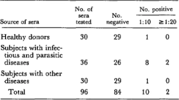
Table 1 summarizes the ELISA test results obtained with the 96 control sera. These re- sults show that 84 (87.5 per cent) of the 96 nonhydatid sera yielded a negative response, while 10 yielded a positive response at a 1 :lO dilution. These latter sera came from one healthy donor, one patient with chronic rheu- matoid polyarthritis, and eight patients with communicable or parasitic diseases (one case of teniasis due to Taenia saginata, two cases of
Table 1. ELISA test results obtained with 96 control sera from subjects without hydatidosis-including
subjects with various other kinds of diseases.
No. of No. positive sera NO.
Source of sera tested negative 1:io 219.0
Healthy donors 30 29 1 0
Subjects with infec- tious and parasitic
diseases 36 26 8 2
Subjects with other
diseases 30 29 1 0
Total 96 a4 10 2
oxyuriasis, two of distomatosis due to Fasciola
hepatica, one of amebiasis, and two of tubercu-
losis). In all, 94 of the 96 control sera yielded responses that were negative, or else were positive only at a titer of 1: 10.
Guisantes et al. l DIAGNOSIS OF HUMAN HYDATIDOSIS 263
In the ELISA test, as in the indirect hemag- glutination (3) and other quantitative tests, it is necessary to establish a titer of diagnostic significance, that being a dilution at which positive responses from nonhydatid sera are nonexistent or very rare (9). The data re- ported here show that we can establish a diag- nostic titer of 1:20 for the antigen used, with a probability of error (false positive diagnosis) of pco.025.
Table 2 summarizes the ELISA test results obtained with 76 sera from hydatidosis cases. Of these, all but three (96 per cent) were posi- tive at the diagnostic (1:20) titer, and two of those three were positive at 1: 10. Sensitivity to sera from pulmonary hydatidosis patients ap- peared slightly less than sensitivity to sera from hepatic hydatidosis patients, but the dif- ference observed was not statistically signifi- cant.
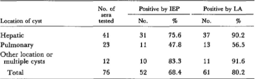
Table 3 shows the IEP and LA test results obtained with the 76 sera from hydatidosis cases, according to the location of the cyst. As may be seen, the overall sensitivity of the IEP
Table 2. ELISA test results obtained with 76 preoperative sera from hydatidosis
patients, by cyst location.
Location of cyst
No of No. positive
sera NO.
tested negative 1:lO 2 120 Hepatic
Pulmonary Other location
41 0 1 40(97.5%)
23 1 1 21(91.3%)
or multiple
cysts 12 0 0 12(100%)
Total 76 1 2 73 (96%)
test was 68.4 per cent, ranging from 47.8 per cent in cases with pulmonary cysts to 83.3 per cent in cases with multiple cysts or nonhepatic and nonpulmonary cysts. Its sensitivity to hepatic cysts (75.6 per cent) was substantially higher than its sensitivity to pulmonary cysts.
Regarding the LA test, its overall sensitivity was 80.2 per cent, and like the IEP test it showed greater sensitivity to hepatic cysts (90.2 per cent) and to multiple or nonhepatic and nonpulmonary cysts (91.6 per cent) than it did to pulmonary cysts (56.5 per cent).
Table 4 compares the ELISA and LA test results obtained with sera from hydatidosis pa- tients. It should be noted that 12 sera yielding negative LA test results gave a positive re- sponse to the ELISA test. In contrast, none of these 76 sera yielded a negative ELISA re- sponse and a positive LA response, all three ELISA-negative sera yielding negative LA re- sults.
Only one of the 96 nonhydatid sera (from a teniasis case) yielded positive LA test results, so that the rate of false positive response was 1.04 per cent. None of the 96 control sera yielded a positive IEP response; that is, none gave rise to diagnostic arc 5 (4).
Table 4. A comparison of the ELISA and LA test results obtained with preoperative sera from 76 hydatidosis patients. Only ELISA titers of 1:20
or greater were considered positive.
Test results
ELISA + ELISA + ELBA - ELISA -
LA+ LA- LA+ LA-
No. of sera 61 12 0 3
Table 3. Immunoelectrophoresis (IEP) and latex agglutination (LA) test results obtained with preoperative sera from 76 hydatidosis
patients, by cyst location.
Location of cyst
No. of sera tested
Positive by IEP
NO. I
Positive by LA
NO. 70
Hepatic Pulmonary Other location or
multiple cysts Total
41 31 75.6 37 90.2
23 11 47.8 13 56.5
12 10 83.3 11 91.6
Discussion
Sensitivip and S’ec$cig
The results obtained indicate that the ELISA method used was highly (96 per cent) sensitive at titers of diagnostic significance. They also show it to be highly specific, only two of the 96 sera from healthy subjects and nonhydatid disease patients having yielded a positive response of diagnostic significance. These two latter sera, it should be noted, came from patients infected with Taenia saginata.
Such a result could have been anticipated, since it is well-known that antigenic sharing exists between this cestode and the hydatidosis agent, Echinococcus granulosus, both of which belong to the Taeniidae family.
Only two of the three patients infected with
Fasciola hepatica yielded a positive ELISA titer,
and these (both 1: 10) were below the diagnos- tic titer of 1:20.
The Diagnostic Titer
Determining a titer of diagnostic signifi- cance tends to increase the specificity of any quantitative serologic method (3). In essence, such a titer can be determined using either of two basic criteria (9); that is, the diagnostic titer should be (a) the lowest at which no re- sponse is observed with nonhydatid sera, or (b) the lowest at which a minimum number of cross-reactions is observed.
We chose to adopt the latter criterion in this study for several reasons. In the first place, cross-reactivity occurred, in the case of sera from teniasis patients where it was logical to find it, when whole hydatid fluid was used as the antigen. Second, when the latter criterion was used, the observed rate of false positive responses (2.1 per cent) was very low. Third, it must be kept in mind that the specificity of any given method will be affected by the cross- reactivity of the control sera used to determine the diagnostic titer (3,9). And finally, because the purified “fraction 5” antigen described by Capron et al. (4) was not used, there is no
reason why a false positive titer higher than any given diagnostic titer could not be at- tained with some other more cross-reactive sera from teniasis cases. It is for this latter reason that, because of its specificity, the IEP test based on observation of diagnostic arc 5 (Capron et al.-4) continues to be the refer- ence test for diagnosis of human infection with
Echinococcus larvae (3,9-12).
Conclusions
Although the ELISA test calls for high- quality reagents and meticulous procedure, and although it may initially seem compli- cated, once a minimum amount of practice has been acquired it is no more difficult than the indirect hemagglutination test. Further- more, it has an advantage over the latter in that it does not require red blood cells, which can be difficult for some laboratories to ob- tain.
The ELISA method described is also rapid (a definitive result is available in 5 or 6 hours), reasonably inexpensive, and repeatable. (Re- garding the latter point, at our laboratory we have on several occasions repeated tests with the same sera, and with each serum have ob- tained the same positive titer or a titer dif- ference no greater than one dilution.)
In view of the frequent difficulties involved in obtaining hydatid fluid and producing stan- dardized antigens by means of immunoelec- trophoretic analysis (31, immunologic labora- tories find tests that are adequately sensitive and specific and that use only small amounts of antigens to be very desirable. In this re- gard, it should be noted that the ELISA test uses minimal amounts of antigen. Moreover, the test described here uses whole hydatid fluid instead of the purified fraction 5 that is harder to obtain.
Since the LA test is a method recommended for diagnosing clinical cases of hydatidosis, as well as for seroepidemiologic surveys, we felt it would be worthwhile to compare ELISA and LA results. As Table 4 shows, the ELISA test detected 12 hydatidosis cases that yielded negative results. (Overall, the LA test de- tected 80.2 per cent of the hydatidosis cases, while the ELISA test detected 96 per cent.) On the other hand, corroborating the results of earlier studies (7,9,10), the rate of false positive LA responses (1.04 per cent) was very low.
To sum up, a test used in seroepidemiologic studies should meet the following criteria: It should be (a) simple, (b) rapid, (c) inexpen- sive, (d) repeatable, (e) sensitive and specific, (f) sparing in its use of antigen, and (g) capa- ble of processing many sera simultaneously. On the basis of our results, we believe that evaluating the ELISA method described in seroepidemiologic studies, by using it in sur- veys assessing large numbers of sera, would yield findings of considerable interest.
Our own results also suggest that the meth- od would be very useful for diagnosing hospi- tal cases of hydatidosis because of its high sen-
sitivity, as indicated by detection of 73 of the 76 cases studied (error probability of false positive results being p < 0.025).
Also, it is well-known that the usual sero- logic tests for human hydatidosis are less sen- sitive to pulmonary cysts than to cysts at other places in the body, a fact corroborated in our study by the LA and IEP test results (see Table 3). In contrast, the ELISA method used demonstrated great sensitivity (91.3 per cent) to sera from patients with pulmonary cysts, a sensitivity similar to that for sera from pa- tients with hepatic cysts (see Table 2). The ELISA test should therefore make a very valu- able contribution to detection of pulmonary hydatidosis, which frequently yields seroneg- ative results in other diagnostic tests.
It should, of course, be remembered that di- agnosis of hydatidosis in man is based on a combination of clinical, serologic, radiologic, and scintillographic data. Therefore, although the rate of false positive responses obtained with the ELISA method used was very low, the possibility of such responses should be kept in mind in diagnosing cases where the ELISA test is positive but the IEP test yields negative results.
Guisantes et al. l DIAGNOSIS OF HUMAN HYDATIDOSIS 265
SUMMARY
The purpose of the study reported here was to evaluate the contribution an indirect enzyme-linked immunosorbent assay (ELISA) method could make in diagnosing cases of human hydatidosis. Accord- ingly, 76 human sera from surgically confirmed hydatidosis cases and 96 control sera were subjected to simultaneous latex agglutination (LA), immuno- electrophoresis (IEP), and ELISA tests.
The ELISA method chosen employs anti-human globulins conjugated with peroxidase and uses a solution of 5-aminosalicylic acid and Hz02 as a substrate. On the basis of positive titers obtained with the control sera, a diagnostic titer of 1:20 was established. Overall, the sensitivity of the ELISA test at titers of diagnostic significance was 96 per cent. No major differences were found between sensitivity to pulmonary as compared to hepatic and other types of hydatidosis cases.
Only two of the 96 nonhydatid (control) sera yielded positive titers higher than 1: 10, and both of
these sera came from patients with teniasis caused by Tae-nia saginata. The overall rate of false positive ELISA responses was thus 2.08 per cent.
By comparison, the overall sensitivity of the LA test was 82 per cent, the percentage of cases de- tected ranging from 56.5 per cent of the pulmonary cases to 90 per cent of the hepatic cases. The overall rate of false positive LA responses was 1.04 per cent. Similarly, the overall sensitivity of the IEP test was 68.4 per cent, the percentage of cases de- tected ranging from 47.8 per cent of the pulmonary cases to 75.6 per cent of the hepatic cases. There were no false positive IEP results.
REFERENCES
(1) Farag, H., D. Bout, and A. Capron. Specific immunodiagnosis of human hydatidosis by the en- zyme-linked immunosorbent assay (ELISA). Bio-
medicine 23~276-278, 1975.
(2) Ambroise-Thomas, P., P. T. Desgeorges, and D. Monget. Diagnostic immuno-enzymologi- que (ELISA) des maladies parasitaires par une mi- cromtthode modilite. Bull WHO 56:797-804, 1978. (3) Varela-Dfaz, V. M., and E. A. Coltorti.
Hidatidosis humana: T&&s para el diagmbico immu-
noltlgico. Monografias Cientilicas y T&nicas, No. 7. Centro Panamericano de Zoonosis OPWOMS, Buenos Aires, 1974.
(4) Capron, A., A. Vernes, and J. Biguet. Le diagnostic immunodlCctrophox&ique de l’hydati- dose. In Le Kyste hydatique du foie: Journt!es Lyonnaises d’Hydatidologie. Simep Edit., Lyon, 1967, pp. 27-40.
(5) Engvall, E., and P. Perlmann. Enzyme- linked immunosorbent assay (ELISA): Quantita- tive assay of immunoglobulin G. Immunochemistry
8:871-874, 1971.
(6) Ruitenberg, E. J., P. A. Steerenberg, and B. J. M. Brosi. Micro-system for the application of ELISA in the serodiagnosis of Trichinella spiralis in- fections. Medikon Nederland 6~30-31, 1975.
(7) Guisantes, J. A., and N. G. A. Picardo. Una nueva variante tCcnica de la prueba de aglutinacion de1 latex para el diagnostic0 de la hidatidosis huma-
na. Rev&a de Medicina de la Universidad de Navarra.
23:13-16, 1979.
(8) Guisantes, J. A., L. A. Yarzabal, V. M. Va- rela-Diaz, M. I. Ricardes, and E. A. Coltorti. Standardization of the immunoelectrophoresis test with whole and purified hydatid cyst fluid antigens for the diagnosis of human hydatidosis. Rev Znst Med Trap Sio Paul0 17:69-74, 1975.
(9) Varela-Diaz, V. M., E. A. Coltorti, M. I. Ricardes, U. Prezioso, P. M. Schantz, and R. Garcfa. Evaluation of immunodiagnostic techni- ques for the detection of human hydatid cyst car- riers in field studies. Am J Trap Med Hyg 25:617-
622, 1976.
(10) Varela-Diaz, V. M., E. A. Coltorti, U. Prezioso, M. H. Lbpez-Lemes, J. A. Guisantes, and L. A. Yarzabal. Evaluation of three immuno- diagnostic tests for human hydatid disease. Am J Trop Med Hyg 241312-319, 1975.
(12) Yarzgbal, L. A., D. T. Bout, F. R. N& quira, and A. Capron. Further observations on the specificity of antigen 5 of Echinococcus granulosus. J Parasitol63:495-499, 1977.
(12) Varela-Diaz, V. M., J. Eckert, R. L. Rausch, E. A. Coltorti, and V. Hess. Detection of