UNIVERSIDADE DE LISBOA FACULDADE DE CIÊNCIAS
DEPARTAMENTO DE BIOLOGIA VEGETAL
Role of EGF mediated laminin
2 chain in uterine
cervix carcinoma
Cristiano Manuel Colaço Ramos
Mestrado em Biologia Molecular e Genética
Dissertação orientada por:
Prof. Doutora Jacinta Serpa (orientação externa)
UIPM (IPO Lisboa Francisco Gentil, EPE)/CEDOC;
Faculdade de Ciências Médicas da Universidade Nova de Lisboa.
Prof. Doutora Rita Zilhão (orientação interna)
Faculdade de Ciências da Universidade de Lisboa.
i
Agradecimentos
Após mais uma etapa na minha vida académica, não posso deixar de agradecer a todos aqueles que de alguma maneira me ajudaram a realizar este trabalho. Ajudaram-me sem dúvida a crescer tanto do ponto de vista científico quanto do pessoal.
Agradeço à Professora Doutora Jacinta Serpa, minha orientadora, por me dar a oportunidade de desenvolver a minha dissertação de mestrado no seu grupo de trabalho. Muito obrigado também por toda a disponibilidade, dedicação, empenho e ensinamentos científicos demonstrados ao longo deste ano para comigo e para com este trabalho.
Agradeço à Professora Doutora Rita Zilhão por ter aceitado ser a minha orientadora interna e por ter demonstrado sempre disponibilidade, dedicação, interesse e pelas palavras de encorajamento durante o mestrado.
Agradeço a todas a minhas colegas do grupo por toda a ajuda, apoio, ensinamentos e palavras de incentivo que tornaram a conclusão deste trabalho possível. Gostaria de dirigir um agradecimento especial à Sofia Gouveia por toda a disponibilidade, paciência, ajuda, ensinamentos e companheirismo que tornaram o início deste trabalho mais simples.
Agradeço a todos os meus colegas da UIPM, que direta ou indiretamente tornaram este trabalho possível.
Agradeço aos meus amigos mais próximos que sempre ouviram os meus desabafos e frustrações, mas que sempre me animaram quando estava mais desmotivado.
Agradeço à minha família pelo apoio incondicional. Aos meus pais e irmã, a quem devo tudo e a quem irei agradecer para toda a vida. Muito obrigado por sempre terem acreditado em mim. Tenho muito orgulho em vocês.
Em último, mas não menos importante, agradeço à Inês Santos por toda a amizade ao longo destes últimos 6 anos, por ter estado incondicionalmente ao meu lado a cada passo. Muito obrigado por toda a compreensão, suporte e motivação, sem este apoio não seria quem sou.
ii
Abstract
Carcinogenesis is a multistep transformation process of normal cells to a neoplastic state. The microenvironment that surrounds cancer cells also act on malignant transformation as a functional network. Uterine cervix carcinoma is the fourth most common malignancy in women worldwide, being diagnosed annually 528 000 new cases and 266 000 related-deaths were observed in 2012. In Portugal, the incidence and mortality rate are relatively high, being the incidence 10.8 and mortality 4.9 cases per 100,000 persons per year. There are several histological types of uterine cervix cancer, however the two most common are squamous cell carcinomas and adenocarcinomas, comprising 75–90 and 10–25 % of all cases, respectively.
In last years, it has been given more importance to tumor microenvironment as an important factor in carcinogenesis and disease progression. Extracellular matrix (ECM) is a major component of microenvironment, being composed by a complex network of glycoproteins such as collagens, laminins, fibronectins and proteoglycans. Laminins are a group of large extracellular glycoproteins and a major constituent of the basement membrane (BM) compartment of ECM. Laminin-332 is a specific subtype of laminins in the BM, having a trimeric structure composed by α3, β3 and 2 chains. High levels of laminin-332 expression were found in several human cancers, being considered a poor prognosis factor and have been related to the invasive ability of several tumors, such as uterine cervix cancer. Furthermore, the cytoplasmic accumulation of 2 chain (LAMC2) has been implicated in uterine carcinoma progression and has been frequently found at the invasive front of tumors, being associated with a poor survival, recurrence, and metastasis. The proteolytic processing of LAMC2 chain affects the dynamics of cellular adhesion and expose the EGF-like repeats of this molecule. EGF is considered the main growth factor acting on uterine cervix cancer.
This work aims to clarify the EGF mediated role of LAMC2 of laminin-332 in the progression of uterine cervix carcinomas, using in vitro models of squamous cell carcinoma (SiHa) and adenocarcinoma (HeLa). In order to achieve our aim, we defined five specific aims to verify: (i) the role of EGF in cell cycle and proliferation; (ii) the effect of EGF stimulation in the expression of LAMA3, LAMB3, LAMC2, LAMC1 and LAMC3; (iii) the role of LAMC2 in EGF effect in cell cycle, proliferation and migration/invasion; (iv) the effect of LAMC2 knockdown in the expression of LAMC1 and LAMC3, and (v) the transcription factors involved in EGF dependent regulation of LAMC2 and LAMC1 expression.
The analyses of human cancer databases confirmed that LAMC2 is upregulated in several cancer types and its expression is also increased in uterine cervix cancer. The cell cycle analyses revealed that both cell lines are EGF-responsive, so we confirmed that EGF is a suitable growth factor to stimulate uterine cervix cancer cells. Our results showed that EGF stimulation results in a shortened duration of G0/G1 cell cycle phase and in an increased percentage of cells in S+G2/M phases, concomitant with increased cyclin D1 levels. It was also found that EGF regulates the expression of LAMC2 in both cell lines (squamous cell carcinoma and adenocarcinoma). Both FOXM1 (in SiHa) and STAT3 (in HeLa) seems to be crucial for LAMC2 regulation.
To investigate the role of LAMC2 in uterine cervix carcinoma, it was performed a LAMC2 silencing through shRNA technology. LAMC2 knockdown showed that EGF stimulates proliferation independently of LAMC2 in SiHa cells, but interestingly in HeLa the pro-proliferative effect of EGF is more efficient in the absence of LAMC2. Moreover, the LAMC2 silencing suppresses SiHa ability to migrate and invade. The matrix metalloproteinases (MMPs) are the most relevant family of proteinases involved in extracellular matrix turnover, acting as tumor microenvironment modulators. The activity of MMP2 and 9 is regulated by EGF and the levels of activity of MMP9 is related to LAMC2 levels, in both cell lines. In SiHa, the MMP2 activity is most affected by LAMC2 knockdown, which results in a
iii decrease of MMP2 activity. So, it seems that the malignant phenotype of squamous cell carcinoma relies more on LAMC2 than the malignant phenotype of adenocarcinoma.
LAMC1 association with cancer is controversial, however, the LAMC2 knockdown induces the transcription of LAMC1 in SiHa at mRNA level. Under control conditions, the same result was observed for LAMC3, although it had a lower significance which disappeared with EGF treatment. In wild type (WT) cell lines upon EGF stimulus the protein levels of LAMC1 increases in HeLa not in SiHa. It seems that this laminin gene can play a role in an EGF rich microenvironment, conferring a mild malignant phenotype in adenocarcinoma (low invasive profile). Again, EGF also plays a role in LAMC1 expression in SiHa and HeLa cells and these regulation seems to be associated to FOXM1 and STAT3, though the real meaning of this action must be unraveled.
We believe this thesis gives relevant insights on the role of regulatory dynamics of LAMC2 by EGF that accounts for the uterine cervix squamous cell carcinoma aggressive phenotype; and it also pointed LAMC1 as a putative key element in uterine cervix cancer progression.
Keywords: uterine cervix cancer, tumor microenvironment, epidermal growth factor (EGF) laminin 2 (LAMC2), cancer cell proliferation, migration/invasion
iv
Resumo
A carcinogénese é um processo complexo e gradual de transformação pelo qual as células normais originam células com potencial neoplásico. Durante este processo, as células cancerígenas adquirem características de malignidade, que resultam da acumulação de várias mutações genéticas e alterações epigenéticas, levando assim, à ativação de oncogenes e inativação de genes supressores de tumores. Essas caraterísticas são designadas de hallmarks do cancro, sendo que as principais são: autossuficiência em relação aos sinais de crescimento, insensibilidade aos sinais de anti-crescimento, resistência à apoptose, elevado potencial replicativo, aumento do potencial angiogénico, evasão ao controlo imunitário, reprogramação metabólica e capacidade de invasão tecidular e metastização. O microambiente que envolve células cancerígenas também atua neste processo como uma rede funcional, que inclui células normais, fatores mediadores e componentes da matriz extracelular.
O carcinoma do colo útero é o quarto tipo de cancro mais comum em mulheres em todo o mundo, sendo diagnosticados anualmente 528 000 novos casos. Em 2012 ocorreram 266 000 mortes relacionadas com este tipo carcinoma. Em Portugal, a taxa de incidência e mortalidade é relativamente alta, sendo a incidência de 10,8 e a mortalidade de 4,9 (taxas por 100.000 indivíduos por ano). Existem vários tipos histológicos de cancro do colo do útero, no entanto, os dois tipos mais comuns são os adenocarcinomas e os carcinomas pavimento-celulares, que compreendem 75-90 e 10-25% de todos os casos, respetivamente.
O vírus do papiloma humano (HPV- human papillomavirus) é transmitido sexualmente e está bem estabelecido o seu papel como agente etiológico de vários tipos de cancro na área anogenital, incluindo o cancro de colo do útero. Aproximadamente 70% dos cancros do colo do útero estão diretamente associados aos sorotipos HPV16 e HPV18. Os genes E6 e E7 dos HPV de alto risco são oncogenes que desregulam o controlo do ciclo celular, podendo originar a transformação maligna das células.
A matriz extracelular (ECM – extracellular matrix) é um componente chave do microambiente tumoral, sendo que nos últimos anos foi reconhecido o seu papel no processo carcinogénico. A ECM é composta por uma rede complexa de macromoléculas como colagénios, lamininas, fibronectinas e proteoglicanos. A sua principal função é o suporte tecidular, no entanto, também participa no controlo de eventos celulares como proliferação celular, adesão, migração, invasão e apoptose.
As lamininas são glicoproteínas extracelulares de elevado peso molecular, sendo um dos principais componentes estruturais dos filamentos de ancoragem presentes nas membranas basais (BM - basement membrane). Todas as lamininas são proteínas heterotriméricas, que contêm três cadeias, denominadas α, β e . Atualmente estão descritas 16 lamininas com diferentes propriedades que resultam da conjugação das diferentes subunidades. A sua expressão é altamente regulada durante o desenvolvimento e a sua distribuição é tecido-específica. A laminina-332 (anteriormente denominada laminina 5) é um subtipo de laminina específica das BM dos epitélios, sendo a sua forma trimérica constituída pelas cadeias α3, β3 e 2. Nos tecidos normais, a laminina-332 interage com as integrinas
31, 61 e 64. Esta interceção é muito importante na adesão celular e por essa razão também está envolvida na migração e a invasão celular.
Atualmente, a importância da laminina-332 é um assunto de destaque em diversas revisões bibliográficas, onde foram descritas novas funções estruturais e reguladoras desta macromolécula em vários carcinomas. Os tumores epiteliais (carcinomas) muitas vezes secretam grandes quantidades de laminina-332 e frequentemente expressam o seu ligando, a integrina 64. O aumento da expressão da laminina-332 tem sido observado em vários tipos de cancro, tendo sido considerado um fator de mau prognóstico. Foi também relacionado com a capacidade invasiva de vários tumores, como o carcinoma do colo do útero. De facto, sabe-se que a laminina-332 pode ativar vias de sinalização, uma vez que, níveis elevados desta proteína podem estimular a ativação da proteína cinase ativada por mitogénio (MAPK -mitogen activated protein kinases) e da fosfatidilinositol 3-cinase (PI3K -phosphatidylinositide
v 3-kinase), levando assim a um aumento da capacidade proliferativa e invasiva das células e à estimulação do crescimento tumoral.
A laminina γ2 humana (LAMC2) é produto da expressão do gene LAMC2 e, como as outras cadeias, possui duas variantes transcricionais, resultantes do splicing alternativo do exão terminal 3'. No entanto, esta é a cadeia que confere especificidade ao trímero da laminin-332. Vários estudos mostraram o aumento da expressão da LAMC2 em cancros humanos, incluindo adenocarcinomas de estômago, pâncreas, tiroide, língua, colo-retal, pulmão, colo do útero, esófago, cabeça e pescoço, pele e pulmão. Observou-se que a acumulação citoplasmática da LAMC2 ocorre frequentemente na frente invasiva dos tumores e está associada a uma diminuição da sobrevivência dos doentes, à recidiva e formação de metástases. Relativamente ao cancro do colo do útero, alguns estudos mostraram que existe uma elevada expressão da cadeia LAMC2 em lesões microinvasivas e invasivas. Apesar da expressão da cadeia LAMC2 ter sido descrita em adenocarcinoma e em carcinomas pavimento-celulares do colo do útero, a sua acumulação ocorre predominantemente nos carcinomas pavimento-celulares.
A formação e remodelação da ECM são ativamente reguladas por proteólise, que contribui para a homeostase dos tecidos. Contudo, no contexto tumoral, podem ocorrer desequilíbrios na proteólise, levando assim, a um crescimento tumoral desregulado, remodelação do tecido, inflamação, invasão e metastização. As MMPs (matrix metalloproteinases) são a família de protéases mais relevante associada à carcinogénese. Estas enzimas estão descritas como moduladoras do microambiente tumoral, uma vez que tem um papel crucial na regulação da matriz extracelular e na regulação de vias de sinalização relevantes para a progressão tumoral. As gelatinases MMP2 e MMP9 são as MMPs mais proeminentemente envolvidas na degradação da BM e, consequentemente, estão envolvidas no desenvolvimento tumoral e na formação de metástases. A hidrólise da cadeia γ2 por MMPs parece estar relacionada com o aumento da migração e invasão celular em carcinomas, uma vez que o processamento da cadeia LAMC2 afeta a dinâmica da adesão celular. A hidrólise da cadeia LAMC2 tem sido associada à atividade da MMP2 em cancros da mama e do colo do útero.
O fator de crescimento epidérmico (EGF-epidermal growth factor) compreende onze polipéptidos, que compartilham um domínio EGF conservado, sendo esta uma das famílias de fatores de crescimento mais relevantes na progressão tumoral. Os vários ligandos podem ser produzidos quer por células cancerígenas quer por células do estroma, e a sua ligação a recetores de cinase de tirosina, conhecidos como recetores do EGF (EGFRs), estimulam vias de sinalização intracelulares específicas. A ativação dos recetores cinase tirosina culminam na ativação as cascatas enzimáticas envolvidas no crescimento e na sobrevivência das células, sendo que as vias MAPK e PI3K são as mais ativadas. Em cancro, a maioria das mutações que afetam o EGFR são mutações nonsense que promovem a sua ativação constitutiva, tornando a sua ativação independente da presença do ligando-EGF. Contudo, os recetores EGFR e HER2 estão frequentemente sobre-expressos, podendo funcionar como oncogenes. No cancro do colo do útero, o EGFR parece ser o recetor do EGF mais relevante. O domínio III da laminina γ2 apresenta segmentos EGF-like, que podem interagir com os recetores de EGF, nomeadamente o EGFR, levado assim à ativação das cinases da via das MAPK.
O principal objetivo deste trabalho foi esclarecer o papel da LAMC2 de laminina-332 na progressão mediada por EGF de carcinomas do colo uterino, utilizando modelos in vitro de carcinoma pavimento-celular (SiHa) e adenocarcinoma (HeLa). Para atingir este objetivo foram delineados 5 objetivos específicos no sentido de verificar: i) o efeito do EGF no ciclo celular e na proliferação; (ii) a relevância da estimulação pelo EGF na expressão da LAMA3, LAMB3, LAMC2, LAMC1 e da LAMC3; (iii) a importância da LAMC2 no efeito do EGF no ciclo celular, proliferação e migração/invasão; (iv) o efeito do knockdown da LAMC2 na expressão da LAMC1 e da LAMC3, e (v) os fatores de transcrição envolvidos na regulação da expressão da LAMC2 e da LAMC1, dependente de EGF.
vi Para a primeira abordagem, foram usadas bases de dados com amostras de doentes, que confirmaram, como já tinha sido descrito por outros autores, que a LAMC2 é sobre-expressa em vários tipos de cancro, incluindo o cancro do colo do útero. De forma a validar o efeito do EGF nos modelos in vitro, foram feitos ensaios de ciclo celular. Estes resultados mostraram que ambas as linhas celulares são responsivas ao EGF, uma vez que proliferam mais após o estímulo. Além disso, os nossos resultados mostraram ainda que a estimulação com EGF resulta num encurtamento da duração da primeira fase do ciclo celular, G0/G1, levando ao aumento do número de células nas fases S+G2/M. Este facto é acompanhado pelo aumento concomitante dos níveis da ciclina D1. Verificou-se ainda, que o EGF também regula a expressão de LAMC2 nas duas linhas celulares (SiHa-carcinoma pavimento-celular e HeLa-adenocarcinoma). Os ensaios de imunoprecipitação da cromatina, deram-nos bons indícios de que o FOXM1 (em SiHa) e o STAT3 (em HeLa) são fundamentais na regulação de LAMC2 pelo EGF.
De forma a compreender melhor o papel da LAMC2 no cancro do colo do útero, a expressão da LAMC2 foi silenciada com shRNA. Os ensaios de proliferação após o knockdown da LAMC2 mostraram que o EGF estimula a proliferação independentemente da LAMC2 nas células SiHa, mas curiosamente nas células HeLa, o efeito proliferativo do EGF é mais eficiente na ausência de LAMC2. Além disso, o silenciamento LAMC2 suprime a capacidade migratória e invasiva das células SiHa, mas não parece afetar a migração das células HeLa. Relativamente à atividade das MMPs, ambas MMP2 e 9 são reguladas pelo EGF e os níveis de atividade de MMP9 estão relacionados com os níveis de LAMC2, em ambas as linhas celulares. A atividade da MMP2 também é afetada pelos níveis da LAMC2, contudo, é afetada de forma oposta comparando ambas as linhas celulares. Nas células SiHa, o knockdown da LAMC2 diminui a atividade de MMP2, enquanto que nas células HeLa a atividade desta enzima é aumentada. Assim, parece que o fenótipo maligno dos carcinomas pavimento-celulares é mais depende da expressão da LAMC2 do que os adenocarcinomas.
Além disso, o knockdown da LAMC2 resultou no aumento da transcrição da LAMC1 nas células SiHa. Em condições controlo, o mesmo resultado foi observado para a LAMC3, contudo, este efeito desaparecia após o estímulo com EGF. Portanto, devia às alterações mais significativas ocorrerem na regulação da LAMC1 esta foi a cadeia que apresentou mais interesse ao nível da carcinogénese. No entanto, nas linhas celulares wild type (WT) sob a influência de EGF os níveis da LAMC1 apenas aumentaram nas células HeLa. Isto sugere que esta cadeia pode desempenhar um papel no fenótipo maligno moderado em adenocarcinoma (perfil de baixa invasão) em microambientes ricos em EGF. Novamente, o EGF também desempenha um papel na expressão de LAMC1 em ambas as linhas celulares, que parece estar associado aos fatores de transcrição FOXM1 e STAT3. O real significado desta regulação há que ser estudado mais em pormenor. De forma a tentar entender o papel da LAMC1 em contexto tumoral analisámos as mesmas bases de dados. Observámos, contudo, que a associação entre a expressão de LAMC1 e cancro do colo do útero é muito controversa.
Assim, esta tese fornece informações relevantes sobre o papel da dinâmica regulatória da expressão da cadeia LAMC2 pelo EGF, podendo explicar o fenótipo mais agressivo do carcinoma pavimento-celular do cancro do colo do útero. Além disso, também sugere que a expressão da LAMC1 pode ser um elemento chave na progressão do cancro do colo do útero. Portanto, novas perspetivas de investigação podem ser delineadas a partir dos nossos resultados, de forma a compreendermos melhor os mecanismos moleculares subjacentes às várias formas de progressão inerentes aos diferentes tipos histológicos do cancro.
Palavras-chave: cancro do colo do útero, microambiente tumoral, fator de crescimento epidérmico
vii
Index
Agradecimentos ... i
Abstract ...ii
Resumo ... iv
Index of figures and tables ... v
List of abbreviations, acronyms and symbols ... vi
1. Introduction ... 1
1.1 Cancer biology ... 1
1.2 Uterine cervix cancer ... 1
1.3 Extracellular matrix and cancer (ECM) ... 1
1.3.1 Laminin-332 ... 2
1.3.2 Laminin-332 and cancer ... 3
1.3.3 Laminin 2 ... 3
1.4 Extracellular matrix proteolysis: Matrix metalloproteinases ... 4
1.5 Growth factors and cancer ... 4
1.6 Regulation of LAMC2 expression ... 5
2. Aim of the project ... 5
3. Materials and methods ... 6
3.1 Cell lines and culture conditions... 6
3.2 Bioinformatics analysis ... 6
3.3 Flow cytometry for cell cycle analysis ... 6
3.4 Cell proliferation curves and calculation of population doubling time (DT) ... 7
3.5 Immunofluorescence ... 7
3.6 RNA extraction, reverse transcription and relative quantifying real-time polymerase chain reaction (qRT-PCR) ... 8
3.7 Wound healing assay ... 8
3.8 Invasion assay ... 8
3.9 Zymography ... 9
3.10 Chromatin immunoprecipitation (ChIP) ... 9
3.11 Statistical analysis ... 9
4. Results ... 10
4.1 LAMC2 is increased in cancer versus normal tissues... 10
4.2 EGF stimulates proliferation of SiHa and HeLa cells ... 11
viii
4.4 Expression of LAMC2 is upregulated by EGF ... 13
4.5 EGF stimulates proliferation independently of LAMC2 in SiHa cells and more efficiently in the absence of LAMC2 in HeLa cells ... 14
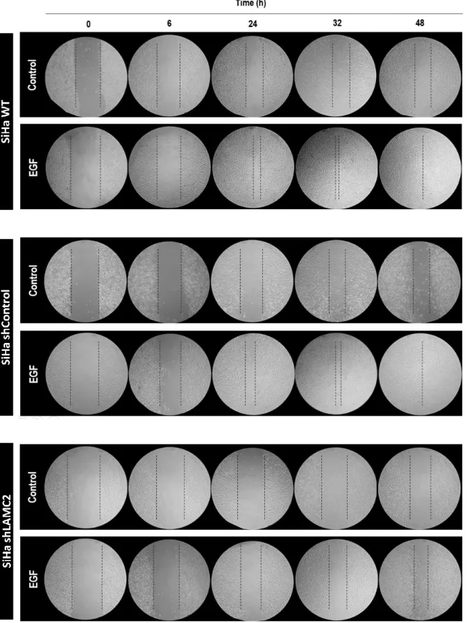
4.6 Silencing of LAMC2 suppresses SiHa migration and matrix invasion ... 15
4.7 MMP activity is increased by EGF ... 17
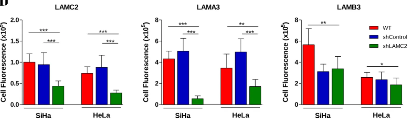
4.8 Knockdown LAMC2 chain affects the expression of LAMA3 and LAMB3 in SiHa and HeLa, at the protein level ... 18
4.9 Knockdown LAMC2 chain increases the expression of LAMC1 and in SiHa, at the mRNA level .. ... 20
4.10 Pulse chase analysis of LAMC2 expression proves the efficacy of shLAMC2 ... 22
4.11 LAMC1 expression is upregulated by EGF in Hela but not in SiHa ... 22
4.12 EGF modulates the FOXM1 and STAT3 binding to LAMC2 and LAMC1 ... 24
4.13 LAMC1 expression is controversial in uterine cervix cancer ... 25
5. Discussion ... 26
5.1 Main conclusions ... 29
5.2 Future perspectives ... 30
6. Bibliography ... 30
v
Index of figures and tables
Figure 1.1 – Main components and organization of extracellular matrix (ECM) ... 2
Figure 1.2 – Schematic illustration of laminin-332 structure ... 3
Figure 4.1 – The LAMC2 expression is significantly increased in tumor samples ... 10
Figure 4.2 – EGF stimulates proliferation of SiHa and HeLa cells ...11
Figure 4.3 – EGF decreases doubling time (DT) and cell cycle duration in SiHa and HeLa cells ... 12
Figure 4.4 – The effect of EGF in α3, β3 and 3 from laminin-332 and 1 and 3 chains expression in SiHa and HeLa cells ... 13
Figure 4.5 – EGF stimulates proliferation independently of LAMC2 in SiHa cells and more efficiently in the absence of LAMC2 in HeLa cells ... 14
Figure 4.6 – LAMC2 is crucial for SiHa cells migration ... 16
Figure 4.7 – LAMC2 is crucial for SiHa cells migration and invasive capacity ... 17
Figure 4.8 – Zymography of SiHa and HeLa media supernatants ... 17
Figure 4.9 – Knockdown of LAMC2 chain affects the expression of LAMA3 and LAMB3 in SiHa and HeLa ... 18
Figure 4.10 – In SiHa cells the knockdown of LAMC2 induced the increase of the expression LAMA3, LAMB3, LAMC1 and LAMC3 ... 21
Figure 4.11 – Pulse chase assay in SiHa and HeLa cells of LAMC2 expression proving knockdown efficacy ... 22
Figure 4.12 – LAMC1 expression is upregulated by EGF in Hela but not in SiHa ... 23
Figure 4.13 – EGF modulates the FOXM1 and STAT3 binding to LAMC2 and LAMC1 promoter regions ... 24
Figure 4.14 – The dynamics of LAMC1 expression is controversial in uterine cervix cancer ... 25
Supplementary Table 1 – Primers sequences used during the experimental work ... 38
vi
List of abbreviations, acronyms and symbols
AA – Antibiotic-antimycotic solutionAP-1 – Activator protein 1
ATCC – American Type Culture Collection BLCA – Bladder urothelial carcinoma BM – Basement membrane
BRCA – Breast invasive carcinoma BSA – Bovine serum albumine CDK – Cyclin-dependent kinase cDNA – Complementary DNA
CESC – Cervical squamous cell carcinoma and endocervical adenocarcinoma ChIP – Chromatin immunoprecipitation
CHOL – Cholangiocarcinoma CO2 – Carbon dioxide
COAD – Colon adenocarcinoma
COADREAD – Colon/rectal adenocarcinoma ddH2O – Sterile ultra-pure water
DMEM – Dulbecco’s modified Eagle media DNA – Deoxyribonucleic acid
dNTPs – Deoxynucleotides DT – Population doubling time DTT – Dithiothreitol
ECM – Extracellular matrix
EDTA – Ethylenediamine tetraacetic acid EGF – Epidermal growth factor
EGFR – Epidermal growth factor receptor ERK – Extracellular signal–regulated kinases ESCA – Esophageal carcinoma
FBS – Fetal bovine serum FGF – Fibroblast growth factor FOXM1 – Forkhead box protein M1 G0 – Cell cycle “resting phase” G1 – Gap 1 cell cycle phase G2 – Gap 2 cell cycle phase GEO – Gene Expression Omnibus
HER-2 – Human epidermal growth factor receptor 2 HGF – Hepatocyte growth factor
HNSC – Head and neck squamous cell carcinoma
HPRT – Hypoxanthine-guanine phosphoribosyltransferase HPV – Human papillomavirus
kDa – Kilodalton
KIRP – Kidney renal papillary cell carcinoma LAMA3 – Human laminin α3 chain
vii
LAMB3 – Human laminin β3 chain LAMC1 – Human laminin γ1 chain LAMC2 – Human laminin γ2 chain LAMC3 – Human laminin γ3 chain LUAD – Lung adenocarcinoma
LUSC – Lung squamous cell carcinoma M – Mitosis cell cycle phase
MAPK – Mitogen-activated protein kinase miR-29 – microRNA 29
MMP – Matrix metalloproteinase mRNA – Messenger RNA NF-kB – Factor nuclear kappa B PAAD – Pancreatic adenocarcinoma PAGE – Polyacrylamide gel electrophoresis PBS – Phosphate buffered saline
PCPG – Pheochromocytoma and paraganglioma PI – Propidium iodide
PI3K – Phosphatidylinositol 3-kinase PRAD – Prostate adenocarcinoma
qRT-PCR – Quantitative real time polymerase chain reaction READ – Rectum adenocarcinoma
RNA – Ribonucleic acid RNA-Seq – RNA sequencing
RSEM – RNA-Seq by Expectation Maximization S – Synthesis cell cycle phase
shRNA – Short hairpin RNA SKCM – Skin cutaneous melanoma STAD – Stomach adenocarcinoma
STAT3 – Signal transducer and activator of transcription 3 STES – Stomach and esophageal carcinoma
TCGA – The Cancer Genome Atlas TGF-β – Transforming growth factor-β TGS – Tris-glycine-sodium dodecyl sulfate THCA – Thyroid carcinoma
TNF-α – Tumor necrosis factor-α
UCEC – Uterine corpus endometrial carcinoma ZEB1 – Zinc finger E-box binding homeobox 1
1
1. Introduction
1.1 Cancer biology
Cancer is a multistep transformation process of normal cells to a neoplastic state, termed carcinogenesis. During this process cancer cells acquire features of malignancy, resulting from the accumulation of several alterations that allow them to become tumorigenic and ultimately malignant. Therefore, carcinogenesis needs genetic mutations and epigenetic alterations that lead to the activation of oncogenes (growth promoting), with dominant gain of function and/or increased expression, and into the inactivation of tumor suppressor genes (growth inhibitory) with recessive loss of function and/or decreased expression 1. In 2000, Hanahan and Weinberg, defined six cancer features so-called hallmarks
of cancer, which are: sustaining proliferative signaling, evasion to growth suppressors, cell death resistance, replicative immortality, angiogenesis capacity, and increase invasive and metastasis ability 2. The microenvironment that surrounds cancer cells also act on malignant transformation as a
functional network, where intervenient cells (malignant and normal) can share chemokines and energy molecules 3.
1.2 Uterine cervix cancer
Uterine cervix cancer is the fourth most common cancer in women worldwide, being diagnosed 528 000 new cases annually and 266 000 related-deaths were observed in 2012 4. In Europe, uterine cervix
carcinoma is the fifth most common cancer among women. Concerning Portugal, this is the sixth most common and deadly cancer among women. Its incidence and mortality are higher than in the other Southern European countries, being respectively estimated in 10.8 and 4.9 (rates per 100,000 person per year). Regarding prevalence, this cancer is the fourth most prevalent in Portugal, affecting most patients (approximately 52.2%) in a period of five years or more 5. There are several histological types of uterine
cervix cancer, however the two most common are squamous cell carcinomas and adenocarcinomas, comprising 75–90 and 10–25 % of all cases, respectively 6.
Human papillomavirus (HPV) is sexually transmitted and it was demonstrated as an etiological agent of several cancers in anogenital area, including uterine cervix cancer 7. It has been firmly established
the biological and epidemiological casual role of HPV in all uterine cervix cancer cases 8, wherein 70%
of these cancers are directly caused by 16 and 18 HPV types 9,10. E6 and E7 genes of high risk HPVs are
oncogenes that deregulate key cell cycle controls, being able to origin the malignant transformation of cells 11.
1.3 Extracellular matrix and cancer (ECM)
Extracellular matrix (ECM) is a relevant component of the tumor microenvironment. In the last years, it has been recognized as an important intervenient in the carcinogenic process. ECM is composed by a complex network of macromolecules, such as collagens, laminins, fibronectins and proteoglycans, giving to this structure distinctive physical and biochemical properties. In homeostasis, its main function is tissue support, however, it also participates in the control of cellular events such as cell proliferation, adhesion, migration, invasion and apoptosis 12.
The ECM deregulation benefits cancer progression, since during carcinogenesis, the remodeling of microenvironment leads to an increased release of ECM-associated growth factors, which will act on cancer and normal cells. In addition, it is also known that tumor cells modulate ECM to facilitate communications and escape the homeostatic control 12,13.
2
Figure 1.1 – Main components and organization of extracellular matrix (ECM). ECM is composed by a complex structure
of macromolecules, such as collagens, laminins, fibronectins and proteoglycans, giving to this network distinctive physical and biochemical properties. Besides tissue support, ECM also participates in the control of cellular events such as cell proliferation, differentiation, adhesion, migration, invasion and apoptosis. ECM molecules regulate the availability of growth factors and cytokines and they also interact with integrins and growth factor receptors. This way ECM can activate specific cytoplasmic signaling transduction pathways, which in turn, regulate the expression of genes involved in these cellular events (adapted from 14).
1.3.1 Laminin-332
Laminins are large extracellular glycoproteins that are a structural component and a major constituent of the anchoring filaments in the hemidesmosomal complex within the basement membrane (BM) compartment of ECM. All laminins are heterotrimeric proteins that contain three chains, termed α, β and . At present, five α, three β and three chains are described 15. There are 16 known laminins with
different properties resulting from the conjugation of different subunits. Their expression is highly regulated during development and these proteins have a tissue specific distribution 15–18. It is known that
both epithelial and mesenchymal cells contribute to the deposition of laminin into the basement membrane 19.
Laminin-332 (formerly termed laminin 5) is an epithelial-BM specific subtype of laminin and it is a trimer composed by α3, β3 and 2 chains. This isoform is present in the BM of the skin and other organs, contributing for the maintenance of epithelial-mesenchymal cohesion 20.In normal tissues, l aminin-332 interacts with integrins 31, 61 and 64 , promoting cell adhesion, migration and invasion, accounting for disease spreading 18,21.
Human laminin-332 precursor molecule is a high-molecular weight of 460 kDa, which upon secretion, originates 440 kDa and 400 kDa isoforms, resulting from extracellular processing of the α3 and γ2-chains 20. The coiled-coil structure is formed by domains I and II of each of the three chains.
Domain III of laminin-332 2 chain is an EGF-like domain, which can interact with EGFR. The globular domain is composed by five repeating segments at the structure base. The first three repeats are EGF-
BASEMENT MEMBRANE
Proteoglycan
Laminin
Epithelium
Fibroblast
Growth factor receptor Growth factor Fibronectin Integrins Laminin-332 EXTRACELLULAR MATRIX Capillary Type IV collagen
3 -like sequences (G1-3 domains) that have binding sites for integrin and growth factors receptors. The last two repeats contain a heparin/proteoglycan binding activity 22.
Figure 1.2 – Schematic illustration of laminin-332 structure: laminin-332 forms a cross-shaped structure containing three
chains (α3, β3 and 2). The main-specific binding sites for each chain are identified (adapted from 23).
1.3.2 Laminin-332 and cancer
The importance of laminin-332 is a highlight subject in diverse reviews where it was described new structural and regulatory functions of this macromolecule in several carcinomas. It has been suggested that this protein is implicated in carcinoma progression and analysis of protein expression has been used as an invasion diagnostic marker and prognostic tool 17,23.
Despite controversial studies, the abnormal expression of laminin and its integrin receptors is used as a mark of certain tumor types. Epithelial tumors (carcinomas) often secrete abundant amounts of laminin-332 and frequently express its ligand α6ß4integrin 24. High levels of laminin-332 expression
were found in several human cancers by immunohistochemical studies, being considered a poor prognosis factor and have been related to the invasive ability of several tumors, such as uterine cervix carcinoma, pancreatic carcinoma, hypopharyngeal cancer, urothelial cancer, small-sized lung adenocarcinoma, malignant glioma, gastric cancer, squamous cell carcinoma of the tongue, colorectal adenoma and hepatocellular carcinoma 18.
Laminin-332 has been identified as an activator of signaling pathways in cell. The high levels of large globular domain 4-5 of 3 chain expression was already demonstrated in carcinoma, in vivo, where it stimulates the mitogen-activated protein kinase (MAPK) and the phosphatidylinositol 3-kinase (PI3K) pathways. This alteration results in an increase of invasive capacity of cells and tumor growth, which could be reverted using blocking-antibodies against this domain of 3 chain 25. Regarding 3 and 2
chains, there are findings relating the cleavage of these chains by specific proteases with the increase in cell migration and invasion in carcinomas. The domain III of laminin 2 fragment is EGF like, being able to bind to EGFR and leading to the activation of extracellular signal–regulated kinases 1/2 (ERK1/2) from MAPK pathway 23.
1.3.3 Laminin 2
Human laminin γ2 chain (LAMC2) is the product of the LAMC2 gene expression and like the other chains it has two transcript variants, resulting from alternative splicing of the 3' terminal exon. The predicted molecular weight of long isoform is 131 kDa while for the short isoform is 122 kDa. However, it has the distinction of being specific to the laminin-332 trimer. Moreover, it is the only chain of the trimer that can be secreted as a monomer, remaining unclear its biological relevance 23.
4 Several immunohistochemistry and in situ hybridization studies showed strong expression of LAMC2 in many human cancers, including adenocarcinomas of the stomach 26, colon 27, pancreas 28,
thyroid 29, tongue 30, colorectal 31, lung 32, uterine cervix 33, and squamous carcinomas of the esophagus 34, head and neck 35, skin 36, lung 37, as well as transitional cell carcinoma of the bladder 38. The
cytoplasmic accumulation of LAMC2 has been frequently found at the invasive front of tumors, being associated with a poor survival, recurrence, and metastasis 22.
The processing of LAMC2 chain affects the dynamics of cellular adhesion. Moreover, LAMC2 chain undergoes proteolytic cleavage by specific enzymes as matrix metalloproteinases (MMPs) (MT1-MMP and MMP2) 39. The proteolysis of LAMC2 occur in the short arm of the molecule, resulting in a 100- or
105-kDa subunits, which harbors EGF-like repeats, allowing it to interact with cell surface receptors 23,
as EGFR, on which depends the activation of cell migration, invasion and proliferation40. This growth
factor-like role of LAMC2 fragments are sustained by the increased levels of both EGFR and LAMC2
23 in cancer cells, creating a positive-feedback loop in which EGF-EGFR binding can enhance the
expression of LAMC2 22.
Regarding uterine cervix cancer, some studies have shown elevated LAMC2 chain expression in microinvasive and invasive lesions 33,41. Despite the expression of LAMC2 chain has been described in
both squamous cell carcinomas and adenocarcinoma, in uterine cervix it is more frequently accumulated in squamous cell carcinoma 42.
1.4 Extracellular matrix proteolysis: Matrix metalloproteinases
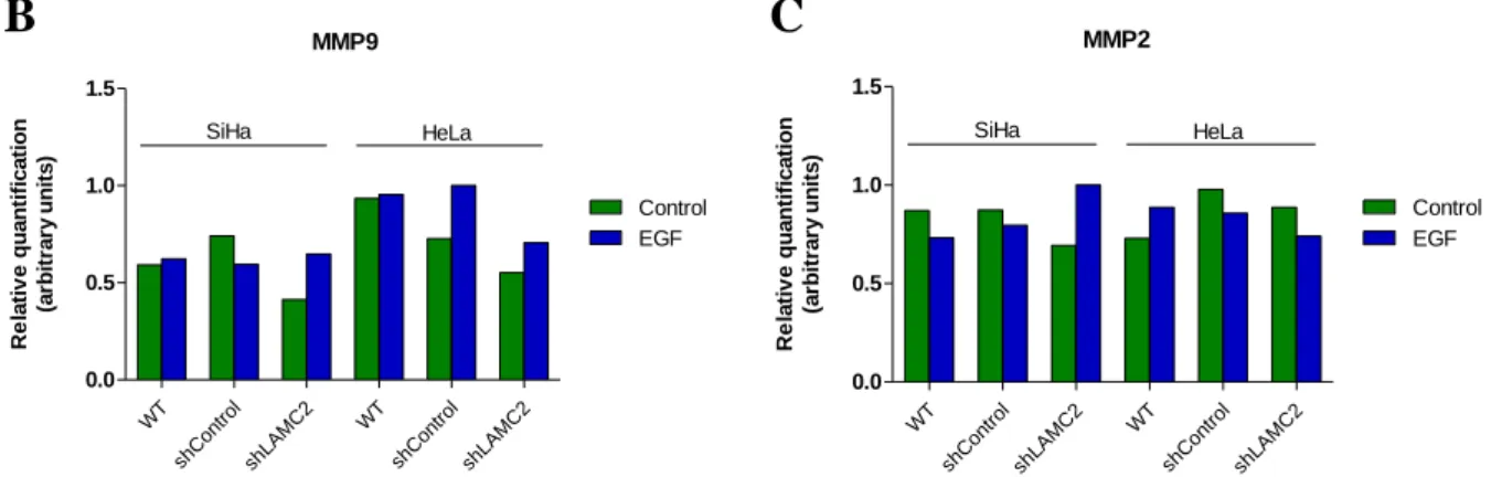
ECM formation and remodeling is regulated by proteolysis and contribute to tissue homeostasis. However, in cancer context proteolysis imbalances lead to deregulated tumor growth, tissue remodeling, inflammation, invasion and metastasis. The MMPs are the most relevant family of proteinases associated with tumorigenesis. Because of their role in extracellular matrix turnover and in the regulation of signaling pathways that are relevant for cancer progression, MMPs have been described as tumor microenvironment modulators 43.
MMPs are a family of zinc-dependent endopeptidases. In the cancer process, besides tumor cells, stromal, endothelial and inflammatory cells are capable of secreting MMPs. Those enzymes are capable of cleaving several macromolecules of the ECM, including laminins 43,44.
Gelatinases MMP2 and MMP9 are the prominent MMPs involved in basement membrane degradation and, consequently, involved in cancer development and metastasis 44. Cleavage of LAMC2 chain have
been associated to MMP2 activity in breast 45 and in uterine cervix cancers 46.
1.5 Growth factors and cancer
Growth factors are a class of compact polypeptides with the capacity to bind to transmembrane receptors harboring kinase activity domains that are localized at the cytoplasmic region of the molecule, stimulating specific intracellular signaling pathways 47. The kinase proteins have the ability to
phosphorylate specific amino acid residues, such as serine, threonine and tyrosine. The tyrosine kinase receptors phosphorylate tyrosine amino acid residues of several intracellular protein, which activates enzymatic cascades involved in cell growth and survival, however, MAPK and PI3K pathways are the mostly activated ones 48. Those growth factors assume an important role in tumor initiation, by
stimulation cell survival and clonal expansion, which permits fixation of oncogenic mutations, as well as tumor progression, invasion, angiogenesis and metastasis 48. Epidermal growth factor (EGF), which
comprises eleven polypeptides sharing a conserved EGF domain, is a relevant growth factor family in tumor progression 48,49.
The EGF ligands can be produced by cancer and stromal cells and it binds to tyrosine kinase receptors known as EGF receptors (EGFRs) 49. Each receptor comprises an extracellular domain to allow ligand
5 binding, a single transmembrane portion, and an intracellular protein tyrosine kinase domain 47. In
human cancers the EGFR and HER2 receptors are frequently overexpressed, which can result in the activation of proto-oncogene transcription factor 50,51. EGFR is frequently affected by non-sense
mutations that promote its constitutive activation, which releases it from the need of EGF stimuli to act
52. Alterations of these receptors were already described in non-small cell lung, bladder, uterine cervix,
ovarian, kidney, pancreatic and head and neck cancer 48. However, in uterine cervix cancer EGFR is so
far the most relevant EGF receptor 51,53.
1.6 Regulation of LAMC2 expression
The ECM-mediated and cytokine-mediated signaling pathways are intrinsically linked and this cross--talk can influence the composition of the basement membrane 54,55. In intestinal epithelial cells, both
transforming growth factor-β1 (TGF-β1) and hepatocyte growth factor (HGF) cytokines stimulate LAMC2 expression, result of activator protein-1 (AP-1) DNA binding sites activation on LAMC2 promoter region 54. Besides that, it was also demonstrated that tumor necrosis factor-α (TNF-α) regulates
LAMC2 transcription by an nuclear kappa B (NF-κB) bound enhancer 56. In colon and pancreatic cancer
cells, LAMC2 is under positive transcriptional control of Smad4, which in turn, is increased by TGF-β 57.
In vitro studies showed that the enhanced expression of LAMC2 is regulated by Wnt/β-catenin signaling pathway in gastric and colorectal cancers 31,58. The frizzled receptor activation after Wnt
binding results in an increase of β-catenin that induces indirectly the transcription of LAMC2 due to transcript factors binding to the AP-1 binding sites of LAMC2 promoter 22,58. Furthermore, unlike the
others laminin-332 chains, the LAMC2 gene expression is activated by zinc finger E-box binding homeobox 1 (ZEB1) transcript factor in colorectal cancers 59.
There are studies that associated the decreased expression of LAMC2 in lung, prostrate, breast and basal cell skin carcinoma to aberrant methylation of the LAMC2 promoter region 22. On the other side,
in gastric cancer, the LAMC2 is frequently overexpressed, being this pattern associated with promoter demethylation and histone modifications 60.
Moreover, recent studies have shown the significant downregulation of miR-29a/b/c in several cancers including uterine cervix cancer 61,62. These microRNAs molecules modulate the expression of
their target genes post-transcriptionally blocking mRNA translation. LAMC2 mRNA was identified as target of miR-29s 61. Thus, the decreasing of miR29 expression levels in cancer cells might result in an
upregulation of LAMC2 expression 22. Altogether, this regulation pathways lead to a significant increase
of LAMC2 release into extracellular compartment and consequently heterotrimerization followed by deposition in BM.
2. Aim of the project
This work aims to clarify the EGF mediated role of LAMC2 of laminin-332 in the progression of uterine cervix carcinomas, using an in vitro model of squamous cell carcinoma and adenocarcinoma.
In order to achieve our aim, we defined five specific aims to verify: (i) the role of EGF in cell cycle and proliferation; (ii) the effect of EGF stimulation in the expression of LAMA3, LAMB3, LAMC2, LAMC1 and LAMC3; (iii) the role of LAMC2 in EGF effect in cell cycle, proliferation and migration/invasion; (iv) the effect of LAMC2 knockdown in the expression of LAMC1 and LAMC3, and (v) the transcription factors involved in EGF dependent regulation of LAMC2 and LAMC1 expression.
6
3. Materials and methods
3.1 Cell lines and culture conditions
SiHa, a human uterine cervix squamous cell carcinoma (HTB-35, ATCC) and HeLa, a human uterine cervix adenocarcinoma (CCL-2, ATCC) cell lines were used as in vitro models of uterine cervix cancer. SiHa cells (-squamous cell carcinoma) are positive for HPV16 and HeLa (adenocarcinoma) are positive for HPV18 63. They were obtained from American Type Culture Collection (ATCC) and cultured in
Dulbecco’s modified Eagle media (DMEM 1x) (41965-039, Gibco, Life Technologies) in a humidified atmosphere of 5% CO2 at 37 °C. The culture medium was supplemented with 10% fetal bovine serum
(FBS) (S 0615, Merck) and 1% of antibiotic-antimycotic solution (AA) (P06-07300, PAN Biotech). Cells were grown to 75 - 100% optical confluence before they were detached with 0.05% trypsin-EDTA 1X. To determine the cell number necessary for each assay a Neubauer counting chamber was used.
For gene expression analysis by qRT-PCR (3.6), cells (2.5 × 10⁵ cells/mL) were plated in 6-well
plates and then exposed to growth factor (EGF- E9644, Sigma-Aldrich, MO, USA). After cells became adherent, they were synchronized under starvation (culture medium without FBS) for 8h. Cells were then cultured in normal (control) medium or exposed to 25ng/mL of EGF. Culture supernatants were analyzed by zymography (3.9).
For cell cycle analysis and proliferation curves (3.3 and 3.4), cells were plated in 48-well, when adherent they were submitted to starvation (~8h) and collected for analysis at 0, 6, 12, 24, 30 and 48h after EGF (25ng/mL) supplementation. Control curves were defined in the same time points using cells cultured in the absence of EGF.
For immunofluorescense (3.5), cells were grown on Millicell® EZ SLIDE, until they reached approximately 80% of confluence, or were attached to a glass slide by cytospin method (1200 rpm for 5 min) (Shandon CytoSpin II Cytocentrifuge). Cells were submitted to starvation and EGF stimulated as described above.
For wound healing assay, cells (2.5 x 10⁵ cells/mL) were plated in 12-well and cultured until reaching
a confluent monolayer. To inhibit cell proliferation, that could mask migration results, they were treated with Mitomycin-C (5 μg/mL) (M4287, Sigma), an antiproliferative agent, for 3 h prior starting the experiment. The effect of EGF was tested by exposing cells in the described above conditions and comparing with cells cultured in the absence of EGF, then cells were analyzed as presented in 3.7.
3.2 Bioinformatics analysis
LAMC2 expression was analyzed in several tumor groups, using data from The Cancer Genome Atlas (TCGA) and Gene Expression Omnibus (GEO). The RNA-Seq data (RSEM) were extracted. All disease groups with normal and tumor samples were considered to the present analysis.
3.3 Flow cytometry for cell cycle analysis
In order to divide, cells need to grow and replicate their DNA in a process known as cell cycle. The four major phases of the cell cycle are Gap 1 (G1), Synthesis (S), Gap 2 (G2), and Mitosis (M). The G1 phase precedes the DNA replication that occurs in S phase, and after G2 phase the cells finally divide in M phase64. G0 is the quiescent phase in which cells are not dividing. Using DNA staining dyes such
as propidium iodide (PI) (P4170, Sigma-Aldrich), the DNA content in the cells at G0/G1, S, and G2/M phases can be accurately quantified. Technically the method/protocol we used by flow cytometry do not allow to distinguish G0 phase from G1 phase and G2 phase from M phase 65.
After exposure to EGF as described in 3.1, cells were harvested and fixed in 70% ethanol at 4 ºC for at least 16 h. Cells were then centrifuged at 1200 rpm for 5 min, the supernatant was discarded and cells
7
were incubated with 100 μL of PI solution (50μg/mL PI, 0.1mg/mL RNase A, 0.05% Triton X-100) for 40min at 37 ºC. After washing with PBS 1X, cells were centrifuged 1500 rpm 10min 4 ºC and the supernatant was discarded. Cells were then suspended in 200 μL of PBS-BSA 0.1%. The flow cytometry analysis was performed in a FACScalibur (Becton Dickinson). Data were analyzed with FlowJo software, after excluding death cells and cell aggregates the univariate model was applied.
3.4 Cell proliferation curves and calculation of population doubling time (DT)
The cell number was calculated using a Neubauer chamber and viable cells were determined using 0.4% (w/v) trypan blue stain at a ratio of 1:4 or using flow cytometer counter. Population doubling time (DT) is the time required for a culture to double the number of cells and it was calculated according to ATCC® Animal cell culture Guide, using the following formula:
3.1 DT = ln2
ln (Xe/Xb) x T
where T is the incubation time in any units, Xb is the cell number at the beginning of the incubation time and Xe is the cell number at the end of the incubation time.
The duration of a particular phase of the cell cycle can be predicted using the following formula 66:
3.2 Tx =𝑙𝑛 (FS+1) ln 2 x DT
where Tx is the duration of cell cycle phase of interest (e.g. G0/G1 phases, S phase, G2/M phases), DT is the duration of cell cycle and FS is the fraction of cells in the cell cycle phase of interest, estimated from the DNA content frequent histogram.
3.5 Immunofluorescence
Immunofluorescence is a technique used to identify specific biomolecules in a cell, using specific antibodies labeled with fluorochromes, directly or indirectly, allowing their visualization and localization in the cell. In this work, indirect immunofluorescence was performed using two antibodies: a primary antibody that binds to the molecule of interest and then a secondary antibody covalently labeled with a fluorophore, which binds to the primary antibody.
Cells were fixed in 2% (w/v) paraformaldehyde (104003, Merck Millipore) for 15 minutes at 4 ºC, blocked with 0.1% (w/v) PBS-BSA for 30 minutes at room temperature, and incubated with primary antibody overnight (diluted in 0.1% (w/v) PBS-BSA, 1:100). Antibodies used were monoclonal anti-LAMA3 (BM165, kindly provided by Dr. Patricia Rousselle, Institut de Biologie et Chimie des Protéines, Lyon, France 67), polyclonal anti-LAMB3 (PA5-21514; Thermo Scientific, Sweden),
monoclonal anti-LAMC2 (MAB19562; Chemicon, Germany), monoclonal anti-LAMC2 (LS-C152903, LifeSpan BioSciences, USA), monoclonal anti-LAMC1 (AMAb91138, Atlas Antibodies, Sweden) . After washes, cells were incubated with the secondary antibodies for 2 hours, at room temperature. Secondary antibodies (diluted in 0.1% (w/v) PBS-BSA, 1:1000) used were: Alexa Fluor® 488 anti- -mouse (A-11001, Invitrogen) and Alexa Fluor® 488 anti-rabbit (A-11034, Invitrogen). Negative controls were stained without primary antibodies to ensure the specificity of the secondary antibody. After washing three times with PBS, samples were counterstained with VECTASHIELD media with DAPI (H-1200, Vector Labs, CA, USA). Cells were examined by standard fluorescence microscopy using a Nikon Instruments Eclipse Ti-S Inverted Microscope (Hamamatsu digital camera C10600 ORCA-R2) and an Olympus IX53 Inverted Microscope dedicated to fluorescence. Images (x200 field) were acquired and processed with NIS-Elements AR-3.2.software and Olympus cellSens software, respectively, and quantified with ImageJ software.
8
3.6 RNA extraction, reverse transcription and relative quantifying real-time
polymerase chain reaction (qRT-PCR)
Real-time PCR is an accurate and sensitive technique that combines both amplification and detection in one step. Gene transcription activity can be evaluated by mRNA quantification, although this analysis can be somehow affected by different mRNA stability and translational rates. The complementary DNA (cDNA) is synthesized from mRNA using a reverse transcriptase. Then cDNA is used as a template in real-time68. The qRT-PCR analysis is normalized for a housekeeping gene, which is constitutively
expressed in cells69.
RNA was extracted using RNeasy Mini Kit Qiagen® (74104, Qiagen), according to the manufacturers’ protocol. RNA concentration of each sample was measured at 260nm, in a NanoDrop 2000 (ND-2000, Thermo Scientific). The RNA was then converted to cDNA, using 1μg of total RNA, by incubation into a buffer with random primers (11034731001, Roche)) at 70 °C (primers annealing temperature), for 10 min, then, at 4 ºC was added a mixture with: First Strand Buffer 5X (Y00146, Invitrogen), reverse transcriptase (SuperScript™ II) (18080-44, Invitrogen), dithiothreitol (DTT)
(Y00147, Invitrogen), RNAse OUT™ (10777-019, Invitrogen), deoxynucleotides (dNTPs) mix (10mM) (28-4065-22V, 28-4065-02V, 28-4065-12V and 28-4065-32V, GE Healthcare), and purified water (ddH2O) up to 12 μL. Relative quantifying PCR was performed using cDNA, specific
pairs of primers for each gene and SYBR
®
Green Master Mix (04707516001, Roche) according to manufacturer’s instructions in Lightcycler® 480 System instrument (05015243001, Roche). Hypoxanthine-guanine phosphoribosyltransferase (HPRT) was used as housekeeping gene. Primer sequences are presented in Supplementary Table 1.3.7 Wound healing assay
Wound healing assay is a method used to measure directional cell migration in vitro, mimicking cell migration during wound healing in vivo 70. This assay was performed in order to identify a potential role
of EGF, as well as the effect of LAMC2, on cell migration.
In each cell monolayer, a scratch was made to the diameter of the well, and the wound healing was followed by acquiring phase-contrast images (x200 field) at the following time points: 0, 6, 24, 32 and 48 hours.
3.8 Invasion assay
Transwell inserts (8-μm pore size) (PI8P01250, Millicell) were used to determine the effect of LAMC2 knockdown on invasion of uterine cervix cell lines in vitro.
For invasion assay, we coated the filter of the transwell inserts with 50 µl of Matrigel. Cells (1x10⁵
per well) were resuspended in 200 µl FBS-free medium and placed in the upper chamber for each group (shControl or shLAMC2). The lower chamber was filled with 500 µl medium containing 10% FBS as a chemoattractant and incubated for 48 h. At the end of the experiment, cells from the upper chamber were removed using a cotton swab. After wash twice into cold PBS 1x, cells on the lower surface of the insert’s filter were fixed using 70% methanol overnight at -20 ºC. Then, cells were stained with 0.5% crystal violet in methanol (25%) for 10 min at room temperature. After washing with 10% methanol, phase-contrast images were taken (x200 field).
9
3.9 Zymography
Zymography is an electrophoretic technique useful for analyzing the activity of hydrolytic enzymes, such as the matrix metalloproteinases, based on the enzyme specific substrates. In this method, performed with native polyacrylamide gel (PAGE), the proteins are separated according to their hydrodynamic size. MMPs activity is detected based on the degradation of gelatin incorporated in PAGE 71.
Media supernatants of cell line were concentrated by using Amicon® Ultra-4 Centrifugal Filter Units (UFC800308, Millipore). After electrophoresis with TGS buffer 1X (161-0772, Bio-Rad) (150V for 90 minutes) in a 12% PAGE with 0.1% (w/v) gelatin, gel was incubated in renaturating buffer (25% TritonX-100 (v/v)) for 1 hour with agitation, and lastly, was incubated overnight at 37°C, in collagenase buffer (6.06 g Tris-base (T6066, Sigma), 11.7 g sodium chloride (1.06404, EMD Millipore), 0.55 g calcium chloride (1.02382, EMD Millipore) and distilled water up to 100 mL (pH 7.6)). Staining was performed with 0.5% (w/v) Coomassie Blue R-250 (27816, Sigma) for 30 minutes and distaining with distilled water.
3.10 Chromatin immunoprecipitation (ChIP)
Chromatin immunoprecipitation (ChIP) is an experimental technique used to investigate the interaction between proteins and chromatin in a cell 72. In this work it was used to determine whether
specific proteins, such as transcription factors, were associated with specific genomic regions. Cells were treated with 37% formaldehyde to crosslink proteins and DNA keeping the chromatin structure, and terminated with 0.125M glycine. After, samples were sonicated and the size of the chromatin fragments was evaluated by electrophoresis gel, in a 1.2% (w/v) agarose, having fragments mainly with a size between 1000bp and 500bp. The chromatin complexes were immunoprecipitated with 1 µL (~2µg/mL) of specific antibodies: rabbitanti-FOXM1 (NBP1-30961, Novus Biologicals, United Kingdom) and rabbit anti-phospho STAT3 (Tyr705) (9145S, Cell Signaling Technology, MA, USA). ChIP assay was performed using OneDay ChIP kit (kch-onedIP-060, Diagenode) according to the manufacturer’s protocol. Primers were designed to amplify a putative FOXM1 and STAT3 binding sites at the LAMC1 and LAMC2 promoters. Amplification of promoter regions sequenced from released DNA was performed by qRT-PCR as described in 3.6. The relative occupancy of binding sites was calculated using the following formula:
3.3 Relative occupancy = 2(CtNegCtl − CtTarget)
3.11 Statistical analysis
All statistical analyses were performed using GraphPad Prism 6.0 software (www.graphpad.com). Data for each study parameter from each group were presented as the mean (normal distribution) or median (non-normal distribution) ± SD. Assays were performed with, at least, 3 replicates per condition. Comparisons between data from each group were statistically analyzed by a two-tailed unpaired Student's t-test. Differences were considered statistically significant at p<0.05.
10
4. Results
4.1 LAMC2 is increased in cancer versus normal tissues
In order to understand the significance of LAMC2 chain in cancer context, LAMC2 mRNA expression data from normal tissue and tumor were analyzed. The dataset was obtained from The Cancer Genome Atlas (TCGA). Considering 27 groups with different cancers, about 59.26% (16 groups) of them exhibited a higher mRNA expression of LAMC2 in tumor samples, comparatively to normal tissues (Figure 4.1 A). Regarding these 16 groups, 87.5% (14) of them showed a highly significant expression of LAMC2 (p<0.0001, for bladder urothelial carcinoma (BLCA), cholangiocarcinoma (CHOL), colon adenocarcinoma (COAD), colon/rectal adenocarcinoma (COADREAD), esophageal carcinoma (ESCA), head and neck squamous cell carcinoma (HNSC), lung squamous cell carcinoma (LUSC), stomach adenocarcinoma (STAD), stomach and esophageal carcinoma (STES), thyroid carcinoma (THCA) and uterine corpus endometrial carcinoma (UCEC), p=0.0378 for kidney renal papillary cell carcinoma (KIRP) and p=0.0050 for rectum adenocarcinoma (READ)). For the purpose of knowing further about the relevance of LAMC2 in uterine cervix cancer, a dataset of squamous cell carcinoma and adenocarcinoma (CESC) were analyzed. In order to get more information, data from Gene Expression Omnibus (GEO) were joined to TCGA data. The expression of LAMC2 in 332 cancer samples was significantly higher (p< 0.0001), comparatively to 27 normal uterine cervix tissues present in both databases (Figure 4.1 B). Moreover, comparing the 3 uterine cervix tumor samples that have matched non-tumor samples, we observed that LAMC2 expression also increased in tumors (p=0.0183) (Figure 4.1 C). TCGA BLCA CH OL CO AD CO ADREA D ESCA HN SC
KIRP LUSC PAA
D
PCPG REA
D
STAD STES THCA UC
EC -5 0 5 10 15 20 25 Tumor *** ** *** *** *** *** *** * *** *** *** *** *** Normal L o g 2 m R N A o f L A M C 2 e x p re s s io n TCGA - CESC Normal Tumor 0 5 10 15 20 25 * n=3 n=3 L o g 2 m RN A o f L A M C2 e x p re s s io n
TCGA + GEO (CESC)
Normal Tumor 0 5 10 15 20 25 *** n=27 n=332 L o g 2 m RN A o f L A M C2 e x p re s s io n
A
B
C
11
Figure 4.1 – The LAMC2 expression is significantly increased in tumor samples. Expression levels of LAMC2 by RNA-
-Seq data (RSEM), available in TCGA and GEO databases. (A) All comparable groups that showed upregulation of LAMC2 expression in tumor samples are represented on graph. Results of bladder urothelial carcinoma (BLCA), cholangiocarcinoma (CHOL), colon adenocarcinoma (COAD), colon/rectal adenocarcinoma (COADREAD), esophageal carcinoma (ESCA), head and neck squamous cell carcinoma (HNSC), kidney renal papillary cell carcinoma (KIRP), lung squamous cell carcinoma (LUSC), pancreatic adenocarcinoma (PAAD), pheochromocytoma and paraganglioma (PCPG), rectum adenocarcinoma (READ), stomach adenocarcinoma (STAD), stomach and esophageal carcinoma (STES), thyroid carcinoma (THCA) and uterine corpus endometrial carcinoma (UCEC). (B) The mRNA expression of LAMC2 is upregulated in squamous cell carcinoma and adenocarcinoma (CESC) of uterine cervix tumor samples compared with the normal tissues revealed by TCGA+GEO dataset. (C) LAMC2 expression also is upregulated in three normal-matched tumor tissues from TCGA dataset. Results are shown as median with interquartile range. *p<0.05, **p<0.01, ***p<0.001.
4.2 EGF stimulates proliferation of SiHa and HeLa cells
Before addressing the role of LAMC2 in cancer features, we tried to understand if EGF was a suitable growth factor to stimulate our cell models. Hence, we addressed the EGF effect on the cell cycle by flow cytometry. Similar percentage of cells in G0/G1 were found in both cell lines in control conditions at the beginning of experience (0 h) and at 16h (Figure 4.2). However, in both cell lines the percentage of cells in G0/G1 significantly decreases (p<0.0001 for both cell lines) with the EGF supplementation with a consequently increase of cells in S+G2/M phases, showing that EGF stimulates cell proliferation.
Figure 4.2 – EGF stimulates proliferation of SiHa and HeLa cells. Cell cycle analysis was performed by flow cytometry,
cells were collected in three groups: controls (0h and 16 h) and 16h in EGF supplied medium. Based on DNA content histograms, the stacked bar graph represents the percentage of cells in different cell cycle phases. Both cells significantly (p<0.0001) responded to EGF treatment, increasing cell proliferation. Results are shown as mean ± SD. *p<0.05, **p<0.01, ***p<0.001.
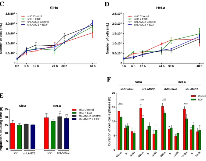
4.3 EGF decreases doubling time (DT) and cell cycle duration in SiHa and HeLa cells
A cell proliferation assay was performed in order to understand how EGF treatment affects the growth population dynamics over 48 hours. For that, except to control groups, cells were supplied with EGF after 8 h on starvation and collected at 6, 12, 24, 30 and 48 hours.
Growth curves showed that cells treated with EGF proliferate slightly more compared to control groups (Figure 4.3 A and B). The population doubling time (DT) is also lower in EGF conditions, demonstrating that EGF tends to accelerate the cell cycle in both cell lines, though not in a statistical significant way (Figure 4.3 C). Furthermore, SiHa cells needed significantly (p=0.0460 for control and p=0.0041 for EGF treatment) less time to duplicate, observed by the lower DT in comparison to HeLa cells. However, there were no significant differences between treatments. Thus, only a biologic tendency of EGF to accelerate the population growth due by the increasing of cell division speed was found.
HeLa 0 h EGF 0 20 40 60 80 100 120 G0/G1 S G2/M 16 h *** % o f c e ll s SiHa 0 h EGF 0 20 40 60 80 100 120 G0/G1 S G2/M 16 h *** % o f c e ll s
12 To determine how EGF could accelerate cell cycle, the duration of each cell cycle phase was calculated (Figure 4.3 D). The results showed that in both cell lines the EGF suppling interfered with the first phase of the cycle, G0/G1. Thus, the treatment significantly (p<0.0001 for both cell lines) decreased the time necessary to complete this phase. Due to this, the expression of possible interveners of this cell cycle phase were also analyzed. Accordingly a significant (p=0.0001 for SiHa and p=0.0002 for HeLa) increase in cyclin D1 mRNA levels was observed upon EGF exposure (Figure 4.3 E). These results indicated that the higher amounts of cyclin D1 in both cell lines, resulted of EGF stimulation, leading to the acceleration of the G0/G1, which, in turn, resulted in increased proliferation.
Figure 4.3 – EGF decreases doubling time (DT) and cell cycle duration in SiHa and HeLa cells. Proliferation curves,
obtained by counting the number of cells, representing SiHa (A) and HeLa (B) population dynamics over 48 hours. Cells treated with EGF (green) proliferate slightly more compared to control groups (red). (C) Doubling time (DT) of SiHa and HeLa cells cultured in control conditions or in EGF supplied medium. EGF treatment tends to decrease DT in both cell lines. (D) Representation of duration of each cell cycle phases with or without EGF supplementation. G0/G1 phases were significantly accelerated (p<0.0001 for both cell lines). (E) The relative CCND1 mRNA quantification in control conditions and after EGF stimulation showed that both cell lines express higher amounts of cyclin D1 in presence of EGF. Results are shown as mean ± SD. *p<0.05, **p<0.01, ***p<0.001. Significant differences: asterisks (*), effect of EGF treatment and hashes (#), comparison between cell lines.
0 10 20 30 Control EGF SiHa HeLa *** *** R e la ti v e CCND1 m R N A e x p re s s io n ( a rb it ra ry u n it s ) SiHa 0 h 6 h 12 h 24 h 30 h 48 h 0 5.0104 1.0105 1.5105 2.0105 EGF Control Nu m b e r o f c e ll s ( m L ) HeLa 0 h 6 h 12 h 24 h 30 h 48 h 0 5.0104 1.0105 1.5105 2.0105 Control EGF Nu m b e r o f c e ll s ( m L ) G0/G 1 S G2/M G0/G 1 S G2/M 0 5 10 15 20 *** *** SiHa HeLa Du ra ti o n o f c e ll c y c le p h a s e s ( h ) 0 10 20 30 40 SiHa HeLa # ## P o p u la ti o n d o u b li n g t im e ( h )