a
1 Chain Mediated Rescue of Murine Laminin
a
2 Chain
Deficiency
Kinga I. Gawlik, Mikael A˚ kerlund, Virginie Carmignac, Harri Elamaa¤, Madeleine Durbeej*
Department of Experimental Medical Science, Muscle Biology Unit, University of Lund, Lund, Sweden
Abstract
Background:Laminina2 chain mutations cause congenital muscular dystrophy with dysmyelination neuropathy (MDC1A). Previously, we demonstrated that laminina1 chain ameliorates the disease in mice. Dystroglycan and integrins are major laminin receptors. Unlike laminina2 chain,a1 chain binds the receptors by separate domains; laminin globular (LG) domains 4 and LG1-3, respectively. Thus, the laminina1 chain is an excellent tool to distinguish between the roles of dystroglycan and integrins in the neuromuscular system.
Methodology/Principal Findings:Here, we provide insights into the functions of laminina1LG domains and the division of their roles in MDC1A pathogenesis and rescue. Overexpression of laminina1 chain that lacks the dystroglycan binding LG4-5 domains ina2 chain deficient mice resulted in prolonged lifespan and improved health. Importantly, diaphragm and heart muscles were corrected, whereas limb muscles were dystrophic, indicating that different muscles have different requirements for LG4-5 domains. Furthermore, the regenerative capacity of the skeletal muscle did not depend on laminin
a1LG4-5. However, this domain was crucial for preventing apoptosis in limb muscles, essential for myelination in peripheral nerve and important for basement membrane assembly.
Conclusions/Significance:These results show that laminina1LG domains and consequently their receptors have disparate functions in the neuromuscular system. Understanding these interactions could contribute to design and optimization of future medical treatment for MDC1A patients.
Citation:Gawlik KI, A˚kerlund M, Carmignac V, Elamaa H, Durbeej M (2010) Distinct Roles for Laminin Globular Domains in Laminina1 Chain Mediated Rescue of Murine Laminina2 Chain Deficiency. PLoS ONE 5(7): e11549. doi:10.1371/journal.pone.0011549
Editor:Antoni L. Andreu, Hospital Vall d’Hebron, Spain
ReceivedMay 12, 2010;AcceptedJune 21, 2010;PublishedJuly 19, 2010
Copyright:ß2010 Gawlik et al. This is an open-access article distributed under the terms of the Creative Commons Attribution License, which permits unrestricted use, distribution, and reproduction in any medium, provided the original author and source are credited.
Funding:Funded by Association Francaise contre les Myopathies, Muscular Dystrophy Association, Anna-Greta Crafoord Foundation for Rheumatological Research, Greta and Johan Kock Foundation and Alfred O¨sterlund Foundation. The funders had no role in study design, data collection and analysis, decision to publish, or preparation of the manuscript.
Competing Interests:The authors have declared that no competing interests exist. * E-mail: madeleine.durbeej-hjalt@med.lu.se
¤ Current address: Department of Medical Biochemistry and Molecular Biology, Biocenter Oulu, Center for Cell-Matrix Research, University of Oulu, Oulu, Finland
Introduction
Congenital muscular dystrophy type 1A (MDC1A) is an autosomal recessive disorder caused by mutations in the gene encoding laminin (LM)a2 chain. The general clinical hallmarks of MDC1A include neonatal onset of muscle weakness, hypotonia often associated with joint contractures, inability to stand and walk, elevated levels of creatine kinase, white matter abnormalities and dysmyelination neuropathy. Histological changes of muscles comprise fiber size variability, massive degeneration and extensive connective tissue infiltration. Most patients die as teenagers since there is no treatment for this devastating disease [1]. Several mouse models for MDC1A exist (e.g. generated LMa2 chain mutants dy3K/dy3Kand dyW/dyW and the spontaneous mutant mouse strain dy/dy) and they adequately mirror the human condition [2–4].
LMs are extracellular proteins formed by a, b and cchains. Together with other extracellular matrix components LMs form specialized extracellular matrices called basement membranes [5]. LM-211 (composed of a2, b1 and c1 chains) is the major LM
isoform expressed in muscle and peripheral nerve. Through interaction with transmembrane receptors it regulates major functions of the neuromuscular system and provides structural support to muscle fibers [6]. In muscle, at least two distinct protein complexes are known to be the key receptors for LMa2 chain; dystroglycan and integrina7b1. Their importance is underscored by the fact that absence of integrin a7 chain, as well as hypoglycosylation of a-dystroglycan cause various forms of congenital muscular dystrophy [7,8]. Furthermore, different studies involving manipulation of the dystroglycan gene in mice revealed an important role for dystroglycan in skeletal muscle [9–11]. Several studies indicated that the function of integrina7 subunit and dystroglycan, being a part of the dystrophin-glycoprotein complex, could overlap [12–14]. However, recent studies show that whereas both dystroglycan and integrina7 chain contribute to force-production of muscles, only dystroglycan contributes to the preservation of sarcolemmal integrity [15].
shown to be important for different aspects of myelination and morphology of peripheral nerves, as revealed by conditional disruption of their genes in Schwann cells [18–20]. Thus, LM-211 is a central player linking these receptors and their functions in the neuromuscular system.
LMa1 chain also binds to dystroglycan, integrin a6b1 and integrina7b1 (and perhaps integrina6b4) [17,21–24]. Yet, it is not expressed in the neuromuscular system [25]. We have previously explored the possibilities of paralogous gene therapy for MDC1A and demonstrated that LMa1 chain is an excellent substitute for LMa2 chain in murine muscle, peripheral nerve and testis [25–28]. Additionally, LMa2 chain deficiency leads to perturbed expression of integrin a7 subunit, and reduced expression of the core protein of a-dystroglycan (but not a -dystroglycan glycosylation), at the sarcolemma [29–31]. Notably, LMa1 chain overexpression restores integrina7 chain expression, indicating that this receptor could be crucial for improvement of muscle function in dystrophic animals [32].
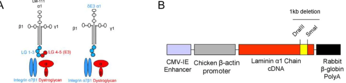
The LMa1 and a2 chains bind dystroglycan and integrins by distinct domains. The a1 chain binds dystroglycan via its C-terminal LG4 domain and integrin binding occurs viaa1LG1-3 [33,34]. This is different from LMa2 chain binding where there is considerable overlap in binding to dystroglycan and integrins. Both a2LG4-5 and a2LG1-3 bind dystroglycan, whereas only a2LG1-3 binds integrins [23,35]. The LMa1 chain can thus be used more efficiently to distinguish between the roles of LM binding to dystroglycan and integrins in the neuromuscular system. Since LMa1 chain functions almost equally well as a2 chain in the neuromuscular system, we used this subunit in order to dissect the roles of the aLG domains and their receptors in MDC1A pathogenesis and rescue. Hence, we produced and characterized animals completely deficient in LMa2 chain, but instead overexpressing a truncated form of LMa1 chain (dy3K/dE3 mice) that lacks the dystroglycan binding site (LG4-5 domains at the C-terminus, also known as the E3 fragment), but retains the integrin binding site (LG1-3, see Fig. 1A) [33,34].
Materials and Methods
Ethics statement
All mouse experimentation was approved by the local (Lund district) ethics committee (permit number M62-09). All mice were maintained in animal facilities according to animal care guidelines.
Transgenic construct
Approximately 1 kb of the C-terminal part was removed from mouse full-length LMa1 chain cDNA to generate truncated
cDNA (dE3LMa1). An in frame deletion between nucleotides 8248–9289 (corresponding to LG4-5 domains) was accom-plished by DraIII-SmaI restriction cutting and fusion of an XhoI site with a BglII site. This DNA was cloned into the pCAGGS vector [25], containing a CMV enhancer and a b -actin promoter.
Transgenic animals
Transgenic mice were generated by microinjections of transgene DNA into the pronucleus of fertilized single-cell C57BL/CBA embryos (Lund Transgenic Core Facility, Lund University, Sweden). Mice carryingdE3LMa1 chain DNA were identified by PCR as described previously [25]. Positive founders overexpressing truncated LMa1 chain in the neuro-muscular system (lines No. 3 and 4) were further bred with dy3K/+ mice [2], followed by sib breeding to generate LMa2 chain deficient animals that expressdE3LMa1 chain (dy3K/dE3 mice). Dy3K/dy3Kmice overexpressing full length LMa1 chain (dy3KLMa1 mice) were previously described [25–28]. Dy/dy mice used for heart studies were obtained from Jackson Laboratory.
Exploratory locomotion and body and muscle weight analyses
Exploratory locomotion was examined in an open field test. A mouse was placed into a new cage and allowed to explore the cage for 5 min. The time that the mouse spent moving around was measured. For all experiments, 10-week-old dy3K/dE3 animals (n = 16) were compared with 10-week-old control mice (wild-type or dy3K/+) (n = 8) and 5-week-old dy3K/dy3K mice (n = 6). For weight analysis dy3K/dE3, control mice and dy3K/dy3K animals were sex- and age-matched (5-week-old) (n = 14, n = 3, n = 11, respectively, for females; n = 8, n = 4, n = 8, respectively, for males). Quadriceps and tibialis anterior muscles from 2-month-old wild-type (n = 3), dy3K/dE3 (n = 3) and 4-week-old dy3K/dy3K
mice (n = 4) were used to estimate the ratio of wet muscle weight to body weight. Muscles from both legs were weighed and average muscle mass was calculated. Unpaired t-test was used for statistical analysis.
Creatine kinase activity
Blood was collected from the tail vein of 2-month-old control mice (wild-type ordy3K/+) (n = 10),dy3K/dE3 (n = 10) and 4-week-olddy3K/dy3Kmice (n = 3) into EDTA-tubes and spun down two times for 5 minutes at 3500 rpm. CK_P_S_cobas method was used by Clinical Chemistry Laboratory at Ska˚ne University
Figure 1. Generation ofdE3LMa1 transgenic animals.(A) Scheme presenting LM-111 structure. Full-length LMa1 chain with LG1-5 domains and truncated LMa1 chain (dE3LMa1) with LG1-3 domains are marked together with their transmembrane receptors. (B) Schematic presentation of transgenic construct with denoted 1 kb deletion (LG4-5). Restriction sites used to engineer the construct are shown.
doi:10.1371/journal.pone.0011549.g001
Hospital to quantify enzyme activity in plasma. Unpaired t-test was used for statistical analyses.
Histology and immunofluorescence microscopy
Skeletal muscle, heart, peripheral nerve and spinal roots cryosections (7mm) from control (wild-type ordy3K/+),dy3K/dy3K,
dy/dy, dy3K/dE3 and dy3KLMa1 mice were either stained with hematoxylin and eosin or subjected to immunofluorescence analysis using following antibodies: rat monoclonal mAb200 against LMa1LG4 domain [25], rabbit polyclonal 1057+against LMa1 LN/LEa domain (N-terminus) (kindly provided by Dr. T. Sasaki) [36], rabbit polyclonal 1100+ against LMa4, (kindly provided by Dr. T. Sasaki), rabbit polyclonal 1113+ against LMa5 (kindly provided by Dr. T. Sasaki), rat monoclonal MTn15 against tenascin-C [25], rabbit polyclonal U31 against integrin a7B subunit (kindly provided by Dr. U. Mayer) [37], mouse monoclonal IIH6 against a-dystroglycan (Upstate Bio-technology), mouse monoclonal F1.652 against embryonic myosin heavy chain (Developmental Studies Hybridoma Bank, Iowa), rabbit polyclonal anti-collagen, type IV (Chemicon), mouse monoclonal 46 against caspase-3 (BD Transduction Laboratories). Mouse on mouse kit (Vector) was used for staining with embryonic myosin heavy chain according to manufacturer instructions. Tissues were fixed with 4% PFA at room temperature (for laminin, tenascin-C, embryonic myosin heavy chain, collagen-IV and caspase-3 stainings), or with acetone at
220uC (for integrina7B) or with 8% formaldehyde, followed by methanol at220uC (fora-dystroglycan). Sections were analyzed using a Zeiss Axioplan fluorescence microscope. Images were captured using an ORCA 1394 ER digital camera with Openlab 3 software. Images were prepared for publication using Adobe Photoshop software.
Immunoblotting
For LM detection proteins were isolated from 100 mg ofdy3K/ dE3 and dy3KLMa1 muscles (3 mice from each group) by brief sonication in 1 mmol/L EDTA in TBS with 1:25 dilution of protease inhibitors (Complete EDTA-free, Roche Diagnostics). For integrin detection proteins were extracted from 100 mg skeletal homogenized muscle powder of 3 wild-type anddy3K/dE3 mice in 1% Triton X-100, 50 mM Tris-HCl, pH 7,4; 1 mM CaCl2, 1 mM MgCl2 and 1:25 dilution of Protease Cocktail
(Complete EDTA-free, Roche Diagnostics). Samples were incu-bated for 1 hour and spun down at 4uC. The supernatants were collected and the protein concentration was determined using BCA assay (Pierce). Dystroglycan was isolated using agarose bound wheat germ agglutinin (Vector) and N-acetyl-D-glucos-amine (Sigma) as described before [32]. Lysates containing LM, integrin and dystroglycan were separated on 5% or 8% polyacrylamide-SDS gels under reducing or non-reducing condi-tions. EHS LM (Invitrogen) was used as a control for LM blotting. Proteins were transferred to nitrocellulose membranes (Amer-sham). Membranes were blocked for 1 hour in 5% non-fat dry milk in 1xTBS with 0.02% Tween-20 and incubated overnight at 4uC with a rabbit polyclonal antibody detecting LMa1LG3 domain (kindly provided by Dr. T. Sasaki); rabbit polyclonal antibody against integrina7B (kindly provided by Dr. U. Mayer); rabbit polyclonal antibody againstb-dystroglycan [25] and mouse monoclonal antibody IIH6 againsta-dystroglycan. Detection was performed with ECL kit (Amersham). Expression of LMa1 chain, integrina7B subunit,a- andb-dystroglycan was normalized toa -actinin expression (detected with mouse monoclonal antibody EA-53, Sigma). Band intensity was measured using ImageJ software. Unpaired t-test was used for statistical analyses.
Quantification of fiber size distribution, central nucleation and fiber number
Diaphragm and limb muscles from at least 3 animals from each group (4–6-week-old wild-type,dy3K/dy3Kanddy3K/dE3 mice) were analyzed. Minimal Feret’s diameter was measured [38] for at least 2600 fibers for each group. The same number of fibers was used for quantification of fibers with centrally located nuclei. An additional group of 4–6-month-olddy3K/dE3 animals was included for quantification of diaphragm fibers. Fibers from quadriceps muscle from 4–6-week-old wild-type (n = 3),dy3K/dy3K(n = 3) and
dy3K/dE3 mice (n = 3) were counted within a square of 646106 pixels2. Unpaired t-test was used for statistical analysis.
Treadmill exercise and Evans blue dye injection
Dy3K/dE3 mice (n = 4) were exercised for 30 min on a treadmill
Exer 6M (Columbus Instruments) at a downhill angle of 15u. During the first 2 min the speed was gradually increased from 7 m/min up to 14–16 m/min. Within 30 min after completed exercise the mice were injected i.p. with Evans blue dye (EBD) (Sigma Aldrich) dissolved in sterile saline (concentration: 0.5 mg EBD/0.05 ml saline; amount: 50ml per 10 g body weight). After
approximately 24 h, muscles were collected and quickly frozen in liquid nitrogen. Unexercised mice were injected with EBD and used as controls. Muscle cryosections (8mm) were fixed in ice-cold acetone at 220uC for 10 min, washed and mounted with FluorSave (Calbiochem). By fluorescence microscopy analysis, EBD uptake into muscle fibers was visualized by red emission.
Cardiotoxin injections
Tibialis anterior muscles from 6 control (wild-type ordy3K/+), 6
dy3K/dy3K and 6 dy3K/dE3 mice were injected with cardiotoxin (10mmol/L in saline). Control and dy3K/dE3 mice were
2–3-month-old. Dy3K/dy3K mice were 3-week-old. Three mice from each group were sacrificed 4 days after injection and the other 3 after 11 days. Both injected and contralateral uninjected tibialis anterior muscles were collected and analyzed.
Electron microscopy and toluidine blue staining
Quadriceps femoris muscles, heart, diaphragm, sciatic nerves and spinal roots from wild-type,dy3K/dy3Kanddy3K/dE3 mice were fixed for 2 hours with 1.5% glutaraldehyde/1.5% paraformalde-hyde, rinsed in So¨rensen’s phosphate buffer, post fixed in 1% OsO4 and then embedded in Epon. Ultra thin sections were
stained with uranyl acetate and lead citrate. Specimens were examined by transmission electron microscopy (Philips CM 10). Three to 4 animals from each group were analyzed.
Results
Generation ofdy3K/dy3Kmice overexpressingdE3LMa1 chain
of sciatic nerve ofdE3 transgenic mice (Figure S1). No staining was detected with the antibody directed towards LG4 domain, indicating the overexpression of truncated LMa1 chain. Staining with both antibodies was detected in LMa1TG mice overexpress-ing full-length LMa1 chain (Figure S1) (described in 25) and it indicated a higher level and more homogeneous expression of LMa1 chain in these animals. Notably, overexpression of truncated LMa1 chain in mice revealed no discernible patholog-ical phenotypes.
Next,dE3 mice from line 3 and 4 were further mated with mice carrying the mutation in Lama2 gene (dy3K/+), to create mice that are devoid of LMa2 chain but instead overexpress dE3LMa1 chain (dy3K/dE3 mice).
Expression of truncated LMa1 chain is upregulated upon LMa2 chain deficiency
We analyzed the expression ofdE3LMa1 chain indy3K/dE3 mice in a similar manner as indE3 mice (only the staining with the antibody against N-terminal domains is shown). Interestingly, upon LMa2 chain deficiency the truncated LMa1 chain was upregulated in all examined tissues (skeletal muscle, diaphragm, heart, peripheral nerve) compared to dE3 mice (Fig. 2A). Also, the expression levels seemed to reach those detected indy3KLMa1 mice overexpressing full-length LMa1 chain. We also noted intracellular staining of truncated LMa1 chain in skeletal muscle (Fig. 2A). Western blot analyses with an antibody against LMa1LG3 domain revealed even higher expression (approxi-mately 2.5-fold) of dE3LMa1 chain in dy3K/dE3 muscles compared to full-length LMa1 chain in dy3KLMa1 muscles (Fig. 2B). Therefore, we can rule out the possibility that the observed phenotype ofdy3K/dE3 mice described below is due to insufficient expression of truncated LMa1 chain. Also, it is clear that the regulatory mechanisms involved in LMa1 chain transgene expression are complex. We also assessed the expression of LMa4 and a5 chains. We and others have previously shown that expression of these two LM chains is upregulated in LMa2 chain deficient basement membranes [25,39] (see also Figure S2). In dy3K/dE3 mice, the muscle basement membrane expression of LMa4 anda5 chains was very similar to that ofdy3K/dy3Kmice (Figure S2). Hence, we suggest that the compensatory increase of LMa4 and LMa5 chains has no beneficial effects indy3K/dE3 muscles (which are analyzed in detail in the next paragraphs).
Expression of integrina7B and dystroglycan indy3K/dE3 tissues
We next evaluated the expression of integrin a7B and dystroglycan in dy3K/dE3 muscles. Expression of integrina7B is reduced at the sarcolemma ofdy3K/dy3Klimb and heart muscle but to a lesser extent indy3K/dy3Kdiaphragm (Fig. 3A). Notably, the expression of integrina7B subunit was restored indy3K/dE3 limb, diaphragm and heart muscle (Fig. 3A). Similarly, also full-length LMa1 chain reconstituted integrin a7B chain at LMa2 chain deficient sarcolemma [32]. We further detected an approximately
4.5-fold upregulation of integrina7B indy3K/dE3 skeletal muscle by immunoblotting experiments (Fig. 3B).
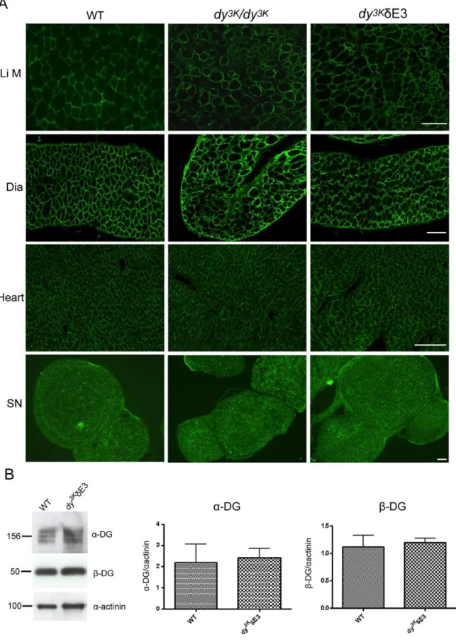
LMa2 chain deficiency does not significantly altera -dystrogly-can glycosylation andb-dystroglycan expression at the sarcolem-ma [32], probably because other ligands (e.g. perlecan) are still present. By immunofluorescence analyses, expression of a -dystroglycan also appeared normal indy3K/dE3 limb, diaphragm and heart muscle and in sciatic nerve (Fig. 4A). In addition, we quantified expression ofa- andb-dystroglycan and they remained the same indy3K/dE3 vs. control skeletal muscle (Fig. 4B).
All in all, these results suggest that integrin a7B is increased, whereas dystroglycans appear normally expressed in dy3K/dE3 muscles.
Dy3K/dy3Kmice withdE3LMa1 transgene have improved overall health
Dy3K/dy3K mice completely deficient in LMa2 chain were previously described [2]. Briefly, these animals suffer from severe muscle wasting, growth retardation, peripheral neuropathy and die approximately 3–5 weeks after birth. As shown in Fig. 5, the overall health ofdy3K/dE3 mice was improved compared tody3K/ dy3Kmice. First, dy3K/dE3 mice live longer. As demonstrated by the survival curve, approximately 75% of dy3K/dE3 animals survive up to 3 months (Fig. 5B). Further estimation ofdy3K/dE3 survival encountered obstacles. Due to hindleg paralysis, several of them were sacrificed according to the guidelines of the ethical permit. Nevertheless, manydy3K/dE3 mice survive much longer than 3 months. Our oldest animals died one year after birth.
Second,dy3K/dE3 animals are bigger thandy3K/dy3Kmice. At 2 weeks of age,dy3K/dy3Kmice can be identified due to their growth retardation whereas dy3K/dE3 mice appeared outwardly normal (data not shown). Furthermore, the majority ofdy3K/dE3 animals at 5 weeks of age can not be distinguished from normal littermates (Fig. 5A). Weight gain for female and male dy3K/dy3K mice was greatly delayed in 5-week-old mice whereas the weight gain for female and male dy3K/dE3 mice was significantly increased compared to dy3K/dy3K mice (Fig. 5C and data not shown). However, dy3K/dE3 mice weigh significantly less than normal littermates (Fig. 5C and data not shown). Beginning from 5 weeks of age, the difference in overall phenotype between most ofdy3K/dE3
and wild-type mice became more evident. Manydy3K/dE3 animals are visibly smaller than control littermates (Fig. 5A, middle panel). However, some of the older dy3K/dE3 animals look outwardly normal and are almost indistinguishable from their littermates (Fig. 5A, left panel). Also, the ratio of quadriceps and tibialis anterior wet weight per body weight was similar in control anddy3K/dE3 mice, whereas the ratio was significantly reduced indy3K/dy3Kmice (Fig. 5D and data not shown). Hence, muscle mass was maintained in proportion to the body size indy3K/dE3 mice. Nevertheless, most ofdy3K/dE3 mice display severe peripheral nerve abnormalities, as demonstrated by temporary hindleg paralysis (either one or occasionally two limbs) (Fig. 5A, arrow). When lifted by the tail, they retract their hindlimbs toward the body. Still,dy3K/dE3 mice perform much better in the locomotion activity test compared to
dy3K/dy3Kanimals (Fig. 5E), indicating that muscle function is largely
Figure 2. Comparison of expression levels of LMa1 chain betweendE3 transgenic mice,dy3K/dE3 anddy3KLMa1 mice.(A) Truncated LMa1 chain is upregulated in skeletal muscle (SM), diaphragm (Dia), peripheral nerve (SN) and heart (He) indy3K/dE3 mice compared todE3 mice expressing LMa2 chain. It reaches the levels of full-length LMa1 chain expression indy3K
LMa1 mice. Three animals from each group were analyzed. Bars, 50mm. (B) Immunoblotting of tissue extracts from wild-type,dy3K/dE3 anddy3KLMa1 skeletal muscle and EHS extract with a rabbit polyclonal antibody against LMa1LG3 domain. Quantification of signals revealed that there is approximately 2.5-fold more of truncated LMa1 chain indy3K/dE3 muscles compared to full-length LMa1 chain indy3K
preserved. Yet,dy3K/dE3 mice move significantly less than control mice and this is supposedly due to the temporary paralysis (Fig. 5E). Finally, we noted that serum kinase activity was significantly elevated indy3K/dE3 mice (Fig. 5F), indicating that muscles may be dystrophic, despite improved general health.
In summary, survival during the first months of life and other features of the overall phenotype ofdy3K/dE3 mice are not greatly dependent on LMa1LG4-5.
DE3LMa1 transgene reduces the dystrophic pathology of skeletal muscles and significantly prevents dystrophic changes in diaphragm and heart
We next examined the morphology of dy3K/dE3 skeletal and heart muscle. When isolating skeletal muscles fromdy3K/dE3 mice (5-week-old and 4-month-old and older), it could be macroscop-ically seen that muscles were only modestly wasted (see also Fig. 5D). However, histological analyses of muscle revealed vast
Figure 3. Restoration and upregulation of integrina7B subunit indy3K/dE3 muscles.(A) Cross-sections of limb muscle (Li M), diaphragm (Dia) and heart from wild-type,dy3K/dy3K
anddy3K
/dE3 mice were stained with antibodies against integrina7B. Bars, 50mm. (B) Immunoblotting of total protein lysates from wild-type anddy3K
/dE3 skeletal muscle and quantitative measurement of integrina7B expression. There is approximately 4.5-fold more integrina7B indy3K/dE3 muscle (p = 0.0231). Results are shown as means6SEM.
doi:10.1371/journal.pone.0011549.g003
Figure 4. Normal expression of dystroglycans indy3K/dE3 muscles.(A) Cross-sections of limb muscle (Li M), diaphragm (Dia), heart and sciatic nerve (SN) from wild-type,dy3K/dy3Kanddy3K/dE3 mice were stained with antibody IIH6 againsta-dystroglycan. Bars, 50mm. (B) Immunoblotting of glycoprotein preparations from wild-type anddy3K
/dE3 skeletal muscle and quantitative measurement ofa- andb-dystroglycan expression. Results are shown as means6SEM. No significant difference in expression ofa- andb-dystroglycan was noted between wild-type anddy3K/dE3 muscle (p = 0.8200 and p = 0.7527, respectively).
regeneration of muscle fibers in limb muscles, demonstrated by the presence of small fibers with centrally located nuclei (Fig. 6A). Approximately 35% and 25% of 4–6-week-olddy3K/dE3 quadri-ceps and triquadri-ceps muscle fibers, respectively, contained centrally located nuclei and the numbers of centrally nucleated fibers did not differ significantly from dy3K/dy3K muscles (data not shown). The number of fibers in randomly selected areas was similar in wild-type anddy3K/dE3 quadriceps muscle, but with a tendency of
more fibers in dy3K/dE3 mice (probably due to the presence of small regenerating fibers). Interestingly, a similar number of fibers was also noted in dy3K/dy3K quadriceps muscle (Figure S3). However, average fiber diameter is smaller (data not shown) and instead muscle contains fibrotic tissue (see Figure 8A). The number of fibers with centrally located was even higher in limb muscles of 4-month-olddy3K/dE3 animals, indicating that pathology worsens over time (Fig. 6A and data not shown). Nevertheless, these results indicate that dy3K/dE3 muscles undergo damage but that the constant regeneration and muscle mass is maintained with age. Moreover, the diaphragm did not undergo degeneration/regen-eration cycles and its morphology appeared near normal in 5-week-old and 4-month-old animals (Fig. 6A–C). Dy3K/dy3K
diaphragm at 4–6-weeks of age displayed about 16% of regenerated muscle fibers with central nuclei. A significant reduction was found indy3K/dE3 diaphragm, both in young and older animals and the numbers did not differ significantly from wild-type diaphragm (Fig. 6B). We also determined the muscle fiber size in 4–6-week-old diaphragm muscle. The fiber size distribution was shifted towards smaller fiber sizes in dy3K/dy3K
animals, compared with wild-type muscles. Notably, the shift was largely prevented indy3K/dE3 muscles (Fig. 6C).
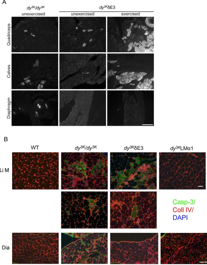
To demonstrate functional benefit conferred by the truncated LMa1 chain in diaphragm, we subjecteddy3K/dE3 mice to downhill treadmill exercise and sarcolemmal integrity was evaluated by Evans blue dye (EBD) accumulation. It has previously been shown that only occasional EBD-positive fibers are found indy/dymuscles [40]. In agreement with these results, we also detected a few EBD-positive fibers in unexerciseddy3K/dy3Kmuscles. We also observed a few EBD-positive fibers in unexerciseddy3K/dE3 limb muscles, but
almost none in dy3K/dE3 diaphragm (Fig. 7A). While it was not possible to exercisedy3K/dy3Kanimals,dy3K/dE3 limb muscles were susceptible to exercise-induced sarcolemmal injury as evidenced by increased uptake of EBD. Interestingly, downhill running induced very little damage indy3K/dE3 diaphragm (Fig. 7A). Although EBD uptake in exerciseddy3K/dE3 limb muscles varied, both between animals and opposing limbs within the same animal, the diaphragm was consistently unaffected. Hence, truncated LMa1 chain prevents exercise-induced injury in diaphragm but not in limb muscles, indicating that different muscles have different requirements for LMa1LG4-5 domains.
The phenomenon of progressive muscle fiber damage in the limbs was further underscored by caspase-3 staining. Apoptosis has
been shown to contribute to the severe dystrophic changes in muscles from MDC1A patients and LMa2 chain deficient mice [2,41,42]. In bothdy3K/dy3K and dy3K/dE3 muscles either single caspase-3 positive apoptotic fibers were detected or apoptosis was more robust (Fig. 7B). In contrast, the muscles from LMa2 chain deficient mice overexpressing full-length LMa1 chain (dy3KLMa1) were free of apoptotic fibers (no caspase-3 staining was observed, Fig. 7B). Interestingly, apoptosis did not take place indy3K/dE3 diaphragms, whereas apoptotic fibers were present in diaphragms from dy3K/dy3K mice (Fig. 7B). This data strongly suggests that LMa1LG4-5 protects limb muscles from apoptosis, most probably via dystroglycan binding, whereas truncated LMa1 chain is sufficient to prevent apoptosis in diaphragm muscle fibers.
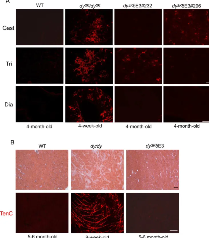
Regardless of apoptotic cell death, muscle replacement with connective tissue, so evident indy3K/dy3Kmice [25], was not very obvious in dy3K/dE3 muscles (Fig. 6A). This tendency was also
demonstrated by tenascin-C staining. Tenascin-C has been shown to be upregulated and extends to the interstitium between muscle fibers indy/dy and dy3K/dy3K mice [25,43]. Some muscles from different dy3K/dE3 animals showed moderate upregulation of tenascin-C (Fig. 8A, two individuals are shown, four animals were analyzed). However, tenascin-C expression was less pronounced than indy3K/dy3Kmuscles. Also, somedy3K/dE3 limb muscles did not display tenascin-C upregulation (Fig. 8A). Moreover, dia-phragm did not show any signs of fibrosis (Fig. 8A).
Cardiomyopathy is not a major feature of MDC1A [1]. However, 2-month-old dyW/dyW hearts show infiltration of connective tissue [44].Dy3K/dy3Kmice probably die too early in order to develop heart fibrosis (data not shown). Therefore, we compared 5–6-month-old dy3K/dE3 hearts with hearts from 8-week-olddy/dymice, which show massive fibrosis in the ventricle wall (Fig. 8B). As demonstrated by hematoxylin and eosin staining,
dy3K/dE3 hearts did not display any fibrotic lesions (Fig. 8). This trend was further confirmed by absence of tenascin-C staining (Fig. 8B).
In summary, LMa1LG4-5 domains are important for securing the mechanical stability of limb muscle fibers in LMa2 chain deficiency, most probably by binding to dystroglycan. Interesting-ly, LMa1LG4-5 domains are not involved in improvement of diaphragm and heart muscle morphology, indicating that other sites of LMa1 chain (most likely integrina7b1 binding modules) are responsible for functional replacement of LMa2 chain in these muscles.
Skeletal muscle regeneration is not impaired indy3K/dE3 mice
Since muscle regeneration seemed to be continuously main-tained indy3K/dE3 limb muscles (Fig. 6A), we next analyzed their regenerative properties in more detail. We injected 2–3-month-old control,dy3K/dE3 mice and 3-week-olddy3K/dy3Ktibialis anterior with cardiotoxin to induce muscle damage and trigger muscle
Figure 5. Overall phenotype ofdy3K/dE3 mice.(A) 5-week-olddy3K
/dE3 mice often have normal size, remain alert and lively with good muscle tone. A wild-type and ady3K/dy3Klittermate are shown for comparison. With age the difference between the body size of normal anddy3K/dE3 mice becomes more evident (middle panel). However some dy3K
/dE3 animals (right panel) remain undistinguishable from littermates at older age. Nevertheless, alldy3K/dE3 mice develop peripheral neuropathy (indicated by arrow). (B) Survival curves ofdy3K/dy3K(n = 8) anddy3K/dE3 mice (n = 44) up to 3 months of age. Curves remain significantly different from each other (p,0.0001). Around 75% ofdy3K/dE3 mice live at least up to 3 months of age. (C) Whole body weights of 5-week-old female control,dy3K/dy3K
anddy3K
/dE3 mice. Body mass is partially recovered in femaledy3K
/dE3 mice. They weigh significantly more thandy3K/dy3Kmice (p,0.0001), but significantly less than control animals p,0.0003). (D) Proportion (in percentage) of the wet weight of quadriceps muscle to the body weight in control,dy3K/dy3K
anddy3K
/dE3 mice. Compared to control mice, the ratio is normal in dy3K/dE3 (p = 0.8001) but significantly reduced indy3K/dy3Kmice (p = 0.0003). (E) Exploratory locomotion of 10-week-old control anddy3K/dE3 mice and 5-week-old dy3K/dy3K
mice.Dy3K
/dE3 mice are significantly more active than dy3K/dy3K
mice (p,0.0001) and less active than control mice (p = 0.0099). (F) Serum creatine kinase (CK) activity in control,dy3K/dy3Kanddy3K/dE3 mice. There is no difference in CK activity betweendy3K/dy3Kand dy3K
/dE3 mice, butdy3K
regeneration. Four days after injection many new fibers had reformed in all mice examined (data not shown). These fibers expressed embryonic myosin heavy chain, indicating an ongoing regeneration (Fig. 9B). Surprisingly, the regeneration process clearly took place in the absence of LMa2 chain (although newly
formed muscle cells indy3K/dy3Ktibialis anterior were rather small) (Fig. 9B). Tibialis anterior from dy3K/dE3 mice also showed normal initial regeneration, comparable to control mice. Most importantly, after 11 days post injection, dy3K/dE3 muscles displayed the regeneration pattern characteristic for control mice
Figure 6. Analyses of muscle morphology and properties.(A) Hematoxylin and eosin staining of quadriceps femoris (Quad), triceps brachii (Tri) and diaphragm (Dia) muscles from 5-week-old and 4-month-old wild-type anddy3K
/dE3 mice. Myopathic changes with groups of centrally nucleated muscle fibers were detected in quadriceps, and to larger extent in triceps of both 5-week-old and 4-month-olddy3K/dE3 mice. Central nucleation was not evident in diaphragm. Connective tissue infiltration was largely prevented in all muscle types. Three animals from each group were analyzed. (B) Quantification of central nucleation in 4–6-week-old wild-type,dy3K/dy3K,dy3K/dE3 and 4–6-month-olddy3K/dE3 diaphragm. The number of fibers with centrally located nuclei is not significantly different between wild-type and young or wild-type and olddy3K
/dE3 diaphragm muscles (p = 0.2163 and p = 0.2707, respectively), whereas the number of regenerating fibers is significantly higher indy3K/dy3Kdiaphragm compared to young and olddy3K/dE3 mice (p = 0.0255 and p = 0.0026). Each bar represents the mean6SEM (p,0.05). At least 3 animals were analyzed. (C) Fiber size distribution in 4–6 week-old wild-type,dy3K/dy3K,dy3K/dE3 diaphragms. Thedy3K/dy3Kdiaphragm fibers are smaller thandy3K/dE3 diaphragm fibers. Bars, 50mm.
doi:10.1371/journal.pone.0011549.g006
Figure 7. EBD staining of unexercised and exercised muscles and caspase-3 immunostaining.(A) Three- to 5-week-olddy3K /dy3K
mice (not exercised) display a few EBD-positive fibers. Also, unexerciseddy3K/dE3 limb muscles display few fibers positive for EBD, whereas hardly any are detected indy3K
/dE3 diaphragm.Dy3K /dy3K
mice were not in the condition to be exercised on the treadmill, but 7–13-week-olddy3K
/dE3 mice were analyzed for EBD uptake upon exercise. Increased uptake of EBD is seen in exerciseddy3K/dE3 limb muscles, but truncated LMa1 chain prevents exercise-induced injury in diaphragm. Bar, 200mm. (B) Robust expression of caspase-3 (green) in the fibers fromdy3K/dy3K
anddy3K
and they were not distinguishable from each other (Fig. 9A). Injected dy3K/dE3 tibialis anterior muscles were tightly packed with big fibers. Also, the expression of embryonic myosin heavy chain was not detected after 11 days (Fig. 9B). This data confirms that regeneration in the presence of truncated LMa1 chain is characterized with high capacity and maintenance. The regener-ation indy3K/dy3Kmice was delayed and not as well-organized as in control anddy3K/dE3 animals, since the muscle fibers in LMa2 chain deficient mice appeared to be less packed and surrounded by connective tissue (Fig. 9A). Also, single fibers still expressed embryonic myosin heavy chain.
In summary, these data provide more insight into mechanism of muscle regeneration in LMa2 chain deficiency and indicate that LMa1 chain deprived of LG4-5 domains ensures proper regeneration. Therefore, binding to dystroglycan is not essential to ensure sufficient muscle regeneration and its maintenance.
LMa1LG4-5 is essential for myelination in peripheral nervous system in LMa2 chain deficiency
MDC1A patients as well asdy3K/dy3Kmice display dysmyelina-tion neuropathy that leads to reduced conducdysmyelina-tion velocity of nerve impulses [45–47]. Unmyelinated axon bundles are prominent especially in spinal roots of LMa2 chain deficient mice. We have demonstrated before that overexpression of full-length LMa1 chain in dy3K/dy3K peripheral nervous system largely corrects myelination defects [27].Dy3K/dE3 mice display hindleg paralysis and motor dysfunction. Morphology analyses of spinal roots and sciatic nerves confirmed that overexpression of truncated LMa1 chain did not correct the phenotype of the proximal part of peripheral nervous system. In spite of the presence of truncated LMa1 chain in both dorsal and ventral roots, large areas with unmyelinated axons (indicating incomplete axonal sorting) were evident indy3K/dE3 mice (Fig. 10). Similar bundles of naked, unmyelinated axons have also been described in dorsal and ventral roots ofdy3K/dy3Kmice [27]. Importantly, this process was fully prevented upon overexpression of full-length LMa1 chain in LMa2 chain deficient peripheral nervous system [27], suggesting a role for LG4-5 domains in myelination processes.
Although myelination took place in the distal part ofdy3K/dE3 peripheral nervous system, sciatic nerve morphology was only partially rescued compared tody3K/dy3Kmice. Bundles of unsorted unmyelinated axons have been reported indy3K/dy3Ksciatic nerve
[27] (see Fig. 10). Smaller, yet clearly visible patches of unsorted axons were also detected in dy3K/dE3 sciatic nerves (Fig. 10 and 11). While occasional unmyelinated axons are present in normal animals (Fig. 11, top panel) and they are known to be part of a healthy nerve, the bundles present indy3K/dE3 nerves were clearly bigger (Fig. 11, top panel) and more frequent (data not shown), than in control mice. Tomacula (thickened myelin sheaths) was observed in dy/dy mice [48] and we also detected these hypermyelinated axons in dy3K/dy3K animals (Fig. 10). Fewer tomacula were seen in dy3K/dE3 mice (Fig. 10). Electron microscopy analyses of 2–4-month-old dy3K/dE3 sciatic nerves revealed a whole spectrum of pathologies. Apart from axons with normal appearance (Fig. 11, top panel, yellow star), many axons with myelin distortion and/or abnormal ovoid shape were detected, especially in the animals affected more severely with
paralysis (Fig. 11, top panel, 3rdoverview photo; middle panel and
bottom panel). The post-myelination pathologies leading to axonal degeneration (Fig. 11A–E) included: myelin degradation, axon demyelination (B,C), myelin intrusions (A), excessive myelin outfoldings (A,D) and redundant loops (H). Degenerated axons often resembled Wallerian degeneration (Fig. 11E) [49]. Many Schwann cells detached from degenerating axons (Fig. 11E, arrow) and showed anomalous, most probably pre-apoptotic phenotype. Further abnormalities included presence of intra-axonal vacuoles (Fig. 11F), myelin infoldings (Fig. 11G), different forms of hypermyelination (Fig. 11I and J) and occasional onion bulbs (several concentric layers of Schwann cell cytoplasm around an axon, leading to demyelination) (Fig. 11K). Schwann cells myelinating more than one axon (satellite axons) were found (Fig. 11F and G). This may point towards defective behavior of Schwann cells and as a consequence a defective myelination process. Many of the described abnormalities have not been associated with LMa2 chain deficiency before. However, redun-dant loop formation is characteristic fordy/dymice [48], and we also found many axons with redundant loops (Fig. 11H, and top panel overview). Redundant loop formation by Schwann cells and collapsing myelin that form ovoid, flat axons could contribute to axonal necrosis [50]. In conclusion, it is possible that upon LMa2 chain deficiency and in the absence of full-length LMa1 chain, Schwann cells acquire pathological properties and perform abnormal myelination. Furthermore, with age these Schwann cells could affect correctly assembled myelin layers, subsequently leading to axonal neuropathy.
These data show that the presence of truncated LMa1 chain did not prevent the possible age-related progression of pathological processes indy3K/dy3Kdistal peripheral nervous system. Therefore, LMa1LG4-5 has a crucial role not only for myelination of the spinal roots, but also for correct myelination, maintenance of myelin, proper axon-Schwann cell interaction and peripheral nerve homeostasis in the distal peripheral nervous system. Various myelin and Schwann cell abnormalities have been shown to contribute to demyelination in different neuropathies [51]. Likewise, the myelin defects described above could influence the severity of observed neuropathy.
Basement membranes are not fully restored in the presence of truncated LMa1 chain
LMa2 chain deficiency results in disrupted basement mem-branes around muscle and Schwann cells [2,25,27,30,46,52,53]. Overexpression of full-length LMa1 chain largely restores basement membranes in the neuromuscular system ofdy3K/dy3K
mice [25,27]. Indy3K/dE3 mice, basement membrane assembly was only partially re-established. Both in sciatic nerves and especially in skeletal muscle, basement membranes had a patchy appearance (Fig. 12, A and D). In diaphragm muscle and heart, despite significant morphological improvement, basement mem-branes were also locally discontinuous (although to a lesser extent than in limb muscle), suggesting that the improvement of the phenotype is not entirely related to intact basement membranes in these organs. Nevertheless, basement membranes in dy3K/dy3K
diaphragm and heart were more disrupted than in dy3K/dE3 animals (Fig. 12, B and C).
indicated ongoing apoptosis in a large group of fibers (top Li M panel), or in single fibers (lower Li M panel). Overexpression of full-length LMa1 chain prevented the cell death in LMa2 chain deficient limb muscles. In contrast to limb muscles, onlydy3K/dy3Kdiaphragm (Dia) contained apoptotic fibers, whereas the overexpression of bothdE3LMa1 and full-length LMa1 chain prevented apoptosis in LMa2 chain deficient diaphragms. DAPI (blue) and an antibody against collagen IV (red) were used to co-visualize apoptotic fibers. Four animals from each group were analyzed. Bars, 50mm. doi:10.1371/journal.pone.0011549.g007
Figure 8. Analyses of fibrosis in skeletal muscle and heart.(A) Different wild-type (4-month-old),dy3K/dy3K
(4-week-old) anddy3K /dE3 (4-month-old) muscles (gastrocnemius, triceps, diaphragm) were stained with an antibody against tenascin-C. Occasionally tenascin-C is present in interstitial matrix of limbdy3K
/dE3 muscles, but it is absent from diaphragm. Note extensive tissue fibrosis indy3K/dy3K
muscles. Fourdy3K
/dE3 animals were analyzed. Bars, 50mm. (B) Hematoxylin and eosin staining (upper panel) of hearts from wild-type (5–6-month-old),dy/dy(8-week-old) anddy3K/ dE3 (5–6-month-old) mice. Hearts fromdy/dymice displayed localized or extensive fibrosis in the ventricular wall.Dy3K
/dE3 hearts did not exhibit any defects and looked as wild-type controls. Tenascin-C immunolabelling confirms the presence of fibrotic lesions indy/dyhearts and their absence in dy3K
In summary, these data show that LMa1LG4-5 is partially required for basement membrane assembly and cell surface anchorage in the neuromuscular system.
Discussion
In this paper, we investigated the roles of LM C-terminal globular domains (and accordingly LM receptors dystroglycan and integrin) in muscle and nerve and analyzed the molecular mechanisms underlying LMa1 chain mediated rescue of LMa2 chain deficiency.
LMa1LG4-5 is dispensable for improvement of
diaphragm and heart muscles, but not limb muscles in LMa1 chain rescued mice
Overexpression of LMa1 chain lacking LG4-5 domains indy3K/
dy3Kmice resulted in significantly prolonged lifespan (at least tripled compared withdy3K/dy3Kmice). Cardiopulmonary complications are often responsible for the early death in dystrophic patients but cardiomyopathy is not a common feature of LMa2 chain deficiency [1]. Considering that a severely dystrophic diaphragm will lead to pulmonary dysfunction, it is quite likely that the improved diaphragm indy3K/dE3 mice accounts for the increased survival, although we
can not completely exclude that the expression of truncated LMa1 in other tissues (e.g. heart) is beneficial. Importantly, integrin a7B subunit is absent fromdy3K/dy3Ksarcolemma, but reconstituted in
dy3K/dE3 muscles. Hence, we propose that prolonged lifespan is secured via LMa1LG1-3 binding, most probably to integrina7b1, in the diaphragm and possibly in the heart.
Interestingly, while LMa1LG4-5 turned out to be dispensable for diaphragm and heart muscle, overexpression of LMa1 chain devoid of LG4-5 did not secure the complete correction of LMa2 chain deficient limb muscles. Although it is not surprising that LMa2 chain deficient peripheral nerve and muscle could respond differently todE3LMa1 overexpression, it is somewhat unexpected that limb muscles and diaphragm would not be spared to the same degree, indicating an important difference in their properties or molecular signature in response to lack of a single protein domain. Our results also point toward diverse roles of LMa1LG4-5 when expressed in different muscle groups. For example, apoptosis has been shown to contribute to LMa2 chain deficient pathogenesis [54,55]. In limb skeletal muscle, LMa1LG4-5 appeared to be critical for prevention of apoptosis of muscle fibers. However, this was not the case in diaphragm. Integrina7b1 has been considered to be the major mediator of myofiber survival [29]. Now, we suggest that also LM binding to dystroglycan prevents apoptosis in limb
Figure 9. Analyses of skeletal muscle regenerative properties subjected to cardiotoxin injection.(A) Hematoxylin and eosin staining of tibialis anterior from control (2–3-month-old),dy3K/dy3K(3-week-old) anddy3K/dE3 (2–3-month-old) 11 days post cardiotoxin injection. Regenerating dy3K
/dE3 muscles morphologically look like regenerating control muscles, whereas regeneration indy3K/dy3K
mice is delayed. (B) Immunostaining revealing the presence of embryonic myosin heavy chain (eMyoH) as the sign of active regeneration (green). Collagen IV (Coll IV) antibody (red) and DAPI nuclear marker (blue) were chosen to co-visualize regenerating fibers. Four-days post injection (upper panel) all analyzed muscles express embryonic myosin. Fibers fromdy3K/dy3K
mice are smaller. Eleven-days post injection (lower panel) control anddy3K
/dE3 tibialis anterior do not express embryonic myosin. Embryonic myosin is occasionally present in somedy3K/dy3Kfibers.Dy3K/dy3Ktibialis anterior does not show regular morphology and displays dystrophic, disorganized pattern with small and big muscle fibers. Three animals from each group were analyzed. Bars, 50mm.
doi:10.1371/journal.pone.0011549.g009
muscle fibers. In support of this notion, dystroglycan binding to LMa2 chain has been shown to protect muscle cells in culture from apoptosis [56]. Yet, in some muscles, (e.g. diaphragm) integrina7b1 could be the key player in apoptosis prevention.
LMa1LG4-5 is not involved in muscle regeneration in LMa1 chain rescued mice
Skeletal muscle regeneration depends on satellite cells, which express both dystroglycan and integrina7b1 [10,57]. In spite of muscle damage and cell death, dy3K/dE3 muscles were able to regenerate and maintain muscle mass, both in normal conditions
and when subjected to cardiotoxin injection. Also, mini-agrin increases the regenerative capacity of LMa2 chain deficient muscles. Since mini-agrin binds dystroglycan (rather that integrin a7b1), it is hypothesized that mini-agrin binding to dystroglycan is responsible for the restored regeneration [58,59] and it has been demonstrated that dystroglycan activity in satellite cells is crucial for the maintenance of regeneration [10]. Yet, integrina7 chain is also involved in skeletal muscle regeneration, asa7 integrin-null mice subjected to cardiotoxin injections exhibit a profound delay in muscle regeneration [57]. Hence, integrin a7 chain is most likely responsible for efficient muscle regeneration in dy3K/dE3
Figure 10. Analyses of myelination in peripheral nervous system.(A) Toluidine blue staining of ventral and dorsal roots and sciatic nerves from 2–4-month-old normal anddy3K/dE3 mice and 5-week-olddy3K/dy3Kanimals. Myelination defects are clearly visible indy3K/dE3 anddy3K/dy3K spinal roots with distinct and wide-spread unmyelinated axons bundles. Occasional unmyelinated axon bundles are also detected in sciatic nerve of dy3K/dE3 anddy3K/dy3Kmice (indicated with arrows). Arrowheads denote tomacula. (B) Truncated LMa1 chain is present indy3K/dE3 spinal roots as demonstrated by immunostaining using the antibody against N-terminal (green) and LG4 domain no staining). Four animals from each group were analyzed. Bars, 25mm.
mice since the dystroglycan binding domain is missing. We propose that the most aggravating step in MDC1A might be the lack of efficient regeneration due to abolished LMa2-integrina7 interaction rather than impaired LMa2-dystroglycan interaction.
LMa1LG4-5 is vital for myelination in peripheral nerve in LMa1 chain rescued mice
None of the neuronal symptoms that occur in LMa2 chain deficiency were ameliorated by dE3LMa1 overexpression. This data together with our previous work [27] indicates a very important role for LMa1LG4-5 in LMa1 chain rescued peripheral nervous system. Interestingly, the phenotype ofdy3K/dy3Kanddy3K/ dE3 peripheral nervous system does not resemble the phenotype of
any conditional knockout mice, where major LM receptors (dystroglycan, integrinsb1 andb4) were depleted from Schwann cells [18–20,60]. Furthermore, genetic inactivation of the a7 integrin chain does not affect peripheral nerve morphology and function [60]. Therefore, those receptors might just regulate the LMa2 chain/LMa1 chain interaction together with other recep-tors. Heparan sulfate proteoglycans syndecans presumably bind LMa1 via the LG4 domain [61] and are enriched in Schwann cells [62], but syndecan-null mice do not display peripheral nerve defects [63]. Also, sulfatides have been shown to bind LMa1LG4-5 [64] and LMa2LG4-5 [65,66] and to be expressed in peripheral nerves [67], where they mediate basement membrane assembly and dystroglycan and integrin signaling [68]. Strikingly, lack of sulfatides
Figure 11. Detailed analyses of morphology and properties of 2–4-month-olddy3K/dE3 sciatic nerves (electron microscopy).Top panel: Unsorted axons in wild-type (WT) anddy3K/dE3 sciatic nerves. Most of bundles of unmyelinated axons are bigger indy3K/dE3 mice (enlarged panels). Apart from unsorted axons (red arrow, overview panel), many compressed, ovoid axons, often with convoluted outfoldings and redundant loops are seen (green arrowhead). Yet, numerous normally shaped and myelinated axons are present (yellow star). Single macrophages were detected (blue arrow). Middle panel: Myelin defects linked to axonal degeneration. (A) Overview of a pathological area with different stages of myelin abnormalities, myelin degradation and axonal degeneration. Red arrowhead - degenerating axon. Blue arrow - degraded interaxonal myelin leading to axon degeneration. Green arrow - axons with vesicular or lamellar myelin debris (intrusions) and dense bodies, often being signs of early stage of degeneration. (B–E) Detailed photos of different forms of degenerating axons found in various areas of sciatic nerve. (B) Degenerating axon with interaxonal myelin debris. (C) An almost completely demyelinated nerve fiber is filled with dilated smooth endoplasmic reticulum and degenerated mitochondria and undergoes degeneration. (D) Granular myelin degeneration with numerous myelin breaks. Arrowhead indicates myelin outfoldings/redundant loop formation. (E) Axonal degeneration forgoes myelin degradation as indicated by loose non-degraded myelin swirls. Schwann cell detached from empty myelin is indicated with arrow. Bottom panels: various axonal and myelin distortions rooting from incorrect myelination process and/or disruption of Schwann cell properties after myelination. (F) One Schwann cell (S) contains thinly myelinated axon (a) with vacuole (v), swollen myelin debris (arrowhead) and thickened myelin sheaths of minute axons (arrow) or myelin outfoldings. (G) Satellite myelinated axon within a bigger axon or excessive intramyelin fold. Myelin outfoldings and satellite myelination seen in F and G may result from impaired myelination process. (H) Redundant loop formation. (I) Hypermyelination due to excessive redundant loop formation. (J) Tomacula. (K) Onion bulb. Arrow indicates an almost demyelinated axon. Bar, 3mm.
doi:10.1371/journal.pone.0011549.g011
and galactocerebrosides (another type of glycolipids) in mice results in similar myelin abnormalities in central nervous system as in
dy3K/dE3 distal peripheral nervous system. Hence, the LM receptor might belong to glycolipids [69–71]. Furthermore, monosialogan-glioside GM1 has been shown to bind LM-111 and promote neurite outgrowth [72]. Therefore, the identification of a peripheral nerve LM receptor is an exciting task.
Basement membrane assembly in LMa1 chain rescued mice requires LMa1LG4-5
In early studies of LMa2 chain deficiency, lack of basement membranes was considered to be deleterious to the muscle fibers [2,52,73,74] and to represent one of the MDC1A pathogenic mechanisms. Consequently, the approach of basement membrane restoration has been hypothesized to be beneficial for the
Figure 12. Basement membranes in the neuromuscular system in the absence of LMa1LG4-5.Electron microscopy of (A) limb skeletal muscle (wild-type anddy3K/dE3); (B) diaphragm (wild-type,dy3K/dy3Kanddy3K/dE3); (C) heart (wild-type,dy3K/dy3Kanddy3K/dE3); (D) sciatic nerve (wild-type and dy3K
/dE3). In dy3K
/dE3 limb skeletal muscle basement membranes had patchy appearance as compared to continuous basement membranes in wild-type samples (A) (arrowheads, in all figures). Stars depict the areas with lack of basement membrane in all figures. Indy3K/dy3K diaphragm basement membranes are either patchy or completely absent. Presence of truncated LMa1 chain partially restores basement membranes in the diaphragm (B). Similarly, in LMa2 chain deficient heart basement membranes are disrupted and partially restored upondE3LMa1 chain overexpression (C). Basement membranes were locally patchy arounddy3K
/dE3 Schwann cells (SCh), but also sometimes continuous throughout longer distances (D). Four animals from each group were analyzed. Bars, 400 nm.
improvement of the dystrophic muscle phenotype [25,28,44,53]. Yet, continuous basement membranes are not strictly required for myelination in peripheral nervous system [46,75]. Likewise, basement membranes are also patchy or less dense indy3K/dE3 mice diaphragm and heart muscle, indicating that continuous basement membranes are not vital for the complete correction of the dystrophic phenotype.
Our data helps to further understand the involvement of LMa1LG4-5 and LG1-3 in basement membrane assembly and point toward interesting basement membrane scaffolding mech-anisms in the neuromuscular system in the absence of LMa 1LG4-5. Exogenous LMa1LG4-5 has been shown to totally abolish the formation of basement membranes in vitro where it selectively blocked the cell-surface accumulation of a LM network [68,76,77]. In our in vivo model, despite lack of LMa1LG4-5, basement membranes showed only partial defects in cell surface anchoring. It is not excluded that integrins or other receptors that bind LMa1LG1-3, partially could compensate for lack of LMa1LG4-5 domain and dystroglycan/sulfatide binding and anchor the LM network to the cell surface. This accumulation, however, is not as efficient as in the presence of full-length LMa1 chain or mini-agrin [25,27,44,53], as basement membranes appear to be continuous only locally in dy3K/dE3 mice. Therefore, it is possible that all LMa1LG domains and the cooperation between different LMa1LG1-5 receptors are important for the assembly of continuous basement membranesin vivo. This hypothesis is further substantiated in McKee et al., where all LG domains were shown to support LM tethering to cell surface [78,79]. However, very recent data by Hanet al., [15] confirms that dystroglycan, but not integrina7b1, is involved in basement membrane anchorage and maintenance (rather than actual assembly) in muscle. Therefore, LMa1LG4-5 binding to dystroglycan could be important not only for basement membrane assembly in the muscle, but also for its maintenance.
Supporting Information
Figure S1 Expression ofdE3LMa1 chain in limb skeletal muscle (SM), peripheral nerve (SN) and heart (He) ofdE3 transgenic mice from lines No. 3 and 4. The two antibodies to detect truncated LMa1 chain were mAb200 and 1057+, which bind LG4 and N-terminal domains, respectively. Mosaic expression of dE3LMa1 chain was detected in transgenic neuromuscular tissues. Wild-type (WT) mice and full-length LMa1 chain transgenic animals (LMa1TG) were used as controls. Bars, 50mm.
Found at: doi:10.1371/journal.pone.0011549.s001 (3.82 MB TIF)
Figure S2 Immunostaining of LMa4 and a5 chains. Cross-sections of quadriceps femoris (Quad), triceps brachii (Tri) and diaphragm (Dia) from 6-week-old wild-type, dy3K/dy3K and dy3K/dE3 mice were stained with antibodies against LMa4 chain (A) anda5 chain (B), respectively. Expression of LMa4 anda5 chains is increased at the muscle basement area in dy3K/dy3K mice and remains increased in dy3K/dE3 muscles. Four dy3K/ dE3 animals were analyzed. Bar, 50mm.
Found at: doi:10.1371/journal.pone.0011549.s002 (3.67 MB TIF)
Figure S3 The numbers of fibers in a randomly selected area is not significantly different between the genotypes.
Found at: doi:10.1371/journal.pone.0011549.s003 (0.20 MB TIF)
Acknowledgments
We thank Drs. Takako Sasaki and Ulrike Mayer for providing antibodies and Dr. Jia-Yi Li for help with dissection of spinal roots.
Author Contributions
Conceived and designed the experiments: KIG MA VC HE MD. Performed the experiments: KIG MA VC HE MD. Analyzed the data: KIG MA VC HE MD. Wrote the paper: KIG MD.
References
1. Voit T, Tome´ FS (2004) The congenital muscular dystrophies. In: Engel A, Franzini-Armstrong C, eds. Myology. New York: McGraw-Hill Inc. pp 1203–1238.
2. Miyagoe Y, Hanaoka K, Nonaka I, Hayasaka M, Nabeshima Y, et al. (1997) Laminina2 chain-null mutant mice by targeted disruption of the Lama2 gene: a new model of merosin (laminin 2)-deficient congenital muscular dystrophy. FEBS Lett 415: 33–39.
3. Kuang W, Xu H, Vachon PH, Liu L, Loechel F, et al. (1998) Merosin-deficient congenital muscular dystrophy. J Clin Invest 102: 844–852.
4. Durbeej M, Campbell KP (2002) Muscular dystrophies involving the dystrophin-glycoprotein complex: an overview of current mouse models. Curr Opin Genet Dev 12: 349–361.
5. Miner JH (2008) Laminins and their roles in mammals. Microsc Res Tech 71: 349–356.
6. Sciandra F, Gawlik KI, Brancaccio A, Durbeej M (2007) Dystroglycan, a possible mediator for reducing congenital muscular dystrophy? Trends Biotechnol 25: 262–268.
7. Mayer U (2003) Integrins: redundant or important players in skeletal muscle? J Biol Chem 278: 14587–14590.
8. Barresi R, Campbell KP (2006) Dystroglycan: from biosynthesis to pathogenesis of human disease. J Cell Sci 119: 199–207.
9. Coˆte´ PD, Moukhles H, Lindenbaum M, Carbonetto S (1999) Dystroglycan: from biosynthesis to pathogenesis of human disease. Nat Genet 23: 338–342. 10. Cohn RD, Henry MD, Michele DE, Barresi R, Saito F, et al. (2002) Disruption
of DAG1 in differentiated skeletal muscle reveals a role for dystroglycan in muscle regeneration. Cell 110: 639–648.
11. Satz JS, Barresi R, Durbeej M, Willer T, Turner A, et al. (2008) Brain and eye malformations resembling Walker-Warburg syndrome are recapitulated in mice by dystroglycan deletion in the epiblast. J Neurosci 28: 10567–10575. 12. Burkin DJ, Wallace GQ, Nicol KJ, Kaufman DJ, Kaufman SJ (2001) Enhanced
expression of thea7b1 integrin reduces muscular dystrophy and restores viability in dystrophic mice. J Cell Biol 152: 1207–1218.
13. Allikian M, Hack AA, Mewborn S, Mayer U, McNally EM (2004) Genetic compensation for sarcoglycan loss by integrina7b1 in muscle. J Cell Sci 117: 3821–3830.
14. Guo C, Willem M, Werner A, Raivich G, Emerson M, et al. (2006) Absence of
a7 integrin in dystrophin-deficient mice causes a myopathy similar to Duchenne muscular dystrophy. Hum Mol Genet 15: 989–998.
15. Han R, Kanagawa M, Yoshida-Moriguchi T, Rader EP, Ng RA, et al. (2009) Basal lamina strengthens cell membrane integrity via the laminin G domain-binding motif of a-dystroglycan. Proc Natl Acad Sci USA 106: 12573– 12579.
16. Previtali SC, Nodari A, Taveggia C, Pardini C, Dina G, et al. (2003a) Expression of laminin receptors in schwann cell differentiation: evidence for distinct roles. J Neurosci 23: 5520–5530.
17. Nishiuchi R, Takagi J, Hayashi M, Ido H, Yagi Y, et al. (2006) Ligand-binding specificities of binding integrins: a comprehensive survey of laminin-integrin interactions using recombinanta3b1,a6b1,a7b1 anda6b4 integrins. Matrix Biol 25: 189–197.
18. Feltri ML, Graus Porta D, Previtali SC, Nodali A, Migliavacca B, et al. (2002) Conditional disruption ofb1 integrin in Schwann cells impedes interactions with axons. J Cell Biol 156: 199–209.
19. Saito F, Moore SA, Barresi R, Henry MD, Messing A, et al. (2003) Unique role of dystroglycan in peripheral nerve myelination, nodal structure, and sodium channel stabilization. Neuron 38: 747–758.
20. Nodari A, Previtali SC, Dati G, Occhi S, Court FA, et al. (2008)a6b4 integrin and dystroglycan cooperate to stabilize the myelin sheath. J Neurosci 28: 6714–6719.
21. Sorokin L, Sonnenberg A, Aumailley M, Timpl R, Ekblom P (1990) Recognition of the laminin E8 cell-binding site by an integrin possessing thea6 subunit is essential for epithelial polarization in developing kidney tubules. J Cell Biol 111: 1265–1273.
22. Lee EC, Lotz MM, Steele GD, Jr., Mercurio AM (1996) The integrina6b4 is a laminin receptor. J Cell Biol 117: 671–678.
23. Talts JF, Andac Z, Gohring W, Brancaccio A, Timpl R (1999) Binding of the G domains of laminina1 anda2 chains and perlecan to heparin, sulfatides,a -dystroglycan and several extracellular matrix proteins. EMBO J 18: 863–870. 24. von der Mark H, Williams I, Wendler O, Sorokin L, von der Mark K, et al.
(2002) Alternative splice variants ofa7b1 integrin selectively recognize different laminin isoforms. J Biol Chem 277: 6012–6016.
25. Gawlik K, Miyagoe-Suzuki Y, Ekblom P, Takeda S, Durbeej M (2004) Laminin
a1 chain reduces muscular dystrophy in laminina2 chain deficient mice. Hum Mol Genet 13: 1775–1784.
26. Ha¨ger M, Gawlik K, Nystro¨m A, Sasaki T, Durbeej M (2005) Laminina1 chain corrects male infertility caused by absence of laminina2 chain. Am J Pathol 167: 823–833.
27. Gawlik KI, Li J-Y, Petersen A˚ , Durbeej M (2006a) Laminina1 chain improves laminina2 chain deficient neuropathy. Hum Mol Genet 15: 2690–2700. 28. Gawlik KI, Durbeej M (2010) Transgenic overexpression of laminina1 chain in
laminina2 chain-deficient mice rescues the disease throughout the lifespan. Muscle Nerve in press.
29. Vachon PH, Xu H, Liu L, Loechel F, Hayashi Y, et al. (1997) Integrins (a7b1) in muscle function and survival. Disrupted expression in merosin-deficient congenital muscular dystrophy. J Clin Invest 11: 1870–1881.
30. Moll J, Barzaghi P, Lin S, Bezakova G, Lochmuller H, et al. (2001) An agrin minigene rescues dystrophic symptoms in a mouse model for congenital muscular dystrophy. Nature 413: 302–307.
31. Jimenez-Mallebrera C, Torelli S, Feng L, Kim J, Godfrey C, et al. (2009) A comparative study of a-dystroglycan glycosylation in dystroglycanopathies suggest that the hypoglycosylation of a-dystroglycan does not consistently correlate with clinical severity. Brain Pathol 19: 596–611.
32. Gawlik KI, Mayer U, Blomberg K, Sonnenberg A, Ekblom P, et al. (2006b) Laminina1 chain mediated reduction of laminina2 chain deficient muscular dystrophy involves integrina7b1 and dystroglycan. FEBS Lett 580: 1759–1565. 33. Andac Z, Sasaki T, Mann K, Brancaccio A, Deutzmann R, et al. (1999) Analysis of heparin,a-dystroglycan and sulfatide binding to the G domain of the laminin
a1 chain by site-directed mutagenesis. J Mol Biol 287: 253–264.
34. von der Mark H, Po¨schl E, Lanig H, Sasaki T, Deutzmann R, et al. (2007) Distinct acidic clusters and hydrophobic residues in the alternative splice domains X1 and X2 ofa7 integrins define specificity for laminin isoforms. J Mol Biol 371: 1188–1203.
35. Smirnov SP, McDearmon EL, Li S, Ervasti JM, Tryggvason K, et al. (2002) Contributions of the LG modules and furin processing to laminin-2 functions. J Biol Chem 277: 18928–18937.
36. Sche´ele S, Falk M, Franze´n A, Ellin F, Ferletta M, et al. (2005) Laminina1 globular domains 4-5 induce fetal development but are not vital for embryonic basement membrane assembly. Proc Natl Acad Sci USA 102: 1502–1506. 37. Cohn RD, Mayer U, Saher G, Herrmann R, van der Flier A, et al. (1999)
Secondary reduction ofa7B integrin in laminina2 deficient congenital muscular dystrophy supports an additional transmembrane link in skeletal muscle. J Neurol Sci 163: 140–152.
38. Briguet A, Courdier-Fruh I, Foster M, Meier T, Magyar JP (2004) Histological parameters for quantitative assessment of muscular dystrophy in mdx mice. Neuromusc Dis 14: 675–682.
39. Patton BL, Miner JH, Chiu AY, Sanes JR (1997) Distribution and function of laminins in the neuromuscular system of developing, adult, and mutant mice. J Cell Biol 139: 1507–1521.
40. Straub V, Rafael JA, Chamberlain JS, Campbell KP (1997) Animal models for muscular dystrophy show different patterns of sarcolemmal disruption. J Cell Biol 139: 375–385.
41. Mukasa T, Momoi T, Momoi MY (1999) Activation of caspase-3 apoptotic pathways in skeletal muscle fibers in laminin a2-deficient mice. Biochem Biophys Res Commun 260: 139–142.
42. Hayashi YK, Tezak Z, Momoi T, Nonaka I, Garcia CA, et al. (2001) Massive muscle cell degeneration in the early stage of merosin-deficient congenital muscular dystrophy. Neuromuscul Disord 11: 350–359.
43. Ringelmann B, Roder C, Hallmann R, Maley M, Davies M, et al. (1999) Expression of laminina1,a2,a4, anda5 chains, fibronectin, and tenascin-C in skeletal muscle of dystrophic 129ReJ dy/dy mice. Exp Cell Res 246: 165–182. 44. Qiao C, Li J, Zhu T, Draviam R, Watkins S, et al. (2005) Amelioration of laminin-a2-deficient congenital muscular dystrophy by somatic gene transfer of miniagrin. Proc Natl Acad Sci USA 102: 11999–12004.
45. Shorer Z, Philpot J, Muntoni F, Sewry C, Dubowitz V (1995) Demyelinating peripheral nerve neuropathy in merosin-deficient congenital muscular dystro-phy. J Child Neurol 10: 472–475.
46. Nakagawa M, Miyagoe-Suzuki Y, Ikezoe K, Miyata Y, Nonaka I, et al. (2001) Schwann cell myelination occurred without basal lamina formation in laminin
a2 chain-null mutant (dy3K
/dy3K
) mice. Glia 35: 101–110.
47. Quijano-Roy S, Renault F, Romero N, Guicheney P, Fardeau M, et al. (2004) EMG and nerve conduction studies in children with congenital muscular dystrophy. Muscle Nerve 29: 292–299.
48. Jaros E, Bradley WG (1979) Atypical axon-Schwann cell relationships in the common peroneal nerve of the dystrophic mouse: an ultrastructural study. Neuropathol Appl Neurobiol 5: 133–147.
49. Lindberg RL, Martini R, Baumgartner M, Erne B, Borg J, et al. (1999) Motor neuropathy in porphobilinogen deaminase-deficient mice imitates the peripheral neuropathy of human acute porphyria. J Clin Invest 103: 1127–1134. 50. Williams RW, Bastiani MJ, Lia B, Chalupa LM (1986) Growth cones, dying
axons, and developmental fluctuations in the fiber population of the cat’s optic nerve. J Comp Neurol 246: 32–69.
51. Sander S, Ouvrier RA, McLeod JG, Nicholson GA, Pollard JD (2000) Clinical syndromes associated with tomacula or myelin swellings in sural nerve biopsies. J Neurol Neurosurg Psych 68: 483–488.
52. Xu H, Christmas P, Wu X-R, Wewer UM, Engvall E (1994) Defective muscle basement membrane and lack of M-laminin in the dystrophic dy/dy mouse. Proc Natl Acad Sci USA 91: 5572–5576.
53. Yurchenco PD, Cheng YS, Campbell K, Li S (2004) Loss of basement membrane, receptor and cytoskeletal lattices in a laminin-deficient muscular dystrophy. J Cell Sci 117: 735–742.
54. Girgenrath M, Dominov JA, Kostek CA, Miller JB (2004) Inhibition of apoptosis improves outcome in a model of congenital muscular dystrophy. J Clin Invest 114: 1635–1639.
55. Girgenrath M, Beermann ML, Vishnudas VK, Homma S, Miller JB (2009) Pathology is alleviated by doxycycline in a laminin-a2-null model of congenital muscular dystrophy. Ann Neurol 65: 47–56.
56. Langenbach KJ, Rando TA (2002) Inhibition of dystroglycan binding to laminin disrupts the PI3K/AKT pathway and survival signaling in muscle cells. Muscle Nerve 26: 644–653.
57. Rooney JE, Gurpur PB, Yablonka-Reuveni Z, Burkin DJ (2009) Laminin-111 restores regenerative capacity in a mouse model fora7 integrin congenital myopathy. Am J Pathol 174: 256–264.
58. Bentzinger CF, Barzaghi P, Lin S, Ruegg MA (2005) Overexpression of mini-agrin in skeletal muscle increases muscle integrity and regenerative capacity in laminin-a2-deficient mice. FASEB J 19: 934–942.
59. Meinen S, Barzaghi P, Lin S, Lochmuller H, Ruegg MA (2007) Linker molecules between laminins and dystroglycan ameliorate laminin-a2-deficient muscular dystrophy at all disease stages. J Cell Biol 176: 979–993.
60. Previtali SC, Dina G, Nodali A, Fasolini M, Wrabetz L, et al. (2003b) Schwann cells synthesize a7b1 integrin which is dispensable for peripheral nerve development and myelination. Mol Cell Neurosci 23: 210–218.
61. Suzuki N, Ichikawa N, Kasai S, Yamada M, Nishi N, et al. (2003) Syndecan binding sites in the laminina1 chain G domain. Biochemistry 43: 12625–12633. 62. Goutebroze L, Carnaud M, Denisenko N, Boutterin MC, Girault JA (2003) Syndecan-3 and syndecan-4 are enriched in Schwann cell perinodal processes. BMC Neurosci 18: 4:29.
63. Alexopoulou AN, Multhaupt HAB, Couchman JR (2007) Syndecans in wound healing, inflammation and vascular biology. Int J Biochem Cell Biol 29: 505–528.
64. Harrison D, Hussain DA, Combs AC, Ervasti JM, Yurchenco PD, et al. (2007) Crystal structure and cell surface anchorage sites of laminina1LG4-5. J Biol Chem 282: 11573–11581.
65. Tisi D, Talts JF, Timpl R, Hohenester E (2000) Structure of the C-terminal laminin G-like domain pair of the laminina2 chain harbouring binding sites for
a-dystroglycan and heparin. EMBO J 19: 1432–1440.
66. Wizemann H, Garbe JH, Friedrich MV, Timpl R, Sasaki T, et al. (2003) Distinct requirements for heparin and a-dystroglycan binding revealed by structure-based mutagenesis of the laminina2 LG4-LG5 domain pair. J Mol Biol 332: 635–642.
67. Mirsky R, Dubois C, Morgan L, Jessen KR (1990) 04 and A007-sulfatide antibodies bind to embryonic Schwann cells prior to the appearance of galactocerebroside; regulation of the antigen by axon-Schwann cell signals and cyclic AMP. Development 109: 105–116.
68. Li S, Liquari P, McKee KK, Harrison D, Patel R, et al. (2005) Laminin-sulfatide binding initiates basement membrane assembly and enables receptor signaling in Schwann cells and fibroblasts. J Cell Biol 169: 179–189.
69. Dupree JL, Coetzee T, Suzuki K, Popko B (1998) Myelin abnormalities in mice deficient in galactocerebroside and sulfatide. J Neurocytol 27: 649–659. 70. Honke K, Hirahara Y, Dupree J, Suzuki K, Popko B, et al. (2002) Paranodal
junction formation and spermatogenesis require sulfoglycolipids. Proc Natl Acad Sci USA 99: 4227–4232.
71. Marcus J, Honigbaum S, Shroff S, Honke K, Rosenbluth J, et al. (2006) Sulfatide is essential for the maintenance of CNS myelin and axon structure. Glia 53: 372–381.
72. Ichikawa N, Iwabuchi K, Kurihara H, Ishii K, Kobayashi T, et al. (2009) Binding of laminin-1 to monosialoganglioside GM1 in lipid rafts is crucial for neurite outgrowth. J Cell Sci 122: 289–299.
73. Sunada Y, Bernier SM, Utani A, Yamada Y, Campbell KP (1995) Identification of a novel mutant transcript of laminina2 chain gene responsible for muscular dystrophy and dysmyelination indy2Jmice. Hum Mol Genet 4: 1055–1061. 74. Colognato H, Yurchenco PD (1999) The laminina2 expressed by dystrophic
dy(2J) mice is defective in its ability to form polymers. Curr Biol 9: 1327–1330. 75. Yang D, Bierman J, Tarumi YS, Zhong Y, Rangwala R, et al. (2005) Coordinate control of axon defasciculation and myelination by laminin-2 and -8. J Cell Biol 168: 655–666.
76. Tsiper MV, Yurchenco PD (2002) Laminin assembles into separate basement membrane and fibrillar matrices in Schwann cells. J Cell Sci 115: 1005–1015. 77. Li S, Harrison D, Carbonetto S, Fa¨ssler R, Smyth N, et al. (2002) Matrix
assembly, regulation, and survival functions of laminin and its receptors in embryonic stem cell differentiation. J Cell Biol 157: 1279–1290.
78. McKee KK, Harrison D, Capizzi S, Yurchenco PD (2007) Role of laminin terminal globular domains in basement membrane assembly. J Biol Chem 282: 21437–21447.