>
Márcia Carina da Silva Faria
Licenciada em BioquímicaOverexpression of RAC1b in follicular cell
derived thyroid cancers: An activator of NF-κB
pathway that contributes to thyroid
tumorigenesis?
Dissertação para obtenção do Grau de Mestre em Genética Molecular e Biomedicina
Orientador: Doutora Ana Luísa Silva,
Instituto Português de Oncologia de Lisboa Francisco Gentil
(IPOLFG)
Júri:
Presidente: Prof. Doutora Paula Maria Theriaga Mendes Bernardes Gonçalves Arguente: Doutor Peter Jordan
Setembro, 2015
Overexpression of RAC1b in follicular cell derived thyroid cancers: An activator of NF-κB pathway that contributes to thyroid tumorigenesis?
Copyright Márcia Carina da Silva Faria, FCT/UNL, UNL
The Faculty of Science and Technology and the New University of Lisbon have the perpetual right, and without geographical limits, to archive and publish this dissertation through press copies in paper or digital form, or by other known form or any other that will be invented, and to divulgate it through scientific repositories and to admit its copy and distribution with educational or research objectives, non-commercial, as long as it is given credit to the author and editor.
A Faculdade de Ciências e Tecnologia e a Universidade Nova de Lisboa têm o direito, perpétuo e sem limites geográficos, de arquivar e publicar esta dissertação através de exemplares impressos reproduzidos em papel ou de forma digital, ou por qualquer outro meio conhecido ou que venha a ser inventado, e de a divulgar através de repositórios científicos e de admitir a sua cópia e distribuição com objetivos educacionais ou de investigação, não comerciais, desde que seja dado crédito ao autor e editor.
I
AGRADECIMENTOS
Aproveito este momento para agradecer a todos os que possibilitaram ou contribuíram para a realização desta tese de Mestrado.
Um especial agradecimento à minha orientadora Doutora Ana Luísa Silva por ter aceite orientar-me na jornada que foi a realização deste trabalho. Por todo o conhecimento e experiência transmitidos, pela autonomia concedida, pela confiança. Pelo incentivo, pelas palavras essenciais nos momentos certos que nunca me deixaram desanimar, por todo o apoio. Por ser um exemplo de competência, empenho, dedicação, esforço e por ser a prova de que a humildade e trabalho dão um sabor especial a cada vitória. Por fazer ciência porque gosta de ciência!
À Doutora Branca Cavaco, coordenadora da Unidade de Investigação em Patobiologia Molecular e do grupo de Endocrinologia Molecular (IPOLFG), por me acolher nesta unidade.
Quero agradecer também à professora Doutora Maria João Bugalho pela oportunidade, por toda a disponibilidade sempre demonstrada e pela simpatia.
A todos os meus colegas de grupo Rita Domingues, Margarida Moura, Liliana Capinha, Tiago Pinheiro, Ana Matias e Inês Marques, pelo companheirismo, disponibilidade e muito boa disposição. Foi muito importante!
Aos restantes elementos da UIPM que direta ou indiretamente contribuíram para este trabalho. Especialmente a todos aqueles que tornaram as pausas para o almoço momentos divertidos e de pura descontração. Foi essencial!
Ao Bruno Cardoso pela disponibilidade, boa disposição e pela ajuda no mundo que é a citometria de fluxo. “Sempre a 200%”! Também à Carolina e à Ana Matias pelas idas ao FACS, por toda a ajuda! Ao Doutor Paulo Matos, por toda a ajuda neste trabalho, pela boa disposição e pela disponibilidade sempre demonstrada.
À coordenação e comissão científica do Mestrado em Genética Molecular e Biomedicina que proporcionam aos alunos todas as condições necessárias para a realização dos seus projetos.
A todos os novos amigos que acompanharam esta nova etapa da minha vida, partilhando comigo todas as emoções. À Helena e Lígia, companheiras de casa, aventuras, diversão, pela presença em todos os momentos, mesmo nos dias maus. Aos amigos de sempre, Ana, Marina, Liliana, João, Leandro e “BNTs” que apesar de não terem partilhado comigo esta nova etapa a 500km da “nossa casa”, estão e estarão sempre presentes. Pela compreensão das ausências!..
O meu mais sincero agradecimento aos meus pais, Lucília e Paulo, e a toda a minha família pelos princípios incutidos, pela motivação e confiança, pela disponibilidade e pelo empenho, trabalho e esforço dedicados à minha formação. Pela compreensão da distância e das prolongadas ausências, por acreditarem que apesar disso estive sempre com eles! Pela paciência e compreensão nos momentos de maior ansiedade.
III
“The beginning of all sciences is the surprise
of things being what they are.”
Aristóteles
V
ABSTRACT
Papillary and Follicular thyroid carcinomas (PTC and FTC) are lesions derived from follicular cells. The standard therapy includes surgery and radioiodine treatment. Despite being usually associated with a good prognosis the rate of recurrence is high and a subset of patients present radioiodine refractory and non-responsive neoplasia. So, the search for new therapeutic alternatives, as well as new diagnostic/prognostic markers is highly relevant.
Our group at IPOLFG has previously shown RAC1b to be overexpressed in PTCs, being this overexpression significantly associated with poorer outcomes. RAC1b is a variant of the small GTPase RAC1 that was shown to have the ability to stimulate the canonical NF-κB regulatory pathway in colorectal cancer. NF-κB activation is associated with the tumorigenic process in several types of cancer, mainly due to its role in apoptosis evasion.
Here, we aimed to extend the study of the role of RAC1b to FTCs and further investigate the molecular mechanisms associated with RAC1b overexpression and downstream signalling in thyroid tumorigenesis.
Using the human papillary thyroid carcinoma derived cell line as in vitro model, we observed that both RAC1 and RAC1b were able to induce a significant increase on NF-κB and cyclin D1 reporter activity. A clear p65 nuclear localization was found in cells transfected with RAC1b-WT, confirming NF-κB canonical pathway activation. Consistently, we were able to observe a RAC1b-mediated decrease in IκBα (NF-κB inhibitor) protein levels.
These findings prompted us to further assess the cellular consequences of RAC1b-mediated NF-κB activation in a thyroid biological system. Our results indicated that RAC1b protected cells against apoptosis and stimulated G1/S progression, through a process involving the NF-κB pathway.
Summing up, presented findings suggest an important role of RAC1b in the progression of follicular cell derived thyroid carcinomas and point out NF-κB activation as one of the molecular mechanisms associated with RAC1b overexpression and downstream signalling in thyroid tumorigenesis.
Key words: Thyroid tumorigenesis; RAC1b; NF-κB canonical pathway; Cyclin D1; Apoptosis; Cell Cycle;
VII
RESUMO
O Carcinoma Papilar (CPT) e o Carcinoma Folicular da Tiróide (CFT) são lesões derivadas de células foliculares. A terapia de referência inclui cirurgia e tratamento com iodo radioativo. Apesar de estarem normalmente associados a um bom prognóstico, o nível de recorrências é elevado dado que um subconjunto de pacientes apresentam neoplasias refratárias ou incapazes de responder ao iodo radioativo. Portanto, a busca por alternativas terapêuticas, bem como novos marcadores de diagnóstico/prognóstico é bastante relevante.
O nosso grupo no IPOLFG identificou anteriormente a sobre-expressão do RAC1b em CPTs, e esta sobre-expressão estava significativamente associada com um pior prognóstico. O RAC1b é uma variante da pequena GTPase RAC1, que demonstrou a capacidade de estimular a via canónica do NF-κB em cancro colo-retal. A ativação do NF-κB está associada com o processo tumorigénico em vários tipos de cancro, maioritariamente devido ao seu papel na evasão da apoptose.
O objetivo do presente estudo foi o de expandir o estudo do papel do RAC1b aos CFT, e ainda investigar os mecanismos moleculares subjacentes à sobre-expressão do RAC1b e sinalização a jusante na tumorigénese da tiróide
Usando uma linha celular derivada de CPT como modelo in vitro, observou-se que quer o RAC1, quer o RAC1b mostraram capacidade de induzir um aumento significativo na atividade do repórter do NF-κB e da ciclina D1. Uma clara localização nuclear da proteína p65 foi observada em células transfetadas com GFP-RAC1b-WT, confirmando a ativação da via canónica do NF-κB. Consistentemente, observou-se uma diminuição mediada pelo RAC1b dos níveis proteicos de IκBα (um inibidor do NF-κB).
Estes resultados levaram-nos a avaliar as consequências celulares da ativação do NF-κB mediada pelo RAC1b no sistema biológico da tiróide. Os nossos resultados indicam que o RAC1b protegeu as células contra a apoptose e estimulou a progressão G1/S, através de um mecanismo que envolve a via do NF-κB.
Resumindo, as presentes conclusões apontam para um importante papel do RAC1b no desenvolvimento de CFTs e indicam a ativação do NF-κB como um dos mecanismos moleculares associados com a sobre-expressão do RAC1b e sinalização a jusante na tumorigénese da tiróide.
Palavras-Chave: Tumorigénese da Tiróide; RAC1b; Via canónica do NF-κB; Ciclina D1; Apoptose; Ciclo celular;
IX
TABLE OF CONTENTS
AGRADECIMENTOS ... I ABSTRACT ... V RESUMO ... VII TABLE OF CONTENTS ... IX LIST OF FIGURES ... XI LIST OF TABLES ... XIII LIST OF SUPPLEMENTARY TABLES ... XIII LIST OF ABBREVIATIONS, SYMBOLS AND CONVENTIONS ... XVI. INTRODUCTION ... - 1 -
1. Tumorigenesis ... - 1 -
2. Thyroid gland: Anatomy and physiology ... - 2 -
3. Thyroid diseases and syndromes ... - 3 -
3.1. Thyroid neoplasia ... - 4 -
3.1.1. Medullary Thyroid carcinoma ... - 5 -
3.1.2. Non-Medullary Thyroid tumours ... - 5 -
3.1.2.1. Follicular thyroid adenoma ... - 6 -
3.1.2.2. Follicular thyroid carcinoma ... - 6 -
3.1.2.3. Papillary thyroid carcinoma ... - 7 -
3.1.2.4. Poorly differentiated thyroid carcinoma ... - 9 -
3.1.2.5. Anaplastic thyroid carcinoma ... - 9 -
4. RAC1 and RAC1b GTPases ... - 10 -
4.1. Organization and regulation ... - 10 -
4.2. Biological functions ... - 11 -
4.3. Association with cancer ... - 13 -
5. Signalling pathways involved in cancer ... - 13 -
5.1. NF-κB signalling pathway ... - 14 -
5.1.1. Association with Tumorigenesis ... - 16 -
5.1.2. Association with RAC1b ... - 17 -
6. Follicular cell derived thyroid cancers, RAC1b and NF-κB pathway-a link to unravel .. - 18 -
Part 1. RAC1b role in FTC Tumorigenesis ... - 18 -
Part 2. RAC1b - An activator of NF-κB pathway that contributes to thyroid Tumorigenesis? ... - 18 -
II. OBJECTIVES ... - 21 -
III. MATERIALS AND METHODS ... 23
-1. Biological Material – patients samples... - 23 -
2. DNA plasmids and constructs ... - 23 -
3. Cell Culture and transfection ... - 23 -
4. RNA extraction ... - 24 -
5. cDNA synthesis and amplification ... - 24 -
X
7. Luciferase reporter assays ... 26
-8. Confocal immunofluorescence microscopy... - 27 -
9. Cell fractionation ... - 27 -
10. Protein extraction with RIPA ... - 28 -
11. Sodium dodecyl sulfate-polyacrylamide gel electrophoresis (SDS-PAGE) and Western Blotting ... - 28 -
12. Cell death analysis by flow cytometry (FACS) ... - 28 -
13. Cell Cycle analysis by flow cytometry (FACS) ... - 29 -
14. Statistical analysis ... - 30 -
IV. RESULTS ... - 31 -
PART 1 ... 31
-1. RAC1b is overexpressed in FTCs ... - 31 -
2. RAC1b overexpression is associated with presence of distant metastasis and poorer clinical outcomes ... - 31 -
PART 2 ... - 34 -
3. RAC1b stimulates NF-κB in K1 cells ... - 34 -
4. RAC1b stimulates Cyclin D1 in the K1 cells ... - 35 -
5. RAC1b stimulates p65 translocation to the nucleus ... - 36 -
6. RAC1b induces a decrease in total IκBα protein expression ... - 39 -
7. RAC1b protects cells against apoptosis induced by staurosporine ... - 40 -
8. RAC1b stimulates cell cycle progression ... - 41 -
9. RAC1b effect on Nthy cell line ... - 42 -
V. DISCUSSION ... - 45 -
VI. CONCLUSION ... - 49 -
VII. PERSPECTIVES ... - 51 -
VIII. BIBLIOGRAPHY ... - 53 -
XI
LIST OF FIGURES
Figure I.1. The hallmarks of cancer. ... - 1 -
Figure I.2. Location and structure of the thyroid gland. ... 3
-Figure I.3. Subtypes of non-meddulary thyroid carcinomas. ... - 6 -
Figure I.4. Diagram of the RAC1b gene. 1, 2, 3, 4, 5, 6 represents exons. ... - 10 -
Figure I.5. RAC1 and RAC1b signalling. ... - 12 -
Figure I.6. Canonical and non-canonical NF-κB activating pathways. ... - 16 -
Figure IV.7. RAC1b overexpression and its association with distant metastasis and clinical outcome... - 32 -Figure IV.8. Luciferase activity of NF-κB regulated reporter in transfected K1 cells. ... - 35 -
Figure IV.9. Luciferase activity of cyclin D1 regulated reporter in transfected K1 cells.. ... 36
Figure IV.10. RAC1b overexpression affects subcellular location of p65NLS protein. ... 38
-Figure IV.10.1. RAC1b overexpression affects subcellular location of p65-NLS protein... - 39 -
Figure IV.11. Total IκBα protein levels in K1 cells assessed by westernblotting.. ... 40
-Figure IV.12. Apoptosis analyzes by flow cytometry (Annexin V and PI staining). ... - 41 -
Figure IV.13. Cell cycle analyzes by flow cytometric (PI staining). ... 42
-Figure IV.14. Luciferase activity of NF-κB and cyclin D1 regulated reporter in transfected Nthy cells. ... 44
XIII
LIST OF TABLES
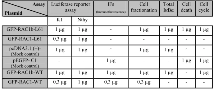
Table III.1. Description of plasmid amounts used in each assay, for each cell line. ... - 24 -
Table III.2. RAC1/1b PCR amplification conditions. ... 25
-Table IV.3. Clinical Data... - 33 -
LIST OF SUPPLEMENTARY TABLES
Table 1. 10x PCR buffer. ... - 64 -Table 2. RIPA buffer. ... - 64 -
Table 3. 5x Sample buffer. ... - 64 -
Table 4. Blot buffer... - 64 -
Table 5. Destain solution. ... - 64 -
Table 6. TBST solution... - 65 -
Table 7. ECL solutions. ... - 65 -
Table 8. Annexin binding buffer 1x... - 65 -
XV
LIST OF ABBREVIATIONS, SYMBOLS AND CONVENTIONS
ATC – Anaplastic thyroid carcinoma
BRAF – Murine sarcoma viral (v-Raf) oncogene homolog B1 Cdc42 – Cell division control protein 42 homolog
cDNA – Complementary DNA C-terminal – Carboxyl-terminal
CTNNB1 - Catenin (Cadherin-associated protein), beta 1 DAPI – 4',6-diamidino-2-phenylindole
ddH2O – Bidestilled water
DMEM:F12 – Dulbecco's Modified Eagle Medium/ Nutrient Mixture F-12 DNA – Deoxyribonucleic acid
dNTPs – Deoxynucleotides DTT – Dithiothreitol
ECL – Enhanced chemiluminescence EDTA – Ethylenediamine tetraacetic acid EHT 1864 – RAC1 inhibitor
EMT – Epithelial-mesenchymal transition FBS – Fetal bovine serum
FMTC – Familial MTC FNA – Fine needle aspiration FTA – Follicular thyroid adenoma FTC – Follicular thyroid carcinoma
FVPTC – Follicular variant of papillary thyroid carcinoma g – Grams
G0 phase – Quiescent phase of cell cycle G1 phase – Gap 1 phase of cell cycle G2 phase – Gap 2 phase of cell cycle GAP – GTPase-activating protein
GDI – Guanine nucleotide dissociation inhibitor GDP – Guanosine diphosphate
GEF – Guanine nucleotide exchange factor GFP – Green fluorescent protein
GTP – Guanosine triphosphate GTPase – Guanosine triphosphatase h – Hours
XVI
Ig – Immunoglobulin
IPOLFG – Instituto Português de Oncologia Lisboa Francisco Gentil IκK – IκB kinase
JNK – Jun NH2-terminal kinase
K1 – Cell line derived from human papillary thyroid carcinoma kDa – Kilodalton
L – Litre
L61 – Constitutively active mutant m – Metre
M – Molar
M phase – Mitosis phase
MAPK – Mitogen-activated protein kinases MEN 2 – Multiple endocrine neoplasia type 2 min – minutes
miRNA – MicroRNA
MMP-3 – Matrix metalloproteinase-3 MTC – Medullary thyroid carcinoma NEMO – NF-κB essential modulator
NF-κB – Nuclear factor kappa-light-chain-gene-enhancer of activated B cells NIK – NF-κB inducing kinase
NIS – Sodium/iodide symporter NLS – Nuclear localization signal
NMTC – Non-medullary thyroid carcinoma N-terminal – Amino terminal
Nthy – Nthy-ori 3-1, cell line derived from human thyroid follicular normal epithelium OPTI-MEM – Improved minimal essential medium
p- p-value
PAK – p21-activated protein kinase
PAX8/PPARγ – paired box gene 8 (PAX8)-peroxisome proliferator-activated receptor γ (PPARγ) PBS – Phosphate-buffered saline
PCR – Polymerase chain reaction
PDTC – Poorly differentiated thyroid carcinoma PI – Propidium iodide
PI3K(CA) – phosphoinositide 3-kinase (Catalytic Alpha) PTC – Pappilary thyroid carcinoma
PTEN – Phosphatase and tensin homolog PVDF – Polyvinylidene difluoride
XVII qRT-PCR – Quantitative RT-PCR
RAC1 – Ras-related C3 botulinum toxin substrate 1 RAS – Rat sarcoma vírus homolog
RET – rearrangement during transfection Rho – Ras homologous
RIPA – Radioimmunoprecipitation assay buffer RNA – Ribonucleic acid
RNAse – Ribonuclease
ROS – Reactive oxygen species rpm – Rotations per minute
RPMI – Roswell Park Memorial Institute medium RT – Room temperature
RTK – Receptor tyrosine kinases RT-PCR – Reverse transcription-PCR S phase – Synthesis phase of cell cycle SB – Sample Buffer
SDS-PAGE – Sodium dodecyl sulfate-polyacrylamide gel electrophoresis T3 – Triiodothyronine
T4 – Tetraiodothyronine TBE – Tris-borate-EDTA
TBST – Tris Buffered Saline Buffer 0,05% Triton X-100 TP53 – Tumour Protein 53
UV – Ultraviolet
WDTC – Well Differentiated Thyroid Carcinoma WT – Wild-type
μ – micro-
% (v/v) – Concentration expressed in volume per volume % (w/v) – Concentration expressed in mass per volume °C – Celsius degree
- 1 -
I.
INTRODUCTION
1. Tumorigenesis
The pursuit for an explanation about the basis of malignant changes has started many years ago with several studies being reported. For example, back to 1974, Loeb and collaborators indicated errors in DNA replication as a possible cause of malignant events. Nevertheless, in the last few years, several scientific advances took place in the cancer research field allowing the scientific community to better understand the biological mechanisms involved in the genesis of tumours – Tumorigenesis.
Modifications in the control and homeostatic balances that regulate a myriad of cellular processes, namely proliferation, differentiation and apoptosis are considered the primary sources of malignant changes (Koeffler et al., 1991). The idea that genetic alterations are required to tumorigenesis is widely accepted, and evidences that tumours accumulate numerous genetic changes, such as chromosomal abnormalities, gene mutations and epigenetic alterations has been shown over the years (Loeb et al., 1974; Koeffler et al., 1991; Bach et al., 2000; Hanahan and Weinberg, 2000; Sieber et al., 2003; MacConaill and Garraway, 2010).
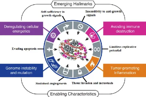
Despite the vast diversity of existing tumours, some intrinsic features may be shared and considered universal features of all tumours. Back to 2000, Hanahan and Weinberg proposed six essential modifications in cell physiology required in the tumorigenesis process (see Figure I.1): self-sufficiency in growth signals, insensitivity to anti-growth signals, evasion to apoptosis, unlimited replicative potential, sustained angiogenesis and ability to invade tissues and metastasize. These features allowed tumour cells to generate their own signals and, in opposition, to escape of anti-proliferative signals. The unlimited replicative potential contributes to the immortalization of tumour cells that guarantee a continuous nutrient supply by create their own blood vessel network.
Figure I.1. The hallmarks of cancer. Adapted from Hanahan and Weinberg (2000) and Hanahan and Weinberg (2011).
- 2 -
In addition to these common and widely accepted features, recently new characteristics were proposed as hallmarks of cancer (see Figure I.1): cellular energy metabolism adaptation and ability to evade immune system (Hanahan and Weinberg, 2011; Serpa and Dias, 2011).
In fact, tumour cells have an altered metabolism relative to normal cells (Serpa and Dias, 2011). The increased proliferative potential of tumour cells requires higher rates of metabolic reactions in order to obtain nutrients, energy and biosynthetic activity (DeBerardinis et al., 2008; Tennant et al., 2009; Tennant et al., 2010). This phenotype allows tumour cells to have an increased rate of glycolysis (conversion of glucose to pyruvate), either in aerobic or anaerobic conditions (Tennant et al., 2009), which compensates the limited levels of oxidative phosphorylation in some regions of solid tumours (Frezza and Gottlieb, 2009).
To progress, tumour cells have to be able to evade immune system. The interaction between the malignant cells and the cells of the immune system has different stages – elimination, equilibrium and escape – and this process is known as cancer immunoediting. In the elimination or cancer surveillance phase, transformed cells are identified and killed by the innate and the adaptive immune system. These events result in an immune selection that is favourable to tumour variants with increased ability to withstand immune factors and decrease immunogenicity. The equilibrium phase is known by the persistence of the tumour cells that were not killed, but also can not expand due to the immune pressure. When the tumour is able to growth due to the immune exhaustion or inhibition, or as result of emerging tumour-cell variants able to evade immune pressure, the escape stage is ongoing (Dunn et al., 2006; Kim et al., 2007).
Several lines of evidence, suggest that human tumorigenesis is a multiset process (Hanahan and Weinberg, 2000) in which normal cells are progressively transformed in malignant derivatives. The progression through the many stages of this process depends on several factors, such as genomic instability, which is inherent to some cancers, or the ability of these tumours to promote inflammation that occasionally support multiple hallmark capabilities (Sieber et al., 2003; Hanahan and Weinberg, 2011).
2. Thyroid gland: Anatomy and physiology
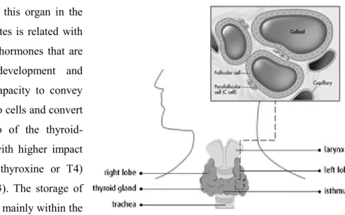
The thyroid gland, part of the endocrine system, is a butterfly-shaped organ consisting of two lobes positioned on either side of the trachea, connected by a structure named isthmus (see Figure I.2). Despite the variance in the size and appearance of the adult gland, - according to functional activity, gender, hormonal status, and iodine intake - the thyroid gland is one of the largest glands in the body, weighing approximately 15 to 25 g (Muro-Cacho and Ku, 2000; Nussey and Whitehead, 2001).
- 3 - The primary role of this organ in the
development of vertebrates is related with its ability to synthesize hormones that are crucial for growth, development and survival, due to their capacity to convey oxygen and nutrients into cells and convert them into energy. Two of the thyroid-synthesized hormones with higher impact are tetraiodothyronine (thyroxine or T4) and triiodothyronine (T3). The storage of thyroid hormones occurs mainly within the follicles, the functional unit of the thyroid gland (Nussey and Whitehead, 2001; Nitsch
et al., 2010). The synthesis of thyroid hormones requires the iodination of tyrosine molecules (Nussey and Whitehead, 2001), and their activity is mediated by thyroid hormone receptors, which exists in different isoforms (Gauthier et al., 1999; Zhu et al., 2010; Kim and Cheng, 2013). In fact, the iodine represents an essential element in thyroid physiology and function, and its uptake in the thyroid gland is mediated by the sodium/iodide symporter (NIS) (Filetti et al., 1999; Kogai et al., 2006).
The thyroid embryogenesis involves an initial step in which this gland develops from the anterior foregut, following a sequence of morphogenetic steps that culminate in the migration of the thyroid bud to its final anatomic position (Fagman and Nilsson 2010a). This developmental process and the final thyroid fate are influenced by the combined expression of specific transcription factors by foregut endoderm cells (Fagman and Nilsson 2010a; Fagman and Nilsson 2010b).
Several cell types, derived from the three germ layers, compose the thyroid gland (Damante and DiLauro, 1994). The most abundant cellular population are the follicular cells that are associated with the production of thyroid hormones and surround the follicles (see Figure I.2) - oval sacs filled with colloid (Ljungberg et al., 1983; Damante and DiLauro, 1994; Muro-Cacho and Ku, 2000). The C cells, or parafollicular cells, are associated with calcitonin production (Ljungberg et al., 1983; Muro-Cacho and Ku, 2000).
3. Thyroid diseases and syndromes
The thyroid gland is subject of a multitude of diseases and syndromes that impair the function of this gland, for example, goitres, hyper- and hypo-thyroidism, thyroiditis, Graves’ disease and cancer (Muro-Cacho e Ku, 2000). Thyroid nodules are considered common and present a risk of malignancy (approximately 5%). The enormous challenge in this field is related with the management of this nodules and the evaluation of treatment needs (Gharib et al., 2007). Generally, the medical approach includes
Figure I.2. Location and structure of the thyroid gland. Adapted from http://www.cancer.ca/en/cancer-information/cancer-type/thyroid/anatomy-and-physiology/?region=bc
- 4 -
the evaluation only of nodules larger than 1 cm or nodules smaller than 1 cm in which the patient revealed suspicious ultrasound findings, a history of head and neck irradiation, or a positive family history of thyroid cancer (Cooper et al., 2006). However, the controversy surrounding this subject prevails (Gharib et al., 2007).
3.1. Thyroid neoplasia
Thyroid tumours account for approximately 1% of worldwide human malignant neoplasms, (Schlumberger, 1998; Biersack and Grünwald, 2005). Nevertheless, they represent the most common (almost 90%) malignancy in the endocrine system (Biersack and Grünwald, 2005; Siegel et al., 2013). Thyroid cancer is most prevalent among women (the incidence ratio male: female is approximately 1:3), is rare in children and the age of diagnosis varies between 20-50 years (Schlumberger, 1998; DeLellis et al., 2004; Biersack and Grünwald, 2005; Vriens, 2009). Despite the higher incidence in women, some reported studies underline a worse prognostic perspective in man (Yao et al., 2011).
Numerous statistical studies have described an increasing incidence of thyroid cancer (Cramer et al., 2010), namely in the United States where it was estimated the appearance of approximately 45000 new cases of thyroid cancer, in 2010 (Jemal et al., 2010). The debate in this field continues on whether or not this increase is only a consequence of a more efficient diagnostic surveillance and pathologic recognition (DeLellis et al., 2004; Cramer et al., 2010). This hypothesis is supported by the reported decrease in mortality rate that prove the impact of more sensitive and efficient diagnostic methods (DeLellis et al., 2004).
The good prognosis associated with most thyroid neoplasia can be explained by its low cell proliferative cellular rate and is also probably related with the advances in the knowledge of factors that influence the malignant progression (DeLellis et al., 2004; Biersack and Grünwald, 2005). The standard therapy for most thyroid carcinomas includes total thyroidectomy followed by radioiodine treatment and close follow up, which is effective for most of the cases. However, a subset of patients with advanced/dedifferentiated malignancy are unresponsive or refractory to radioiodine management, which are closely related to high recurrence and mortality levels (Sonkar et al., 2010; Soares et al., 2014).
The development and progression of thyroid malignancies are influenced by diverse factors, that can be environmental, hormonal or genetic as well as by the interaction between all of the above (DeLellis et al., 2004).
Thyroid neoplasia can be divided in different groups with remarkably different features. Firstly, it is possible to distinguish benign lesions, known as adenomas (follicular thyroid adenoma – FTA), from malignant lesions, called carcinomas (Muro-Cacho and Ku, 2000). Thyroid carcinomas comprise two groups based on cell type from which they develop: thyroid cancers that arise from follicular cells (thyroid hormone-producing) that are called non-medullary thyroid carcinomas (NMTCs) and represent approximately 95% of all tumours; carcinomas that arise from C cells (calcitonin-producing) and are known as medullary thyroid carcinomas (MTCs), representing 3–5% of all tumours (Muro-Cacho and
- 5 - Ku, 2000; Capezzone et al., 2008; Nikiforov and Nikiforova, 2011). The large majority of NMTCs are well-differentiated and classified as papillary (papillary thyroid carcinoma-PTC) or follicular (follicular thyroid cancinoma-FTC). The minor portion of NMTCs show a poorly differentiated (poorly differentiated thyroid carcinoma-PDTC) or undifferentiated (anaplastic thyroid carcinoma-ATC) phenotype (Liu et al., 2006; Morani et al., 2014).
3.1.1. Medullary Thyroid carcinoma
The medullary thyroid carcinoma arises from C cells (calcitonin-producing) and accounts for a modest percentage of all thyroid tumours (3-10%) (Dvorakova et al., 2008; Nikiforov and Nikiforova, 2011). Most of MTC are sporadic (75% of all cases), however 25% of the cases can be associated with a familial predisposition (autosomal dominant). The familial forms can occur either as familial MTC (FMTC – isolated event) or as part of multiple endocrine neoplasia type 2 (MEN 2) syndromes. The MEN 2 syndromes and FMTC are usually associated with germline gain of function mutations in the RET proto-oncogene, which can also occur somatically, associated with the sporadic form of MTC (Dvorakova et al., 2008). This form may also be associated with RAS point mutations that seem to work as alternative genetic events for the activating mutations of RET in sporadic MTC (Moura et al., 2011).
3.1.2. Non-Medullary Thyroid tumours
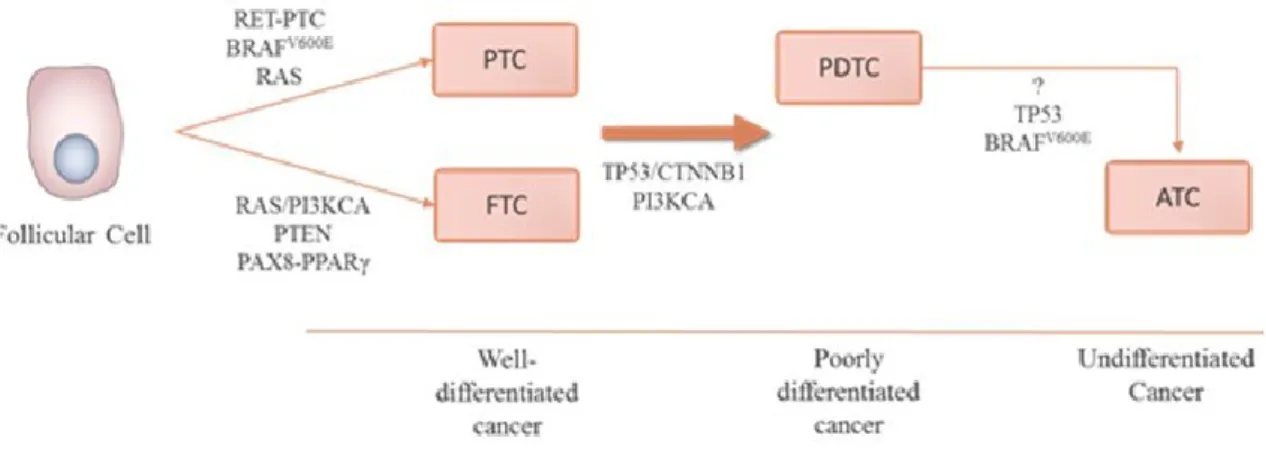
As previously denoted, non-medullary thyroid tumours are lesions with origin in the follicular cells representing about 95% of all thyroid malignancies. The lesions can be grouped in concordance with the differentiation degree of follicular cell - Well Differentiated Thyroid Carcinoma (WDTC), Poorly Differentiated Thyroid Carcinoma (PDTC) and Anaplastic Thyroid Carcinoma (ATC). The WDTC group are composed by two different histologic types of tumours – papillary and follicular (see Figure I.3) (Muro-Cacho and Ku, 2000; Liu et al., 2006; Capezzone et al., 2008; Morani et al., 2014).
Tumours associated with less differentiation of follicular cells are usually the most aggressive and also associated with poor prognosis due to the lack of therapies available (Kondo et al., 2006). While these malignancies can arise de novo, some reported studies support the hypothesis that all of these tumours share an evolutionary link, in which papillary and follicular thyroid carcinomas may give rise to the poorly differentiated and anaplastic forms, through a stepwise process of dedifferentiation of papillary and follicular carcinomas (Bhaijee and Nikiforov, 2011; Caronia et al., 2011; Nikiforov and Nikiforova, 2011). The majority of known genetic alterations associated with papillary and follicular thyroid carcinoma fall into one of four mutually exclusive groups (Bhaijee and Nikiforov, 2011): BRAF [Murine sarcoma viral (v-raf) oncogene homolog B1] and RAS (Rat sarcoma vírus homolog) point
mutations, and RET/PTC (RET- rearrangement during transfection) and PAX8/PPARγ [paired box
gene 8 (PAX8)-peroxisome proliferator-activated receptor γ (PPARγ)] rearrangements. It is known that thyroid cancer initiation and progression involve an accumulation of different types of genetic
- 6 -
alterations. The same probably happens in the dedifferentiation process in which some mutations that represent early events in thyroid cancer progression (such as BRAF and RAS mutation), that are found either in WDTC or in PDTC/ATC, may predispose WDTC to dedifferentiation.
Figure I.3. Subtypes of non-meddulary thyroid carcinomas. Adapted from Nikiforov and Nikiforova (2011) and Sastre-Perona and Santisteban (2012).
3.1.2.1. Follicular thyroid adenoma
Benign tumours of thyroid gland are designated by follicular thyroid adenomas (FTA). This group of tumours is characterized by their encapsulated form (usually a fibrous capsule of variable thickness) and they are round or oval, measuring between 1 and 3 cm. This lesion can occur in normal glands or in association with thyroiditis, nodular hyperplasia or other lesions. Typically FTAs present follicular cell differentiated phenotype (Muro-Cacho and Ku, 2000; DeLellis et al., 2004; Liu et al, 2006). It has been also proposed that FTA may serve as a precursor for some follicular carcinomas (Nikiforov and Nikiforova, 2011).
3.1.2.2. Follicular thyroid carcinoma
Follicular thyroid carcinoma is a type of malignant tumours that represents approximately 10-15% of all thyroid cancers (Bhaijee and Nikiforov, 2011; Sobrinho-Simões et al., 2011). According to the WHO (World Health Organization) Classification of Tumours of Endocrine Organs, FTC can be defined as ‘a malignant epithelial tumour showing follicular cell differentiation and lacking the diagnostic nuclear features of papillary thyroid carcinoma’ (see DeLellis et al., 2004). These are commonly solid tumours presenting as an isolated focus (unifocal), encapsulated, characterized by haematogenous spread, exhibiting less lymph node involvement (than PTCs), and with size higher than 1 cm (Schlumberger, 1998; DeLellis et al., 2004; Kondo et al., 2006; Romitti et al., 2013). As PTCs, follicular carcinomas can also be divided in different variants, namely the conventional type and the oncocytic type (oxyphilic or Hürthle cells tumours) (Schlumberger, 1998; Bhaijee and Nikiforov, 2011; Bonora et
- 7 - al., 2010). The invasion grade of the capsule and vessels (infiltrative growth) is a distinguishing feature between follicular carcinomas and follicular adenomas. The classification of tumours according to its pattern of invasion recognized two groups – those minimally invasive and the widely invasive carcinomas (Schlumberger, 1998; Biersack and Grünwald, 2005; Bonora et al., 2010).
The most common genetic alterations associated with FTCs (70-75% of cases) are RAS point mutations and PAX8/PPARγ rearrangements which are mutually exclusive (DeLellis et al., 2004; Castro et al., 2006; Bhaijee and Nikiforov, 2011; Sobrinho-Simões et al., 2011).
The prognosis of FTC depends on several features, namely the age of the patients at the diagnosis (usually higher comparing to PTCs), tumour size and stage, invasion rate, extensiveness of surgery and responsiveness to radioactive iodine (Muro-Cacho and Ku, 2000; DeLellis et al., 2004; Kondo et al., 2006).
One of the biggest challenges in this field is related with the pre-surgical discrimination between malignant follicular thyroid cancer and benign follicular thyroid adenoma. Although the gold standard for evaluating thyroid nodules is fine needle aspiration (FNA) biopsy, this method is unable to reliably distinguish FTC from FTA, since the key feature associated with this malignancy is its invasiveness rate (capsular and/or vascular penetration) (Schlumberger, 1998; Barden et al., 2003; DeLellis et al., 2004). Thyroid unilateral lobectomy is still used as a standard approach which allows a postsurgical diagnosis as FTC or FTA. Depending on the postsurgical conclusion, patients are managed in different ways - only cancer patients require completion total thyroidectomy, adjuvant radioiodine treatment, and long-term follow-up (Pfeifer et al., 2013). The big challenge is to find a reliable method to discriminate FTC and FTA in FNA material. Over the years numerous studies proposed different immunohistochemical markers. Pfeifer and collaborators (2013) reported a panel of five genes (ELMO1, EMCN, ITIH5, KCNAB1, SLCO2A1) with a 72% specificity grade; 94% accuracy was found in a six gene set (TFF3/PLAB/TG/ADM3/HGD1/LGALS3) (Krause et al., 2008). Many other studies were performed, namely studies assessing the use of miRNAs as potential distinguish markers (Stokowy et al., 2014) or studies where metabolomics approach were used (Deja et al., 2013). Despite all the efforts, no panel of molecular markers with confidence levels of specificity and sensitivity that allows the preoperative discrimination between FTCs and FTAs has been defined yet.
3.1.2.3. Papillary thyroid carcinoma
PTC is the most common malignant epithelial thyroid tumour presenting differentiation of follicular cell – PTC integrates the WDTC group. Papillary tumours account to nearly 80% of all thyroid cancers (DeLellis et al., 2004; Caronia et al., 2011). This tumour type is usually associated with good prognosis and long-term survival rates, due to a successful combination of radioiodine and levothyroxine treatment following total thyroidectomy. However, up to 10% patients would eventually die of the disease mostly due to the high levels of recurrences, since a subset of patients with advanced stage carcinomas have
- 8 -
radioiodine refractory and non-responsive malignancies (Sonkar et al., 2010; Soares et al., 2014). Older age at diagnosis, male gender, large tumour size and extra-thyroidal growth represent the most relevant poor prognostic factors (DeLellis et al., 2004; LiVolsi, 2011; Soares et al., 2014).
Most of papillary lesions are firm masses with the size ranging from less than 1 mm to several centimetres. The prominent papillae are a specific feature of this type of lesion, such as enlargement, oval shape, elongation and overlapping of the nuclei (DeLellis et al., 2004).
Papillary thyroid neoplasia is influenced by environmental, genetic and hormonal factors. Environmental factors comprise genotoxic (DNA damage due to radioactive iodine) and non-genotoxic effects (due to iodine deficiency). The thyroid dependency of iodine makes it vulnerable to radiation, being the PTC the subtype mostly linked to this factor (DeLellis et al., 2004).
PTC can be divided into numerous histopathologic variants based on specific growth patterns, cell types and stromal changes (Lloyd et al., 2011) – for example, follicular variant of papillary thyroid carcinoma (FVPTC) preserves nuclear features similarly to PTC, but exhibits a predominantly micro-follicular histologic growth pattern (McFadden et al., 2014).
Point mutations (such as mutations in the RAS and BRAF genes) and the RET/PTC and PAX8/PPARγ chromosomal rearrangements are common genetic alteration in this cancer subtype, that usually involve the effectors of the MAPK (Mitogen-activated protein kinases) and PI3K-AKT (PI3K– phosphoinositide 3-kinase; AKT – Protein Kinase B) pathways (Nikiforov and Nikiforova, 2011). Rearrangements in the RET proto-oncogene, known as RET/PTC, can be detected in PTCs with its prevalence varying between different patient cohorts – from 20% of all cases to much higher percentages (DeLellis et al., 2004; Nikiforov and Nikiforova, 2011). The V600E mutation on the BRAF proto-oncogene is the most frequent genetic alteration in PTC, representing 40-50% of cases (Nikiforov and Nikiforova, 2011). V600E – a transversion resulting in the substitution of glutamic acid for valine - is the most common BRAF mutation (Lee et al., 2007; Caronia et al., 2011). RAS point mutations can also be found, but the reported prevalence is much smaller – 10-20% according to Nikiforov and Nikiforova (2011). The indicated numbers can be much different in the others variants of PTC, namely on FVPTC, where a different and less prevalent type of BRAF mutation (K601E) can be found. Also, PAX8/PPARγ rearrangements and RAS point mutation have a higher prevalence than in classic PTC subtype – 40% and 25% of all FVPTC cases, respectively (Castro et al., 2006).
BRAFV600E mutation in PTC
BRAF is a serine/threonine kinase of the RAF family of proteins and it’s considered a strong activator of the MAPK pathway (involved in the regulation of several cellular responses) (Davies et al., 2002; Lee et al., 2007). BRAF mutations are not specific of thyroid tumours and are also highly prevalent in malignant melanoma, colorectal carcinoma and serous ovarian cancer (Garnett and Marais, 2004). BRAFV600E mutation is generally accepted as the most frequent genetic alteration in PTCs. This mutation
constitutively activates BRAF kinase, which works as a prolonged stimuli of the MAPK pathway, culminating in an increase of cell proliferation and defective apoptotic levels (Davies et al., 2002; Soares
- 9 - et al., 2003; Wan et al., 2004). The association between BRAF mutation and morphological and functional alterations related with PTC phenotype has been reported, for example, the presence of papillary structures that are a characteristic of the classical variant of PTCs are a phenotypic feature closely associated with BRAFV600E-positive tumours (Lee et al., 2007; Rusinek et al., 2011). Significant
correlation between BRAF mutations and extra-thyroidal invasion, lymph node metastasis, and tumour stage has been also reported in PTC by Chakraborty and collaborators (2012).
3.1.2.4. Poorly differentiated thyroid carcinoma
Poorly differentiated thyroid carcinoma is a rare entity that represents less than 10% of all NMTC (Biersack and Grünwald, 2005; Bonora et al., 2010). As already mentioned, this is a neoplasia originated from thyroid follicular cells, representing an intermediate morphological and biological behaviour between well-differentiated thyroid carcinomas and anaplastic thyroid carcinomas (Muro-Cacho and Ku, 2000; DeLellis et al., 2004; Biersack and Grünwald, 2005). This type of malignancy affects more males than WDTC and the average age of onset is higher, at approximately 50 years old. Lymph nodes and distant metastasis are highly frequent. The survival rates are low in the first three years after the diagnosis, and lower after five years, precisely due to metastasis occurrence (30-80%) (DeLellis et al., 2004; Biersack and Grünwald, 2005; Kondo et al., 2006). Kondo and collaborators (2006) documented RAS and TP53 (Tumour protein 53) mutations as the most frequent genetic alterations in PDTC (18-27% and 17-38% of all cases, respectively). BRAF point mutation are also present in some cases (0-13%) supporting the hypothesis of a WDTC to PDTC transition.
3.1.2.5. Anaplastic thyroid carcinoma
Anaplastic carcinoma is a highly malignant tumour composed by undifferentiated cells in a partial or total undifferentiated form. This type of malignancy represents less than 5% of all NMTC cases, being the least common form (Muro-Cacho and Ku, 2000; DeLellis et al., 2004; Bonora et al., 2010). ATCs are highly aggressive and lethal tumours with a very low survival rate – the mean duration of survival is only months (DeLellis et al., 2004; Biersack and Grünwald, 2005; Kondo et al., 2006; Soares et al., 2011). These tumours can coexist with other thyroid carcinomas or hyperplasia (30% of the cases), and are usually non-encapsulated tumours, extensively invading the perithyroid tissues. The three main morphological patterns are squamoid, pleomorphic giant cell and spindle cell (Kondo et al., 2006; Soares et al., 2011). The mean age at the time of initial diagnosis is about 60 years old and there is a higher frequency on females (Muro-Cacho and Ku, 2000; DeLellis et al., 2004; Kondo et al., 2006; Soares et al., 2011). Mutations in the tumour suppressor gene p53 are known as the most common genetic events in this type of malignancy (67-88%) (Kondo et al., 2006).
- 10 -
4. RAC1 and RAC1b GTPases
4.1. Organization and regulationRAC1 is a member of the Rho (Ras homologous) family of small guanosine triphosphatases (GTPases) – a subgroup of the RAS superfamily of GTP (Guanosine triphosphate)-binding proteins. This Rho GTPases family includes other members such as Rho and Cdc42 (Cell division control protein 42 homolog). RAC1 protein is encoded by RAC1 gene and can exist in two different conformational states - an inactive GDP (Guanosine diphosphate)-bound form and an active GTP-bound form (Jordan et al., 1999; Matos et al., 2000; Wennerberg and Der, 2004; Jaffe and Hall, 2005).
The interconversion between the two states occurs through a cycle of guanine exchange and GTP hydrolysis, wherein GTP binding induces a conformational change that involves two important regulatory regions, termed Switch I and Switch II. Consequently, the switch regions provide a surface that, in the active state, enables their interaction with downstream effectors, allowing these GTPases to function as molecular switches (Wennerberg et al., 2005). This cycling process is tightly regulated by several groups of proteins: Rho-GEFs (Guanine exchange factors) which promote exchange of GDP for GTP; Rho-GAPs (GTPase activating protein) that enhance the hydrolysis of bound GTP to GDP and inorganic phosphate, regulating in the inactivation of the GTPases; GDP dissociation inhibitors (GDI) are also regulatory proteins that sequester Rho GTPases in the cytoplasm in an inactive GDP-bound state, preventing exchange of GDP to GTP (Jordan et al., 1999; Bernards and Settleman, 2004; Singh et al., 2004).
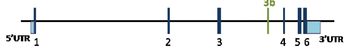
Figure I.4. Diagram of the RAC1 gene. 1, 2, 3, 4, 5, 6 represent exons. Adapted from Matos et al., 2000. Back to 1999, Jordan and collaborators performed a RT-PCR (Reverse transcription polymerase chain reaction) based assessment of RAC1 expression in colorectal samples (tumours and normal mucosa) that leads to the identification of a new RAC1 splicing variant, termed RAC1b. This variant was also found to be expressed in breast carcinomas (Schnelzer et al., 2000). The RAC1b isoform results from an alternative splicing event that leads to the inclusion of an additional exon (exon 3b). This additional exon is inserted between exons 3 and 4 (see Figure I.4) of RAC1 and contains 57 additional nucleotides that result in an in-frame insertion of 19 amino acid residues between codons 75 and 76, in the vicinity of the switch II domain (Jordan et al., 1999).
- 11 - RAC1b is considered a highly activatable variant of RAC1: despite the lower levels of expression compared to RAC1, RAC1b exists predominantly in the active GTP-bound state. This is essentially due to RAC1b disability to interact with Rho-GDI, which keeps this GTPase constitutively membrane-bound, a location that benefits the interaction with activators, and consequently promotes the active GTP-bound state (Matos et al., 2003). Moreover, RAC1b shows impaired intrinsic activity and increased GDP to GTP exchange rates, although this variant can still be downregulated by activated GAPs and it is influenced by GEFs action (Schnelzer et al., 2000; Matos et al., 2003; Fiegen et al., 2004; Singh et al., 2004). Also, RAC1b's additional amino acids seem to confer to this variant a selective downstream signalling, since several pathways activated by RAC1, are not activated by RAC1b (Matos et al., 2003).
4.2. Biological functions
The Rho family of small GTPases is known to play an important role in several cellular processes, namely in actin cytoskeleton reorganization – Cdc42 regulates the formation of filopodia; RAC1 is involved in the lamellipodia formation; RhoA promotes the formation of stress fibres (Fukuda et al., 2002; Matos et al., 2003; Singh et al., 2004).
RAC1, in particular, has the ability to interact with specific effectors, inducing activation of numerous signalling cascades that culminate in different physiological outcomes, namely cytoskeletal dynamics alteration, progression through the cell cycle and cell proliferation, apoptosis, migration and cell-cell adhesion, membrane trafficking and superoxide production (Matos et al., 2000; Jaffe and Hall, 2005; Bosco et al, 2009). There are several downstream effectors and signalling proteins influenced by activated RAC1, namely WAVE (WASP-family verprolin-homologous protein) complex, PAK (p21 activating kinase) and LIM. For example, GTP-bound RAC1 binds to PAK1, stimulating its protein kinase activity, which allows the interaction with other proteins, leading to the modulation of several biological activities, such as the mentioned actin reorganization (Jaffe and Hall, 2005; Bosco et al, 2009; Nagase and Fujita, 2013).
The action of activated RAC1 also includes the stimulation of transcription factors such as the
activation of the Jun NH2-terminal kinase (JNK) cascade or the transcription factor NF-κB (Nuclear
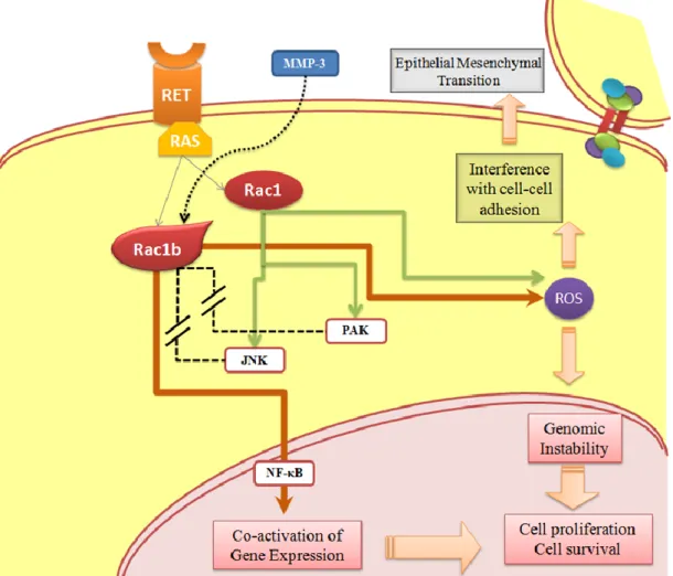
factor kappa-light-chain-gene-enhancer of activated B cells). The RAC1-related activation of the NF-κB pathway involves the production of reactive oxygen species (ROS), and initiates an anti-apoptotic transcriptional response, leading to an increased cyclin D1 expression and consequently promoting the cell cycle progression (Hinz et al., 1999; Joyce et al., 1999; Matos and Jordan, 2006).
As already mentioned, compared to RAC1, RAC1b, shows a selective downstream signalling. Unlike RAC1, GTP-bound RAC1b ability to induce actin cytoskeleton reorganization is impaired – RAC1b is unable to induce lamellipodia formation. RAC1b is also unable to interact with two other well-established RAC1 signalling pathways, in particular, this splice variant is incapable to activate PAK1 effector and stimulate the JNK cascade (Matos et al., 2003; Fiegen et al., 2004; Singh et al.,
- 12 -
2004). On the other hand, at least in the colorectal biological system, RAC1b retains the capacity to stimulate the NF-κB pathway, although its action is limited to the classical RelA-dependent NF-κB pathway, contrary to RAC1 that also stimulates the alternative pathway (RelB-dependent). The RAC1b stimulation of NF-κB classical pathway increases the cell cycle progression and cell survival, reducing the apoptotic rates (Matos and Jordan, 2005; Matos and Jordan, 2006). RAC1b was also shown to have the ability to mediate epithelial-mesenchymal transition (EMT) in breast (Radisky et al., 2005).
- 13 -
4.3. Association with cancer
Malignant transformation is characterized by dramatic alterations in cell function and properties. This process includes the gain of migratory and invasive properties of tumour cells, increased proliferation levels and decreased apoptotic rates. RAC1 and RAC1b have been implicated in several of these cellular processes, namely in cell survival by increasing responses for apoptosis evasion and by stimulating cell cycle progression. The ability of these GTPases to interfere with these cellular functions could represent a tumorigenic property that allows the initiation of tumour formation, which, in turn, depends on the outcome of uncontrolled proliferation and anti-apoptotic mechanisms (Matos and Jordan, 2005; Fritz and Kaina, 2006; Bid et al., 2013; Zhou et al., 2013).
In agreement, a study using mouse mammary epithelial cells, reported that matrix metalloproteinase 3 (MMP-3) induces RAC1b expression, which promotes an increase in cellular ROS (reactive oxygen species). EMT, oxidative damage to DNA and genomic instability are some of the consequences of ROS increased levels, which are ultimately involved in tumour formation, invasion and metastasis (Radisky et al., 2005).
In fact, the elevated expression of RAC1b has been documented in colorectal (Jordan et al., 1999), breast (Schnelzer et al., 2000), lung (Zhou et al., 2013) and thyroid cancer (Silva et al., 2013). Specifically in colorectal carcinoma, the alternative splice variant of RAC1 was shown to functionally
cooperate with BRAFV600E mutation to sustain the proliferation and survival of tumour cells (Matos et
al., 2008). In a previous study, a group at IPOLFG (Silva et al., 2013) reported for the first time RAC1b expression in thyroid tissue. Furthermore, the IPOLFG team shown that RAC1b is overexpressed in PTCs compared to normal thyroid tissue and that RAC1b overexpression is significantly associated with BRAFV600E mutation and poor clinical outcome in PTC. Similar to that reported in colorectal cancer,
these findings point to an important role of RAC1b in PTC development, which may also be associated with a functional cooperation with BRAFV600E mutation.
5. Signalling pathways involved in cancer
The tumorigenesis process is often characterized by the accumulation of point mutations and changes in gene expression in pathways regulating specific aspects of cell proliferation and survival. Deregulation of some crucial pathways allows tumour cells to increase cell cycle progression, evade apoptosis and invade surrounding tissue. Many of these pathways that regulate cellular processes are controlled by specific genes. Alterations in the sequence, expression or epigenetics of these genes can alter these signalling cascades, inducing modifications in the cellular processes. For example, alteration in TP53 gene (a gene involved in one of the major signalling pathways associated with apoptosis), induces alterations in this specific TP53 pathway, increasing tumour cells protection against apoptosis (Vogelstein and Kinzler, 2004).
- 14 -
In addition to TP53 pathway, there are many other signalling pathways described as being involved in the tumorigenic process of several tumours, such as JNK, PI3K, RTK (Receptor tyrosine kinases), cyclin D1 or NF-κB. NF-κB, in particular, was already implicated in several cancer types and has been studied as a potential therapeutic target (Debatin, 2004; Vogelstein and Kinzler, 2004; Dhillon et al., 2007).
5.1. NF-κB signalling pathway
NF-κB (Nuclear factor kappa-light-chain-gene-enhancer of activated B cells) is a protein complex that plays a pivotal role in transcriptional control, cytokine production and in the regulation of several other cellular processes. In mammalian cells, the NF-κB family is composed of five members that form homodimers or heterodimers with each other – RelA (p65), RelB, c-Rel, p50 and p52. These proteins have an unique N-terminal Rel homology domain (approximately 300 amino acid) that allows dimer
formation, binding to DNA and to NF-κB inhibitors (Karin and Ben-Neriah, 2000; Liang et al., 2004;
Solt and May, 2008; Madonna et al., 2012). Conversely, the C-terminal domain differs between Rel (RelA, RelB and c-Rel) and NF-κB proteins (p50 and p52) – RelA, RelB and c-Rel exhibit transactivating function, while p50 and p52 contain inhibitory sequences associated to this C-terminal
domain (Karin and Ben-Neriah, 2000; Madonna et al., 2012). The Rel homology domain also contains
a nuclear localization signal (NLS) (Huang et al., 2000; Karin and Ben-Neriah, 2000; Karin et al., 2002). Unlike Rel proteins, p50 and p52 proteins are produced by proteolytic removal of C-terminal sequences of two inhibitory precursor proteins – NF-κB1/p105 and NF-κB2/p100, respectively. This removal allows the final proteins to enter the nucleus, usually as heterodimers with one of the Rel proteins that bears a transactivation domain (Karin et al., 2002; Bassères and Baldwin, 2006).
The NF-κB dimers remain transcriptionally unstimulated as long as they are sequestered in the cytoplasmic compartment. In this form, NF-κB proteins are usually associated with members of the IκB family (consist of an N-terminal regulatory domain followed by a series of ankyrin repeats), such as IκBα, IκBβ, IκBε or the inhibitory precursor proteins – NF-κB1/p105 and NF-κB2/p100 (Bassères and Baldwin, 2006; Solt and May, 2008). Some studies indicated that IκB proteins act by masking the nuclear localization signal, which prevents the NF-κB dimers translocation into the nuclear compartment (Huang et al., 2000; Karin and Ben-Neriah, 2000).
Their activation can happen as consequence of a diversity of stimuli, such as those activating membrane B cell receptors (BCR) or tumour necrosis factor receptors (TNFR). Moreover, several extracellular stimuli can also stimulate NF-κB pathway, namely inflammatory cytokines, viral and bacterial infections, oxidative and DNA-damaging agents, UV light and osmotic shock (Chen et al., 2012; Karin et al., 2002).
The regulation of the NF-κB dimers can be performed through two main pathways. Both signalling pathways that activate NF-κB transcriptional activity have as pivotal regulator, the IκB kinase (IκK - inhibitors of κB kinases) complex, which is composed by two catalytic subunits (IκKα and IκKβ) and a
- 15 - non-catalytic regulatory subunit named NEMO (NF-κB essential modulator) or IκKγ. NEMO plays a crucial role in both pathways, whereas IκKβ is particularly important in the classical pathway and IκKα is also part of the non-canonical regulatory pathway. IκKs are involved in the phosphorylation of NF-κB inhibitor proteins at specific serine residues (Liang et al., 2004; Waes, 2007; Solt and May, 2008; Sun, 2011).
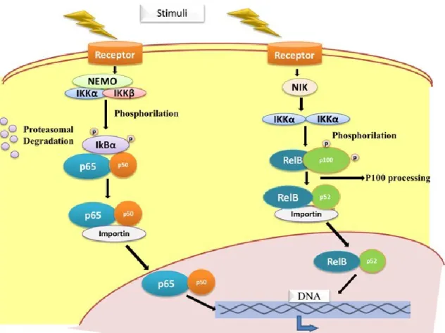
The first regulatory pathway (see Figure I.6), termed canonical or classical NF-κB activation pathway, applies to dimers composed by RelA, c-Rel and p50. It is usually triggered in response to microbial and viral infections and exposure to proinflammatory cytokines. The IκKβ has a particularly important role in this classical pathway by phosphorylating IκBα, which is subsequently ubiquitinated and degraded by the 26S proteasome. The degradation of NF-κB inhibitor, culminates with the accumulation of the dimeric NF-κB transcription factor (mostly composed by p50 and p65) into the nucleus, where it is able to bind κB site and induces the transcription of different target genes, most of them implicated in inflammation, survival and proliferation responses (Karin and Ben-Neriah, 2000; Karin et al., 2002).
The second regulatory pathway (see Figure I.6), named non-canonical pathway, affects preferentially NF-κB2/p100 and RelB. The TNF (Tumour necrosis factor) cytokine family provides the most important stimuli for this pathway, which selectively activates the IκKα subunit along with another protein kinase called NIK (NF-κB inducing kinase). This activation induces a phosphorylation-dependent proteolytic removal of C-terminal sequences of NF-κB2 inhibitory precursor protein, which allows RelB/p52 dimers to translocate to the nucleus and recognize specific kB sites in the promoter of target genes (Karin and Ben-Neriah, 2000; Karin et al., 2002; Sun, 2011).
Each pathway have distinct regulatory functions, although some target genes are common to all NF-κB proteins and are usually related to immunoregulatory and inflammatory processes, anti-apoptotic responses and cell cycle regulation (Shishodia and Aggarwal, 2002; Liang et al., 2004; Matos and Jordan, 2005).
- 16 -
Figure I.6. Canonical and non-canonical NF-κB activating pathways. 5.1.1. Association with Tumorigenesis
NF-κB plays three essential roles (Madonna et al., 2012). It is involved in the proinflammatory response, including immune, inflammatory and acute phase responses. It has already been established that this pathway sustains survival, maturation and recirculation of lymphocytes as well as T and B cell-mediated immune responses (Liang et al., 2004). The ability to evade apoptosis is one of the most studied function of NF-κB pathway, which is known to induce the transcription of several anti-apoptotic proteins, such as Bcl-XL (B-cell lymphoma-extra-large), tumour necrosis factor receptor-associated factor 1 and 2 (TRAF1 and TRAF2, respectively), and the inhibitor-of-apoptosis (IAP) protein 1 and 2, which allows NF-κB to control the activity of the caspase family of enzymes (essential for the apoptotic cellular cascade) (Shishodia and Aggarwal, 2002; Liang et al., 2004; Solt an May, 2008; Madonna et al., 2012).
Ultimately, NF-κB is involved in the regulation of cell cycle progression through its ability to induce the expression of cyclin D1 (Hinz et al., 1999; Joyce et al., 1999; Matos and Jordan, 2005). Cyclin D1 works as a key sensor and integrator of extracellular signals of cells in G0 and early G1 phases, allowing the progression through the cell cycle, to S phase (Baldin et al., 1993; Stacey, 2003; Fu et al., 2004). Consequently, aberrant cyclin D1 expression is a critical event in cancer development that leads to
- 17 - uncontrolled progression of cell cycle, usually induced by other cancer-related signals such as NF-κB activation or β-catenin (Hinz et al., 1999; Joyce et al., 1999; Fu et al., 2004; Liu et al., 2014).
Given the extent of biological mechanisms associated with NF-κB pathways and the overlap of these mechanisms with some hallmarks of cancer, it is expectable to find deregulation of this pathway in association with several types of malignancies. Research work in this field documented several changes in the upstream pathways that lead to NF-κB activation to become deregulated in cancer (Staudt, 2010). Moreover, some studies reported that mutations which are usually associated with cancer initiation and development, such as RAS mutation, can lead to IκB inactivation and consequently to the upregulation of NF-κB pathway (Mizumoto et al., 2011; Chen et al., 2012).
In fact, it has been reported over the years that increased activation of the NF-κB pathway is involved in some forms of cancer such as melanoma (Madonna et al., 2012), lung cancer (Chen et al., 2012), glioblastoma (Nogueira et al., 2011), colorectal cancer (Wang et al., 2009), thyroid cancer (Namba et al., 2007; Bauerle et al., 2010) and ovarian cancer (White et al., 2011).
Given that, NF-κB pathway appears to have a crucial role in a vast diversity of tumours, the inhibition of this signalling cascade starts to be seen as a promising strategy for anti-cancer therapies.
5.1.2. Association with RAC1b
The ability of RAC1 to stimulate the NF-κB pathway is shared with its splice variant RAC1b, which was shown to be capable to promote IκB phosphorylation and nuclear translocation of RelA, in NIH
3T3mouse embryonic fibroblast cell line (Matos et al., 2003). Moreover, a study in colorectal cells
demonstrated that RAC1b only stimulates the canonical NF-κB regulatory pathway, differing from RAC1 which is capable of stimulate both the classical and the alternative pathway. The activation of the non-canonical pathway by RAC1 could interfere, in a potential negative feedback regulation, with the RAC1-induced classical cascade. The selective RAC1b induction of the canonical regulatory pathway permits this GTPase to circumvent the negative feedback from the RelB pathway, which allows a continuously stimulation of the p50/RelA NF-κB-mediated transcription. Also in colorectal cells, the expression of RAC1b was shown to stimulate transcription from a luciferase reporter promoter containing consensus NF-κB binding sites, through a ROS dependent cascade (Matos and Jordan, 2006).
In opposition, Singh and collaborators (2004) reported that RAC1b failed to activate the NF-κB transcription factor or stimulate cyclin D1 expression, in NIH 3T3 cell model (fibroblasts).
Whether RAC1b contributes to NF-κB-mediated cellular processes (namely proliferation or apoptosis evasion) and to malignant progression in cell systems other than colorectal cancer remains to be clarified.
- 18 -
6. Follicular cell derived thyroid cancers, RAC1b and NF-κB
pathway – a link to unravel
Part 1. RAC1b role in FTC Tumorigenesis
Our group has recently reported RAC1b expression in thyroid tissue (Silva et al, 2013). In fact, this variant was found to be overexpressed in PTCs compared to normal tissue, and to be associated with a subset of PTCs with a poor clinical outcome. These findings not only point to an important role of RAC1b in PTC development but also raise the possibility that RAC1b overexpression might be associated with other types of thyroid carcinoma, namely with other subtypes derived from follicular cells (non-medullary thyroid carcinomas). The second most frequent form of thyroid carcinomas is the FTC, representing about 15% of all cases. Despite the good prognosis usually associated to this type of malignancy, some patients present advanced tumours, non-responsive to radioiodine therapy.
Hence, the broadening of the study of this GTPase to other follicular cells derived neoplasia, such as the FTCs, could provide novel information about the role of RAC1b in the thyroid tumorigenesis. This might as well contribute to the definition of prognostic biomarkers for this tumoural subtype, allowing an individual case management of risk level.
On the other hand, since one major challenge in the clinical management of FTC patients is related with the inability to discriminate between benign and malignant follicular tumours at the preoperative stage, a comparative analysis of RAC1b expression between FTC and FTA may clarify the importance of this GTPase as a potential marker to be included in a broader panel of biomarkers to be used as diagnostic tool.
Part 2. RAC1b - An activator of NF-κB pathway that contributes to thyroid Tumorigenesis? The potential association of RAC1b overexpression with PTC development, magnified the importance of investigating the molecular mechanisms associated with RAC1b overexpression and downstream signalling in thyroid tumorigenesis. In fact, the identification of precise alterations on signalling pathways associated with specific cancer processes would be pivotal for the understanding of molecular mechanisms behind tumour progression and for the development of diagnostic and prognostic biomarkers. In thyroid cancer there is, undoubtedly, the emerging need for a better understanding of the molecular events involved in the initiation and progression of malignancy as well as its association with failure in the response to iodine treatment and high recurrence rates. Understanding these mechanisms should contribute to individual case management optimization.
A possible rational approach to investigate the molecular mechanisms associated with RAC1b overexpression and downstream signalling in thyroid tumorigenesis is to select a signalling pathway previously implicated in the tumorigenic process as a starting point. This selection was based in two previously published facts: NF-κB activation has been also reported to play an important role in thyroid
- 19 - cancer (Namba et al., 2007; Bauerle et al., 2010); RAC1b selectively induces the canonical regulatory NF-κB pathway in colorectal cancer (Matos and Jordan, 2006). Hence, we considered relevant to study the role of RAC1b on NF-κB activation in thyroid tissue, and to further assess the effect of this potential RAC1b-mediated NF-κB stimulation in terms of cell growth and survival. Furthermore, given the role of Cyclin D1 as a cell cycle regulator, its association with some cancer events and the capacity of NF-κB to induce cyclin D1 expression, it is reasonable to consider Cyclin D1 as another target potentially affected by RAC1b overexpression.
- 20 -