UNIVERSIDADE DE LISBOA
Faculdade de Medicina Veterinária
“Understanding the dynamics of African swine fever spread at
the interface between wild boar and domestic swine in Sweden”
João Filipe Dias Gomes de Morais Costa
CONSTITUIÇÃO DO JÚRI
Doutor Carlos Manuel Lopes Vieira Martins Doutor Fernando Jorge Silvano Boinas Mestre Telmo Renato Landeiro Raposo Pina Nunes
ORIENTADOR Doutora Fernanda Cetrangolo Dórea CO-ORIENTADOR Mestre Telmo Renato Landeiro Raposo Pina Nunes
2017 LISBOA
UNIVERSIDADE DE LISBOA
Faculdade de Medicina Veterinária
“Understanding the dynamics of African swine fever spread at
the interface between wild boar and domestic swine in Sweden”
João Filipe Dias Gomes de Morais Costa
DISSERTAÇÃO DE MESTRADO INTEGRADO EM MEDICINA VETERINÁRIA
CONSTITUIÇÃO DO JÚRI
Doutor Carlos Manuel Lopes Vieira Martins Doutor Fernando Jorge Silvano Boinas Mestre Telmo Renato Landeiro Raposo Pina Nunes
ORIENTADOR Doutora Fernanda Cetrangolo Dórea CO-ORIENTADOR Mestre Telmo Renato Landeiro Raposo Pina Nunes
2017 LISBOA
“Train while they sleep, study while they have fun, persist while they rest, and then live what they dream”
Acknowledgements
I would like to acknowledge COST Action CA15116 –ASF-STOP for funding a Short Term Scientific Mission and the Erasmus Program for partially funding my traineeship at the National Veterinary Institute in Uppsala, Sweden.
To my supervisor Fernanda Dórea, from the National Veterinary Institute, for all the guidance through this scientific journey. Thank you for all your time, help and for always pushing me hard and demanding the best of me. Thank you for this journey.
To Karl Ståhl, from the National Veterinary Institute, for the opportunity to learn and develop as professional at the National Veterinary Institute. Your enthusiasm, scientific knowledge and positive attitude towards science was always an inspiration for me.
To my co-supervisor, Professor and friend, Telmo Nunes. Thank you for all your precious guidance and for helping me to discover the brave new world of epidemiology. With your help I had the opportunity to develop new skills, get outside my comfort zone and overcome some limitation that I had.
To the National Veterinary Institute, especially to all the members of the Department of Disease Control and Epidemiology and the Department of Pathology and Wildlife Diseases, my sincere acknowledge for receiving me with open arms. A special acknowledge to Cecilia Hultén, Jenny Frosling, Thorsten Mörner, Estelle Ågren, Erik Ågren, Maria Nöremark and Thomas Rosendal for all the good moments shared. To Marie Söjlund and Per Wallgren for their expert opinion.
To my Professor Carlos Martins for all the contagious enthusiasm about ASF and all the knowledge shared with me.
To my parents for my values, for our love and for being my role models for life. Everything that we accomplish, we accomplish together. I would also like to thank my uncles and all family for all the support and special moments that we spend together.
To me dear friend Carlos Nelson Parente and his family for all the friendship and inspirational attitude. “Never quit, never surrender”.
To my Emergency Department Chief Nurse and friend, Fernando Sousa for all your help during the time I was a nurse at the Hospital Beatriz Ângelo. Your humanity and friendship made this journey possible.
To all my faculty friends that I shared this seven-years journey, especially to Bruno Moreira, Nélio Cebola, Diogo Cachitas e Franscico Malcata.
To the Ibrahim family and to Cocca family, for making me feel at home and among family during my stay in Sweden.
To Tita for this journey, for giving me a purpose, for all the love… ”alone we go faster, but together we go further!
“Understanding the dynamics of African swine fever spread at the interface between wild boar and domestic swine in Sweden”
Abstract
African swine fever (ASF) is a viral disease with devastating consequences that is currently spreading across the Baltic States and Poland mainly driven by wild boar cases. Countries in Europe are urged to strengthen preparedness. In Sweden, a disease spread model has been used to evaluate the risk of transmission among domestic pigs, but further investigation is needed to assess the potential spill over of ASF virus from wild boars to the domestic pig population.
This study aimed to characterize the opportunities for transmission of ASF in the interface between domestic pigs and wild boars in Sweden, providing a review of relevant information to carry out a qualitative assessment. A risk characterization using scenario trees was used, allowing similar characterizations in other countries.
Five potential transmission pathways were identified: direct contact; indirect contact; infected swill; environmental contamination/local spread; vector borne transmission. The risks identified were: geographical overlap of both populations and the domestic pig outdoor access for direct contact; human risk activities and farm biosecurity for indirect contact; swill feed ban compliance for infected swill; distance, farm biosecurity and type for environmental contamination/local spread.
This work provides a potential framework for a risk assessment of ASF in this interface, not only in Sweden but also in other countries.
Keywords: African swine fever, wild boar, domestic pig, transmission, interface, risk characterization.
“Compreendendo a dinâmica de dispersão da Peste Suína Africana no interface entre o javali e o porco doméstico na Suécia”
Resumo
A Peste Suína Africana é uma doença viral com consequências devastadoras, atualmente em dispersão nos Países Bálticos e na Polónia, particularmente devido à sua presença no javali. É, desta forma, urgente o estabelecimento de medidas preventivas por parte dos países europeus. Na Suécia, tem sido utilizado um modelo de dispersão da doença para avaliar o risco de transmissão entre porcos domésticos, sendo ainda necessários estudos que avaliem o potencial de transmissão do vírus da peste suína africana dos javalis para a população de porcos domésticos.
Este estudo pretendeu caracterizar as oportunidades de transmissão da Peste Suína Africana no interface entre javalis e porcos domésticos na Suécia, providenciando uma revisão da informação relevante para a realização de uma avaliação qualitativa. Foi utilizada uma caracterização do risco recorrendo a “árvores de evento”, permitindo assim que caracterizações semelhantes sejam realizadas noutros países.
Foram identificadas cinco potenciais vias de transmissão: contacto direto, contacto indireto, restos alimentares contaminados, contaminação ambiental/dispersão local, transmissão mediada por vectores. Os riscos identificados foram: sobreposição geográfica das duas populações e o acesso ao exterior por parte dos porcos domésticos no contexto do contacto direto; atividades humanas de risco e biossegurança da exploração no contexto de contato indireto; conformidade com a proibição da alimentação com restos alimentares no contexto dos restos alimentares contaminados; distância, biossegurança da exploração e o tipo de exploração na contaminação ambiental/dispersão local;
Este trabalho fornece uma base informativa para a realização de uma análise de risco da deste interface, não só na Suécia como também em outros países.
Palavras-chave: Peste Suína Africana, javalí, porco doméstico, transmissão, interface, caracterização de risco.
Table of contents
Acknowledgements ... II Abstract ... IV Resumo ... V List of figures ... VIII List of tables ... IX List of Abbreviations and Symbols ... X
INTRODUCTION ... 1
1. 1.1 Etiology of ASF ... 2
1.2 Genotyping and geographical distribution ... 2
1.3 Inactivation and viral resistance ... 3
1.4 Hosts ... 4
1.5 Pathogenesis of ASF infection ... 5
1.6 Clinical forms ... 6
1.6.1 Peracute forms ... 6
1.6.2 Acute Forms ... 6
1.6.3 Sub acute forms ... 7
1.6.4 Chronic forms ... 7
1.7 Specific treatment and Vaccine ... 7
1.8 Diagnosis ... 8
1.8.1 Viral identification ... 9
1.8.1.1 Haemadsorption Test (HAD) ... 9
1.8.1.2 Antigen detection by fluorescent antibody test (FAT) ... 10
1.8.1.3 Antigen Elisa ... 10
1.8.1.4 Polymerase-Chain Reaction (PCR) ... 10
1.8.2 Serologic tests ... 11
1.8.2.1 Enzyme- Linked Immunosorbent Assay (ELISA) ... 11
1.8.2.2 Indirect fluorescent antibody test (IFA) ... 12
1.8.2.3 Immunoblotting ... 12
1.9 ASF Epidemiology ... 12
1.9.1 Basic definitions ... 12
1.9.2 OIE status ... 13
1.9.3 History and current epidemiology of ASF in Africa ... 14
1.9.4 History of ASF in Europe ... 15
1.9.5 Current European epidemics ... 16
1.9.6.1 Transmission cycle in Africa ... 19
1.9.6.2 Transmission cycle in Europe ... 20
1.9.7 Prevention and control ... 21
1.9.7.1 Preventive measures ... 21
1.9.7.2 Control ... 22
1.9.7.3 Surveillance ... 22
1.9.8 ASF and Sweden ... 24
OBJECTIVES ... 26
2. MATERIALS AND METHODS ... 26
3. 3.1 Transmission pathways at the interface WB-DP ... 26
3.2 Risk Description and Swedish Risk Characterization ... 27
RESULTS ... 28
4. 4.1 Transmission pathways at the interface WB-DP ... 28
4.1.1 Literature review process ... 28
4.1.2 Descriptive Results ... 28
4.1.2.1 Direct Contact ... 29
4.1.2.2 Indirect Contact ... 30
4.1.2.3 Infected Swill ... 32
4.1.2.4 Environmental contamination / Local Spread ... 33
4.1.2.5 Vector-Borne Transmission ... 34
4.2 Risk description and Swedish risk characterization. ... 36
4.2.1 Direct Contact ... 36
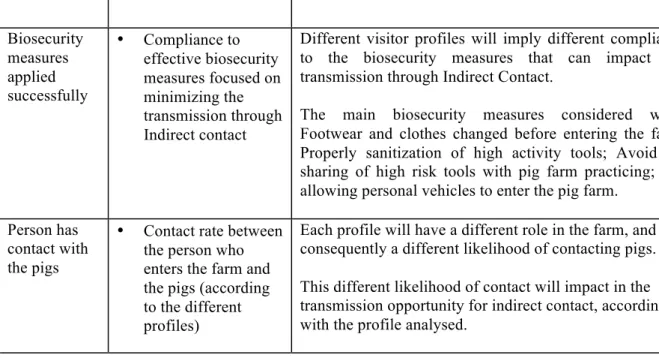
4.2.2 Indirect Contact ... 40
4.2.3 Infected Swill ... 44
4.2.4 Environmental Contamination / Local Spread ... 47
DISCUSSION ... 49 5. CONCLUSION ... 53 6. BIBLIOGRAPHY ... 54 7.
List of figures
Figure 1 – Transmission pathways at the interface between WB-DP. ... 29
Figure 2 - Scenario Tree detailing the events that can lead to transmission of ASF through direct contact between WB and DP. ... 36
Figure 3 - "Number of pigs per Km2 in 21 Swedish Counties as of June, 2015". Reproduced from SVA (2015) ... 38
Figure 4 – Number of WB shot by Swedish County. WB hunting data source: Swedish Association For Hunting and Wildlife Management, 2017; Swedish County data: Sverige Länsgränser (2017) ... 38
Figure 5 – Scenario Tree detailing the events that can lead to transmission of ASF through Indirect Contact between WB and DP. ... 40
Figure 6 – WB baiting station. ... 42
Figure 7 - Scenario Tree detailing the events that can lead to transmission of ASF through infected swill transmission pathway between WB and DP. ... 45
Figure 8 - Scenario Tree detailing the events that can lead to transmission of ASF through environmental contamination /local spread between WB and DP. ... 47
List of tables
Table 1 – List of information needed to characterize risks associated with the transmission of ASF between WB and DP through direct contact, as well as a description of these risks in Sweden. ... 37 Table 2 - List of information needed to characterize risks associated with the transmission of
ASF between WB and DP through Indirect Contact, as well as a description of these risks in Sweden. ... 41 Table 3 – List of information needed to characterize risks associated with the transmission of
ASF between WB and DP through infected swill, as well as a description of these risks in Sweden. ... 45 Table 4 – List of information needed to characterize risks associated with the transmission of
ASF between WB and DP through environmental contamination / local spread, as well as a description of these risks in Sweden. ... 48
List of Abbreviations and Symbols
ASF – African swine fever
ASFV – African swine fever virus
COST - European Cooperation in Science and Technology
CSF – Classic swine fever CVR - Central variable region
DIFT – Direct immunofluorescence test
DG SANCO - The Directorate-General for Health and Food Safety DNA - Deoxyribonucleic acid
dNTP - Deoxynucleotides DP – Domestic pig(s)
DTU-DADS - Technical University of Denmark - Davis Animal Disease Simulation model
EDTA - Ethylenediaminetetraacetic acid EFSA – European Food Safety Authority
ELISA - Enzyme-Linked Immunosorbent Assay
EURL-ASF – European Union Reference Laboratory for ASF EU – European Union
EVIRA - Finnish Food Safety Authority
FAO – Food and Agriculture Organization of the United Nations FAO EMPRES-I – FAO Global Animal Disease Information System
FAT – Fluorescent antibody test HAD - Haemadsorption
IFA - Indirect fluorescent antibody test
KRAV - Kontrollföreningen för Ekologisk Odling (Control Association for Alternative Cultivation)
OIE – Office International des Epizooties (World Organisation for Animal Health)
PCR – Polymerase chain reaction
PRRS - Porcine reproductive and respiratory syndrome RF – Russian Federation
RT-PCR – Real Time PCR
UPL PCR – Universal Probe Library PCR
SVA - Statens Veterinärmedicinska Anstalt (National Veterinary Institute)
Traineeship description
The curricular traineeship was accomplished under the Erasmus Program at the National Veterinary Institute (Statens Veterinärmedicinska Anstalt [SVA]), Uppsala, Sweden, from the 3rd of October 2016 until the 31st of March 2017, in a total period of 6 months. This training was performed at the Department of Disease Control and Epidemiology with a total amount of 750 hours, supervised by Dra. Fernanda Dórea, Dr. Karl Ståhl and co-supervised by Professor Telmo Nunes (FMV- Universidade de Lisboa).
This project with the title “Understanding the dynamics of African swine fever spread at the interface between wild boar and domestic swine in Sweden” was a part of the research study conducted during the traineeship at the SVA.
During this period I was able to develop different research skills, such as literature review methodology, identification of information gaps, collection of information and data from different sources, collection of information through expert consultation, poster presentation, oral presentation and open discussion of the present results. A special focus was given to the methodology of each of this research steps.
I also had the opportunity to participate in the international conference “African swine fever - recent research advances and strategies to combat the disease in Europe” from the COST Action CA15116: Understanding and combating African swine fever in Europe (ASF-STOP) in Pulawy, Poland. In addition to having attended the lectures and discussions of the working groups 2 and 4, “ASF-Wild boar” and “ASF Epidemiology” respectively, I also had the chance to present the first results of my literature review in a scientific poster.
In parallel with the research study, practical skills of data analysis and geographical projections using the software language R were developed. It included data aggregation from different databases, structuring the data for posterior classification and analysis. However, these were only practical exercises which results were not used in this study.
During my training period, I was able to regularly visit the SVA’s Department of Wildlife and Pathology, motivated by a deepest interest in wildlife and wildlife infectious diseases. During my visits to this department I could follow some of the daily clinical rounds where the responsible pathologist discuss the findings and the clinical features of the animals subjected to necropsy that day. I reserved also some time to follow and perform necropsies in wildlife carcass under the close supervision of the responsible pathologist. Beyond the practical skills, it also helped me understand the close relation between the sample collection in the pathology room and the analysis of that data by an epidemiologist. A good example of this relationship
was the surveillance program of Chronic wasting disease in where samples are routinely collected from dead cervids during necropsy in order to provide data for survey in Sweden.
INTRODUCTION 1.
African swine fever (ASF) is a viral disease considered as one of the most important of all swine diseases, affecting both domestic and wild pigs (Gallardo et al., 2015b; Sánchez-Vizcaíno, Mur, Gomez-Villamandos, & Carrasco, 2015a).
The ASF complexity is due to several aspects such as: wide variety of clinical forms; the existence of different genotypes; the absence of an effective vaccine or specific treatment available; existence of both wild and domestic cycles; different epidemiological behaviours and scenarios in different geographical regions (Gallardo et al., 2015b; Woźniakowski, Frączyk, Niemczuk, & Pejsak, 2016).
The spread and presence of ASF in a country or region leads to massive economic losses due to trade and transboundary restrictions, high mortality and the stamping out of animals (Gavier-Widén et al., 2015; Woźniakowski et al., 2016). Since the reintroduction of the disease in Europe in 2007, the disease has been spreading through different epidemiological scenarios until arriving to the EU (European Union) territory in 2014. Since then, the disease has been spreading mainly through wild boar (WB) populations and the eradication seems more difficult each day, with a plausible risk of becoming endemic (Bosch et al., 2016). Better understanding of the possible drivers of ASF spread is therefore crucial for all the EU members. In Sweden, a previously published foot-and-mouth disease spread model, DTU-DADS (Technical University of Denmark - Davis Animal Disease Simulation model) (Boklund, Halasa, Christiansen, & Enøe, 2013; Halasa, Boklund, Stockmarr, Enøe, & Christiansen, 2014), has been adapted to ASF and is currently being adjusted to the Swedish livestock structure by the National Veterinary Institute (Statens Veterinärmedicinska Anstalt [SVA]). The current model starts from one infected domestic pig (DP) farm and makes no assumption as to how that farm got infected. While its plausible that WB could be a source of infection, this epidemiological model does not account for ASF spread in WB population. More information is therefore needed to understand the potential role of WB in an ASF epidemic. Different authors in different epidemiological contexts have documented the spill over of ASF from WB to DP. However a contextualization of the risks in a national level is still missing
1.1 Etiology of ASF
The African swine fever virus (ASFV) is the etiologic agent of an African swine fever (ASF), a fatal and contagious disease that affects members of the Suidae family (Tulman et al., 2009). The ASFV is a double stranded DNA (Deoxyribonucleic acid) arbovirus, previously classified as a member of the family Iridoviridae, and currently classified as the sole member of genus Asfarvirus from the family Asfarviridae (International Committee on Taxonomy of Viruses & King, 2012; Tulman et al., 2009).
According to the International Committee on Taxonomy of Viruses & King (2012), this virus is morphologically composed by a “nucleoprotein core structure, 70–100nm in diameter, surrounded by an internal lipid layer and an icosahedral capsid, 170–190 nm in diameter, and an external lipid-containing envelope”. Inside the lipid envelope is the capsid, which has an icosahedrical symmetry (International Committee on Taxonomy of Viruses & King, 2012). Its genome is composed by “a single molecule of linear, covalently close-ended, DsDNA 170-190 Kbp in size”. The genomic differences that can happen between strains result from “the gain or loss of members of the multigene families (MGF) located in the left and right variable regions” (Dixon, Chapman, Netherton, & Upton, 2013).
The ASF virion has more than 50 proteins and among them enzymes and factors needed for early mRNA transcription and processing are present. Some of the virus-encoded proteins act by modulating the host response to infectious virus (International Committee on Taxonomy of Viruses & King, 2012).
The proteins responsible for the induction of antibodies after natural infectious are: the viral capsid, protein p72; two structural proteins, p30 and p54 and the polyprotein pp62 (Gallardo, Blanco, Rodriguez, Carrascosa, & Sanchez-Vizcaino, 2006; reviewed by Gallardo et al., 2015b; Pastor, Laviada, Sanchez-Vizcaino, & Escribano, 1989).
1.2 Genotyping and geographical distribution
The molecular characterization of the virus allowed to understand geographic patterns of the viral spread and, therefore, contributed in a decisive way to better understanding the epidemiology of the disease ( Bastos et al., 2003; Costard et al., 2009; Costard, Mur, Lubroth, Sanchez-Vizcaino & Pfeiffer, 2013).
The current approach for molecular discrimination uses the genotyping of the B646L gene. It can use either the sequencing of the central variable region (CVR) of closely related isolates, or the PCR (Polymerase chain reaction) combination of several other gene regions in order to distinguish viral sub-groups (Bastos et al., 2003; Costard et al., 2009).
Until recently it was recognized the existence of 22 different genotypes (p72 genotypes) among the known viral isolates (Lubisi, Bastos, Dwarka, & Vosloo, 2005; Boshoff, Bastos, Gerber, & Vosloo, 2007). However, a new genotype has been recently identified in the eastern African country of Ethiopia, accounting as the 23rd p72 ASFV genotype (Achenbach et al., 2016).
All of the identified ASF genotypes circulate in Africa, with the genotype I being predominant in the eastern regions of Africa. In Europe are present at the moment two different genotypes. The genotype I is endemic in Sardinia, and, since 2007, the genotype II is also present, after the introduction and spreading from the Caucasus region (Reviewed by: Costard et al., 2009 and Gallardo et al., 2015b)
As the virus spread across Eastern Europe some molecular changes occurred. However, despite the genetic variability among the ASFV isolates circulating in Europe, Gallardo et al. (2014) considered the existence of only one ASFV variant of the genotype II circulating in Europe since 2007. The same study shows that the isolates collected from outbreaks in Lithuania and Poland in February 2014, were 100% homologous with the virus from Eastern Europe.
The isolates of the ASFV variant circulating in the Caucasus and Eastern Europe were characterized by high virulence and high mortality both in DP and WB (Blome et al., 2012; Gabriel et al., 2011). Due to this, chronic forms of ASFV and carrier states, which could contribute to long-term persistence of the disease, were ruled out. However, recently WB data collected from the north region of Estonia (Nurmoja et al., 2017), showed lower mortality and clinically healthy antibody positive WB. Subsequent experiments involving the inoculation of WB with the viral isolates collected from this region, led to the survival of one experimental animal. This animal was commingling with other three animals and no transmission was observed under the experimental conditions between the first and this latter animals. These results relaunch the discussion of viral attenuation and endemicity in the present European epidemics.
1.3 Inactivation and viral resistance
The ASFV is considered to be resistant in the environment, especially at low temperatures. It’s able to survive years at 200 and at 40C (European Food Safety Agency [EFSA], 2010; International Committee on Taxonomy of Viruses, 2012; Office International des Epizooties (OIE), 2013).
A similar relation between ASF viral survival time and environmental temperatures is seen in organic materials. In a published article assessing infectious doses (Davies et al., 2015), the
ASFV remained infectious 8.48 days at 40C and 3.71 days at 370C in faeces, and 15.33 days
at 40C and 2.88 days at 370C in urine. The same authors estimate that “the half-life of ASFV
DNA was found to be 32.54 days at 4°C decreasing to 19.48 days at 37°C”.
Regarding pH, the virus was found to survive to treatments at pH4 and pH13 (International Committee on Taxonomy of Viruses & King, 2012). However according to OIE (2013) the ASFV inactivation occurs at a pH lower then 3.9 or higher then 11.5 in serum-free medium. The ASF virion is susceptible to several chemical products such as ether, chloroform and deoxycholate. It is also inactivated by: 8/1000 sodium hydroxide (30minutes), hypochlorites – 2.3% chlorine (30 minutes), 3/1000 formalin (30 minutes), 3% ortho-phenylphenol (30 minutes) and iodine compounds (International Committee on Taxonomy of Viruses & King, 2012; OIE, 2013).
In meat products, ASFV is sensitive to heat process, but it can persist from several weeks to months if the meat is stored frozen or uncooked. It is inactivated in 3 hours at 500, 70 minutes at 560 and in 20 minutes at 600 (EFSA, 2010a; Mebus et al., 1997). In frozen meat products, ASFV can survive over long periods (months or years) when frozen or stored at 4°C (Dixon et al., 2005 in EFSA, 2010).
In salted dried products, the results could reflect the viral sensitivity to different food technological methods. The virus survive in Iberian and Serrano hams for 140 days, but no infectious virus was found in Parma Ham after 300 days or in cooked or canned hams when processed at 70°C (EFSA, 2010a; Farez & Morley, 1997).
1.4 Hosts
A susceptible animal is the one that becomes infected by the transmission of the agent from an infectious animal (Dohoo et al., 2009). All the susceptible hosts for ASF belong to the Suidae family, with the exception of the argasid ticks from the genus Ornithodoros spp. This argasid tick is considered to be a biological vector and reservoir for ASFV (Sanchez-Botija, 1963; Costard et al., 2013).
The members of the Suidae proved to be susceptible to ASF are: the domestic pigs (Sus scrofa f: domesticus); european wild boars (Sus scrofa scrofa); warthogs (Phacochoerus africanus); bush pigs (Potamochoerus spp.); giant forest hog (Hylochoerus meinertzhageni) (Jori & Bastos, 2009). Only the domestic pig and the wild boar had proven to develop disease with clinical signs and mortality due to infection, while the infected African wild suids develop subclinical and asymptomatic long term persistent infections, acting as virus reservoirs (Jori & Bastos, 2009; Penrith & Vosloo, 2009; Woźniakowski et al., 2016).
The European and the African contexts represent different epidemiological scenarios, due to the presence of different hosts and vectors and due to the differences in the types of pig production. In the African context various species of wild suids act as reservoirs for the disease, condition that does not seem to occur in Europe (Bellini et al., 2016).
1.5 Pathogenesis of ASF infection
In the host the common viral entry points are the oral and nasal route. However, other routes were described, such as cutaneous, subcutaneous, tick bites and scarification (Reviewed by: Gallardo et al., 2015b). In transmission experiments with WB, both oral and intramuscular infection route lead to 100% lethality among this species (Gabriel et al., 2011; Guinat et al., 2014; Pietschmann et al., 2015).
The tonsils, the dorsal pharyngeal mucosa to the mandibular or retropharyngeal lymph nodes are the sites initial targets for viral infection. From there it spreads through blood stream and in 8 hours after the infection occurs the first vireamia. Between 15 to 24 hours afterwards, a second viraemia leads to the viral spreading from the primary sites, and the virus reach almost every tissue of the body. Around 30 hours after the virus enters the body it can be found in almost every organ. The most common are: spleen, kidneys, bone marrow, liver, lungs and the endothelium of the organs associated with the mononuclear phagocytic system (Reviewed by: Blome et al., 2013).
The ASFV infects and replicates on the cytoplasm of the mononuclear phagocyte system cells, predominantly, but not exclusively, in the macrophages and the monocytes lineage. This leads to the release of pro-inflammatory cytokines and to coagulopathy disorders. No differences in cell tropism or organ distribution had been showed comparing moderate and highly virulent strains. However, more severe tissue destruction has been associated with increased virulence (Oura, Powell, & Parkhouse, 1998; Reviewed by: Blome et al., 2013) According to the stages of infection described by Dohoo, Martin & Stryhn (2009), the latent period is the period of time when the infectious agent is present but the individual is not capable of transmitting the infection. For pig-to-pig transmission, the latent period of ASF was experimentally estimated to be 4 days using an isolate (Georgia 2007 strain) from the current genotype circulating in the present European epidemics (Guinat et al., 2016a) and 3 to 6 days with a less virulent isolate (Malta 1978) (de Carvalho Ferreira et al., 2013a). For WB-to-WB transmission, the duration of the latent period was estimated to be 4 days using a highly virulent strain (Armenia 2008) (Pietschmann et al., 2015).
The infectious period is defined as the period of time where the host is capable of transmitting the agent (Dohoo et al., 2009). The ASF’s experimentally estimated infectious period for
pig-to-pig transmission has a minimum duration of 3 to 6 days by direct and indirect contact and a maximum duration of 3 to 14 days. These results were obtained using a highly virulent and current strain (Georgia 2007 strain) (Guinat et al., 2016a). For WB-to-WB transmission, with another isolate of the current European genotype II (Armenia 2008 strain), Pietschmann et al. (2015) pointed an infectious period of 4 to 10 days.
Different results have been reported for the incubation period, which is defined by Dohoo et al. (2009), as the length of time between the infection and the onset of the clinical signs. While Gallardo et al. (2015b) published that the incubation period last from 4 to 19 days, Blome et al. (2012) reported a duration of 2 to 7 days, seldom up to 14 days, during an experiment with a high virulent strain.
1.6 Clinical forms
Animals of all ages are equally susceptible to ASF, unlike classic swine fever (CSF) that affects mainly young animals. The infection with different viral strains could be related with the different clinical course of the disease. Highly virulent strains are related with peracute, acute forms and high mortality, while moderately virulent strains are related with acute and sub-acute forms (Reviewed by: Gallardo et al., 2015b).
1.6.1 Peracute forms
The peracute clinical forms are caused by highly virulent ASFV strains. These clinical forms are characterized by: high fever (body temperature can raise to 41-420C), anorexia, inactivity, hyperpnoea and cutaneous hyperaemia. The death of the animals comes suddenly in 1 to 4 days after the onset of the clinical signs. No lesions are evident in the organs (Reviewed by: Sánchez-Vizcaíno et al., 2015a).
1.6.2 Acute Forms
These forms are caused by highly or moderately viral strains of the virus. They are characterized by: fever (body temperature of 40-420C), anorexia, apathy, mucoid nasal discharge, epistaxis, incoordination, erythema and cyanosis of the skin (Reviewed by: Gallardo et al., 2015b and Sánchez-Vizcaíno et al., 2015a)
The erythema is usually located in the skin of the ears, tails, extremities, chest, abdomen and perianal region (Reviewed by: Gallardo et al., 2015b and Sánchez-Vizcaíno et al., 2015a). The cyanosis commonly onsets 1 to 2 days before death and is located predominately on the ears, abdomen and perianal region. Small foci of cutaneous necrosis and/or subcutaneous hematomas may occur (Reviewed by: Gallardo et al., 2015b and Sánchez-Vizcaíno et al., 2015a). Signs of functional failure of the internal organs can also be present, mainly in the
digestive system with vomiting and haemorrhagic diarrhoea (Reviewed by: Gallardo et al., 2015b and Sánchez-Vizcaíno et al., 2015a). The reproductive signs are usually in form of abortion in pregnant sows (EFSA, 2009).
Between 90 to 100% of the animals die in shock, usually 1 week after the onset of the fever with foam being found sometimes around the mouth and nose (Sánchez-Vizcaíno et al., 2015a; Villeda, Williams, Wilkinson, & Vinuela, 1993).
At the necropsy, the acute form can present itself by: hyperaemic splenomegaly; haemorrhages in organs (usually visceral lymph nodes and kidney); and haematic free fluids in the body cavities (Reviewed by: Gallardo et al., 2015b and Sánchez-Vizcaíno et al., 2015a).
1.6.3 Sub acute forms
The sub acute forms are usually related with moderately virulent strains. Fever is usually persistent or fluctuating, lasting up to 20 days. In general, the affected animals show similar clinical signs as seen in the acute form, however they are usually less severe. Sometimes abortion can be the first clinical sign (Reviewed by: Sánchez-Vizcaíno et al., 2015a)
The mortality rate is between 30 and 70% after 20 days post-infection. At the necropsy the lesions are usually milder than those described in the acute form (Reviewed by: Gallardo et al., 2015b and Sánchez-Vizcaíno et al., 2015a).
1.6.4 Chronic forms
The chronic forms are caused by an infection with a low virulent strain. It has no specific clinical signs, but can lead to necrotic lesions on the skin and to arthritis (Sánchez-Botija, 1982 in Sánchez-Vizcaíno et al., 2015a), delayed growth, emaciation, lameness, respiratory signs, abortion and low mortality (Manso Ribero, Nunes Petisca, Lopes Frazão, & Sobral, 1963; Sánchez-Botija, 1982; Arias et al., 1986).
At the necropsy, it’s usual the absence of vascular lesion and the presence of injuries in which the bacteria are involved, such “as fibrinous pleuritis and/or pericarditis, pleural adhesions, necrotic pneumonia, fibrinous arthritis/periarthritis and necrotic skin lesions, as well as necrotic areas on the tonsils and the tongue” (Moulton and Coggins, 1968; Arias et al., 1986). 1.7 Specific treatment and Vaccine
Until now, neither commercial vaccine nor specific treatment is available and therefore, avoiding the introduction of the disease in free areas is still a keystone of prevention (Bellini et al., 2016; Sánchez-Vizcaíno et al., 2012).
For several years different strategies and approaches were employed, without the production of an effective vaccine (Gallardo et al., 2015b; Sánchez-Vizcaíno et al., 2012). In the 60’s the use of live attenuated vaccines led to chronic clinical forms on the Iberian Peninsula due to the infection with low virulence ASFV (Manso Ribeiro et al, 1963; Sánchez-Vizcaíno et al., 2012).
Until now, all the different attempts to produce the vaccine only led to the delay of the onset of the clinical signs and consequently the delay of the death of the animals. Only partial protection against homologous virus was achieved. None of the published studies could reach the adequate and required properties of an effective vaccine (Reviewed by: Sánchez-Vizcaíno et al., 2012).
In the present European epidemics, especially in the EU affected members, where the disease spread had been driven mainly through WB populations new challenges arise. Biosecurity measures to avoid the introduction and limit the disease expansion are the corner stones of the ASF prevention and management. However, other approaches are being considered, such as WB reproduction control (Penrith & Vosloo, 2009; Woźniakowski et al., 2016).
1.8 Diagnosis
The clinical diagnosis of ASF isn’t easy due to a wide spectrum of clinical signs in the different hosts, which can also mimic other diseases. The possible differential diagnoses with ASF are: CSF; porcine reproductive and respiratory syndrome (PRRS); swine erysipelas; septaecemic salmonellosis; porcine dermatitis; nephropathic syndrome; and other septaecemic conditions like poisoning (Gallardo et al., 2015b; Woźniakowski et al., 2016).
The exclusive clinical diagnosis is often a hard task, especially if a small number of animals are affected (Sánchez-Vizcaíno et al., 2015a). However, the investigations of the clinical signs and high fatality rate in pigs have a key role in the early diagnosis of ASF in passive surveillance programs (Directorate General for Health and Food Safety [DG SANCO], 2013). Due to the clinical diagnosis complexity, the laboratory diagnosis is a key tool in the ASF prevention and control. A sensitive laboratory diagnosis together with an appropriate interpretation provides precious information for designing effective preventive and control programs (Gallardo et al., 2015b). The laboratory diagnosis addresses two different, but complementary, perspectives: the identification of the agent and the antibody detection through serologic tests (OIE , 2012).
It’s essential to perform both approaches in parallel, in order to provide a complete view of the epidemiological context in study. When ASF occurs with highly virulent strains and with peracute clinical forms, it’s common that the animals’ death occur before the onset of
antibodies production. Despite this fact, serological tests of hunted and/or found dead animals are essential, since it can help to have a complete picture of the timeline of the outbreaks (Gallardo et al., 2015b; Guinat et al., 2016a; Woźniakowski et al., 2016).
1.8.1 Viral identification
In laboratory diagnosis of ASFV, the virus is isolated in porcine macrophage cultures obtained from blood, bone marrow and lungs. Infected macrophages show cytopathic and often haemadsorption effect that are used also to quantify the virus through “in vitro” viral titration (Malmquist & Hay, 1960).
Currently, the available validated diagnostic tests for virus detection are (Gallardo et al., 2015b): Virus isolation/Haemadsorption (HAD); FAT test; Antigen ELISA (Enzyme-Linked Immunosorbent Assay) K2; PCR conventional-Aguero; Real time PCR-King; UPL PCR [Universal Probe Library PCR]; PCR Multiplex; PCR Tignon; PCR tetracore; Tetracore/ARS. Isolation is recommended when there is a suspicious case of ASF in a country considered free. If tissues are unsuitable for virus isolation and antigen detection, PCR is recommended. The samples from the suspected animals should contemplate: blood in anticoagulant (Ethylenediaminetetraacetic acid [EDTA]); spleen; lymph nodes; tonsil; kidney (OIE, 2013) PCR tests are the first choice for early detection of ASF viral genome, according to Gallardo et al. (2015a), since they are an excellent, highly sensitive and rapid technique for the ASFV detection. They are useful under a wide range of circumstances, especially if the tissues are not suited for viral isolation and/or for antigen detection (OIE, 2012).
1.8.1.1 Haemadsorption Test (HAD)
According to the OIE Manual of Diagnostic Tests and Vaccines for Terrestrial Animals (2012), the virus isolation through haemadsorption test is based on the principle of adhesion of pig erythrocytes to the surface of the pig monocyte or macrophage cells infected with ASFV.
The majority of ASF strains produce “the haemadsorption reaction (HAD) due to adsorption of pig red blood cells on ASFV infected leukocytes”. While a positive result in this test confirms the ASF diagnosis, a small number of non-haemadsorbing virus have been isolated, some of which produce a typical acute clinical form of ASF. This reaction is of particular importance since none of the other swine virus is capable of producing a haemadsorption reaction in leukocyte cells (European Union Reference Laboratory for ASF [EURL-ASF], 2013a).
Given to its long and laborious procedure, this method isn’t recommend as a first choice for primary diagnosis. This test is recommended as a reference for confirming primary positive
results through other methods (ELISA, PCR, Direct Immunofluorescence tests). False positive reactions could occur due to poorly conserved samples (EURL-ASF, 2013a).
1.8.1.2 Antigen detection by fluorescent antibody test (FAT)
The FAT method is based on the UV light microscopic detection of viral antigens on impression smear of organ samples from suspicious animals using positive and negative controls. The intracellular antigen (inclusion bodies) are detected using anti-ASF specific sera labelled with fluorescein isothiocyanate (Reviewed by: Oura et al., 2013).
A positive test together with relevant clinical signs and lesions, provide a diagnosis of ASF. However, it has a decreased sensitivity in subacute and chronic forms of ASF that could be related with the formation of antigen-antibody complexes in the tissues of infected pigs. This complexes block the interaction between the ASFV antigen and the conjugate (Sánchez-Vizcaíno, 2006 in OIE, 2012). The FAT can also help in the distinction of cytophatic effects produced by the ASF from other virus (OIE, 2012).
1.8.1.3 Antigen Elisa
This Enzyme-Linked Immunosorbent Assay (ELISA) method is used for detecting ASFV antigen, however it’s only recommended for the acute forms of the ASF, since its sensitivity is believed to be lower than the PCR method. However this method has lower setting costs than the PCR methodology and it can be used in laboratories that have already ELISA technology (Oura et al., 2013; Steiger, Ackermann, Mettraux, & Kihm, 1992).
1.8.1.4 Polymerase-Chain Reaction (PCR)
The polymerase chain reaction (PCR) is a molecular method that allows the detection of specific DNA through the enzyme-based amplification of a short viral genome fragment. This method use primers from a highly conserved region of the genome, allowing detecting and identifying a wide range of isolates. The enzyme DNA polymerase generates multiple copies of the DNA that add deoxynucleotides (dNTPs) to a template piece of DNA (EURL-ASF, 2013b).
The real-time polymerase chain reaction (RT-PCR) is a modification of the conventional PCR, which allows the automated detection of the amplified product. This method is the gold standard diagnostic test for the detection of the ASFV genome, and it is used in all the OIE regional reference laboratories. It’s a rapid method with sensitivity and specificity closed to 100%. It allows the viral detection before the onset of clinical signs in the infected animals (EURL-ASF, 2013c).
The sensitivity, specificity and quickness of these methods (conventional PCR and RT-PCR), allow control measures to be implemented in shorter times, when compared with other virus isolation methods (Oura et al., 2013).
1.8.2 Serologic tests
The serologic tests aim to the detection of specific antibodies in serum or in tissue samples (OIE, 2012). With no vaccine available, the detection of antibodies in the animals implies the presence of infection (Sánchez-Vizcaíno et al., 2015a). The antibodies develop in 7 to 10 days post-infection and persist for a long period of time. Despite the fact that antibodies induced by ASF infection don’t have a protective effect, they are valuable tools of diagnostic, since they can provide valuable information about the endemic situation and the ASF carrying states (Sánchez-Vizcaíno & Mur, 2013 in Gallardo et al., 2015a).
The detection of antibodies have an epidemiological relevance since, in regions affected with ASF low virulent or avirulent strains and in the lack of evident clinical signs, the serological tests could be the only form of detecting the infected animals (OIE, 2012; Woźniakowski et al., 2016). This could be very important especially in endemic areas, where the confirmation of suspicious cases or outbreaks could be done using serologic test such as ELISA in combination with indirect fluorescent antibody test (IFA) or immunoblotting (Sánchez-Vizcaino et al., 2006 in OIE, 2012). Until recently, highly virulent strains, like the one circulating in the present European epidemic, were related with low percentage of antibody detection due to the early death before the development of detectable antibody response (EFSA, 2014; Guinat et al., 2014). However, in a recent published study, healthy antibody-positive animals (WB) were found in hunting bags in Estonia, which lead to a new scientific discussion (Nurmoja et al., 2017).
The ASF antibodies detection tests are, therefore, recommended for subacute and chronic forms as well as for large-scale tests and/or eradication programs (EURL-ASF, 2013e).
1.8.2.1 Enzyme- Linked Immunosorbent Assay (ELISA)
This ELISA is a method based on the formation of a complex between labelled antibodies and antigens. This labelling process with an enzyme allows that the resulting conjugated complex has an enzymatic and immunologic activity. The resulting compound is labelled with an enzyme and insolubilized, and therefore the antigen-antibody reaction could be immobilized (EURL-ASF, 2013d).
The recommended OIE’ ELISA for ASF is an indirect ELISA assay method. The antigen is fixed in the plate and adding the samples of the suspicious animals with specific anti-ASF antibodies, it will be formed an ASFV antigen-antibody complex. After other several steps,
whose description is out of the scope of this work, is added a marker to the complex to help identify the specific complexes in the sample (EURL-ASF, 2013d).
The European Reference Laboratory for ASF states that this method has a sensitivity of 95,8% and a specificity of 97,3%. Together with the fact that it can be used in a large number of samples in a short period of time, make this method the OIE recommended test for international trade (EURL-ASF, 2013d).
In addition an immunoblotting test, an indirect fluorescent antibody test (IFA) or an imunoperoxidase test can be performed in case of a doubtful result or a positive case in a poorly conserved sample (OIE, 2012).
1.8.2.2 Indirect fluorescent antibody test (IFA)
Indirect immunofluorescence test is briefly based on the use of glass slides carrying fixed ASFV infected cells (viral cells infected with adapted ASFV). Positive samples are revealed by UV light microscope upon incubation with anti-swine immunoglobulin sera labelled with fluorescein isothiocyanate (EURL-ASF, 2016).
This method is used as a confirmatory test, especially in ASF free areas with positive cases on the ELISA method and also for analysing samples from endemic areas with inconclusive results with other methods (OIE, 2012).
1.8.2.3 Immunoblotting
The immunoblotting is a fast and sensitive test for the detection and characterization of proteins (EURL-ASF, 2013d). This method is based in the antigen-antibody complex that is formed when ASF viral proteins that have been placed on antigen strips react with specific antibodies from samples (OIE, 2012).
This method is recommended by OIE as a confirmation test for samples that doubly tested positive with the ELISA method or for samples poorly preserved (OIE, 2012)
According to the European Reference Laboratory for ASF, this method has a sensitivity and specificity of 98% (EURL-ASF, 2013d).
1.9 ASF Epidemiology 1.9.1 Basic definitions
The OIE Terrestrial Animal Health Code (2012) (OIE, 2016) defines domestic pig as a permanently captive and farmed free-range pig, and distinguishes wild suids in WB and feral
pigs. This latter can be defined as “a domestic pig that is living in the wild, either having been released or having escaped from confinement” (Jori & Bastos, 2009).
A suspicious case of ASF is defined as a “pig or pig carcass exhibiting clinical symptoms or showing post mortem lesions or reactions to laboratory tests carried out in accordance with the diagnostic manual which indicate the possible presence of ASF” (EU, 2002). The same document distinguishes a case from a suspected case of ASF which can be defined as the case “ in which clinical symptoms or post mortem lesions of African swine fever have been officially confirmed, or in which the presence of the disease has been officially confirmed as the result of a laboratory examination carried out in accordance with the diagnostic manual” (EU, 2002).
1.9.2 OIE status
The OIE Terrestrial Animal Health Code (OIE, 2016), on its chapter 15, regarding ASF, states that in order to determine the ASF status of a country, zone or compartment, the following criteria should be fulfilled, both in DP and WB:
• “ASF is notifiable in the whole country, and all clinical signs suggestive of ASF are subjected to appropriate field and laboratory investigations;
• “an ongoing awareness program is in place to encourage reporting of all cases suggestive of ASF”;
• “The Veterinary Authority has current knowledge of, and authority over, all domestic pigs in the country, zone or compartment”;
• “The Veterinary Authority has current knowledge about the species, population and habitat of wild pigs in the country or zone.”
A Country, zone or compartment can only be considered free of ASF if it’s historically free of ASF, or if the free status resulted from an eradication programme (OIE, 2016).
The same code defines that, in order to be historically free of ASF, a country, zone or compartment should never had a case of ASF, the eradication has been achieved or the disease or infection has not occurred for at least 25 years. However, in order to be considered historically free, additional criteria should be met: the disease must have been considered notifiable in the last 10 years; an early detection must have been in place in all the relevant epidemiological species; measures to prevent the introduction must have been applied; no vaccination should be performed; no evidence of the disease or infection in wildlife (OIE, 2016).
When the free status is achieved through an eradication program, some criteria must be fulfill in order to the declared the free status of ASF (OIE, 2016):
• “ There has been no outbreak of ASF during the past three years; this period can be reduced to 12 months when there is no evidence of tick involvement in the epidemiology of the infection”;
• “No evidence of ASFV infection has been found during the past 12 months”; • “Surveillance has been in place in domestic pigs for the past 12 months”;
The absence of ASF infection in any wild pig must also be demonstrated through surveillance in the country or zone declared as free. The follow requirements should also be met (OIE, 2016):
• “There has been no clinical evidence, nor virological evidence of ASF in wild pigs during the past 12 months”;
• “No seropositive wild pigs have been detected in the age class 6–12 months during the past 12 months”;
The recovery of the free status can occur in a country, zone or compartment if surveillance has been carried out with negative results through (OIE, 2016):
• “Three months after the last case where a stamping-out policy is practiced and in the case where ticks are suspected to be involved in the epidemiology of the infection, followed by acaricide treatment and the use of sentinel pigs” or;
• “Where a stamping-out policy is not practise, the criteria to be follow is the same as the free status achieved through an eradication program”;
• “Based on surveillance, ASF infection has been demonstrated not to be present in any wild pig population in the country or zone”.
1.9.3 History and current epidemiology of ASF in Africa
ASF was first described in the eastern African country of Kenya, in 1921 by the veterinary pathologist R. Eustace Montgomery (Plowright, 1986). This swine disease was described as an acute haemorrhagic fever with mortality rate close to 100%. It was also observed that the infection in DP occurred when these came in close contact with some wild suids species, especially warthogs (Reviewed by: Costard et al., 2009). These findings were soon confirmed in South Africa and Angola and, until today, it has been spreading through the African continent despite all the control and eradication efforts.
This disease has been considered endemic in the large regions of the sub-Saharan, with different epidemiological scenarios involving different hosts and reservoirs (Costard et al., 2009; Penrith & Vosloo, 2009). In the eastern and southern regions of Africa, ASF is known to be present in African wild hosts such as warthogs (Phacochoerus aethiopicus), bushpigs (Potamochoerus porcus) and soft ticks from the genus Ornithodoros, and spilling over to the DP. Beside this sylvatic cycle, the infection can also be due to a domestic cycle, occurring between infected and susceptible DP and transmitted through infected swill or as a result of animal movements (Reviewed by: Costard et al., 2009). In West Africa, where the warthog is not present, the domestic cycle seems to be the only responsible for the disease spread and persistence. The formal and informal trade of animals and direct contact are believed to be the major drivers of ASF spread and dissemination (Reviewed by :Costard et al., 2013).
1.9.4 History of ASF in Europe
The first outbreak of ASF in Europe occurred in Portugal in 1957, which match with the first report of the disease outside the African continent. The source of viral introduction was considered to be infected catering waste, which was used as feed in DP farms in the Lisbon region (Reviewed by:Costard et al., 2009). Despite this first introduction of the disease resulted in eradication, a second introduction in 1960 led to an endemic state in the Iberian Peninsula that lasted until mid 1990’s (Costard et al., 2009; Sánchez-vizcaíno et al., 2006 in OIE, 2012). In the Iberian Peninsula, the eradication was only achieved after extensive and costly programmes funded by European Union. In this region ASF circulated in a cycle involving domestic pig, WB and the argasid tick Ornithodoros erraticus (Costard et al., 2013).
Portugal was considered to be free of ASF in 1993. A delimitated zone, mainly in Alentejo region, was adopted by the EU commission with imposed exportation restriction. In 1999 ASF re-emerged in a single outbreak having as the most probable cause the re-infection through vector borne transmission from an argasid tick (Boinas, Wilson, Hutchings, Martins, & Dixon, 2011; Sánchez-Vizcaíno et al., 2009). In Spain no outbreak has been reported since 1994, which has been recently confirmed by serological survey in WB in the Doñana National Park, one of the last ASF seropositive regions among the WB populations (Mur et al., 2012) From the Iberian Peninsula, the disease spread also to the Caribbean region (Cuba (1971 to 1984), Dominican Republic (1978 to 1981) and Haiti (1979 to 1984)) and to South America. In Brazil, the disease was introduced in 1978 from Portugal or Spain, by transcontinental flights and/or animal products (Lyra, 2006 in Costard et al., 2013). The only exception was
the italian island of Sardinia, where ASF remains endemic since its introduction in 1978 (Sánchez-vizcaíno et al., 2006 in OIE, 2012 ; Sánchez-Vizcaíno et al., 2009).
1.9.5 Current European epidemics
After a successful eradication effort in the Iberian Peninsula in the 1990’s, ASF only remained endemic in the Sardinia Island. Here the disease circulates in a cycle that includes free ranging DP and WB without the existence of the soft tick acting as a vector or reservoir (Reviewed by: Sánchez-Vizcaíno et al., 2009).
Beside this endemic region of Sardinia, the disease re-emerged in Europe in 2007 in Georgia through the disposal of transcontinental infected waste in the Poti Port. The disease was reported in this country to OIE on 5 June 2007. Viral isolation and sequence analysis showed a close homology with the viral strain that circulated in that time in the southeast Africa. Only a small percentage of the outbreaks were notified and, since August 2007, there is no official reported outbreak (FAO EMPRES-i, 2017; Rowlands et al., 2008).
In late August 2007, ASF was reported in Armenia. Since then and until the end of the same year, 13 additional outbreaks were reported. Between December 2007 and 2010, no new cases were reported. However, in 2010 four new outbreaks were reported and, in 2011, eleven additional outbreaks occurred. In the WB populations of this country were declared three outbreaks that occurred in two distinct provinces. According to the Food and Agriculture Organization of United Nations (FAO), through its online Global Animal Disease Information System (FAO EMPRES-i, 2017), no new cases where in Armenia since 2011.
In November 2007, ASF arrived at the Russian Federation (RF). The first reported case was a WB found dead in the Shatoy’skoe Ushel’e region of the Chechen Republic (EFSA, 2014). After this introduction, the disease moved east spreading through WB populations. Between 2008 and 2010, an endemic area was established in southern and northern Caucasian federal states of RF. In this endemic area an epidemiological disease cycle between WB and free ranging DP was established. In the Caucasus region, DP are traditionally kept outdoors, which allowed the ASF transmission by direct contact between DP and WB in both directions. In three years 177 outbreaks were reported in the regions of Republic of North Ossetia, Chechen Republic, Republic of Kabardino-Balkaria, Krasnodarskii and Stravapol’skiy Kray (EFSA, 2014; Oganesyan et al., 2013). Between 2011 and 2013, while outbreaks were still occurring in the Caucasus districts, new cases arose in previously free territories. The disease started to move north towards the European part of the RF, reaching the regions surrounding Moscow, Tverskaya Oblast and Finland border, Leningradskaya Oblast and Murmanskaya Oblast (Oganesyan et al., 2013). Since 2012, a new endemic zone was established in the Tver region
near Moscow, with a total of 34 outbreaks occurring between May 2011 and August 2012 (Sánchez-Vizcaíno et al., 2013). According to FAO (FAO EMPRES-i, 2017), since 2009, outbreaks have been notified in the RF both in DP and WB populations. The geographical leap that occurred from the first endemic zone to the north regions is thought to been consequence of anthropologic activities, such as the importation of infected meat or meat products (Beltrán-Alcrudo et al., 2009).
While ASF was spreading in the Russia Federation, new cases were also being notified in other countries, such as Azerbaijan, a country with a low density of DP, which are clustered in Christian communities. The only case reported in Azerbaijan involved the death and destruction of a total of 4,734 pigs. Suggestive cases of ASF were noticed, in 2013, through national media without official confirmation (EFSA, 2014; FAO EMPRES-i, 2017)
In the mid 2012, the first outbreak was notified in Ukraine at the Zaporozhye region, east of the Crimea Peninsula in a DP. The second case notified involved a WB, found in the riverside near the border with the RF. This is believed to have been the result of high hunting pressure near the border. Since the introduction until now, 202 outbreaks have been confirmed, with around 90% of the cases occurring in DP (FAO EMPRES-i, 2017).
In June 2013, ASF spread into Belarus. The first outbreak occurred in a DP farm in the western region of Grosno, which border with Poland. In July 2013, a second outbreak, also in DP, was reported in a region close to the Russian border (EFSA, 2014). Since then there has been no official notification of ASF outbreaks (FAO EMPRES-i, 2017).
On the 24th January 2014, the ASF genotype II that had been circulating in Eastern Europe
entered in the EU territory in Lithuania. Since 1978 that ASF had been endemic in the Italian island of Sardinia but having ASFV genotype I as the responsible for the disease. The first two cases reported in Lithuania involved WB, located 40km from the border with Belarus. Genotype diagnosis found that the strain implicated in these cases had 100% homology with the case in the region of Grodno, Belarus. From January 2014 until July 2014, no new cases were reported. In July 2014, two new outbreaks were reported in areas located 160-180 km from the previous cases. Both outbreaks occurred in DP holdings, with one of them involving a large commercial farm that led to the death and/or destruction of almost 20,000 animals. The source of ASFV introduction in this case was related with human activities (EFSA, 2015). According to recent published data (EFSA 2017), until 2016, four clusters of cases were formed in Lithuania, with a limited annually spatial expansion. From the first outbreak until 17th of May 2017, 664 outbreaks were reported in OIE, with only 5,7% of them involving DP (FAO EMPRES-i, 2017).
Poland was the second EU member to declare a case of ASF in 14th of February 2014. The
first case occurred in a WB found dead that tested positive for ASF DNA, 900 meters from the Belarus border. Three days later a new fresh WB carcass tested positive for ASFV three km from the Belarus border and 15km from the previous case. From mid February until mid April 2014, the Polish authorities actively search for ASF, sampling and testing 1,033 samples from DP and 2,868 samples from shot WB, and all of them were negative (EFSA, 2015). However, in May 2014, a new case was found in a WB carcass recovered from the river in the border of Poland with Belarus. In the following 12 months, 40 cases of positive ASF were confirmed within a radius of 30km from the second case found (Pejsak et al., 2014). The first case of ASF in a DP occurred in late July 2014 in a pig holding with 8 pigs. From the first case until the date of this review (17th of May, 2017) 222 outbreaks were confirmed in Poland, with nearly 90% of these confirmed outbreaks occurring in WB (FAO EMPRES-i, 2017). Since the epizootic began in Poland it had showed limited geographic spread in the WB populations, with most of the cases notified in the areas near the Belarus borders (EFSA, 2017). Results of a genetic analysis of the ASFV, together with epidemiological findings, confirm that the virus detected in both Poland and Lithuania was probably originated from Belarus (Gallardo et al., 2014).
The next EU member to be affected with a case of ASF was Latvia. On the 26th of June 2014, in the southern part of the country near the border with Belarus, three WB were found dead and tested positive for ASF. In the next day the first case in DP was also confirmed in a backyard farm with three pigs. The disease spread out fast, and one month later was detected in the central part of the country and in the regions bordering the RF (Ludza and Rezeknes). In late July a new WB case and a DP farm case with 58 pigs, were confirmed. In this latter, the source of introduction was considered to be contaminated grass used as a feed (EFSA, 2015). Since the first case until recently, it was suggested by EFSA (2017) the existence of four different clusters of cases. The epidemiological data suggest that the spread of ASF in WB population covered an area of almost 70% of the total territory of the country (EFSA, 2017). Until the date of this revision (17th of May, 2017) were confirmed a total of 1,184 outbreaks, with the majority of the cases involving WB (96.2%) and only a small percentage involving DP (3,8%). The outbreaks in DP, lead to the death and/or destruction of 1035 animals (FAO EMPRES-i, 2017).
Estonia was the latest EU country where ASF was introduced in the present epidemics. In September 2014, a WB piglet was found dead and tested positive for ASF, 6 km from the Latvia border. Five days after, a dead WB was also found 25 km from the previous outbreak, and tested positive for ASFV. In the same month, another WB was found dead and tested
positive, 220 km from the previous case (EFSA, 2015). EFSA, in a scientific publication from 2017, considered the existence of four clusters of ASF cases in Estonia, including the cluster of two cases in WB on Saaremaa Island. In the particular case of this Island, human intervention was considered to be responsible for the ASF introduction. Due to the natural barriers between this island and the mainland, it was not possible the ASF introduction through WB migration from the mainland. Since the first case until the date of this review (17th May, 2017) 1,054 outbreaks were confirmed in Estonia with 98.3% occurring in WB and only 18 outbreaks in DP. The DP outbreaks resulted in the death of 62 DP and in the destruction of 21,508 animals (FAO EMPRES-i, 2017).
According to EFSA (2017) the rate of geospatial spread among the EU countries affected with ASF was relatively slow (between 1 and 2 km/month). The same publication states that epidemiological data suggests that all the cases in WB in every country show the spatial-temporal pattern of small-scale epidemics.
The latest ASF spread dissemination in Europe occurred in October of 2016 in Moldavia. In this country two outbreaks of ASF in DP were confirmed in the northwest region of Edinet. No new cases were reported until March 2017, when two new outbreaks were confirmed with two weeks apart in DP. One of the outbreaks occurred in the Soroca region, which is close to the Ukranian border. All ASF outbreaks in Moldavia were reported to occur in DP, and led to the death and/or destruction of 29 animals in total (FAO EMPRES-i, 2017)
1.9.6 Transmission cycles
ASF is transmitted and maintained on a specific area through a transmission cycle that reflects the specific epidemiological context. This may vary according with the presence of different hosts, biological reservoir (ticks) or even with different commercial pig production systems (Costard et al., 2009; Sánchez-Vizcaíno et al., 2012).
1.9.6.1 Transmission cycle in Africa
Several authors have described a sylvatic cycle of ASF, which involves a transmission cycle between wild suids, Ornithodoros genus soft ticks and occasionally domestic pigs in southern and eastern Africa (Reviewed by: Costard et al., 2009, 2013).
This cycle involves the presence of warthog (Phacochoerus africanus), in which young suckling animals get infected by being parasitized by infected argasid ticks from the Ornithodoros moubata complex. This young warthogs produce a transitory viraemia that it’s the source of infection to naïve ticks during the blood meals. This allows the virus to recirculate in the epidemiological context. The warthogs remain asymptomatically infected for life, and the virus can be detected in their lymphoid organs. The older warthogs do not