..
..
..
..
..
..
..
..
..
..
..
..
..
..
..
..
..
..
.
Omalizumab induced Takotsubo syndrome:
case report
Ineˆs Aguiar-Ricardo
1*, Afonso Nunes-Ferreira
1, A
ˆ ngela Roda
2, and
Luis Bras-Rosario
11
Cardiology Department, Santa Maria University Hospital (CHLN), Lisbon Academic Medical Centre, and Centro Cardiovascular da Universidade de Lisboa, Faculdade de
Medicina, Portugal; and2Centro Hospitalar Lisboa Norte EPE, Hospital de Santa Maria, Servic¸o de Dermatologia, Lisboa, Portugal
Received 19 June 2018; accepted 23 November 2018
Background Omalizumab is a humanized monoclonal anti-immunoglobulin E antibody, approved for the treatment of spontan-eous chronic urticaria, with high efficacy and an excellent safety profile. Although its adverse effects are rare, aller-gic reactions and cardiovascular events were previously described.
... Case summary The authors describe the case of a 75-year-old woman, followed at the outpatient dermatology clinic due to
spon-taneous chronic urticaria, treated with omalizumab 300 mg every 4 weeks. After the 11th administration of omalizumab, the patient developed an episode of thoracalgia associated with electro- and echocardiographic abnor-malities. Coronary angiogram excluded coronary artery disease, and left ventriculography demonstrated mid-apical akinesia and basal hyperkinesia, consistent with the Takotsubo syndrome (TS).
... Discussion Takotsubo syndrome was already reported in association with other monoclonal antibodies. However, to our
knowledge, this is the first case of TS following the administration of omalizumab.
䊏 䊏 䊏 䊏 䊏 䊏 䊏 䊏 䊏 䊏 䊏 䊏 䊏 䊏 䊏 䊏 䊏 䊏 䊏 䊏 䊏 䊏 䊏 䊏 䊏 䊏 䊏 䊏 䊏 䊏 䊏 䊏 䊏 䊏 䊏 䊏 䊏 䊏 䊏 䊏 䊏 䊏 䊏 䊏 䊏 䊏 䊏 䊏 䊏 䊏 䊏 䊏 䊏 䊏 䊏 䊏 䊏 䊏 䊏 䊏 䊏 䊏 䊏 䊏 䊏 䊏 䊏 䊏 䊏 䊏 䊏 䊏 䊏 䊏 䊏 䊏 䊏 䊏 䊏 䊏 䊏 䊏 䊏 䊏 䊏 䊏 䊏 䊏 䊏 䊏 䊏 䊏 䊏 䊏 䊏 䊏 䊏 䊏 䊏 䊏 䊏 䊏 䊏 䊏 䊏 䊏 䊏 䊏 䊏 䊏 䊏 䊏 䊏 䊏 䊏 䊏 䊏 䊏 䊏 䊏 䊏 䊏 䊏 䊏 䊏 䊏 䊏 䊏 䊏 䊏 䊏 䊏 䊏 䊏 䊏 䊏 䊏 䊏 䊏 䊏 䊏 䊏 䊏 䊏 䊏 䊏 䊏 䊏 䊏 䊏 䊏 䊏 䊏 䊏 䊏 䊏 䊏 䊏 䊏 䊏 䊏 䊏 䊏 䊏 䊏 䊏 䊏 䊏 䊏 䊏 䊏 䊏 䊏 䊏 䊏 䊏 䊏 䊏 䊏 䊏 䊏 䊏 䊏 䊏 䊏 䊏 䊏 䊏 䊏 䊏 䊏 䊏 䊏 䊏 䊏 䊏 䊏 䊏 䊏 䊏 䊏 䊏 䊏 䊏 䊏 䊏 䊏 䊏 䊏 䊏 䊏 䊏
Keywords Takotsubo syndrome
•
Omalizumab•
Case report•
Left ventricular dysfunctionIntroduction
A Takotsubo syndrome (TS) is an acute, often reversible, dilated car-diomyopathy that clinically mimics an acute myocardial infarction and is usually associated with emotional or physical stress. There is con-siderable evidence that sympathetic stimulation is central to its pathogenesis, however, the precise pathophysiological mechanisms of TS are not completely understood.
There are rare cases described that associate TS with adverse drug reactions. This is the first known reported case of TS after ad-ministration of omalizumab.
Learning points
•
Omalizumab may be a cause of Takotsubo syndrome.•
It is of crucial importance to maintain adequatepharmacovigi-lance in order to detect adverse drug effects at an early stage.
* Corresponding author. Tel:þ351 21 780 5000, Email:[email protected]
Handling Editor: Vijay Kunadian
Peer-reviewers: Domenico D’Amario and Rami Riziq Yousef Abumuaileq Compliance Editor: Mark Philip Cassar
Supplementary Material Editor: Peysh A. Patel
VCThe Author(s) 2019. Published by Oxford University Press on behalf of the European Society of Cardiology.
This is an Open Access article distributed under the terms of the Creative Commons Attribution Non-Commercial License (http://creativecommons.org/licenses/by-nc/4.0/), which permits non-commercial re-use, distribution, and reproduction in any medium, provided the original work is properly cited. For commercial re-use, please contact [email protected]
..
..
..
..
..
..
..
..
..
..
..
..
..
..
..
..
..
..
..
..
..
..
..
..
..
..
..
..
..
..
..
..
..
..
..
..
..
..
..
..
..
..
..
..
..
..
..
..
..
..
..
..
..
..
..
..
..
..
..
..
..
..
..
..
..
..
..
..
..
..
..
..
..
..
..
..
..
..
..
..
..
..
..
..
..
..
.
Timeline
Case presentation
A 75-year-old-woman with a history of Type 2 diabetes, hyperten-sion, dyslipidaemia, and hypothyroidism was being followed up by the dermatology team for spontaneous chronic urticaria resistant to oral antihistamines, which was controlled with four weekly subcutaneous omalizumab 300 mg (Urticaria Activity Score Over 7 Days of 5).
Thirty minutes after the 11th administration of omalizumab, the pa-tient complained of oppressive chest pain, lasting 30 min, without radi-ation, and no other associated symptomatology. On physical examination, she was hypertensive with blood pressure of 190/ 80 mmHg and heart rate of 70 b.p.m. Cardiac auscultation revealed pres-ence of S1, S2, and S4 with no murmurs noted and lung auscultation was normal. The rest of the physical examination was unremarkable, namely with no other signs of heart failure, angioedema, or cutaneous lesions.
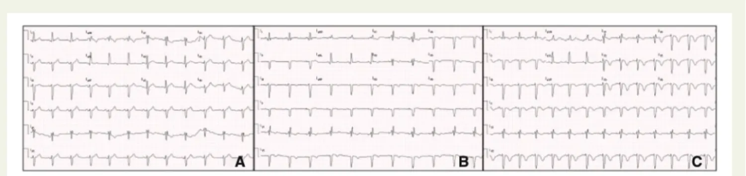
An electrocardiogram (EKG) was performed, showing a left anter-ior fascicular block de novo, besides the complete right bundle branch block already present. The 30-min EKG evolved with deep inversion
of the V2–V6 T wave and loss of R waves in these leads (Figure1).
Laboratory tests showed troponin T values of 535 ng/dL (normal <14 ng/dL), creatine kinase-muscle/brain (CK-MB) of 248 U/L (nor-mal <192 ng/dL), and C-reactive protein of 4.9 mg/dL (nor(nor-mal <0.5 mg/dL), without other significant changes.
A transthoracic echocardiogram was remarkable for akinesia of all the medial and apical segments of the left ventricle with apical bal-looning, sparing the base of each wall, causing a reduced ejection
fraction of 30%. The patient was administered loading doses of aspirin and clopidogrel and underwent coronary angiography, which excluded significant coronary artery disease. Ventriculography showed the existence of extensive mid-apical akinesia and basal
hyperkinesia and confirmed the diagnosis of TS (Figure2).
The patient was admitted to the cardiology ward for monitoring. Bisoprolol and ramipril were started at low dose and uptitrated.
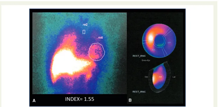
The hospitalization was uneventful, with normalization of troponin, CK-MB levels, and inflammatory markers at Day 8. Repeat trans-thoracic echocardiography showed a global left ventricular ejection fraction of 56% at time of discharge. Serum and urinary catechol-amines were not increased. Myocardial scintigraphy identified a
decreased myocardial123I-meta-iodobenzylguanidine (MIBG) uptake
in the lateral, inferolateral and apical walls, suggesting cardiac
adren-ergic nervous dysfunction (Figure3). A low late heart-to-mediastinum
ratio and a high washout rate were documented, a pattern usually observed in MIBG imaging in TS. The patient was discharged with bisoprolol 5 mg and ramipril 2.5 mg in addition to the previous medi-cations. Omalizumab was discontinued.
The patient was evaluated 1 month after hospital discharge and the cardiac MRI performed at this time was normal (namely without alterations of the segmental contractility and a left ventricular ejec-tion fracejec-tion of 66%), demonstrating the transient behaviour and complete resolution of this pathology.
Urticaria symptoms remained well controlled with oral antihist-amines, after omalizumab withdrawal.
Discussion
Takotsubo syndrome is included in non-classified non-familial
car-diomyopathies1 and represents between 1.7% and 2.2% of
patients with suspected acute coronary syndrome.2 The
patho-physiology of TS has not been fully elucidated, although the most
accepted mechanism is a catecholaminergic excess,3–5 which in
turn causes microvascular dysfunction at the level of the coronary arteries causing vasospasm and transient decrease of blood flow to the myocardium, responsible for the dysfunction of ventricular segmental contractility. In fact, most reported cases are associated with sympathetic triggers such as the typical emotional or physical stress. However, there is increasing evidence that the pathophysi-ology of this disease may be more complex, involving direct myo-cardial injury.
In this case, and according to the new 2015 Heart failure Association of the European Society of Cardiology Takotsubo
Syndrome Diagnostic criteria,6the diagnosis of TS was made based
on the evidence of left ventricular mid and apical segments akinesis with apical ballooning; new electrocardiogram abnormalities; posi-tive but relaposi-tively small elevation in cardiac troponins; absence of sig-nificant coronary artery disease or plaque rupture on angiograph; and recovery of ventricular systolic function on cardiac imaging at
follow-up. An InterTAK Diagnostic Score7of 43 [female sex 25 points,
ab-sence of ST-segment depression (except in lead aVR) 12 points, and QTc prolongation 6 points] was suggestive of TS.
One may hypothesize about risk factors predisposing this patient to TS. In fact, TS most frequently affects older and post-menopausal
women.8 Women above the age of 55 seem to have a five-fold
September 2016 Diagnosis of chronic urticaria resistant to oral anti-histamines and initiation of medication with omalizumab
26 July 2017 Admitted to the hospital to have the 11th adminis-tration of omalizumab
Thirty minutes later, the patient complained of op-pressive chest pain
Electrocardiogram showed alterations of repolarization
Transthoracic echocardiogram showed mid-apical akinesia of the left ventricle with apical balloon-ing and reduced ejection fraction of 30% Coronary angiogram excluded atherosclerotic
cor-onary disease
31 July 2017 Cardiac meta-iodobenzylguanidine (MIBG) scintig-raphy showed decreased myocardial123I-MIBG uptake in the lateral, inferolateral, and apical walls
All the investigations suggested Takotsubo syndrome
3 August 2017 Full recovery and discharge
August 2017 Cardiac MRI showed complete resolution February 2018 Last follow-up. Patient in good clinical condition
..
..
..
..
..
..
..
..
..
..
..
..
..
..
..
..
..
..
..
..
..
..
..
..
..
..
..
..
..
..
increased risk of developing TS than younger females.9Reduced
oes-trogen levels in menopausal women may render the heart more vul-nerable to catechomaninergic stress, explaining the higher frequency
of TS in this population.10 Further studies are needed to better
understand these associations.
After excluding the most frequent triggers, including previous his-tory of emotional or physical stress, the administration of omalizu-mab appeared to be the main triggering factor of TS in this case. Indeed, TS has been increasingly associated with adverse drug
events,11even though none so far has described the association with
omalizumab.
Omalizumab is a monoclonal antibody that binds selectively to serum free immunoglobulin E, avoiding binding to its receptors and consequently inhibiting the inflammatory response induced by aller-gens, so is an effective drug in severe asthma and chronic
spontan-eous urticaria.12
Observational studies,13which included patients with moderate to
severe asthma treated with omalizumab and a control group, showed that patients receiving this monoclonal antibody had a higher inci-dence at 5 years of cardiovascular events (acute myocardial
infarction, unstable angina, transient ischaemic stroke, and cerebral thromboembolism), although cardiovascular death was similar in both groups. These adverse effects were not confirmed in
subse-quent studies.14,15Regarding these controversies, coupled with study
limitations such as baseline discrepancies in asthma severity and car-diovascular risk factors between the two groups, as well as the ele-vated dropout rate, further evidence is needed to definitively confirm the cardiovascular risk and safety profile of omalizumab.
There are reported cases of TS secondary to the administration of several other monoclonal antibodies used in oncology such as
rituxi-mab,16–18bevacizumab,19transtuzumab,20and cetuximab.21The
oc-currence of TS was in these cases attributed more often to cardiotoxic direct effect, mostly via free radicals-induced cardiac
myocyte damage and death.17 However, other pathophysiological
hypotheses were raised as a paraneoplastic phenomenon or stress associated with neoplasia.
A recent review examined 15711 published cases of
drug-dependent TS and showed that 68.2% of the cases were associated with excess catecholamines [either by exogenous administration (36.2%) or by drugs with a potential adrenergic effect (32%)], 8.9%
Figure 1Electrocardiograms showing loss of R wave progression and T wave inversions in the anterior leads. EKG at presentation (A) showing a left anterior fascicular block de novo, besides the complete right bundle branch block already present. The 30-min EKG (B) evolving with inversion of V1-V6 wave and the 1 hour EKG showing deep T wave inversion and loss of R wave in all precordial leads (C).
Figure 2Left ventriculogram of patient in diastole (panel A) and systole (panel B) showing persistent mid-apical left ventricular akinesia with hyper-kinesis of the remaining basal walls.
..
..
..
..
..
..
..
..
..
..
..
..
..
..
..
..
..
..
..
..
..
..
..
..
..
..
..
..
..
..
..
..
..
..
..
..
..
..
..
..
..
..
..
..
..
..
..
had a probable vasospastic aetiology, and in a significant percentage (20.4%) of the cases it was not possible to determine the most likely pathophysiological mechanism.
Although the pathophysiology of TS in the present case remains unconfirmed, we cannot exclude a possible role of a cardiotoxic ef-fect of omalizumab in a predisposed patient with reduced oestrogen levels.
Even though it is rare, there are some reported cases of anaphyl-actic reactions following chronic administration of omalizumab. Price
and Hamilton22have most likely hypothesized an anaphylactic
reac-tion to an omalizumab excipient—polysorbate. Coors et al.23
attempted to explain a case of anaphylaxis secondary to polysorbate and failed to demonstrate an immune response secondary to poly-sorbate but proved that this substance was capable of inducing mast cell degranulation.
Thus, in the present case, even though an anaphylactic reaction did not take place, the pathophysiological mechanism could be explained by a polysorbate-mediated mast cell degranulation with the release of mediators such as histamine, norepinephrine, epinephrine, which
are inducers of coronary spasm.24
Besides that, it is not possible to exclude a cross-link omalizumab-specific IgG bound to macrophages through low-affinity receptor (FcRIII). The large antigen load afforded by the 300-mg injection may have been enough to cross-link omalizumab-specific IgG bound to macrophages through low-affinity FcRIII, causing the activation and degranulation of the mastocytes.
The fact that serum and urinary cathecolamine levels were normal may be due to the short half-life of these circulating hormones that can be degraded before evaluation of the hormones.
When applying the Naranjo scale25to our case, which assesses the
likelihood of an adverse drug reaction, the causality between the
administration of omalizumab and the development of TS is classified as possible (three points).
At a time when monoclonal antibodies have an increasing range of indications and are in exponential use, it is of crucial importance to maintain adequate pharmacovigilance in order to detect at an early stage potential adverse drug effects.
Conclusion
This is the first case described in the literature of TS after administra-tion of omalizumab. As the aetiopathogenesis of TS is still under de-bate and the pathophysiology link with many drugs’ side effects remain to be investigated, this case may provide valuable insights into the pathogenesis of TS. As our knowledge grows, the list of possible TS triggers may also grow. More TS reported cases and thorough registries are needed to better describe the triggers and to better understand this syndrome.
Supplementary material
Supplementary materialis available at European Heart Journal - Case Reports online.
Slide sets: A fully edited slide set detailing this case and suitable for
local presentation is available online asSupplementary data.
Consent: The authors confirm that written consent for submis-sion and publication of this case report including image(s) and associated text has been obtained from the patients in line with COPE guidance.
Figure 3MIBG scintigraphy showing the heart/mediastinum ratio of 1.55 (A) and decreased myocardial123I-MIBG uptake in the lateral, inferolat-eral, and apical walls (B).
..
..
..
..
..
..
..
..
..
..
..
..
..
..
..
..
..
..
..
..
..
..
..
..
..
..
..
..
..
..
..
..
..
..
..
..
..
..
..
..
..
..
..
..
..
..
..
..
..
..
..
..
Conflict of interest: none declared.
References
1. Elliott P, Andersson B, Arbustini E, Bilinska Z, Cecchi F, Charron P, Dubourg O, Kuhl U, Maisch B, McKenna WJ, Monserrat L, Pankuweit S, Rapezzi C, Seferovic P, Tavazzi L, Keren A. Classification of the cardiomyopathies: a position state-ment from the European Society Of Cardiology Working Group on Myocardial and Pericardial Diseases. Eur Heart J 2007;29:270–276.
2. Gianni M, Dentali F, Grandi AM, Sumner G, Hiralal R, Lonn E. Apical ballooning syndrome or Takotsubo cardiomyopathy: a systematic review. Eur Heart J 2006; 27:1523–1529.
3. Wittstein IS, Thiemann DR, Lima JAC, Baughman KL, Schulman SP, Gerstenblith G, Wu KC, Rade JJ, Bivalacqua TJ, Champion HC. Neurohumoral features of myocardial stunning due to sudden emotional stress. N Engl J Med 2005;352: 539–548.
4. Paur H, Wright PT, Sikkel MB, Tranter MH, Mansfield C, O’Gara P, Stuckey DJ, Nikolaev VO, Diakonov I, Pannell L, Gong H, Sun H, Peters NS, Petrou M, Zheng Z, Gorelik J, Lyon AR, Harding SE. High levels of circulating epinephrine trigger apical cardiodepression in a beta2-adrenergic receptor/Gi-dependent manner: a new model of Takotsubo cardiomyopathy. Circulation 2012;126: 697–706.
5. Menezes MN, Silva D, Almeida AG, Pinto FJ, Brito D. A rare case of concomitant stress (Takotsubo) cardiomyopathy and acute myocardial infarction. Rev Port Cardiol 2015;34:499.e1–493.
6. Lyon AR, Bossone E, Schneider B, Sechtem U, Citro R, Underwood SR, Sheppard MN, Figtree GA, Parodi G, Akashi YJ, Ruschitzka F, Filippatos G, Mebazaa A, Omerovic E. Current state of knowledge on Takotsubo syndrome: a position statement from the taskforce on Takotsubo Syndrome of the Heart Failure Association of the European Society of Cardiology. Eur J Heart Fail 2016; 18:8–27.
7. Ghadri JR, Cammann VL, Jurisic S, Seifert B, Napp LC, Diekmann J, Bataiosu DR, D’Ascenzo F, Ding KJ, Sarcon A, Kazemian E, Birri T, Ruschitzka F, Lu¨scher TF, Templin C. A novel clinical score (InterTAK Diagnostic Score) to differentiate takotsubo syndrome from acute coronary syndrome: results from the International Takotsubo Registry. Eur J Heart Fail 2017;19:1036–1042. 8. Ghadri J-R, Wittstein IS, Prasad A, Sharkey S, Dote K, Akashi YJ, Cammann VL,
Crea F, Galiuto L, Desmet W, Yoshida T, Manfredini R, Eitel I, Kosuge M, Nef HM, Deshmukh A, Lerman A, Bossone E, Citro R, Ueyama T, Corrado D, Kurisu S, Ruschitzka F, Winchester D, Lyon AR, Omerovic E, Bax JJ, Meimoun P, Tarantini G, Rihal C, Y.-Hassan S, Migliore F, Horowitz JD, Shimokawa H, Lu¨scher TF, Templin C. International expert consensus document on Takotsubo Syndrome (part I): clinical characteristics, diagnostic criteria, and pathophysi-ology. Eur Heart J 2018;39:2032–2046.
9. Deshmukh A, Kumar G, Pant S, Rihal C, Murugiah K, Mehta JL. Prevalence of Takotsubo cardiomyopathy in the United States. Am Heart J 2012;164:66–71.e1.
10. Boland TA, Lee VH, Bleck TP. Stress-induced cardiomyopathy. Crit Care Med 2015;43:686–693.
11. Kido K, Guglin M. Drug-induced takotsubo cardiomyopathy. J Cardiovasc Pharmacol Ther 2017;22:552–563.
12. Schulman ES. Development of a monoclonal anti-immunoglobulin E antibody (omalizumab) for the treatment of allergic respiratory disorders. Am J Respir Crit Care Med 2001;164:S6–S11.
13. Iribarren C, Rahmaoui A, Long AA, Szefler SJ, Bradley MS, Carrigan G, Eisner MD, Chen H, Omachi TA, Farkouh ME, Rothman KJ. Cardiovascular and cere-brovascular events among patients receiving omalizumab: results from EXCELS, a prospective cohort study in moderate to severe asthma. J Allergy Clin Immunol 2017;139:1489–1495.e5.
14. Rodrigo GJ, Neffen H, Castro-Rodriguez JA. Efficacy and safety of subcutaneous omalizumab vs placebo as add-on therapy to corticosteroids for children and adults with asthma: a systematic review. Chest 2011;139:28–35.
15. Lai T, Wang S, Xu Z, Zhang C, Zhao Y, Hu Y, Cao C, Ying S, Chen Z, Li W, Wu B, Shen H. Long-term efficacy and safety of omalizumab in patients with persist-ent uncontrolled allergic asthma: a systematic review and meta-analysis. Sci Rep 2015;5:8191.
16. Kanamori H, Tsutsumi Y, Mori A, Kawamura T, Obara S, Shimoyama N, Tanaka J, Asaka M, Imamura M, Masauzi N. Delayed reduction in left ventricular function following treatment of non-Hodgkin’s lymphoma with chemotherapy and rituxi-mab, unrelated to acute infusion reaction. Cardiology 2006;105:184–187. 17. Smith SA, Auseon AJ. Chemotherapy-induced Takotsubo cardiomyopathy. Heart
Fail Clin 2013;9:233–242, x.
18. Ng KH, Dearden C, Gruber P. Rituximab-induced Takotsubo syndrome: more cardiotoxic than it appears? BMJ Case Rep 2015;2015. pii: bcr2014208203. 19. Franco TH, Khan A, Joshi V, Thomas B. Takotsubo cardiomyopathy in two men
receiving bevacizumab for metastatic cancer. Ther Clin Risk Manag 2008;4: 1367–1370.
20. Khanji M, Nolan S, Gwynne S, Pudney D, Ionescu A. Tako-Tsubo syndrome after trastuzumab—an unusual complication of chemotherapy for breast cancer. Clin Oncol (R Coll Radiol) 2013;25:329.
21. Kim L, Karas M, Wong SC. Chemotherapy-induced Takotsubo cardiomyopathy. J Invasive Cardiol 2008;20:E338–E340.
22. Price KS, Hamilton RG. Anaphylactoid reactions in two patients after omalizu-mab administration after successful long-term therapy. Allergy Asthma Proc 2007; 28:313–319.
23. Coors EA, Seybold H, Merk HF, Mahler V. Polysorbate 80 in medical products and nonimmunologic anaphylactoid reactions. Ann Allergy Asthma Immunol 2005; 95:593–599.
24. Hung M-J, Hu P, Hung M-Y. Coronary artery spasm: review and update. Int J Med Sci 2014;11:1161–1171.
25. Naranjo CA, Busto U, Sellers EM, Sandor P, Ruiz I, Roberts EA, Janecek E, Domecq C, Greenblatt DJ. A method for estimating the probability of adverse drug reactions. Clin Pharmacol Ther 1981;30:239–245.