Induction of Osteogenic and Chondrogenic Differentiation in
Adipose Derived Stem Cells (ASCs) Using Magnetic Stimuli
By
João Miguel Aguiar Furtado Pinto Lima
Induction of Osteogenic and Chondrogenic Differentiation in
Adipose Derived Stem Cells (ASCs) Using Magnetic Stimuli
Thesis presented to Escola Superior de Biotecnologia of the Universidade Católica Portuguesa to fulfill the requirements of Master of Science degree in Biomedical Engineering
By
João Miguel Aguiar Furtado Pinto Lima
Place: 3 Bs Research Group
Supervision: Doutora Manuela E. Gomes
“Somewhere, something incredible is waiting to be known.” Carl Saga
V
Resumo
O osso e a cartilagem são dois tecidos de grande importância no corpo humano, sendo a sua funcionalidade comprometida por lesões ou doenças relacionadas com o envelhecimento, afetando milhares de pacientes em todo o mundo. Impulsionadas por uma urgente necessidade médica para melhorar a reabilitação e regeneração destes tecidos, têm sido desenvolvidas estratégias alternativas que visam promover a regeneração do osso e da cartilagem.
Como os tratamentos tradicionais para o osso e para a cartilagem apresentam pouca eficácia em fornecer uma solução a longo prazo, não restaurando completamente as funções do tecido e/ou com graves efeitos secundários, a engenharia de tecidos (ET) apresenta estratégias alternativas que vão ao encontro de soluções regenerativas. As células estaminais são um mecanismo endógeno de reparação e regeneração tecidular, tendo a capacidade de se diferenciar em fenótipos celulares de várias linhagens. Assim, apresentam grande interesse e desempenham um importante papel nas abordagens de ET e medicina regenerativa. As células estaminais humanas derivadas do tecido adiposo (hASCs) demonstram características promissoras para ET, uma vez que são relativamente fáceis de recolher, têm uma elevada taxa de proliferação e a capacidade de se diferenciar em linhagens de osso e cartilagem.
O crescente interesse na aplicação de nano partículas magnéticas (MNPs) através da atuação remota de um campo magnético externo para influenciar o comportamento celular, revela-se em estratégias capazes de estimular os processos intracelulares segundo um nível à escala celular, como a proliferação e a diferenciação. Esta tese foca-se no estudo de nano partículas magnéticas de óxido de ferro no processo de diferenciação das hASCs nas linhagens de osso e cartilagem. Foram realizados ensaios de viabilidade e proliferação até 28 dias em cultura e utilizaram-se corantes específicos de osso e cartilagem para verificar a diferenciação celular nos fenótipos osteogénico e condrogénico. As MNPs selecionadas e sobre a influência de um campo magnético não afetaram negativamente a viabilidade ou proliferação celulares. Apesar da presença destas partículas ativadas com o campo magnético influenciarem a diferenciação das hASCs, a maior influência foi verificada ao nível da diferenciação osteogénica, ao nível da produção de uma matriz mineralizada. Assim sendo, os resultados obtidos sugerem que a aplicação de MNPs sob a influência de um campo magnético externo revelam potencial para serem utilizadas em terapias celulares direcionadas para o osso e a cartilagem.
VII
Abstract
Bone and cartilage are two crucial tissues of the human body. Their functionality impairment caused by injuries or age-related diseases affects a multitude of people worldwide. Driven by the urgent medical necessities to improve these tissues rehabilitation and regeneration, extensive efforts have been dedicated in strategies to promote bone and cartilage tissue regeneration.
As traditional treatments for bone and cartilage healing fail to provide an effective long-term solution, without restoring the tissue functions and/or with severe side effects, tissue engineering (TE) arises as a promising alternative approach. As stem cells are an endogenous mechanism of tissue repair and regeneration, having the capacity to differentiate into cell phenotypes of a particular lineage, they have great interest and play an important role in TE and regenerative medicine approaches. Among them, human Adipose Derived Stem Cells (hASCs) demonstrate promising characteristics for TE as they are relatively easy to harvest, are highly proliferative and can differentiate towards bone and cartilage lineages.
Growing interest on the use of magnetic nanoparticles (MNPs) through the application of a remote magnetic field to influence cell behavior, as these strategies can stimulate intracellular processes at a cellular level, such as proliferation and differentiation. This thesis focuses on the study of iron oxide magnetic nanoparticles in the differentiation process of hASCs towards bone and cartilage lineages. Proliferative and viability assays were assessed for up to 28 days and selective stainings of bone and cartilage tissues were performed to infer on the commitment of hASCs to bone and cartilage phenotypes. The selected MNPs under a MF do not negatively affect cellular viability and proliferation, as expected. Although MNPs and MF has influences hASCs differentiation, MNPs under the MF have a greater impact in osteogenic differentiation, especially in terms of mineralized ECM production. Thereby, the attained results suggests that the application of MNPs under an external MF stimulation show the potential in cell based therapies for bone and cartilage tissues.
IX
Acknowledgments
Many people contributed to this dissertation and facilitated the long path from the beginning until the end. For that, I want to express my gratitude. First of all, I want to acknowledge my work group in 3 Bs, especially my supervisor Prof. Dr. Manuela Gomes and Dr. Márcia Rodrigues, for their continuous support, encouragement words and wise advice. The discussions within the group meetings also helped me to extend my vision regarding all science matters. To Ana Gonçalves, for her kindness and supportive attitude, always being willing to help. To Teresa Oliveira, for providing her wisdom on histological tests, and her always kind words. To all other colleagues in 3 Bs, for helping me to integrate in the day-to-day environment of the laboratory, and for brighten my days spent there.
I would also thank my friends and family, for all the support and friendship, which always make me smile. Especially Maria, for her professional and personal advises and opinions, that makes me a better person. Last but not the least, I want to acknowledge my parents for all the lifetime advises, knowledge and support.
XI
Table of Contents
Resumo ... V Abstract ... VII Acknowledgments ... IX Table of Contents ... XI List of Figures: ... XIII List of Abbreviations: ... XV Thesis Organization... XVIIChapter 1 ... 1 General Introduction ... 1 ... 1 1. General introduction ... 3 1.1. Bone ... 5 1.2. Cartilage tissue ... 6 1.3. Tissue engineering ... 9
1.3.1. Cell sources in TE – ASCs ... 12
1.3.2. Stem cells differentiation – stimulation by biochemical, mechanical and other factors... 13
Chapter 2 ... 19
Materials and Methods... 19
... 19
2. Materials and methods ... 21
2.1. Isolation and expansion of Human Adipose Derived Stem Cell (hASCs) ... 21
2.2. Chondrogenic and osteogenic differentiation of hASCs – culture medium ... 22
2.3. Mechanical stimulation with magnetic nanoparticles (MNPs) ... 23
2.4. Chondrogenic and osteogenic differentiation of hASCs – under the influence of MNPs and an external magnetic force field ... 23
2.4.1. Osteogenic differentiation of hASCs ... 23
2.4.2. Chondrogenic differentiation of hASCs ... 24
2.5. Assessment of cellular viability, proliferation and differentiation of hASCs ... 25
XII
2.5.2. Assessment of cell proliferation by DNA assay ... 25
2.5.3. Histological Characterization ... 27
2.6. Statistic analysis ... 29
Chapter 3 ... 31
The effect of magnetic stimulation on the osteogenic and chondrogenic differentiation of human stem cells derived from the adipose tissue (hASCs) ... 31
3.1. Abstract... 33
3.2. Introduction ... 33
3.3. Materials and Methods ... 35
3.3.1. Materials ... 35
3.3.2. Human adipose derived stem cells isolation and expansion ... 35
3.3.3. MNPs and external magnetic field stimulation in the differentiation process of hASCs ... 36
3.3.4. Assessment of metabolic activity (MTS assay) ... 37
3.3.5. Determination of cell content by DNA quantification ... 37
3.3.6. Histological analysis ... 38
3.3.7. Statistical analysis ... 39
3.4. Results ... 40
3.4.1. MTS assay ... 40
3.4.2. dsDNA Assay ... 41
3.4.3. Bone selective staining – Alizarin Red ... 43
3.4.4. Cartilage Selective Stainings ... 45
3.5. Discussion ... 47
3.5.1. Osteogenic differentiation study ... 48
3.5.2. Chondrogenic differentiation study ... 49
3.6. Conclusions ... 50
Chapter 4 ... 53
Final Remarks/Future Work ... 53
... 53
4.1. Final remarks/future work ... 55
XIII
List of Figures:
Figure 1.2.1 - (A) Chondrocytes (arrow) dispersed in abundant matrix; (B) Endochondral ossification 7
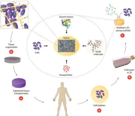
Figure 1.3.1 - TE strategies involving cells, nanoparticles, growth factors and other small molecules incorporated in a scaffold 10
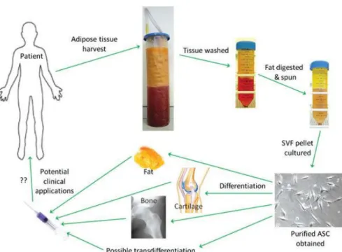
Figure 1.3.1.1 - Process from in vitro to clinical application of ASCs. Abbreviations: ASC, adipose-derived stem cells; SVF, stromal vascular fraction 13
1.3.3.1 - Natural microenvironment surrounding the cell. ECM function as a mechanical support and both microscale and nanoscale signals control cell behavior 16
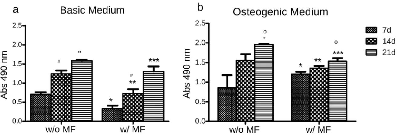
Figure 3.4.1.1: Viability of hASCs in the presence of magnetic nanoparticles (MNPs) undergoing osteogenic differentiation. 44
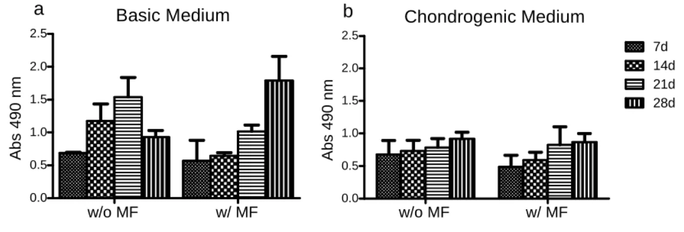
Figure 3.4.1.2: Viability assessment of hASCs cultured in a pellet system in the presence of MNPs undergoing chondrogenic differentiation 45
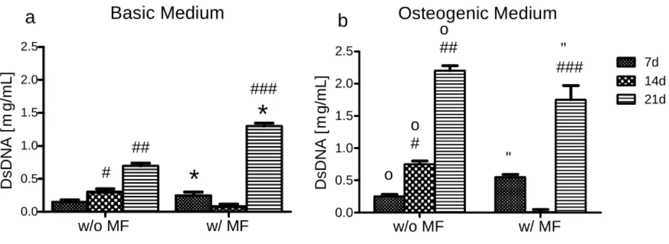
Figure 3.4.2.1: Cell content (proliferative capacity) of hASCs in the presence of MNPs undergoing osteogenic differentiation 46
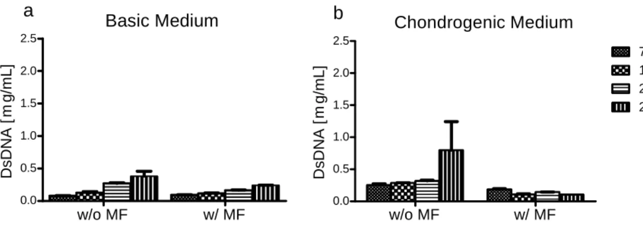
Figure 3.4.2.2: Cell content of hASCs cultured in the presence of MNPs undergoing chondrogenic differentiation 47
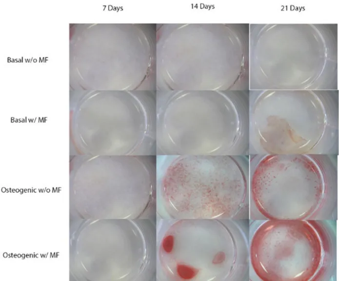
Figure 3.4.3.1: Alizarin Red staining on hASCs cultured in the presence of MNPs 48
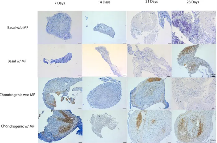
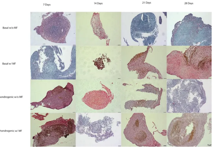
Figure 3.4.4.1: Toluidine Blue staining of hASCs cultured in a pellet system in the presence of MNPs 49
Figure 3.4.4.2: Safranin-O staining of hASCs cultured in a pellet system in the presence of MNPs 50
XV
List of Abbreviations:
ºC – Degree Celsius TE – Tissue Engineering Ab - Antibiotic-Antimycotic
ACI - Autologous Chondrocyte Implantation ASCs – Adipose Derived Stem Cell
BM-MSCs – Bone Marrow MSCs
D- MEM – Dulbecco’s Modified Eagle Medium DMSO – Dimethyl Sulphoxide
DNA - Deoxyribonucleic Acid ECM – Extracellular Matrix ESC - Embryonic Stem Cell FBS – Fetal Bovine Serum GAG – Glycosaminoglycan
hASC – Human Adipose Stem Cell iPS - Induced Pluripotent Stem Cell MF – Magnetic Field
MNP – Magnetic Nanoparticle mL - Mililiter
MSC - Mesenchymal Stem Cell
MTS – 3-(4,5-dimethylthiazol-2-yl)-5(3-carboxymethonyphenol)-2-(4-sulfophenyl)-2H-tetrazolium
PBS – Phosphate Buffered Saline PG – Proteoglycans
SB – Sodium Bicarbonate
SVF – Stromal Vascular Fraction TGF – Transforming Growth Factor µL - Microliter
XVII
Thesis Organization
This dissertation is organized into four chapters. This chapter, “General Introduction”, presents the subject and main objectives of this research work, as well as a contextualization of bone and cartilage properties, tissue engineering field, magnetic particles and stem cells selected for this study. Chapter 2 focuses on the materials and methodologies used in this experimental work. On chapter 3, the study on the influence of magnetic stimulation on the osteogenic and chondrogenic differentiation of human stem cells from the adipose tissue (hASCs) is presented in a publishing format. Finally, some future work perspectives are presented on Chapter 4.
1
Chapter 1
3
1. General introduction
Motivation
In United States of America alone, it is estimated that musculoskeletal injuries and associated diseases like arthritis, osteoporosis, osteonecrosis, bone fracture, bone tumor or trauma costs $250 billion annually, and affect hundreds of millions of people across the world [1]. Musculoskeletal conditions have been categorized as the number one reason why patients visit a doctor, by the American Academy of Orthopedic Surgeons (AAOS) [1].
Bone tissue has a high intrinsic regeneration capacity, but large bone defects need treatments, such as surgical procedures whose outcomes are not successful in a long-term basis. Currently, the most common strategies used to heal bone defects are autografting, which involves the harvest of bone tissue from the patient followed by the transplantation to the defect site and implantation of medical devices, such as metallic implants [2, 3], but these strategies don’t truly mimic the functions of natural bone and fail to restore complete tissue function.
Cartilage, on the other hand, has limited regeneration potential, due to the lack of vascularization, which makes injuries and the degeneration of this tissue difficult to overcome. Like bone tissue, current strategies relate to the use of autografts, allografts or prostheses that are not ideal. Limitation of autologous donor sites, risks of disease transmission and immunosuppression, risks of infection and extrusion of the prosthesis as well as lack of complete functionality are some drawbacks of these strategies [4].
Thus, the development of novel bone and cartilage-like substitutes that mimic the native tissue functionality are crucial for the quality of life of many patients all over the world.
As current clinical strategies for cartilage and bone defect healing are not completely effective, tissue engineering (TE) arises as a good alternative for the development of novel approaches to treat and regenerate bone and cartilage defects. Traditional TE strategies often include cells (e.g. stem cells), scaffolds (e.g. hydrogels, sponges, or meshes) and/or the incorporation of stimulatory growth factors and bioreactors. TE is an expanding field and the improvement of TE strategies is crucial towards the ideal complete functional healing of any defect. One important input to the
4
biomedical field is the application of magnetic nanoparticles (MNPs), tracked back to the fifty’s. Since then, these nanoparticles have been used as support materials for enzyme immobilization, drug delivery and tools for targeted cell separation. Recently, the use of nanoscale approaches is increasing, directing the focuses into cellular biological processes, which includes cellular differentiation. MNPs can bind to receptors in the cell surface and transmitting nanoscale forces at the ligand-receptor bond which can activate cellular signaling pathways that are known as mechanotransduction pathways. As bone and cartilage are mechano-sensitive tissues, a potential strategy could include the application of MNPs to promote healing of injured tissue.
Main Objective
The application of iron oxide nanoparticles for magnetic cell manipulation and induction of a particular cellular behavior or process has been recently approached in tissue engineering (TE) and cell therapy. Despite the current use of these MNPs in the biomedical field, as contrast agents for MRI or drug delivery carriers, their use is still controversial and MNPs require more studies to improve the understanding on the influence of these particles in cell behavior. Moreover, the study of MNPs in the TE field could open a comprehensive range of new solutions by stimulating cells at a nanoscale and at a cellular level, inducing cellular processes such as cell migration and differentiation.
Thus, the goal of this work was to evaluate the influence of the magnetic nanoparticles (Fe3O4) on the chondrogenic and osteogenic differentiation potential of human Adipose Derived Stem cells (hASCs). For that purpose, MNPs were internalized by the hASCs through the external application of a magnetic force field. hASCs differentiation in the presence of MNPs was stimulated by the presence/absence of standard biochemical supplements to the culture medium through the continuous application of a magnetic field (MF).
Cells were weekly evaluated for proliferation and viability. The bone or cartilage lineage commitment was assessed by specific markers associated to cartilage or bone tissue for up to 28 days.
5
1.1. Bone
Bone tissue is a natural organic–inorganic composite [5] created in a complex and dynamic process that initiates with the migration and recruitment of osteoprogenitor cells followed by their proliferation, differentiation and matrix formation along with remodeling of the bone [6].
Bone formation is accomplished through two distinct phases: i) by endochondral ossification, where cartilage segments are replaced by bone tissue, and ii) by intramembranous ossification, in which cartilage plays no role and bone is formed directly by condensations of mesenchymal cells. Bone formation occurs throughout life and a careful balance has to exist in the formation and loss of bone tissue [7].
The osteoblast lineage is comprised by osteoprogenitor cells, osteoblasts and osteocytes and it is responsible for bone formation. Osteoprogenitor cells, derived from mesenchymal stem cells (MSCs), have the ability to divide and differentiate in osteoblasts. The latter are responsible for bone formation and can be differentiated in osteocytes, which are responsible to maintain the bone matrix [8]. Osteoclasts are the ones in charge of bone resorption.
Bone tissue is composed by 80% of dense cortical bone that surrounds the entire bone structure and 20% of trabecular bone, a much less dense bone that constitutes the inner region of the epiphysis and metaphysis. At a molecular level, bone is composed by collagenous proteins arranged in fibrillar bundles, forming the lamellae, and a mineral component, hydroxyapatite (Ca10(OH)2(PO4)6), that helps to maintain bone mechanical integrity. Cortical and trabecular bone are composed by the haversian systems that contain bone nerve and blood supply, and help the nutrients supply through this very dense tissue, osteons and layers of lamellae. These microstructures form the macrostructure of cortical and trabecular bone.
Bone vascularity is crucial for the maintenance of cellular survival, active remodeling, skeletal integrity and regeneration capacity [9]. Macroscopically, bones can be classified as long, flat or cuboid bones [10].
Bone is the main supporting system in the human body. Its special features are the result of a unique combination of minerals and soft tissue that provides excellent tensile and loading strength. The inorganic mineral phase is responsible for its stiffness; and the organic phase, constituted by cells and collagen fibers, is responsible for elasticity, maintenance and strength [11]. Collagen fibers present in bone are fibrillar type I collagen, the most abundant, and some minor collagen
6
types like collagen type III and V. The collagenous matrix plays an important role regarding tissue mechanical strength [12]. Along with a supportive function, bone also displays a critical role in the regulation of calcium and phosphate blood levels. Also, bone tissue is crucial for organs and other tissues protection. Finally, bone harbors the bone marrow [11].
Mechanical factors are permanently present in the bone tissue due to its body supportive function. Mechanical stimulation is described to influence the cellular and tissue differentiation patterns and thus enhance bone’s regenerative activity. In vivo mechanical stimulus has demonstrated potential to induce bone repair in canine metaphyseal trabecular bone [13].
Although bone tissue exhibits a naturally high regenerative capacity, particularly in younger people, in large bone defects there is a need for major intervention, such as surgical procedures, to repair tissue damage. As mentioned earlier, autografting and implantation of metallic devices are currently the most common strategies used for bone repair [2,3] with numerous drawbacks, as inducing stress shielding, stiffness, increasing the risks for infections, and chronic pain. Also, autografting is associated with donor site morbidity and requires multiple surgeries at the injury site [2, 3]. However, no tissue-substitutes have been described capable of fully mimicking the functions of natural bone. Thus, new approaches, such as TE and regenerative medicine strategies, are then important to overcome tissue impairment and provide alternative solutions for bone regeneration.
1.2. Cartilage tissue
Cartilage is a type of connective tissue made up of cells called chondrocytes distributed in a highly specialized extracellular matrix (ECM) occupying 95% of its total volume. (Figure 1.2.1 A).
Cartilage matrix is composed mainly by water (70%), collagens (mainly type II collagen, 70% of the dry weight) and proteoglycans (PGs) (20% of the dry weight). From these 20 % PGs, 90% represents highly negatively charged molecules, the glycosaminoglycans (GAGs) [14]. These PGs play a crucial role in cartilage function leading to the hydrated gel-like structure of cartilage and resistibility to compression and deformation in joints [15]. The matrix is an important functional
7
element of cartilage and the chondrocytes, although sparse, are essential for producing and maintaining the ECM.
Figure 1.2.1: (A) Chondrocytes (red arrow) dispersed in abundant matrix [16]; (B) Endochondral ossification: A – hyaline cartilage in fetus; B - primary ossification center; C - bony collar; D - degenerating cartilage matrix; E - secondary ossification center; F - medullary cavity; G - epiphyseal growth plate; H - spongy bone; I – articular cartilage [Image adapted from Copyright© 2006 Pearson education Inc., Publishing by Benjamin Cummings].
Cartilage is avascular [16] and the absence of vasculature makes the composition of the ECM vital to the survival of the chondrocytes within it. The viability of this tissue depends on the diffusion of substances between blood vessels in the surrounding connective tissues and chondrocytes within the matrix. This characteristic is due to the high GAGs to collagen fibers ratio. The resilience of this tissue and resistance to tension results from the close interactions between two classes of molecules that have distinct biophysical properties. The meshwork of collagen fibrils resists tension and the large quantity of heavily hydrated proteoglycan aggregates resists shearing, allowing cartilage to bear weights and permits smooth movement at joints. Cartilage also plays an important role in bone development though endochondral ossification (Figure 1.2.1 B), the process by which most of the long bones of the skeleton are formed and continue to grow in childhood [17].
8
Because cartilage has limitations regarding its regeneration capacity, degeneration of this tissue and associated injury or disease, such as osteoarthritis, becomes a more serious and difficult problem to handle. Partial chondral defects or full scale osteochondral defects may be caused by trauma, biomechanical imbalance, or genetic predisposition [18]. The most common response to these full thickness defects is the formation of a fibrocartilaginous repair tissue, which has relatively low amounts of type II collagen and aggrecan, and relatively high amounts of type I collagen, a protein that usually is not significantly present in normal adult articular cartilage [19]. There are three types of cartilage; elastic, fibrocartilage and hyaline cartilage.
Elastic cartilage is located in the pinna of the external ear, in the external acoustic meatus, auditory tube, cartilages of the larynx (e.g epiglottis). Its function is to provide flexible support. It has perichondrium (layer of connective tissue that surrounds cartilage of developing bone) and does not undergo calcification. It is composed of chondroblasts and chondrocytes and an ECM rich in type II collagen fibrils, elastic fibers and aggrecan.
On the other hand, fibrocartilage is found in the intervertebral discs, symphysis pubis, articular discs (sternoclavicular and temperomandibular joints), menisci (knee joint), triangular fibrocartilage complex (wrist joint) and in the intersection of tendons. Its role is to resist deformation under stress. It does not have perichondrium but can undergo calcification. It is composed of chondrocytes and fibroblasts, and the ECM contains Type I and II collagen fibers and versican (a PG secreted by fibroblasts) [16].
Hyaline cartilage is another type of cartilage and is located in fetal skeletal tissue, epiphyseal plates, articular surface of synovial joints, costal cartilage of rib cage, the nasal cavity, larynx (e.g thyroid cartilage), rings of trachea and plates in bronchi. Its functions consist of: resisting compression, providing cushioning, smooth, and low-friction surface for joints, providing structural support in the respiratory system and forming the foundation for development of fetal skeleton and further endochondral bone formation and bone growth. It can alter its properties in response to differences in loading [20]. This cartilage undergoes calcification during endochondral bone formation and with aging. Its main cells are chondroblasts and chondrocytes. The ECM of hyaline cartilage is mainly composed of type II collagen fibrils and aggrecan. Damaged hyaline cartilage located in the articular surfaces of bones typically degenerates over time, and may progress to osteoarthritis [21].
9
Hyaline cartilage is often the ultimate cartilage type in cartilage TE, because articular cartilage has a higher damage and deterioration incidence, as it undergoes continuous stress forces over lifetime.
1.3. Tissue engineering
The concept of tissue engineering (TE) relates to an interdisciplinary field that applies the principles of engineering and life sciences to develop biological substitutes that restore, maintain or improve tissue function [5, 22-24]. The knowledge of the structure-function relationships in normal and pathological tissue is crucial for the development of tissue-like substitutes with proper functionality. Not only is the knowledge of the cells in a tissue critical, but also the microenvironment surrounding cells. The extracellular matrix (ECM) is a highly organized system that regulates essential cellular functions such as morphogenesis, differentiation, proliferation, adhesion and migration [25].
Three main strategies in TE are usually followed, alone or combined: i) the use of tissue-inducing substances, like growth factors; ii) scaffolds, which can support and guide tissue development; and iii) cells to establish implant-host tissue interactions and replace limited functions of the tissue [5, 22, 23] (figure 1.3.1). Thus, TE is a promising alternative to organ transplantation. Organ transplantation has many limitations, like the immune responses to allografts or the large gap between the tissues and organs needed to be transplanted and the ones available for patients in need [25, 26]. Therefore, TE has been increasingly studied for bone related approaches, as engineering functional tissue-like substitutes would surpass the need for multiple surgeries associated with the clearance of metallic stabilizers (metallic pins, screws, plates, or rods), used to align and stabilize the bone; and graft harvesting, reducing the recovery time, costs and treatment associated risks, as infections and immune rejections. Also, the successful integration of a tissue-like substitute in the existing tissue may improve the transition between the two [3]. One of the major challenges of TE for bone, is the fact that bone is extremely complex to mimic, as bone is a highly vascularized and vital organ in constant remodelation. Also, bone tissue is subjected to considerable physiological effort and mechanical stress in everyday activities [11]. The incorporation of scaffolds in the TE strategies is very common, as it allows cells to form a continuous structure via cell colonization, adhesion, proliferation, differentiation and deposition of
10
ECM in a supportive architecture. Scaffolds intended to mimic 3D environment for bone have to be highly porous to allow for cell ingrowth and efficient mass transport of nutrients, oxygen, growth factors, and waste products. Pores must also facilitate vascularization, and cell colonization at the core of the scaffold.
Figure 1.3.1- TE strategies involving cells, nanoparticles, growth factors and other small molecules incorporated in a scaffold. a. Cells of interest are isolated from the patient. b. These cells are expanded in a suited culture medium. c. Cells can be seeded in a scaffold, in combination with growth factors, nanoparticles and other small molecules. d. Bioreactors can be used to provide the desired tissue organization. e. Implantation on the defect occurs, in order to regain healthy tissue. Image obtained from [25].
Although scaffolds are considered to be an important element in TE, methodologies to reconstruct tissue without the use of scaffolds are arising and their potential assessed. Kazunori Shimizu and colleagues obtained a 3D tissue construct using magnetite cationic liposomes, being able to produce 3D structures without the use of scaffolds [27]. Microspheres for bone regeneration [28] and the recently explored cell sheet approach, for instance, applying bone marrow MSCs (BM-MSCs) sheets injected in bone defects [29] proved to be a good mechanism for alternative and
11
efficient developments in the TE field, as these cell sheets experience bone formation and osteogenic potential can be rescued.
Cartilage limited regeneration has been also one of the drawbacks to address in cartilage TE. In the attempt to solve this issue in the last couple of decades, several approaches were considered [30].
As for bone tissue, many strategies in cartilage TE include the use of scaffolds or hydrogels, growth factors and mechanical loading. Hydrogels provide stem cells a 3D context that can be supplemented with biochemical and biomechanical clues to direct stem cell differentiation [30]. Cell sheets are also an interesting alternative of cartilage TE and demonstrated some success, as cell sheets were successfully fabricated as layered articular chondrocytes, which maintained chondrocyte phenotype [31, 32].
ECM is essential not only by its components, but also by the biophysical factors, such as the stiffness of ECM and extrinsic mechanical factors as well as cell shape changes to direct their differentiation paths. Other strategies focus on scaffold-free approaches, comprising the use of cells, growth factors and mechanical loading, including magnetic stimulation. Recently, stem cells research has emerged, and the evidence of the role of physical stimuli in directing cartilage differentiation has been explored [30].
Growth factors have a huge impact in cell behavior, including the differentiation process, both during initial development and long-term tissue maintenance. Also, mechanical loading proved to be critical for normal development and maintenance of cartilage function. Among different forms of mechanical forces, magnetic stimulation has raised an increased interest in recent years and promising outcomes [30].
Magnetic stimuli has been studied regarding its ability to guide MSCs aggregates that could be manipulated to produce large continuous and functional cartilage tissue substitutes [33]. Another study showed that chondrogenic differentiation of BM-MSCs has been successful achieved, corroborated by an increase in the cartilage specific GAGs and PGs production, using a 0.4 T magnetic field [34].
12
1.3.1. Cell sources in TE – ASCs
Cells have a key role in tissue regeneration. The choice of the cell source is crucial to achieve a stable, long-term beneficial solution to the regeneration process of any tissue defect.
Different cell types with different intrinsic properties are currently being studied in tissue engineering.
Current clinical solutions to repair cartilage defects include autologous chondrocyte implantation (ACI) [35, 36], mosaicplasty, and microfracture [37]. Although autologous chondrocytes have been the first cell type to be thoroughly explored for cartilage TE, these strategies are limited in their ability to regenerate functional cartilage, both in composition and mechanic component. Despite ACI good results at short-time, like pain relief and decrease in swelling, further cellular expansion after cartilage biopsy is needed, as a consequence of the reduced availability of cells. Also, autologous chondrocytes have limited proliferation potential and local morbidity, experience loss of phenotype during in vitro expansion, and as they age the number of cell divisions in vitro and the ability to synthesize ECM components decreases, often resulting in the formation of fibrous tissue [35, 36]. Thus, the use of stem cells have, indeed, many advantages over chondrocytes and are seen as the future of the regenerative medicine [18].
Stem cell niches, where stem cells reside, have been described in the majority of adult tissues in the body [35]. Stem cells must meet certain requirements, including self-renewal capacity, long-term viability and multilineage potential [18, 38, 39].
Although embryonic stem cells (ESCs) and induced pluripotent stem (iPS) cells have a higher differentiation capacity than adult stem cells, the use of adult stem cells can surpass limitations like the ethical questions behind ESCs and the production costs, as well as safety concerns such as mutagenesis and tumorigenesis, associated to the use of iPS [40].
Thus, adult stem cells are an interesting and promising choice for cell-based studies. Among the Mesenchymal Stem Cells (MSCs), Adipose Derived Stem Cells (ASCs) are abundant cells that can be gathered from liposuction aspirates or subcutaneous adipose tissue fragments and are relatively easy to expand in vitro. ASCs were first identified in 2001 [41] and showed potential to undergo adipogenic, neurogenic, osteogenic and chondrogenic [39, 41-44] differentiation in vitro [45]. They are genetically stable in long-term culture and showed immune-modulatory behavior, with potential for allogenic approaches [46]. ASCs have great similarity with Bone Marrow MSCs
13
(BM-MSCs), because both relates to the stromal cell fraction of adipose tissue and bone marrow, respectively, making their surface protein and morphology very resembling [41]. The use of BM-MSCs have, however, some drawbacks, like the fact that the harvesting of BM-BM-MSCs from bone marrow is a painful procedure, with possible donor site morbidity and the genetically long-term stability is lower compared to ASCs [41]. Also, ASCs can be harvested in higher numbers from a more available source, as opposed to bone marrow cells [46]. ASCs potential applications in cell based strategies are represented in figure 1.3.1.1.
Figure 1.1.1.1 – Process from in vitro to clinical application of ASCs. Abbreviations: ASC, adipose-derived stem cells; SVF, stromal vascular fraction. Image obtained from [45].
1.3.2. Stem cells differentiation – stimulation by biochemical, mechanical and other
factors
The use of stem cells for tissue repair requires a deep understanding on how cells respond to stimuli, so this knowledge can direct cell fate. A careful control of the biochemical and biophysical signaling environments is increasingly regarded as crucial to stem cell studies.
14 1.3.2.1. Biochemical factors
The role of biochemical stimuli like growth factors, hormones, and small molecules as well as the role of the physical environment and mechanical stimuli, are being investigated for cellular differentiation purposes.
Growth factors, which usually circulate in the extracellular matrix (ECM), bind to integrins or other ECM receptors, influencing cell fate [18].
Among the growth factors families, TGF-β superfamily is the most studied whose members participate in cartilage and bone formation, beyond other lineages. TGF-β induces chondrogenesis in embryonic mesenchymal cells and is generally a stimulator of anabolic activities in connective tissue cells. It also increases DNA synthesis in chondrocyte cultures [47].
Furthermore, TGF-β has been described to increase type II collagen expression and the accumulation of specific proteoglycans [18]. TGF-β viral transduction of BM-MSCs has been accomplished and shown to consistently induce chondrogenesis while avoiding terminal differentiation [48]. Specifically, TGF-β1 is the most extensively used factor for inducing
chondrogenesis in directed differentiation of MSCs. [49] TGF-β1 has successfully induce chondrogenesis in hASCs, in combination with dexamethasone. [50] A combination of TGF-β and dexamethasone have shown potential to upregulate chondrogenic markers [49]. Dexamethasone is important for chondrogenic differentiation, but it is also crucial for osteogenic lineage commitment. Some studies indicate that at least a 3-week period of continuous treatment of a confluent monolayer of BM-MSCs with dexamethasone, combined with ascorbic acid and β-glycerophosphate, is required for osteogenic differentiation. [51] Ascorbic acid is vital as a cofactor for enzymes that hydroxylate proline and lysine in pro-collagen. So, in the absence of ascorbic acid collagen chains are not able to form a proper helical structure and the secretion of Collagen I into the extracellular matrix (ECM) is compromised [51]. Many studies have shown that in vitro proliferation and differentiation of BM-MSCs into skeletal tissues depends on the presence of ascorbic acid in the culture medium. [52] Usually, a low ascorbic acid concentration enhances cell proliferation, and it is essential for the expression of osteoblastic markers and mineralization. [52] β-glycerophosphate, on the other hand, serves as a phosphate source for bone mineral and induces osteogenic gene expression by extracellular related kinase phosphorylation [51].
15
Therefore, dexamethasone, ascorbic acid and β-glycerophosphate are important culture supplements for osteogenic studies.
1.3.2.2. Mechanical stimulation
Under the physiological environment, cell contact with the ECM and perceive various forces from the surrounding environment. The stimuli provided by mechanical environments of the native musculoskeletal tissues can facilitate the proliferation and differentiation of cells into specific lineages [53].
Bone structural integrity and mass are in constant balance towards the mechanical stress and strain that it is subjected to. So, mechanical loading is essential for osteogenesis and the maintenance of skeletal homeostasis [54].
Friedrich Pauwels was the first researcher to propose a relationship between mechanical stimulation and tissue differentiation [18]. He claimed that stress and strain that deformed cells could induce the formation of fibrous tissue, compression of the cell by hydrostatic pressure would produce hyaline cartilage and these two factor combined would produce fibrocartilage. Fibrous tissue formation is also necessary before ossification and bone formation, being crucial for bone tissue development. Also, mechanical forces could induce the synthesis of ECM components in both bone and cartilage [18]. A study conducted in rats demonstrated a decrease in bone formation, by the decline in BM-MSCs proliferation and differentiation, as well as a decrease in the synthesis of osteotropic growth factors and the extracellular matrix component, osteopontin, induced by skeletal absence of mechanical forces [55].
In articular cartilage, the composition, morphology and mechanical properties could be improved when mechanical stimulation was provided by a bioreactor [56].
The precise mechanism by which cells respond to mechanical stimuli is still not fully understood, but it is known that some membrane receptors transduce applied forces into biochemical signals or directly transmit stress forces from the ECM to intracellular structures. Mechanical stimulation has proven to influence gene expression of collagen I and III, improving the stiffness of MSCs-collagen sponge constructs, which demonstrates to be favorable to bone tissue formation [57].
16
1.3.3. Magnetic particles and their application in tissue engineering and regenerative medicine
Nanotechnology is an expanding field, namely in nanomedicine, where the nanoparticles are being increasingly used as drug delivery systems, diagnosis tools, in therapy techniques, biomaterials and in TE [58, 59]. It is estimated that nanoscale applications in the medical field could reach $70 - $160 billion by 2015 worldwide [59].
As the many applications suggest, nanoscale in TE provide many advantages, like the possibility for a cellular level stimulation directly or indirectly through the cell microenvironment. ECM is a natural web of hierarchically organized nanofibers that provides cell support and directs cell behavior via cell-ECM interactions. Its role in storing, releasing and activating a wide range of biological factors, along with ECM participation in cell-cell and cell-soluble factor interactions is crucial for cell viability pathways [59]. In vivo, interactions of cell with the ECM occur through nanoscale transmembrane integrin receptors binding to specific ligands. These adhesions induce intracellular signaling cascades that influence most aspects of the cell behavior (figure 1.3.3.4) [60].
The combination of nanoscale properties with magnetism is being increasingly studied. Magnetism has been considered an important phenomenon in human life. Its application reports to the Egyptians, where they used magnetite as an antidote to the swallowing of rust. Miniaturization of electromagnets, development of superconducting electromagnets and introduction of strong permanent magnets have extended the use of magnetic fields to areas such as biomedicine or TE [61].
Figure 1.3.3.1 –Schematic representation of the natural microenvironment surrounding the cell. ECM function as a mechanical support and both microscale and nanoscale signals control cell behavior. Image obtained from [60].
The combination of magnetism and its properties to nanotechnology culminated in the emergence of magnetic nanoparticles. Currently magnetic nanoparticles are used as a class of
non-17
invasive imaging agents for magnetic resonance imaging, [62] for instance, in the diagnosis of hepatic lesions and lymph node metastasis. In TE, approaches are being performed using MNPs as magneto-mechanical stimulation/activation of cell arrays, and as mechano-sensitive ion channels, magnetic cell-seeding procedures, and controlled cell differentiation and proliferation [63]. MNPs dimensions range from a few nanometers to some dozens nanometers, much smaller than the dimensions of cell (10-100µm). These nanoparticles are usually composed of a magnetic element (iron, nickel, cobalt) and their oxides [58, 64]. MNPs have some specific properties that makes them extremely promising. As they are magnetic, they comply with Columb’s law (which describes electrostatic interaction between electrically charged particles), and so they are affected by an external magnetic field (MF). The possibility to influence the cell behavior aloof, together with the fact that, cell internalization is possible due to the nanoscale dimensions of the particles, gives MNPs an increased biomedical potential.Moreover, MNPs can be coated with biological molecules to bind specific biologic entities, being able to target specific sites. One of the most used transitional metal to incorporate in nanoparticles is iron, mainly as iron oxide nanoparticles.
Iron has some crucial properties for its biological role. Iron uptake, both in the body and in the cells, is regulated by total available iron. Iron normally circulates in the body bounded to transferrin, a very high affinity iron protein. This link makes iron non-reactive, but also difficult to extract [65]. The iron that is not readily utilized is stored bounded to cytosolic ferritin, a protein that perform the detoxification and iron storage. Eukaryotic cells, and most of the prokaryotic ones, necessitate iron for their survival and proliferation. Some hemoproteins, iron-sulfur (Fe-S) proteins and some other proteins that use iron in their functional groups constitutes the major iron requirement in the body. Deficiency in cellular iron levels prevents cell growth and leads to cell death [65].
Iron easily capture/donates one electron to interchange its oxidative state from +2 and +3. This interesting feature, however, has some drawback when excessive “free” iron levels are present in cells, because a fraction of the iron is reduced and reacts with hydrogen peroxide (H2O2) or lipid peroxides to generate ferric iron, hydroxide (OH− ) and the reactive hydroxyl radical (OH). or lipid radicals such as alkoxyl radical (LO• ) and alkylperoxyl radical (LOO•).. The accumulation radicals have devastating consequences in cell structures, like lipid membranes and some proteins and nucleic acids. Thus, iron homeostasis has to be carefully maintained by the cell and iron
18
concentration should be carefully considered in TE approaches to avoid potential cytotoxicity effects [65].
Iron can be used in a MNP, in the form of iron oxide. Recently, iron oxide MNPs has been used for cell and micro tissue assembly, when applying MF. The use of MNPs has many potential applications like stem cell differentiation, preparation of artificial blood vessels with small diameter, preparation of skeletal muscles and bone tissue formation [66]. Ito and colleagues also used magnetite cationic liposomes containing MNPs to enhance MSCs expansion and proliferation [67]. In the particular case of superparamagnetic iron oxide particles, these particles have shown no viability or proliferation ablation onto adipose derived stem cells , a good outcome for the inclusion of these particles in biomedical applications [68].Thus, the incorporation of MNPs in the TE field can provide a challenging strategy to future therapies in regenerative medicine.
19
Chapter 2
21
2. Materials and methods
This chapter describes all the techniques as well as all materials, reagents and cells used in this work. These procedures are also described in the following chapter that corresponds to an experimental manuscript for publication. Nevertheless, a detailed explanation as well as the justification for the use of the selected methodologies is provided below.
2.1. Isolation and expansion of Human Adipose Derived Stem Cell
(hASCs)
Human Adipose Derived Stem Cells (hASCs) have a high rate of proliferation and multilineage differential capacity and are the most abundant and accessible source of adult stem cells being able to differentiate into bone, cartilage, tendon and fat lineages [38, 69].
In this study, hASCs were isolated from tissue removed during an elective cosmetic liposuction procedure, following a protocol previously established with the Department of Plastic Surgery of Hospital da Prelada. Samples were collected following informed consent and the protocol of ethics. Firstly, an enzymatic digestion of the tissue samples was performed, using 0.05 % collagenase II (C6885, Sigma), in Dulbecco’s Phosphate Buffered Saline (DPBS, 21600-044, Invitrogen). This enzymatic digestion requires an incubation of 45 minutes, at a temperature of 37ºC and under soft stirring. Posteriorly, a filtration was performed using a 100 µm filter mesh (sigma), and, after several centrifugations at 1200 rpm for 10 minutes at 20 ºC, the stromal vascular fraction (SVF) was rinsed using lysis buffer (155 mM Ammonium chloride (NH4Cl), 10 mM; Potassium bicarbonate (KHCO3), 0.1 mM; Ethylenediamine tetraacetic acid (EDTA), pH = 7.3) in order to eliminate blood red cells. This SVF is then plated in cell culture flasks and after 2-3 days the non-adherent cells were removed by repeated PBS rinsing steps. Adherent cells (hASCs) were then cultured and expanded in basic medium (alpha Minimum Essential Medium (α-MEM 1200 063 Gibco, Invitrogen), 10% Fetal Bovine Serum (FBS, 10270 Gibco Invitrogen), 1% Antibiotic-Antimycotic (Ab, 15240 062 Gibco Invitrogen) and Sodium Bicarbonate (SB, S5761 NaHCO3, sigma), until further usage [70, 71].
22
After isolation and expansion, cells used in this study were cryopreserved in a solution containing 90% FBS and 10 % dimethyl sulphoxide (DMSO CryoSure, 11-32-30216, Wak-Chemie Medical GMBH).
Before initiating the differentiation studies, hASCs were thawed and cultured in 150 cm2 T flasks. Basic medium, as previously described, was used for expansion of hASCs. When cells in the T-flasks reached approximately 80% of confluence, cells were trypsinized and passed to other T-flasks, using 0.05% Triplex (25300-062, Invitrogen). hASCs were expanded up to passage 2 until a sufficient number of cells was achieved for the experimental setup.
2.2. Chondrogenic and osteogenic differentiation of hASCs – culture
medium
For the chondrogenic and osteogenic differentiation of hASCs, two media was used, namely standard chondrogenic medium and osteogenic medium.
Basic medium, as previously described in section 2.1, was used as a control of cell differentiation.
Chondrogenic medium was composed by Dulbecco’s Modified Eagle Medium (D-MEM), SB (S5761-NaHCO3, Sigma), 1% Ab (15240-062, Invitrogen), 35 mM L-proline (P5607, sigma) , 17 mM L-ascorbic acid (013-12061, sigma), 0.1 M Sodium pyruvate (11360-039, alfagene), ITS+1 Liquid Media Supplement (41400-045-insulin-transferrin-selenium-liquid media supplement, Sigma), 1 mM Dexamethasone (D1756, sigma) and 10 ng/mL of human TGF-β1.
Osteogenic medium was used to induce osteogenic differentiation of hASCs and was composed of α-MEM, 10% FBS, 1% Ab, SB, 10 mM of β-Glicerophosphate, 50 µg/mL Ascorbic Acid and 10-9 M Dexametasone.
23
2.3. Mechanical stimulation with magnetic nanoparticles (MNPs)
MNPs were used to study the influence of these particles in the stimulation of the osteogenic and chondrogenic differentiation of hASCs through mechanical stimulation provided by a magnetic field (MF). The MNPs selected for this experiment were commercially available at Micromod (Germany) and had a [iron (II/III) oxide (Fe3O4, magnetite)] core. They were about 100 nm in diameter and were cross linked with dextran and labelled with a red florescence labeling. MNPs were used for both chondrogenic and osteogenic differentiation of hASCs. In all experiments, the concentration used was 370 µg/mL.
Two conditions were considered: static and dynamic conditions. Static conditions refer to cells cultured in the absence of MF and were defined as a control. Dynamic conditions represent cultured cells stimulated by a MF. MF was induced using a magnetic device (Nanotherics magnefect-nano II duo ®) with an oscillation frequency of 2 Hz and 0.2 mm of displacement.
2.4. Chondrogenic and osteogenic differentiation of hASCs – under the
influence of MNPs and an external magnetic force field
2.4.1. Osteogenic differentiation of hASCs
hASCs were seeded in 24 well plates, as a monolayer culture system. The density used was 1.000 cells/cm2. 500 µL of basic or osteogenic media were used in each well. Medium was changed twice a week. In static conditions, the plates were incubated at 37 ºC in 5% CO2, while in dynamic conditions, the plates were previously placed in a magnetic device (Nanotherics magnefect-nano II duo ®) before incubated at 37 ºC in 5% CO2. Cells were kept in culture for up to 21 days and samples were removed and characterized (9 for each timepoint) at days 7, 14 and 21.
24
2.4.2. Chondrogenic differentiation of hASCs
2.4.2.1. Chondrogenic pellet preparation
To mimic pre-cartilage conditions during embryonic development, cells were cultured as aggregates, forming the pellets. Cryotubes were used as a support to sustain the pellets formation with 250.000 cells and 400 µL of basic medium. Cryotubes were then centrifuged one time at 2000 rpm for 5 minutes, received a smooth blow and then centrifuged again, under the same conditions. After one day (48 hours since the beginning of the experiment), MNPs were added to the medium containing the pellets and they were again centrifuged two times at 2000 rpm for 5 minutes, with a blow being given between the two centrifugations. After another day (72 hours), medium was withdrawn and fresh medium (1 mL) added. After fresh medium addiction, pellets were again centrifuged.
After pellets assembling, cells were moved into 96 well plates. 300 µL of basic or chondrogenic medium were added to cultured wells, and medium was changed twice a week. Half of the wells coitaining the pellets were cultured in basic medium (300 µL). In the other half chondrogenic medium (300 µL) was used. Medium was changed twice a week.
As for the chondrogenic differentiation procedure, pellets were incubated at 37 ºC in 5% CO2, under static conditions. In dynamic conditions, the plates were previously placed in a magnetic device described previously and incubated at 37 ºC in 5% CO2. Cells were kept in culture for up to 28 days and samples removed and characterized at days 7, 14, 21 and 28 days, 9 samples were considered for each timepoint/assay.
Static and dynamic conditions were used to ascertain the influence of the MF hASCs differentiation with MNPs. In dynamic conditions, magnetic fields was individually applied to each well of the plates where cells were cultured.
25
2.5. Assessment of cellular viability, proliferation and differentiation of
hASCs
2.5.1. Assessment of cell viability by MTS assay
The 3-(4,5-dimethylthiazol-2-yl)-5(3-carboxymethonyphenol)-2-(4-sulfophenyl)-2H-tetrazolium (MTS) is a colorimetric method used to determine the viability of cells. It is based on the reduction of MTS to a water soluble formazan product by the dehydrogenase enzyme found in mitochondria of viable cells, which can be quantified by reading the absorbance at 490 nm in a microplate reader (Bio-tek, synergie HT) [72, 73].
To perform the MTS assay, a solution composed by a reagent MTS (Promega Corporation) and a FBS free DMEM medium without phenol red was prepared at a ratio of 1:5.
Pellets in the chondrogenic differentiation study were rinsed using a PBS solution and the MTS solution added, 300 µL to each well. The plate was incubated for 3 hours at 37 ºC and a humidified atmosphere containing 5% CO2. After the incubation, 100 µL (readings made in triplicate) was transferred to a 96 well plate and read in a microplate reader at an absorbance of 490 nm.
In cells that underwent the osteogenic differentiation, MTS solution was added directly to the 24 well plate wells (where hASCs were initially seeded), after medium removal and PBS rinsing. 500 µL of MTS medium was added to each well. The plates were incubated at 37ºC and a humidified atmosphere containing 5% CO2 for 3 hours. Then 100 µL of each well (readings made in triplicate) were transferred to a 96 well plate. This 96 well plate was then read in a microplate reader at an absorbance of 490 nm.
2.5.2. Assessment of cell proliferation by DNA assay
There are many methods available for the quantification of the DNA, like the use of fluorochromes or the uptake of radioactively-labeled DNA precursors such as [(3)H]thymidine. An effective method to quantify double-strain DNA (dsDNA) in a solution is using Picogreen, a fluorochrome that selectively binds to dsDNA. This dye is excited at 480 nm and emits at 520 nm
26
when bound to dsDNA. It has the advantage of having virtually no background because the unbound dye has very little fluorescence, unlike the bounded dye, that has a high fluorescence. It is very stable to photo-bleaching, making its handling more flexible [74].
The proliferative capacity of the hASCs was assessed by a fluorimetric dsDNA quantification kit (P7589-picogreen, Molecular Probes, Invitrogen).
2.5.2.1. Cell preparation
Prior to the DNA quantification, cells were prepared by the following procedure:
Cells of the osteogenic differentiation study were rinsed in PBS, and then detached from the bottom of the well before 1 mL of ultrapure water was added to each well. The water containing cellular DNA was then transferred to 1.5 mL microtubes.
Pellets of the chondrogenic differentiation study were transferred to 1.5 mL microtubes, after medium withdraw and PBS rinse, and 1 mL of ultrapure water was added to each 1.5 mL microtube.
After this procedure, cells were stored at -80 ºC, until usage.
2.5.2.2. dsDNA quantification
Prior to the analysis, all samples were thawed and sonicated for 15 minutes to ensure that all DNA was released from the cells.
To create a standard curve, standards solutions were used ranging from 0 to 2 µg/mL.
Samples and standards were mixed with a picogreen solution previously diluted in TE buffer at a ratio of 1:200. Opaque 96 wells white plates (Labclinics) were used and triplicates for each sample/standard were made. The plate with the samples was incubated for 10 minutes in the dark and fluorescence was measured using a microplate ELISA reader) at an excitation of 485/20 nm and an emission of 528/20 nm. dsDNA concentration in each sample was interpolated from the standard curve made.
27
2.5.3. Histological Characterization
2.5.3.1. Sample preparation
As a preparation for the stainings, samples both for osteogenic and chondrogenic studies were rinsed with PBS and then fixed using 10 % formalin (Bio-Optica Milano S.p.a) solution for 24 hours at room temperature. Histological fixation is performed to preserve cellular structures and prevent the loss of components during technical procedures [14]. It also protects tissue sections from microbial activity, osmotic damage, dehydration and autolysis. Formalin meets the criteria of a good fixative, as it converts tissue into a macromolecular network, in which lipids, protein, and other cellular constituents are linked together indissolubly, keeping the integrity of the functional groups, preventing the affectation of the staining’s properties [75].
2.5.3.2. Staining for the assessment of osteogenic differentiation
2.5.3.2.1. Alizarin Red staining for the assessment of osteogenic differentiation:
Alizarin Red has been used in textile industry since antiquity. In histological techniques, it has been used for decades to effectively identify calcium salts by reacting with calcium and other cations. [76, 77] Alizarin solution was prepared dissolving alizarin red compound in distilled water (0.02g/mL). pH was then adjusted to approximately 4.2 with 10% ammonium hydroxide. The wells, containing the cells, were rinsed with PBS and then the alizarin red solution was added. The solution was left 10 minutes in contact with the cells so the reaction between the alizarin red solution and the calcium salts could occur. After that, wells were rinsed again with PBS to remove the non-bonded staining and stained cells were visualized on a Stereo Microscope (Zeiss, Stami 2000-C) and images taken.
28
2.5.3.3. Stainings for the assessment of chondrogenic differentiation:
Samples of the chondrogenic study were stained by toluidine blue, safranin-O and alcian blue, for GAGs and PGs detection.
They were then dehydrated using increasing alcohol concentration solutions and embedded in parafin in a tissue processor (Microm, tspin tissue STP120-2). Pellet sections were cut in a microtome (Microm, HM355S Inopat), with a size of 3.5 µm, and stored for further usage.
Immediately before each stain, samples were deparaffinized using xylene and hydrated using decreasing alcohol concentration solutions.
After the staining protocol, the slides were dehydrated with a soaring alcohol concentration solutions. Slides were mounted and left to dry overnight. They were then ready to visualize in a transmitted and reflected light microscope (Zeiss, Imager Z1M).
2.5.3.3.1. Toluidine Blue
Toluidine Blue is a cationic dye that, in both orthochromatic and methacromatic phases, is known to stain GAGs and PGs, naturally present in cartilage ECM. The staining is proportional to the amount of GAGs in cartilage [78].
Toluidine Blue solution was prepared proceeding to the dissolution of 1% of toluidine blue in distilled water containing 0.5 g of sodium borate.
Thus, toluidine Blue solution was added to cover the fixed section of the pellet for about 2 minutes and then washed with water.
2.5.3.3.2. Safranin-O
Safranin-o is a cationic dye constituted by a mixture of dimethyl phenosafranin and trimethyl phenosafranin [79]. It is a widely used staining for cartilage GAGs and PGs. It is stoichiometric when the amount of GAGs in the tissue is not too low. [80].
29
Safranin-O solution was prepared by dissolving safranin-O compound in distilled water (0.001g/mL). A fast green solution was also made, dissolving the fast green dye in distilled water (0.002 g/mL).
Slides containing the pellets were immersed in 0.02% fast green staining solution for 5 minutes. 3 dips were performed in 1% acetic acid solution. Slides were immersed in 0.1% safranin O staining solution for 5 hours.
2.5.3.3.3. Alcian Blue
Alcian blue is a soluble form of copper phthaloeyanine. Copper phthaloeyanine has very interesting properties as a pigment, but is extremely insoluble and inert. The insolubility plays an important role as it prevents the stain to be subsequently removed when is inside the tissue. However, it has to be carried to the tissues by isothiouronium, or other cationic groups, in an aqueous solution [81].Alcian blue was also used to assess the chondrogenic differentiation of the cells within the pellets, as this marker stains GAGs present in cartilage tissue.
Alcian blue solution was prepared by dissolving the alcian blue dye in acetic acid (0.3g/mL) and add distilled water to fulfill 97% of the solution.Pellets sections were immerse with 1% alcian blue solution for 1 hour and then washed with running tap water for 5 minutes.
2.6. Statistic analysis
All data is represented as mean ± standard deviation (SD). Statistical analysis was performed by using two-tailed Student’s t-test, considering a confidence interval of 95%. P-values lower than 0.05 were considered significant.
31
Chapter 3
The effect of magnetic stimulation on the osteogenic and
chondrogenic differentiation of human stem cells derived from the
adipose tissue (hASCs)
This chapter is based on the following publication: “The effect of magnetic stimulation on the osteogenic and chondrogenic differentiation of human stem cells derived from the adipose tissue (hASCs)”, João M. Lima, Ana I. Gonçalves, Márcia T. Rodrigues, Rui L. Reis, Manuela E. Gomes, submitted to the journal of Journal of Magnetism and Magnetic Materials, 2014.
33
3.1. Abstract
The use of magnetic nanoparticles (MNPs) towards the musculoskeletal tissues has been the focus of many studies, regarding MNPs ability to promote and direct cellular stimulation and orient tissue responses. This is thought to be mainly achieved by mechano-responsive pathways, which have the ability to promote changes in cell behavior, as proliferation and differentiation rates, in response to external mechanical stresses. The use of MNPs based strategies in tissue engineering (TE) thus may have the potential to open a wide range of solutions for cell therapy on bone and cartilage strategies to accomplish tissue regeneration.
The present work aimed at studying the influence of MNPs on the osteogenic and chondrogenic differentiation of human adipose derived stem cells (hASCs). Different culture conditions were considered, as standard media for osteogenic and chondrogenic differentiation and presence/absence of an external MF, to determine if the MNPs alone could affect the osteogenic or chondrogenic phenotype of the hASCs. The obtained results suggest that MNPs do not negatively affect the viability nor the proliferation levels of hASCs during the time in culture. Furthermore, alizarin red staining indicate an enhancement in ECM mineralization in the presence of MNPs and under the influence of a MF. Although not as evident as for the osteogenicdifferentiation, toluidine blue and safranin-O stainings suggests the presence of glycosaminoglycans (GAGs) and proteoglycans (PGs) in the presence of MNPs and under a magnetic field.
Thus, MNPs and the influence of an external MF has potential to induce differentiation towards the osteogenic and chondrogenic lineages.
3.2. Introduction
Bone and cartilage defects are a clinical problem that affects a multitude of people worldwide. Bone tissue has innate regeneration capacity, granting this tissue the capacity to self-repair minor injuries. However, large bone defects are not fully healed without treatments, such as surgical procedures, whose outcomes have no success in a long-term basis. Cartilage, on the contrary, exhibits a limited regeneration capacity due to the lack of vascularization hindering its healing
