i
Dissertação: Artigo de Investigação Médica
Mestrado Integrado em Medicina
CHARACTERIZATION AND PREVALENCE OF EPISODES OF
MAST CELL MEDIATOR RELEASE TRIGGERED BY
ANALGESIC/ANTIPYRETIC AND NON-STEROIDAL
ANTIINFLAMMATORY DRUGS IN MASTOCYTOSIS PATIENTS
Tiago Emanuel Azenha Rama
Orientador:
José Manuel Soares Malheiro Romão
Co-orientadores:
Luis Escribano Mora Almudena Matito Bernechea
ii
Artigo de Investigação Médica
CHARACTERIZATION AND PREVALENCE OF EPISODES OF
MAST CELL MEDIATOR RELEASE TRIGGERED BY
ANALGESIC/ANTIPYRETIC AND NON-STEROIDAL
ANTIINFLAMMATORY DRUGS IN MASTOCYTOSIS PATIENTS
Autor:
Tiago Emanuel Azenha Rama, estudante do Mestrado Integrado em Medicina do Instituto de Ciências Biomédicas de Abel Salazar – Universidade do Porto
Rua de Jorge Viterbo Ferreira nº228, 4050-313, Porto, Portugal Correio Electrónico - tiagorama@gmail.com
Orientador:
José Manuel Soares Malheiro Romão, Licenciado em Medicina, Professor Associado Convidado do ICBAS-UP, Chefe de Serviço de Anestesia, Centro Hospitalar do Porto, Hospital de Santo António, Porto, Portugal.
Co-orientador:
Doutor Luis Escribano Mora, Doutor em Medicina, Investigador Associado do Centro de Investigação do Cancro, Universidade de Salamanca, Espanha.
Co-orientadora:
Doutora Almudena Matito Bernechea, Doutora em Medicina, Assistente graduada de Alergologia, Hospital Virgen del Valle, Toledo, Espanha.
iii “Só se nos detivermos a pensar nas pequenas coisas chegaremos a compreender as grandes.” José Saramago
iv
Índice
Índice de Tabelas e Figuras ... 1
Agradecimentos ... 2
Lista de Abreviaturas ... 3
Abstract ... 4
Introduction ... 5
Materials and methods ... 7
Results ... 10
Conclusion ... 20
References ... 21
1
Índice de Tabelas e Figuras
Table I. WHO defined diagnostic criteria for Systemic Mastocytosis. ... 8 Table II. “B” and “C” findings. . ... 8 Table III. Categories of adult mastocytosis grouped by the pattern of tolerance to NSAID/COX inhibitor drugs. ... 11 Table IV. Basal MC mediators related symptoms of adult mastocytosis patients grouped according to the pattern of tolerance to COX inhibitors drugs. ... 11 Table V. Frequencies of NSAIDs and other COX inhibitors as elicitors of MC mediators related symptoms in adult mastocytosis grouped by type of tolerance to these drugs. 12 Table VI. Clinical findings during reactions to NSAIDs and other COX inhibitors in adult patients, according to the pattern of intolerance to COX inhibitor drugs. ... 12 Table VII. Frequencies of NSAIDs and other COX inhibitors as elicitors of anaphylaxis. ... 13 Table VIII. Epidemiological, clinical and laboratory characteristics of adult mastocytosis patients grouped according to the pattern of tolerance to COX inhibitors drugs. ... 14 Table IX. Epidemiological, clinical and laboratory features of pediatric, according to the pattern of tolerance to COX inhibitors. ... 17
Figure 1. Receiver Operating Characteristics (ROC) curve analysis of serum basal tryptase levels for the identification of NSAIDI patients. ... 15 Figure 2. Receiver Operating Characteristics (ROC) curve analysis of BMMC burden for the identification of NSAIDI patients. ... 15
2
Agradecimentos
À Marta, a quem devo imenso, agradeço pelo inestimável apoio e paciência. Agradeço, profundamente, pelo companheirismo e pela força que me transmitiu, sem a qual não teria sido possível percorrer este caminho.
Ao Prof. Dr. José Romão, agradeço por ter aceitado orientar esta tese, pela disponibilidade que sempre demonstrou, pelo espírito crítico, rigor científico e linguístico, e pela confiança que depositou em mim ao longo deste trabalho.
À Doutora Almudena Matito, agradeço pelo apoio e disponibilidade constantes, pelos ensinamentos e pelo exemplo de rigor, como forma de estar na Medicina.
Ao Dr. José Mário Morgado, agradeço por toda a ajuda, amizade e pela forma crítica como me ajudou a encarar as adversidades naturais da investigação científica.
Ao Doutor Luis Escribano, a quem, ao longo dos anos, deixei de ver como um orientador, mas como um amigo e mentor, agradeço por ter aceitado ser co-orientador, mais uma vez. Agradeço pelas oportunidades que me concedeu, pela disponibilidade, por todo o conhecimento que me permitiu adquirir, e, derradeiramente, pela amizade.
3
Lista de Abreviaturas
ASA - Acetylsalicylic Acid
ASM – Aggressive Systemic Mastocytosis AUC - Area under curve
BM – Bone marrow
BMMC – Bone marrow Mast cell CI - Confidence interval
cM - Cutaneous Mastocytoma CM – Cutaneous Mastocytosis COX – Cyclooxygenase
DCM - Diffuse Cutaneous Mastocytosis GI - Gastrointestinal
HLA – Human Leukocyte Antigen Ig - Immunoglobulin
ISM – Indolent Systemic Mastocytosis
ISMs- - Indolent Systemic Mastocytosis without skin lesions
ISMs+ - Indolent Systemic Mastocytosis with skin lesions
LT - Leukotriene MC – Mast Cell
MIS – Mastocytosis in the skin NPV - Negative predictive value
NSAID – Non-steroidal antiinflammatory drug
NSAIDI – Intolerants to Non-steroidal antiinflammatory drugs PDGRF - Platelet-derived growth factor receptors
PG – Prostaglandin
PPV - Positive predictive value
REMA – Spanish Network on Mastocytosis ROC - Receiver operating characteristic sBT – serum basal tryptase
SM-AHNMD – Systemic Mastocytosis with associated clonal hematological non-mast cell lineage disease
SSM – Smouldering Systemic Mastocytosis
WDSM – Well-Differentiated Systemic Mastocytosis WHO - World Health Organization
4
Abstract
Background: Mastocytosis are a group of diseases characterized by an accumulation
of aberrant Mast Cells. Nonsteroidal antiinflammatory drugs are frequently avoided in mastocytosis patients, due to concerns about the safety of these drugs in these diseases, as they may elicit mast cell mediated symptoms. In the general population, hypersensitivity reactions to nonsteroidal antiinflammatory drugs are thought to derive from the depletion of Prostaglandin E2 and resulting Leukotriene release.
Objectives: To determine the prevalence of mediator release symptoms triggered by
nonsteroidal antiinflammatory drugs in mastocytosis patients, and to assess associations with medical findings.
Methods: Medical records of 417 adults and 137 pediatric were reviewed. Groups
defined by the tolerance patterns to nonsteroidal antiinflammatory drugs and other cyclooxygenase inhibitors were compared for epidemiological, clinical, laboratory and imaging variables.
Results: Tolerance patterns to nonsteroidal antiinflammatory drugs and other
cyclooxygenase inhibitors were as follows: 79% of the patients tolerated, 5% were intolerant, 4% had mast cell-related symptoms elicited by one drug, but tolerated other drugs and 3% presented mast cell-related symptoms secondary to one drug and avoided others. In the adult sample, intolerance was found to be more frequent in patients with aggressive systemic mastocytosis (p=0.019), flushing (p=0.003), pruritus (p=0.005), prior anaphylaxis (p=<0.001), multilineage KIT mutation (p=0.002), diffuse osteosclerosis (p=<0.001), serum basal tryptase levels above 48 ng/mL (p=0.013), and bone marrow burden above 0.012% (p=0.006). Drug challenges were performed in 51 adult and 21 pediatric patients. Positive challenges were reported in 3 adults, and no children.
Conclusions: In mastocytosis, the frequency of allergic/pseudoallergic reactions to
cyclooxygenase inhibitors seems to be higher than in the general population, albeit lower than previously thought. Such reactions may be more frequent in aggressive and under progression forms of the disease. Mastocytosis should not be considered as a contraindication for the administration of these drugs. Serum basal tryptase levels above 48 ng/mL, and bone marrow burden above 0.012% may be surrogate markers for cyclooxygenase inhibitor intolerance, in mastocytosis.
Keywords: Mastocytosis, mast cells, mast cell-mediator release-associated
5
Introduction
Mastocytosis encompasses a heterogeneous group of rare diseases characterized by an accumulation of aberrant mast cells (MC) in different tissues and organs, such as the skin, bone marrow (BM) and gastrointestinal (GI) tract, among others (1, 2). These diseases may affect both children and adults, independently of gender(1, 2). The prevalence of mastocytosis is estimated to range from 9 to 13 cases per 100,000 (3, 4).
World Health Organization (WHO) defines seven categories of mastocytosis: cutaneous mastocytosis (CM, when limited to the skin), extracutaneous mastocytoma, indolent systemic mastocytosis with skin lesions (ISMs+), aggressive systemic
mastocytosis (ASM), SM associated with other clonal hematological non-MC lineage diseases (SM-AHNMD), MC leukemia (MCL) and MC sarcoma (5). In addition, two other subvariants of indolent forms of the disease have been recognized: well-differentiated SM (WDSM) (6) and ISMs- (7).
Patients suffering from mastocytosis may present signs and symptoms resulting from release of MC mediators, infiltration of tissues by MC, or both (8). The clinical presentation is quite variable and may range from a complete lack of signs and symptoms, to recurrent anaphylaxis (9). Activated MC release a myriad of vasoactive, proinflammatory, chemotactic and immunomodulatory mediators, both prestored in granules and produced de novo (9). Triggers for the activation of MC have been largely described in literature, and include Hymenoptera stings, stress, dentition and drugs, such as opioids, or nonsteroidal antiinflammatory drugs (NSAIDs) and other COX inhibitors (9, 10).
According to the WHO Collaborating Centre for Drug Statistics Methodology, NSAIDs include butylpyrazolidines (e.g. phenylbutazone), acetic acid derivatives and related substances (e.g. diclofenac, etodolac, ketorolac, aceclofenac), oxicams (e.g. piroxicam, meloxicam), propionic acid derivatives (e.g. ibuprofen, naproxen, dexketoprofen), fenamates (e.g. mefenamic acid), coxibs (celecoxib, etoricoxib) and others non-otherwise classified (e.g. nimesulide, nabumetone, clonixin lysine). Salycilates (e.g. acetylsalicylic acid – ASA -, diflunisal) are included in analgesics and antipyretics. This group also includes drugs with a weak COX-inhibitory capacity: pyrazolones (metamizole, propyphenazone) and, an anilide derivative, paracetamol (11).
6 NSAIDs inhibit the activity of cyclooxygenases (COX) resulting in antipyretic, pain control and antiinflammatory effects. As opposed to NSAIDs, paracetamol exerts a weak inhibitory effect on COX-1, and is devoid of antiinflammatory effects. Regarding its mechanism of action, some authors have recently reported that, in the majority of situations, it is mostly a COX-2 inhibitor (12). Moreover, paracetamol may exert its analgesic and antipyretic effects by inhibiting the production of PGE2 in central nervous
system (12).
In Spain, the prevalence of allergic reactions to NSAIDs ranges from 1% to 3% in the adult general population (13). However, these reactions are infrequent in the pediatric population (14). Differences in the prevalence of reactions reported in different studies, both in adult and pediatric populations, result from differences in the diagnostic criteria, as they can be based on anamnesis, or on drug challenges (15). Prevalence of MC mediator-related symptoms reported in mastocytosis adult patients in association with NSAIDs is 14%, being much lower in pediatric mastocytosis (2%) (16). However, these drugs represent 8% (17) to 11% (18) of triggers for anaphylaxis, in adults. Therefore, the administration of these drugs in mastocytosis patients should be cautious, being frequently and strictly avoided in this group of diseases (19).
Allergic and pseudoallergic reactions to NSAIDs, and other COX inhibitors can be elicited by a specific drug, by a specific group or by structurally unrelated drugs (20). Pyrazolones are a frequent cause of IgE-mediated hypersensitivity reactions (20). The remaining COX inhibitors usually elicit the symptoms through a non-IgE dependent mechanism (pseudoallergic reactions). These reactions are most likely associated with inhibition of the COX-1 pathway of the arachidonic acid metabolism (14-18), leading to depletion of protective PGE2 (21, 22). PGE2 exerts inhibitory effects on IgE-FcɛRI MC
activation and relaxation effects on bronchial smooth muscle by binding to the E prostanoid receptor 2 (EP2) (23). Moreover, PGE2 release inhibits the expression of
leukotriene (LT) C4 synthase, thus increasing the production of cysteinyl-leukotrienes
(24). The release of these mediators causes clinical signs and symptoms that may involve the skin (e.g. pruritus, urticaria, exanthema, angioedema, etc), the airway (e.g. bronchospasm), or multiple organs, inducing anaphylaxis (21, 25).
Patients with pseudoallergic reactions to NSAIDs may present with symptoms elicited by COX-1 inhibitors, while tolerating weak COX-1 inhibitors (e.g. paracetamol) and COX-2 selective inhibitors, since they induce a less marked decrease of PGE2,
resulting in a less significant production of leukotrienes (20, 26). In mastocytosis, the mechanism for NSAID-related release of MC mediators is not yet clear. However, COX inhibition may play a similar role to the one described for the general population.
7 The aim of this study is to determine the prevalence of MC mediator release symptoms triggered by NSAIDs in mastocytosis patients and to assess clinical and laboratory findings most frequently associated with, or predictive of such reactions.
Materials and methods
This retrospective study included 417 adults and 137 pediatric mastocytosis patients, followed up by the Spanish Network on Mastocytosis (REMA) until October 2014. Collected variables included:
1) epidemiological: gender, and age;
2) clinical: category of mastocytosis; type of skin lesions in children, age at disease onset, presence of atopy, food allergy, asthma, rhinoconjunctivitis and/or atopic dermatitis; allergy to Hymenoptera venom; basal MC mediator-related symptoms (pruritus, flushing, gastrointestinal symptoms) and anaphylaxis; tolerance to NSAIDs and other COX inhibitors; MC mediator symptoms triggered by NSAIDs and other COX inhibitors;
3) laboratory: serum basal tryptase (sBT), total serum IgE, specific IgE (when available), presence of multilineage KIT mutation, BMMC burden;
4) imaging studies: computed tomography scan, dual energy X-ray absorptiometry, skeletal X-ray survey and/or magnetic resonance imaging, for the diagnosis of diffuse osteosclerosis.
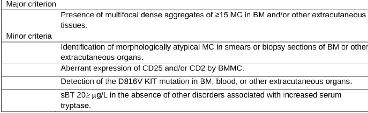
Diagnosis of mastocytosis was based on well-established criteria for morphology (27), histopathology, immunohistochemistry (28) and flow cytometry immunophenotyping (29). Cutaneous mastocytosis was diagnosed in the presence of a biopsy-proven mastocytosis in skin, and absence of enough criteria for SM - presence of the major criterion, plus one minor criterion, or in the presence of, at least, three minor criteria (Table I), as defined by the WHO (5). Patients with cutaneous involvement that were not submitted to BM studies were classified as having MIS, since the systemic involvement could not be ruled out. SSM was diagnosed in the presence of criteria for SM plus two or more “B” findings, in the absence of “C” findings (Table II). ASM was diagnosed in the presence of criteria for SM, plus at least one “C” finding, and in the absence of MC leukemia (MCL). MCL was diagnosed in the presence of, at least, 20% MC in BM and, at least, 10% MC in peripheral blood.
8 Table I. WHO defined diagnostic criteria for Systemic Mastocytosis (5).
Major criterion
Presence of multifocal dense aggregates of ≥15 MC in BM and/or other extracutaneous tissues.
Minor criteria
Identification of morphologically atypical MC in smears or biopsy sections of BM or other extracutaneous organs.
Aberrant expression of CD25 and/or CD2 by BMMC.
Detection of the D816V KIT mutation in BM, blood, or other extracutaneous organs. sBT 20g/L in the absence of other disorders associated with increased serum tryptase.
MC: Mast cells; BM: bone marrow; sBT: serum basal tryptase
Table II. “B” and “C” findings (5).
“B” Findings
BM biopsy showing >30% infiltration by MC (focal, dense aggregates) and/or serum total tryptase level >200 ng/mL.
Signs of dysplasia or myeloproliferation, in non-MC lineage(s), but insufficient criteria for definitive diagnosis of a hematopoietic neoplasm (AHNMD), with normal or slightly abnormal blood counts.
Hepatomegaly without impairment of liver function, and/or palpable splenomegaly without hypersplenism, and/or lymphadenopathy on palpation or imaging.
“C” Findings
Palpable splenomegaly with hypersplenism.
BM dysfunction manifested by one or more cytopenia(s) (ANC <1.0 x10⁹/L, Hgb <10
g/dL, or platelets <100 x 109/L), but no obvious nonmast cell hematopoietic malignancy.
Palpable hepatomegaly with impairment of liver function, ascites, and/or portal hypertension.
Malabsorption with weight loss due to gastrointestinal mast cell infiltrates. Skeletal involvement with large osteolytic lesions and/or pathological fractures. ANC: absolute neutrophil count; Hgb: hemoglobin.
Pediatric mastocytosis skin lesions were classified as previously described in the literature: maculopapular (MPCM), plaque type, nodular/mastocytoma (solitary or multiple), diffuse (DCM), and telangiectatic cutaneous mastocytosis (red and brown telangiectatic macules) (30).
Age at disease onset was defined either at the moment when skin lesions first appeared, age at first anaphylaxis episode, or when B or C findings were confirmed, in patients who presented without skin lesions (5).
The presence of KIT mutations in highly purified BMMC and other hematopoietic lineages was assessed, in order to ascertain the presence of MC restricted or multilineage KIT mutation (31).
Peripheral blood count and differential, routine biochemistry including serum basal tryptase (sBT) (Phadia ImmunoCAP Tryptase System - Phadia, Uppsala, Sweden/Thermo FisherScientific Inc.) and total serum IgE (ThermoFisher Scientific and
9 CAP-FEIA system Unicap 100; Phadia, Uppsala, Sweden) were performed at referral and during follow-up. Specific IgE antibodies were measured through ImmunoCAP (ThermoFisher Scientific and CAP-FEIA system Unicap 100; Phadia, Uppsala, Sweden), whenever there was a high suspicion of IgE-mediated symptoms, if a specific assay was commercially available. Cutaneous tests (skin prick and intradermal tests) were performed using specific triggers (e.g. Hymenoptera venom, foods and drugs). Atopy was diagnosed in patients presenting symptoms suggestive of allergic inflammation (rhinitis, bronchospasm, food allergy, etc), in the presence of allergen-specific IgE, or positive cutaneous tests (32).
Allergic/pseudoallergic reactions to NSAIDs and other COX inhibitors were assessed for each patient. Initial evaluation was based on anamnesis of reactions potentially caused by these drugs, considering time relation between the administration and the onset of symptoms, severity of symptoms, and later tolerance to the specific drug. Skin prick tests were performed if an underlying IgE-mediated mechanism was suspected, and drug challenge tests were performed using the potentially-involved drug or other COX inhibitor. The latter were carried out having the risk-benefit ratio in consideration, and only when patients gave their consent. The elicitor COX inhibitor was used when the level of suspicion for causality was low, and if symptoms were mild. Other COX inhibitors, most likely to be tolerated, were used when causality was almost certain and/or in the presence of moderate to severe symptoms. Patients who had symptoms after taking 2 or more, structurally unrelated NSAIDs or COX inhibitors, were classified as NSAID intolerants (NSAIDI) – idiosyncratic reactions. Those who revealed no symptoms following the administration of NSAIDs/other COX-inhibitors, excluding paracetamol, were considered tolerant. Patients who presented with symptoms elicited by only one NSAID/COX-inhibitor were further divided into two groups: one including those that avoided only that particular drug, or group, and which tolerated other NSAIDs after that episode (group 1), and another comprising those who avoided all NSAIDs/COX-inhibitor, excluding paracetamol, from that moment on (group 2). Finally, patients who avoided NSAIDs, or other COX-inhibitors excluding paracetamol, since the onset of mastocytosis were excluded from the statistical analysis.
Statistical analysis
Median and range values were calculated for continuous variables, while frequencies were determined for categorical variables. The Kruskall Wallis or Mann-Whitney U, and 2 orcoefficient tests were used to assess the statistical significance of
10 differences between groups, respectively, for continuous and categorical variables. Receiver operating characteristic (ROC) curve analysis was used in order to define the optimal cut-off values, and predictive values of continuous variables for intolerance to NSAIDs/other COX inhibitors. p values <0.05 were considered to be statistically significant. Statistical analysis was performed using IBM SPSS for Windows (version 23.0) and Microsoft Excel 2010.
Results
Adult patients
A total of 417 adult mastocytosis patients were included, 221 (53%) were females and 196 (47%) were males. Median age was 48 years (range: 19 to 85), and median age at onset of the disease was 33 years (range: 0 - 82). Ten patients (2%) had CM, 27 (7%) had MIS, 207 (50%) had ISMs+, 126 (30%) had ISMs-, 18 (4%) had
WDSM, 13 (3%) ASM, 2 (1%) MCL and 8 (2%) ISMs+-AHNMD (2 hypereosinophilic
syndromes with partial deletion of PDGFRα, 3 idiopathic hypereosinophilias, 1 chronic lymphoid leukemia, 1 non-Hodgkin B-cell lymphoma, 1 myeloproliferative/myelodisplastic syndrome).
Atopy was present in 40% of patients. Prevalences of allergic diseases were 8% for drug allergy (other than NSAIDs), 14% for food allergy and 20% for Hymenoptera venom allergy. Furthermore, rhinoconjunctivitis, asthma and atopic dermatitis were found in, respectively, 11%, 6% and 1% of adult patients.
Regarding basal MC mediator related symptoms, frequencies were: pruritus 52%, flushing 43%, gastrointestinal symptoms 37%. Furthermore, anaphylaxis occurred in 46% of patients. Concerning clinical findings among categories of mastocytosis, pruritus and flushing were more frequent in WDSM, with, respectively, 83% and 67% (p<0.001 and p=0.003), gastrointestinal symptoms in ASM, having occurred in 69%, (p=0.005), and anaphylaxis in ISMs-, with 94% (p <0.001).
Regarding patterns of tolerance to NSAIDs: 329 patients (79%) tolerated NSAIDs, and 48 (12%) had reactions. Specifically, 20 (5%) were intolerant to NSAIDs (NSAIDI), 15 (4%) patients had MC-related symptoms elicited by one COX inhibitor, but tolerated other groups of drugs (group 1) and 13 (3%) patients presented MC-related symptoms secondary to one NSAID and avoided others (group 2). The remaining 40
11 patients never took NSAIDs or other COX inhibitors, other than paracetamol. All but 4 cases in the NSAIDI group, tolerated paracetamol.
Table III shows patterns of tolerance to COX inhibitors, according to the category of mastocytosis. Table IV shows the frequency of basal MC mediators-related symptoms in groups. Interestingly, pruritus, flushing and anaphylaxis were significantly more frequent in all groups in which reactions to COX inhibitors occurred.
Table III. Categories of adult mastocytosis grouped by the pattern of tolerance to NSAID/COX inhibitor drugs.
NSAIDI
(n=20)
Intolerants to one COX inhibitor NSAID tolerants (n=329) p value Group 1 (n=15) Group 2 (n=13) CM 1 (5%) 0 1 (8%) 8 (2.5%) NS MIS 0 1 (7%) 0 23 (7%) NS ISMs+ 12 (60%) 4 (27%) 8 (62%) 168 (51%) NS ISMs- 4 (20%) 7 (47%) 2 (15%) 101 (31%) NS WDSM 0 2 (13%) 1 (8%) 14 (4%) NS SSM 0 0 1 (8%) 2 (0.5%) NS SM-AHNMD 0 0 0 7 (2.%) NS ASM 3 (15%) 1 (7%) 0 6 (2%) 0.019
Results expressed as number of patients out of total number of patients in the group and percentage between brackets.
ASM: Aggressive Systemic Mastocytosis; CM:Cutaneous Mastocytosis; ISMs+: Indolent Systemic
Mastocytosis presenting with skin lesions; ISMs-: Indolent Systemic Mastocytosis without skin lesions;
MIS: Mastocytosis in the Skin; NSAIDI: Nonsteroidal antiinflammatory drugs intolerants; SSM. Smoldering Systemic Mastocytosis; SM-AHNMD: Systemic mastocytosis with associated clonal hematological non-mast cell lineage disease; WDSM: Well-Differentiated Systemic Mastocytosis.
Table IV. Basal MC mediators related symptoms of adult mastocytosis patients grouped according to the pattern of tolerance to COX inhibitors drugs.
NSAIDI (n=20)
Intolerants to one COX inhibitor NSAID tolerants (n=329) p value Group 1 (n=15) Group 2 (n=13) Flushing 15 (76%) 7 (46%) 9 (69%) 129 (39%) 0.003 Pruritus 16 (80%) 11 (73%) 10 (77%) 163 (50%) 0.005 GI symptoms 8 (40%) 5 (33%) 7 (54%) 117 (36%) NS Anaphylaxis 17 (85%) 10 (67%) 10 (83%) 137 (41%) <0.001 Results expressed as number of patients out of total number of patients in the group and percentage between brackets
NS: not significant; NSAIDI: Intolerants to nonsteroidal antiinflammatory drugs; GI: Gastrointestinal
Table V depicts the frequency of each COX inhibitor as elicitor of MC mediator related symptoms and Table VI describes the specific MC related symptoms elicited by COX inhibitors in every group - no statistically significant differences were found.
12 Table V. Frequencies of NSAIDs and other COX inhibitors as elicitors of MC mediators related symptoms in adult mastocytosis grouped by type of tolerance to these drugs.
Drug/group
NSAIDI (n=20)
Intolerants to one COX inhibitor
Total Group 1 (n=15) Group 2 N=13) Ibuprofen 13 (65%) 1 (7%) 6 (46%) 20 (42%) ASA 11 (55%) 3 (20%) 6 (46%) 20 (42%)
Metamizole and other pyrazolones 10 (50%) 8 (53%) 1 (8%) 19 (40%)
Diclofenac 5 (25%) 3 (20%) 0 8 (17%) Coxibs 4 (20%) 0 0 4 (8%) Paracetamol 4 (20%) 0 0 4 (8%) Clonixin 2 (10%) 0 0 2 (4%) Dexketoprofen 2 (10%) 0 0 2 (4%) Nabumetone 1 (5%) 0 0 1 (2%) Naproxen 1 (5%) 0 0 1 (2%)
Results expressed as number of patients out of total number of patients in the drug/group, and percentage between brackets
NSAIDI: Intolerants to nonsteroidal antiinflammatory drugs; ASA: acetylsalicylic acid
Table VI. Clinical findings during reactions to NSAIDs and other COX inhibitors in adult patients, according to the pattern of intolerance to COX inhibitor drugs.
NSAIDI (n=20)
Intolerants to one COX inhibitor
Group 1 (n=15) Group 2 (n=13) Pruritus 5 (25%) 4 (27%) 0 Hives 5 (25%) 4 (27%) 3 (23%) Angioedema 6 (33%) 3 (20%) 3 (23%) Conjunctivitis 2 (10%) 0 1 (8%) Rhinitis 2 (10%) 0 2 (15%) Wheezing 1 (5%) 1 (7%) 1 (8%) Dyspnea 7 (35%) 3 (20%) 1 (8%) Abdominal cramping 1 (5%) 0 1 (8%) Diarrhea 2 (10%) 0 3 (23%) Flushing 11 (55%) 6 (40%) 4 (31%) Tachycardia 8 (40%) 2 (13%) 4 (31%) Presyncope 0 2 (13%) 3 (23%) Syncope 6 (33%) 3 (20%) 3 (23%) Anaphylaxis 13 (65%) 8 (53%) 10 (77%)
Results expressed as number of patients out of total number of patients in the group and percentage between brackets
NSAIDI: Intolerants to nonsteroidal antiinflammatory drugs
Anaphylaxis caused by NSAID/or other COX inhibitors occurred in 31 (8%) patients, among patients who took these drugs, being the cause for 16% of anaphylaxis on the total sample. The drug most frequently associated with anaphylaxis caused was ASA, as depicted in Table VII.
13 Table VII. Frequencies of NSAIDs and other COX inhibitors as elicitors of anaphylaxis.
Drug/group Anaphylaxis
ASA 13 (42%)
Metamizol and other pyrazolones 11 (35%)
Ibuprofen 10 (32%) Diclofenac 7 (23%) Paracetamol 3 (10%) Coxibs 2 (6%) Clonixin 1 (3%) Dexketoprofen 1 (3%) Nabumetone 1 (3%) Naproxen 0
Results expressed as number of patients out of total number of patients who had anaphylaxis caused caused by NSAID/Cox inhibitors and percentage between brackets
ASA: acetylsalicylic acid
In the NSAIDI group, 1 patient reacted to 5 different COX inhibitors (including 1 coxib) while tolerating paracetamol, 4 patients reacted to 4 COX inhibitors (including coxibs and paracetamol in 3 cases, while 1 case never took paracetamol) and 2 patients reacted to 3 COX inhibitors (paracetamol was involved in one of them, while the other patient tolerated this drug). The remaining 13 cases reacted to 2 NSAIDs, and all tolerated paracetamol.
Three out of four patients who presented with MC mediators-related symptoms induced by paracetamol were females, and all 4 had a prior history of anaphylaxis not triggered by COX inhibitors.
A total of 51 patients underwent drug challenges with coxibs – celecoxib (n=49) and etoricoxib (n=2) -, and meloxicam (n=10). The pattern of tolerance to COX inhibitors among them was: NSAIDI in 14 cases, group 1 in 5 patients, group 2 in 7 cases, NSAID tolerance in 9 patients, and 16 cases never took COX inhibitors, except paracetamol. Three NSAIDs patients had a positive challenge, 2 with etoricoxib and 1 with celecoxib, 2 of which did not tolerate paracetamol.
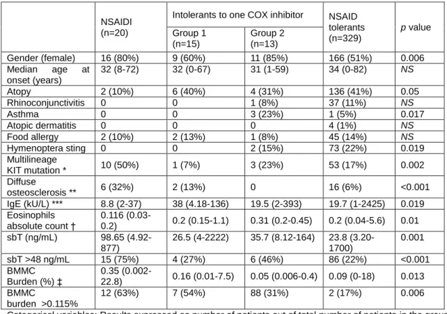
Table VIII shows epidemiological, clinical and laboratory characteristics of patients grouped by pattern of tolerance to COX inhibitors. The NSAIDI group had higher sBT values (p=0.001), percentage of BMMC burden (p=0.013), and frequencies of multilineage KIT mutation and diffuse osteosclerosis (p=0.002 and p<0.001, respectively). On the other hand, values of total IgE and eosinophils (p=0.02 and p=0.01, respectively) were shown to be lower in the NSAIDI group.
14 Table VIII. Epidemiological, clinical and laboratory characteristics of adult mastocytosis patients grouped according to the pattern of tolerance to COX inhibitors drugs.
NSAIDI (n=20)
Intolerants to one COX inhibitor NSAID tolerants (n=329) p value Group 1 (n=15) Group 2 (n=13) Gender (female) 16 (80%) 9 (60%) 11 (85%) 166 (51%) 0.006 Median age at onset (years) 32 (8-72) 32 (0-67) 31 (1-59) 34 (0-82) NS Atopy 2 (10%) 6 (40%) 4 (31%) 136 (41%) 0.05 Rhinoconjunctivitis 0 0 1 (8%) 37 (11%) NS Asthma 0 0 3 (23%) 1 (5%) 0.017 Atopic dermatitis 0 0 0 4 (1%) NS Food allergy 2 (10%) 2 (13%) 1 (8%) 45 (14%) NS Hymenoptera sting 0 0 2 (15%) 73 (22%) 0.019 Multilineage KIT mutation * 10 (50%) 1 (7%) 3 (23%) 53 (17%) 0.002 Diffuse osteosclerosis ** 6 (32%) 2 (13%) 0 16 (6%) <0.001 IgE (kU/L) *** 8.8 (2-37) 38 (4.18-136) 19.5 (2-393) 19.7 (1-2425) 0.019 Eosinophils absolute count † 0.116 (0.03-0.2) 0.2 (0.15-1.1) 0.31 (0.2-0.45) 0.2 (0.04-5.6) 0.01 sbT (ng/mL) 98.65 (4.92-877) 26.5 (4-2222) 35.7 (8.12-164) 23.8 (3.20-1700) 0.001 sbT >48 ng/mL 15 (75%) 4 (27%) 6 (46%) 86 (22%) <0.001 BMMC Burden (%) ‡ 0.35 (0.002-22.8) 0.16 (0.01-7.5) 0.05 (0.006-0.4) 0.09 (0-18) 0.013 BMMC burden >0.115% 12 (63%) 7 (54%) 88 (31%) 2 (17%) 0.006
Categorical variables: Results expressed as number of patients out of total number of patients in the group and percentage between brackets; Continuous variables: Results expressed as median in the group and range between brackets
BM: bone marrow; NS: not significant; NSAIDI: Intolerants to nonsteroidal antiinflammatory drugs; MC: mast cells; sBT: serum baseline tryptase;
*Analyzed in 355 patients ** Analyzed in 337 patients ***Collected in 268 patients † Collected in 192 patients
‡ Assessed by flow cytometry, performed in 332 patients
ROC curve analysis showed that levels of sBT≥48 ng/mL (Figure 1) and percentage of BMMC burden ≥0.12% (Figure 2) were able to predict intolerance to NSAIDs in adult mastocytosis patients, with a sensitivity of 75% and 68%, a specificity of 73% and 62%, respectively. AUC for both IgE and eosinophil absolute count was found not to be statistically significant.
15 Figure 1. Receiver Operating Characteristics (ROC) curve analysis of serum basal tryptase levels for the identification of NSAIDI patients.
sbT cut-off AUC 95% CI p value Sensitivity Specificity PPV NPV 48 ng/mL 0.73 0.622-0.882 <0.001 75% 73% 14% 98%
AUC: Area under curve; CI: Confidence interval; NPV: Negative predictive value; NSAIDI: Intolerants to nonsteroidal antiinflammatory drugs; PPV: Positive predictive value; sbT: Serum basal tryptase.
Figure 2. Receiver Operating Characteristics (ROC) curve analysis of BMMC burden for the identification of NSAIDI patients.
BMMC cut-off AUC 95% CI p value Sensitivity Specificity PPV NPV
0.16% 0.675 0.517-0.832 0.011 68% 62.3% 10% 97%
AUC: Area under curve; CI: Confidence interval; BMMC: Bone marrow mast cell; NPV: Negative predictive value; NSAIDI: Intolerants to nonsteroidal antiinflammatory drugs; PPV: Positive predictive value.
16 Interestingly, in the NSAIDI group, the presence of sBT ≥48 ng/mL and BMMC burden ≥0.12% were both associated with the presence of multilineage KIT mutation (p=0.001 and p=0.002, respectively) and/or diffuse osteosclerosis (p<0.001 and p=0.002, respectively).
Pediatric patients
A total of 137 mastocytosis pediatric patients were included, 67 (49%) of which were girls and 70 (51%) were boys, with a median age of 10 years (range: 1 – 17). Thirteen (10%) patients had cutaneous mastocytoma (cM), 5 (4%) had WDSM, and the remaining 118 (86%) had MIS. Distribution of patterns of skin lesions were as follows: 87 (64%) patients had a maculopapular (MPCM) form, 19 (14%) had nodules, 10 (7%) had plaques (PCM), and 8 (6%) had diffuse cutaneous mastocytosis (DCM). Regarding basal MC mediators related symptoms, 68 (50%) children had pruritus, 52 (38%) had flushing, and 40 (29%) had GI symptoms. Furthermore, a total of 7 (5%) had prior history of anaphylaxis. The median value of sBT was 5.3 ng/mL (range: 1.1-149).
One hundred and twenty (88%) pediatric mastocytosis patients tolerated paracetamol and 50 (36%) patients never received NSAIDs. Eighty-three (61%) patients received ibuprofen (among them, 7 and 2 also received metamizole and ASA, respectively), 3 (2%) only received metamizole, and the remaining patient (0.7%) only received ASA.
A total of 79 (58%) cases were included in the NSAIDs tolerant group. Seven patients were included in group 2 – all reactions were elicited by ibuprofen -, while the remaining case was classified as group 1 – the reaction was induced by metamizole.
Patterns of tolerance to NSAIDs, according to the form of mastocytosis are specified in Table IX. Interestingly, statistically significant differences were not found among these groups regarding epidemiological, clinical and laboratory characteristics.
17 Table IX. Epidemiological, clinical and laboratory features of pediatric, according to the pattern of tolerance to COX inhibitors.
Intolerants to one COX inhibitor NSAID tolerants n=79 p value Group 1 n=1 Group 2 n=7 Female (gender) 1 (100%) 4 (57%) 34 (43%) NS
Median age at onset (Years) 0 0 (0-8) 0 (0-7) NS
MPCM 1 (100%) 5 (71%) 51 (65%) NS Plaque CM 0 0 8 (10%) NS Nodular CM 0 0 6 (8%) NS cM 0 1 (14%) 9 (11%) NS DCM 0 1 (14%) 2 (3%) NS WDSM 0 0 3 (4%) NS sbT (ng/mL) 9.9 5.7 (1.1-149) 5.6 (1.1-68.7) NS Flushing 0 3 (43%) 27 (34%) NS Pruritus 1 (100%) 5 (71%) 42 (53%) NS GI symptoms 0 2 (29%) 23 (30%) NS Anaphylaxis 0 1 (14%) 3 (4%) NS
Categorical variables: Results expressed as number of patients out of total number of patients in the group and percentage between brackets; Continuous variables: Results expressed as median in the group and range between brackets;
cM: Cutaneous Mastocytoma; CM: Cutaneous Mastocytosis; DCM: Diffuse Cutaneous Mastocytosis; WDSM: Well-Differentiated Systemic Mastocytosis; GI: gastrointestinal; NSAIDI: Intolerants to nonsteroidal antiinflammatory drugs; sbT: serum basal tryptase.
Drugs challenges were performed in 25 patients. All challenges were negative, thus proving absence of intolerance to ibuprofen in 21 cases, metamizole in 2 cases and celecoxib in 2 cases. The latter drug was used in 14 and 17 year old patients.
Discussion
In this study, we have shown that MC mediator release episodes caused by drugs that inhibit the COX occur in 12% and 9% of, respectively, adult and pediatric mastocytosis patients that received such drugs. Also, 5% of adults and no pediatric patients were shown to be intolerant to NSAIDs. Thus, allergic/pseudoallergic symptoms elicited by NSAID are significantly more common in mastocytosis, when comparing with the general population. Moreover, the prevalence of such reactions is similar to the one found is asthma, which rounds 4% to 11%, but still inferior to the one
18 described in chronic urticaria, which may reach 27% to 35% (33) and MC activation plays a key role in both diseases.
In mastocytosis the risk of MC mediated symptoms caused by such COX inhibitors is not as high as previously thought, notwithstanding the fact that it manifested as anaphylaxis, in 8% of adults, which represented 16% of triggers for anaphylaxis. Interestingly, in the adult series, those who presented with symptoms derived from MC mediator release had a higher frequency of prior anaphylaxis. This finding was particularly more striking in NSAIDI and group 2 patients. Group 2 patients had other resemblances with NSAIDI: both displayed a female gender preference, high frequencies of flushing and anaphylaxis, and ASA and ibuprofen were the main implicated drugs, in both. As such, at least some patients in group 2 may be NSAID intolerants. Moreover, prevalence of asthma was higher in group 2, which would be more expected in NSAID intolerants, similarly as to what occurs in the general population.
Adult patients with anaphylaxis caused by Hymenoptera sting are less prone to NSAID intolerance. In fact, 97% of patients with Hymenoptera venom allergy tolerated NSAIDs. These patients were reported to be mainly male, less symptomatic, and to present with lower sbT levels, absence of multilineage KIT mutation, and a low BMMC burden (34), opposing features found in adult NSAIDI patients.
As occurs in the general population (35), propionic acid derivatives were found to be the most frequent elicitors for NSAID MC mediated reactions. This finding may be explained by the fact that these are the most frequently prescribed NSAIDs, in all age groups (35). In the adult series, they were followed by ASA and pyrazolones. The latter, are commonly prescribed in Spain, and are reported to be a frequent cause of IgE-mediated reactions to drugs (35). In this study, pyrazolones were the most frequent cause for group 1 intolerance, but were also frequently involved in NSAID intolerance (idiosyncratic reactions). This finding is unusual since pyrazolones are often considered to be weak COX-1 inhibitors (36, 37). Moreover, hypersensitivity to pyrazolones is strongly associated with HLA-DQ and HLA-DR expression (33), which has been found to occur in ISM, ASM and MCL patients (38). Thus, the mechanism through which metamizole and other pyrazolones induce MC mediator release in mastocytosis may be complex and multifactorial.
Mastocytosis patients were previously reported to be prone to intolerance to ASA while being tolerant to NSAIDs (35). In our study, this finding is confirmed in only 3 patients (20% of group 1).
Hypersensitivity reactions to NSAIDs in general population, were reported not to be associated with increased sBT levels (39). However, in this study, sBT levels above
19 48 ng/mL and a BMMC burden above 0.12% are proposed as surrogate markers to predict intolerance to NSAIDs in mastocytosis patients. Additionally, multilineage KIT mutation and diffuse osteosclerosis are more frequent in the NSAIDI group. Higher sBT levels, BMMC burden, presence of multilineage KIT mutation and diffuse osteosclerosis are associated with progression to aggressive categories of the disease (40, 41). As such, the non-indolent forms of mastocytosis may be at superior risk for intolerance to NSAIDs, as shown by a significantly higher frequency of intolerance to NSAIDs and other COX inhibitors in ASM patients. Moreover, patients with both CM and WDSM, presenting without the D816V KIT mutation in the BMMC had a very low frequency of intolerance to NSAIDs. These findings may be explained by a lack of upregulation of the transcription of genes involved in the arachidonic acid metabolism in MC without the D816V mutation, that is present in the MC of ASM and ISM, and absent in CM and WDSM (42).
In our study, reactions to paracetamol and coxibs were found to be infrequent in adult patients, and absent in children. These results are coherent with previous studies in non-mastocytosis-related intolerance to NSAIDs (20, 26). Tolerance to paracetamol, coxibs and/or to meloxicam was confirmed in the vast majority of intolerant patients. However, paracetamol elicited MC mediator release in 20% of NSAID intolerant patients. This frequency is superior to the one described in the literature for aspirin-intolerant asthma patients, rounding 7% (25). Our results show that MC related mediator symptoms caused by paracetamol and COX inhibitors only occur in NSAIDI, similarly to which occurs in the general population (16).
This study has some limitations. As drug challenges were not routinely carried out, due to safety concerns or refusal of patients, there is a lack of evidence that the drugs in study actually caused MC mediator release mediated symptoms. In fact, drug challenges were performed with the purpose of finding safer alternative drugs. Data obtained in pediatric patients are less informative, as the sample size was smaller, and the administration of NSAIDs was more limited.
There is still a high percentage of both adult and pediatric patients that have never taken NSAIDs due to fear of serious reactions. This fact has significant implications on patients’ quality of life, as the management of pain may become a source of anxiety, for both patient and health care professionals. However, our results show that NSAIDs do not trigger MC activation in mastocytosis as frequently as widely assumed. We recommend that patients who require NSAIDs and who have never taken them should be submitted to drug challenges with the drug(s) that offers the safest profile, for each case. In our experience, coxibs and meloxicam should be used in adults, and ibuprofen in children. Patients which have tolerated specific NSAIDs
20 following the onset of the disease, do not require further testing, and may be instructed to take a previously tolerated drug(s) (43).
Conclusion
Based on the retrospective analysis of a large series of adults and children with mastocytosis, we may conclude that symptoms associated with MC mediator release COX inhibitors are more frequent in mastocytosis, than in the general population. No clinically dramatic or fatal outcomes have been reported, in this series. As such, mastocytosis should not be considered as an absolute contraindication for the administration of NSAIDs, or other COX inhibitors. Nevertheless, it is strongly recommended that only drugs with the safest profile are used. Drugs to be preferably chosen in adults include those previously tolerated by the patient, coxibs, meloxicam and paracetamol. In children, ibuprofen may be used. If tolerance to COX inhibitors is unknown, a controlled drug challenge is recommended, and the drug should be chosen among those mentioned above.
Intolerance to NSAIDs was associated with aggressive categories of the disease, or with data suggesting possible future progression of the disease (multilineage KIT mutation and diffuse osteosclerosis). Hence, in these mastocytosis categories, NSAID administration should be cautiously planned, and the drug should be chosen carefully.
sBT levels above 48 ng/mL, and a BMMC burden above 0.12% emerge as potentially useful surrogate markers, as predictors of NSAIDs intolerance in mastocytosis. However, further studies are necessary to validate their routine usage.
21
References
1. Akin C, Valent P. Diagnostic criteria and classification of mastocytosis in 2014. Immunol Allergy Clin North Am. 2014 May;34(2):207-18. PubMed PMID: 24745670. 2. Valent P, Horny H, Li C, Longley J, Metcalfe D, Parwaresch R, et al. Mastocytosis (Mast cell disease). . In: JAFFE ES, HARRIS NL, STEIN H, VARDIMAN JW, editors. World Health Organization (WHO) classification of tumours Pathology & genetics Tumours of haematopoietic and lymphoid tissues. 1. Lyon: IARC Press; 2001. p. 291-302.
3. Cohen SS, Skovbo S, Vestergaard H, Kristensen T, Moller M, Bindslev-Jensen C, et al. Epidemiology of systemic mastocytosis in Denmark. Br J Haematol. 2014 Aug;166(4):521-8. PubMed PMID: 24761987.
4. van Doormaal JJ, Arends S, Brunekreeft KL, van der Wal VB, Sietsma J, van Voorst Vader PC, et al. Prevalence of indolent systemic mastocytosis in a Dutch region. J Allergy Clin Immunol. 2013 May;131(5):1429-31 e1. PubMed PMID: 23219169. 5. Horny H, Valent P, Metcalfe D, Bennett J, Bain B, Akin C, et al. Mastocytosis. In: Swerdlow SH, Campo E, Harris NL, Jaffe ES, Pileri SA, Stein H, et al., editors. WHO classification of tumours of haematopoietic and lymphoid tissues. 4 ed. Lyon: IARC Press; 2008. p. 54-63.
6. Akin C, Escribano L, Núñez R, García-Montero A, Angulo M, Orfao A, et al. Well-differentiated systemic mastocytosis: a new disease variant with mature mast cell phenotype and lack of codon 816 c-kit mutations. Journal of Allergy and Clinical Immunology. 2004;113(2):S327.
7. Alvarez-Twose I, Gonzalez de Olano D, Sanchez-Munoz L, Matito A, Esteban-Lopez MI, Vega A, et al. Clinical, biological, and molecular characteristics of clonal mast cell disorders presenting with systemic mast cell activation symptoms. J Allergy Clin Immunol. 2010 Jun;125(6):1269-78 e2. PubMed PMID: 20434205.
8. De la Hoz B, Gonzalez de Olano D, Alvarez I, Sanchez L, Nunez R, Sanchez I, et al. [Guidelines for the diagnosis, treatment and management of mastocytosis]. Anales del sistema sanitario de Navarra. 2008 Jan-Apr;31(1):11-32. PubMed PMID: 18496577. Guias clinicas para el diagnostico, tratamiento y seguimiento de las mastocitosis.
9. Escribano L, Akin C, Castells M, Orfao A, Metcalfe DD. Mastocytosis: current concepts in diagnosis and treatment. Ann Hematol. 2002 Dec;81(12):677-90. PubMed PMID: 12483363. Epub 2002/12/17. eng.
10. Matito A, Alvarez-Twose I, Morgado JM, Sanchez-Munoz L, Orfao A, Escribano L. Anaphylaxis as a clinical manifestation of clonal mast cell disorders. Curr Allergy Asthma Rep. 2014 Aug;14(8):450. PubMed PMID: 24947681.
11. WHO Collaborating Centre for Drug Statistics Methodology. Anatomical therapeutic chemical (ATC) classification index including defined daily doses (DDD) for
plain substances. Oslo: WHO; 2015. Available from:
http://www.whocc.no/atc_ddd_index/.
12. Graham GG, Davies MJ, Day RO, Mohamudally A, Scott KF. The modern pharmacology of paracetamol: therapeutic actions, mechanism of action, metabolism, toxicity and recent pharmacological findings. Inflammopharmacology. 2013 Jun;21(3):201-32. PubMed PMID: 23719833.
13. Sastre J, Cuesta J, Díaz M, Igea J, Olaguibel J, Sellers G. Factores epidemiológicos, clínicos y socioeconómicos de las enfermedades alérgicas en España. Sociedad Española de Alergia e Inmunología Clínica. 2005:249-79.
14. Ponvert C, Scheinmann P. [Allergic and pseudoallergic reactions to analgesics, antipyretics and non-steroidal antiinflammatory drugs]. Arch Pediatr. 2007
22 May;14(5):507-12. PubMed PMID: 17442550. Les reactions allergiques et pseudoallergiques aux antalgiques, antipyretiques et anti-inflammatoires non steroidiens.
15. Porto Arceo JA. [Special features of NSAID intolerance in children]. Allergol Immunopathol (Madr). 2003 May-Jun;31(3):109-25. PubMed PMID: 12783761. Particularidades de la intolerancia AINEs en ninos.
16. Sanchez-Matas I, Matito-Bernechea A, Gonzalez de Olano D, Alvarez-Twose I, Sanchez-Munoz L, de la Hoz Caballer B, et al., editors. Prevalence of hypersensitivity reactions to nonsteroidal anti-inflamatory drugs in 212 patients with mastocytosis in Spain. Allergy; 2009.
17. Brockow K, Jofer C, Behrendt H, Ring J. Anaphylaxis in patients with mastocytosis: a study on history, clinical features and risk factors in 120 patients. Allergy. 2008 (Feb);63(2):226-32. PubMed PMID: 18186813. Epub 2008/01/12. eng. 18. González de Olano D, de la Hoz Caballer B, Núñez López R, Sánchez Muñoz L, Cuevas Agustín M, Diéguez MC, et al. Prevalence of allergy and anaphylactic symptoms in 210 adult and pediatric patients with mastocytosis in Spain: a study of the Spanish network on mastocytosis (REMA). Clin Exp Allergy. 2007 Oct;37(10):1547-55. PubMed PMID: 17883734.
19. Ben-Amitai D, Metzker A, Cohen HA. Pediatric Cutaneous Mastocytosis: a review of 180 cases. IMAJ. 2005;7(May):320-2.
20. Çelik GE, Pichler WJ, Adkinson Jr NF. Drug Allergy. In: Adkinson Jr NF, Bochner BS, Burks AW, Busse WW, Holgate ST, Lemanske Jr RF, et al., editors. Middleton's allergy: principles and practice. 8th ed: Elsevier Health Sciences; 2013.
21. Mastalerz L, Setkowicz M, Sanak M, Szczeklik A. Hypersensitivity to aspirin: common eicosanoid alterations in urticaria and asthma. J Allergy Clin Immunol. 2004 Apr;113(4):771-5. PubMed PMID: 15100686. Epub 2004/04/22. eng.
22. Mastalerz L, Sanak M, Gawlewicz-Mroczka A, Gielicz A, Cmiel A, Szczeklik A. Prostaglandin E2 systemic production in patients with asthma with and without aspirin hypersensitivity. Thorax. 2008 Jan;63(1):27-34. PubMed PMID: 17584993.
23. Safholm J, Manson ML, Bood J, Delin I, Orre AC, Bergman P, et al. Prostaglandin E inhibits mast cell-dependent bronchoconstriction in human small airways through the E prostanoid subtype 2 receptor. J Allergy Clin Immunol. 2015 May 9. PubMed PMID: 25962903.
24. Steinke JW, Negri J, Liu L, Payne SC, Borish L. Aspirin activation of eosinophils and mast cells: implications in the pathogenesis of aspirin-exacerbated respiratory disease. J Immunol. 2014 Jul 1;193(1):41-7. PubMed PMID: 24890720. Pubmed Central PMCID: 4065844.
25. Szczeklik A, Stevenson DD. Aspirin-induced asthma: advances in pathogenesis, diagnosis, and management. J Allergy Clin Immunol. 2003 May;111(5):913-21; quiz 22. PubMed PMID: 12743549. Epub 2003/05/14. eng.
26. Daham K, James A, Balgoma D, Kupczyk M, Billing B, Lindeberg A, et al. Effects of selective COX-2 inhibition on allergen-induced bronchoconstriction and airway inflammation in asthma. J Allergy Clin Immunol. 2014 Aug;134(2):306-13. PubMed PMID: 24461582.
27. Sperr WR, Escribano L, Jordan JH, Schernthaner GH, Kundi M, Horny HP, et al. Morphologic properties of neoplastic mast cells: delineation of stages of maturation and implication for cytological grading of mastocytosis. Leuk Res. 2001 Jul;25(7):529-36. PubMed PMID: 11377677.
28. Horny HP, Valent P. Diagnosis of mastocytosis: general histopathological aspects, morphological criteria, and immunohistochemical findings. Leuk Res. 2001 Jul;25(7):543-51. PubMed PMID: 11377679.
29. Sanchez-Munoz L, Teodosio C, Morgado JM, Escribano L. Immunophenotypic characterization of bone marrow mast cells in mastocytosis and other mast cell disorders. Methods in cell biology. 2011;103:333-59. PubMed PMID: 21722810.
23 30. Hartmann K, Henz BM. Classification of cutaneous mastocytosis: a modified consensus proposal. Leuk Res. 2002 May;26(5):483-4; author reply 5-6. PubMed PMID: 11916523. Epub 2002/03/28. eng.
31. Garcia-Montero AC, Jara-Acevedo M, Teodosio C, Sanchez ML, Nunez R, Prados A, et al. KIT mutation in mast cells and other bone marrow hematopoietic cell lineages in systemic mast cell disorders: a prospective study of the Spanish Network on Mastocytosis (REMA) in a series of 113 patients. Blood. 2006 Oct 1;108(7):2366-72. PubMed PMID: 16741248.
32. Johansson SG, Bieber T, Dahl R, Friedmann PS, Lanier BQ, Lockey RF, et al. Revised nomenclature for allergy for global use: Report of the Nomenclature Review Committee of the World Allergy Organization, October 2003. J Allergy Clin Immunol. 2004 May;113(5):832-6. PubMed PMID: 15131563.
33. Kowalski ML, Makowska JS, Blanca M, Bavbek S, Bochenek G, Bousquet J, et al. Hypersensitivity to nonsteroidal antiinflammatory drugs (NSAIDs) - classification, diagnosis and management: review of the EAACI/ENDA(#) and GA2LEN/HANNA*. Allergy. 2011 Jul;66(7):818-29. PubMed PMID: 21631520.
34. Alvarez-Twose I, Zanotti R, Gonzalez-de-Olano D, Bonadonna P, Vega A, Matito A, et al. Nonaggressive systemic mastocytosis (SM) without skin lesions associated with insect-induced anaphylaxis shows unique features versus other indolent SM. J Allergy Clin Immunol. 2013 Aug 3. PubMed PMID: 23921094.
35. Woessner KM, Castells M. NSAID single-drug-induced reactions. Immunol Allergy Clin North Am. 2013 May;33(2):237-49. PubMed PMID: 23639711.
36. Pierre SC, Schmidt R, Brenneis C, Michaelis M, Geisslinger G, Scholich K. Inhibition of cyclooxygenases by dipyrone. Br J Pharmacol. 2007 Jun;151(4):494-503. PubMed PMID: 17435797. Pubmed Central PMCID: 2013970.
37. Hinz B, Cheremina O, Bachmakov J, Renner B, Zolk O, Fromm MF, et al. Dipyrone elicits substantial inhibition of peripheral cyclooxygenases in humans: new insights into the pharmacology of an old analgesic. The FASEB journal : official publication of the Federation of American Societies for Experimental Biology. 2007 Aug;21(10):2343-51. PubMed PMID: 17435173.
38. Teodosio C, García-Montero AC, Jara-Acevedo M, Sánchez-Muñoz L, Álvarez-Twose I, Núñez R, et al. Mast cells from different molecular and prognostic subtypes of systemic mastocytosis display distinct immunophenotypes. Journal of Allergy and Clinical Immunology. 2010;125(3):719-26.e4.
39. Seitz CS, Brockow K, Hain J, Trautmann A. Non-steroidal antiinflammatory drug hypersensitivity: association with elevated basal serum tryptase? Allergy, asthma, and clinical immunology : official journal of the Canadian Society of Allergy and Clinical Immunology. 2014;10(1):19. PubMed PMID: 24782901. Pubmed Central PMCID: 4002884.
40. Escribano L, Alvarez-Twose I, Sanchez-Munoz L, Garcia-Montero A, Nunez R, Almeida J, et al. Prognosis in adult indolent systemic mastocytosis: a long-term study of the Spanish Network on Mastocytosis in a series of 145 patients. J Allergy Clin Immunol. 2009 (Sep);124(3):514-21. PubMed PMID: 19541349. Epub 2009/06/23. eng. 41. Matito A, Morgado JM, Alvarez-Twose I, Laura S-M, Pedreira CE, Jara-Acevedo M, et al. Serum tryptase monitoring in indolent systemic mastocytosis: association with disease features and patient outcome. PLoS One. 2013;8(10):e76116. PubMed PMID: 24155887. Pubmed Central PMCID: 3796517.
42. Teodosio C, Garcia-Montero AC, Jara-Acevedo M, Sanchez-Munoz L, Pedreira CE, Alvarez-Twose I, et al. Gene expression profile of highly purified bone marrow mast cells in systemic mastocytosis. J Allergy Clin Immunol. 2013 Feb 9. PubMed PMID: 23403045. Epub 2013/02/14. Eng.
43. Bonadonna P, Pagani M, Aberer W, Bilo MB, Brockow K, Oude Elberink H, et al. Drug hypersensitivity in clonal mast cell disorders: ENDA/EAACI position paper. Allergy. 2015 Mar 31. PubMed PMID: 25824492.
24
Resumo Circunstanciado
Introdução
As mastocitoses são um grupo de doenças raras, caracterizadas pela acumulação de mastócitos anormais, em diferentes tecidos e órgãos (1, 2). Estas doenças surgem em indivíduos de todas as idades, sem preferência de género (1, 2). Estima-se que a sua prevalência esteja compreendida entre os 9 e os 13 casos por cada 100.000 habitantes (3, 4). A sintomatologia das mastocitoses decorre da libertação de mediadores mastocitários, da infiltração tecidual, ou de ambos (8). Quando ativados, os mastócitos secretam uma miríade de mediadores vasoativos, proinflamatórios, quimiotáticos e imunomoduladores (9). Os fatores despoletadores da ativação mastocitária incluem picadas por Himenópteros, stress, erupção dentária, e fármacos, tais como os opióides e os anti-inflamatórios não esteroides (AINEs) e outros inibidores da COX (10).
De acordo com a classificação ATC (11), os anti-inflamatórios incluem as butilpirazolidinas, derivados do ácido acético, derivados do ácido propiónico, fenamatos, coxibes e outros. Os fármacos analgésicos e antipiréticos incluem os salicilatos e outros, com reduzida capacidade inibitória das COX, tais como o paracetamol.
Os AINEs inibem a ação das ciclooxigenases (COX). Deste mecanismo de ação resultam efeitos antipiréticos, controlo da dor e efeitos anti-inflamatórios. Por sua vez, o paracetamol e outros inibidores fracos da COX-1, atuam ao nível do sistema nervoso central, exercendo, apenas, efeitos antipiréticos e analgésicos (12).
Em Espanha, a prevalência de reações alérgicas a AINEs varia entre 1% e 3%, nos adultos, sendo muito pouco frequente nas crianças (13, 14). Nas mastocitoses, a
25 prevalência de reações a AINEs decorrentes da ativação de mastócitos ronda os 14%, nos adultos, e 2% nas crianças (16). Contudo, neste grupo de doenças, estes fármacos são os fatores despoletadores de 8% (17) a 11% (18) das crises anafiláticas. Assim, a prescrição de AINEs deve ser cautelosa, razão pela qual o seu uso é, frequentemente, evitado nas mastocitoses.
As reações alérgicas e pseudoalérgicas provocadas por AINEs/fármacos com ação inibidora das COX podem ser induzidas por um único fármaco, por um grupo de fármacos, ou por fármacos estruturalmente não relacionados (20). As pirazolonas são, frequentemente, causa de reações de hipersensibilidade mediada por IgE (20). Porém, os restantes fármacos com ação inibidora das COX provocam reações não mediadas por IgE – pseudoalérgicas. Estas reações estão, provavelmente, relacionadas com a inibição da COX-1, resultante depleção de prostaglandina (PG) E2 e consequente
libertação de leucotrienos (21, 22). A libertação destes mediadores causa sinais e sintomas que podem variar entre sintomas cutâneos leves e choque anafilático (21, 25).
Os doentes que apresentam reações pseudoalérgicas a AINEs podem tolerar fármacos inibidores fracos da COX-1 e inibidores seletivos da COX-2 (20, 26). Nas mastocitoses, o mecanismo através do qual os AINEs induzem a libertação de mediadores mastocitários não é claro, mas poderá ser semelhante ao já descrito.
Com este estudo, pretende-se determinar a prevalência de reações provocadas pela libertação de mediadores despoletada por AINEs/outros fármacos com ação inibidora das COX, mas mastocitoses. Por outro lado, pretende-se descrever achados clínicos e laboratoriais, mais frequentemente, associados com estas reações.
Material e Métodos
Neste estudo retrospetivo, foram incluídos 417 adultos e 137 doentes pediátricos com mastocitose, seguidos pela Red Española de Mastocitosis (REMA), até Outubro de 2014. As variáveis em estudo foram:
1) epidemiológicas: género e idade;
2) clínicas: tipo de mastocitose, idade de aparecimento da doença, presença de atopia, alergias alimentares, asma, rinoconjuntivite, dermatite atópica, alergia a veneno de Himenópteros, sintomatologia basal (prurido, flushing e sintomatologia gastrointestinal), tolerância e sintomas despoletados por AINEs
26 e outros fármacos com ação inibidora das COX, história de reações anafiláticas;
3) laboratoriais: triptase sérica basal, IgE sérica total, IgE específica, presença de mutação do KIT com envolvimento de várias linhagens celulares, carga mastocitária da medula óssea;
4) medicina nuclear/radiológicas: presença de osteosclerose difusa.
As reações alérgicas/pseudoalérgicas foram avaliadas para cada doente. Esta avaliação incluiu a anamnese da reação, seguida de testes cutâneos, na suspeita de reações mediadas por IgE, ou de testes de provocação. Nas situações em que o nexo de causalidade era baixo, ou em que os sintomas eram leves, foi usado o fármaco, potencialmente, responsável pela reação. Nas restantes foram usados outros fármacos, com maior potencial de tolerância. Os doentes que apresentaram reações provocadas por, pelo menos, dois AINEs/inibidores das COX foram classificados como intolerantes a AINEs. Aqueles que não apresentaram reações foram considerados como tolerantes a AINEs. Por fim, os doentes que apresentaram reações provocadas por apenas um fármaco foram agrupados em 2 grupos: o grupo 1, que incluiu doentes que apresentaram reações a um fármaco, tolerando outros fármacos, e o grupo 2, que incluiu doentes que reagiram a um fármaco e que nunca mais tomaram outros AINEs. Os doentes que nunca tomaram AINEs, ou outros fármacos com ação inibidora das COX, à exceção do paracetamol, foram excluídos da análise estatística.
Análise Estatística
Para as variáveis contínuas, foram determinadas a mediana e a amplitude total. No caso das variáveis categóricas foram determinas as frequências. A presença de diferenças estatisticamente significativas entre os grupos, foi avaliada através dos testes de Kruskall Wallis ou U de Mann-Whitney, e do 2 ou teste de coeficientes,
respetivamente, entre grupos de variáveis contínuas e categóricas.Os valores de corte e os respetivos valores preditivos, para os níveis de triptase sérica basal e da carga mastocitária da medula óssea foram determinados através de curvas ROC. Os valores de p<0.05 foram considerados estatisticamente significativos. A análise estatística dos dados colhidos durante estudo foi realizada através do IBM SPSS para Windows, versão 23.0, e do Microsoft Excel 2010.
27
Resultados
Doentes adultos
Foram incluídos 417 adultos com mastocitose, 221 (53%) dos quais eram mulheres e 196 (47%) eram homens. Dez doentes (2%) tinham mastocitose cutânea (CM), 27 (7%) tinham mastocitose na pele (MIS), 207 (50%) tinham mastocitose sistémica indolente com lesões cutâneas (ISMs+), 126 (30%) tinham mastocitose
sistémica indolente sem lesões cutâneas (ISMs-) (7), 18 (4%) tinham mastocitose
sistémica bem-diferenciada (WDSM) (6), 13 (3%) tinham mastocitose sistémica agressiva (ASM), 2 (0.5%) tinham leucemia de mastócitos (MCL) e 8 (2%) tinham ISMs+ associada a hemopatia clonal não relacionada com a linhagem mastocitária
(AHNMD).
Verificou-se a presença de atopia em 40% dos doentes, prurido em 52%, flushing em 43%, e sintomas gastrointestinais em 37%. Adicionalmente, 46% dos doentes apresentavam história de anafilaxia.
Trezentos e vinte e nove doentes (79%) toleravam AINEs, 20 (5%) eram intolerantes (NSAIDI), 16 (4%) estavam incluídos no grupo 1 e 13 (3%) pertenciam ao grupo 2. Os 40 doentes restantes nunca tomaram AINEs, ou outros fármacos com ação inibidora das COX, com a excepção do paracetamol. À exceção de 4 doentes intolerantes a AINEs, verificou-se que todos os doentes toleravam o paracetamol.
Os padrões de tolerância a inibidores das COX, de acordo com o tipo de mastocitose estão presentes na tabela III. Verificou-se que estas reações são mais frequentes na ASM. A tabela IV explicita a frequência de sintomas basais nos diferentes grupos. A tabela V contém as frequências de episódios provocados por cada um dos fármacos, em cada um dos grupos. Na tabela VI, podem ser observadas as frequências de cada sinal, ou sintoma associado a reações de libertação de mediadores mastocitários, causadas por fármacos com ação inibidora das COX. Não foram encontradas diferenças estatisticamente significativas entre os grupos.
A tabela VIII apresenta as variáveis epidemiológicas, clínicas e laboratoriais, em relação a cada um dos grupos.
O grupo de doentes intolerantes a AINEs apresentou valores de triptase basal sérica superiores (p=0.001), assim como maior carga mastocitária na medula óssea (p=0.013); e maiores frequências de mutação do KIT com envolvimento de múltiplas
28 linhagens e da presença de osteosclerose difusa (respetivamente, p=0.002 e p<0.001).
A análise com curvas ROC permitiu aferir que níveis de triptase sérica basal superiores a 48 ng/mL e uma carga mastocitária na medula óssea superior a 0.12% permitem prever a intolerância a AINEs, nos adultos com mastocitose (figuras 1 e 2). Adicionalmente, a presença de ambos está associada à presença de envolvimento de múltiplas linhagens (p=0.001 e p=0.002, respectivamente) e, ou de osteosclerose difusa (p<0.001 e p=0.002, respectivamente).
Foram realizados testes de provocação com coxibes e, ou meloxicam em 51 doentes, dos quais: 14 pertenciam ao grupo dos intolerantes, 5 ao grupo 1, 9 ao grupo de tolerantes, 7 ao grupo 2 e 16 ao grupo de doentes que nunca tomaram fármacos com ação inibidora das COX, à exceção do paracetamol. Os testes foram considerados positivos em 3 doentes (6%). Todos pertenciam ao grupo dos intolerantes.
Doentes pediátricos
Foram incluídas 137 crianças, 49% das quais do género feminino e 51% do género masculino. Destes, 13 (10%) apresentavam mastocitoma cutâneo solitário (cM), 5 (4%) tinham mastocitoses sistémicas bem-diferenciadas (WDSM), e os restantes 118 (86%) tinham MIS. No que concerne à frequência dos diferentes tipos de lesões cutâneas: 87 (64%) doentes apresentavam uma forma maculopapular (MPCM), 19 (14%) tinham nódulos, 10 (7%) tinham placas (PCM), e 8 (6%) tinham uma mastocitose cutânea difusa (DCM). No que diz respeito à sintomatologia basal: 68 (50%) crianças referiam prurido, 52 (38%) apresentavam episódios de flushing, e 40 (29%) apresentavam sintomatologia GI. Acresce que 7 (5%) tinham história de reações anafiláticas. Os valores de triptase sérica basal variaram entre 1.1 e 149 ng/dL, com uma mediana de 5.3 ng/dL.
Um total de 120 doentes já tinha tomado paracetamol, sendo que nenhum destes apresentou qualquer tipo de reação mediada por mastócitos. Oitenta e sete doentes já tinham tomado AINEs ou outros analgésicos/antipiréticos, como o ibuprofeno, o metamizol e o ácido acetilsalicílico.
Ocorreram reações em apenas 8 doentes (9%), das quais 7 foram provocadas por ibuprofeno e 1 foi causada por metamizol. Não foram encontradas diferenças estatisticamente significativas entre os grupos definidos pelo padrão de tolerância a