Universidade de Aveiro Ano 2013
Departamento de Biologia
Ana Rita Marques
Almeida
Efeito combinado da radiação UV e
xenobióticos no peixe zebra
Combined effects of Ultraviolet radiation
and xenobiotics on zebrafish
DECLARAÇÃO
Declaro que este relatório é integralmente da minha autoria, estando devidamente referenciadas as fontes e obras consultadas, bem como identificadas de modo claro as citações dessas obras. Não contém, por isso, qualquer tipo de plágio quer de textos publicados, qualquer que seja o meio dessa publicação, incluindo meios eletrónicos, quer de trabalhos académicos.
_____________________
(Ana Rita Marques Almeida)Universidade de Aveiro Ano 2013
Departamento de Biologia
Ana Rita Marques
Almeida
Efeito combinado da radiação UV e
xenobióticos no peixe zebra
Combined effects of Ultraviolet radiation
and xenobiotics on zebrafish
Dissertação apresentada à Universidade de Aveiro para cumprimento dos requisitos necessários à obtenção do grau de Mestre em Biologia Aplicada no ramo Microbiologia Clínica e Ambiental, realizada sob a orientação científica da Doutora Paula Inês Borralho Domingues, Bolseira de Pós-Doutoramento do Departamento de Biologia da Universidade de Aveiro, co-orientação do Doutor Newton Carlos Marcial Gomes, Investigador auxiliar do Centro de Estudos do Ambiente e do Mar (CESAM) da Universidade de Aveiro.
O Júri
Presidente João António de Almeida Serôdio
Professor auxiliar do departamento de Biologia da Universidade de Aveiro
Susana Patrícia Mendes Loureiro
Investigador auxiliar do CESAM – Centro de Estudos do Ambiente e do Mar, Universidade de Aveiro
Paula Inês Borralho Domingues
Investigador de Pós-Doutoramento do Departamento de Biologia da Universidade de Aveiro
Agradecimentos
Agradeço:À minha orientadora Dr.ª Inês Domingues por todo apoio, motivação, paciência e ajuda dada ao longo de todo o trabalho. Foram muito importantes.
Ao meu co-orientador Newton Gomes por toda a disponibilidade e sugestões dadas.
À minha família, pai e mãe, por todo o esforço, amor e dedicação para que conseguisse terminar esta etapa.
Ao João pela companhia, amor e incentivo dado ao longo de todos estes anos.
À Dr.ª Susana Loureiro por toda a ajuda e sugestões dadas.
À Tânia pela paciência, ajuda e todas as horas “perdidas” a tentar ensinar-me. Agradeço-lhe muito por isso.
Ao pessoal dos “peixes”, Thayres, Jorge e Violeta por toda a ajuda, companhia, boa disposição e trocas de conhecimentos.
A todo o pessoal do LEMAN, Ana, Patrícia, Rossana, Rita, Francisco e Fernando por toda a ajuda e incentivo.
E finalmente, agradeço aos meus amigos e a todas as pessoas que ao longo destes anos passaram pela minha e deixaram a sua marca.
Palavras-chave
Resumo
Peixe-Zebra, Xenobióticos, comunidades bacterianas naturais, efeitos combinados.
Hoje em dia, as alterações climáticas são um problema imperativo e múltiplas medições feitas nos últimos anos mostram um aumento de toda a radiação solar, especialmente a radiação Ultravioleta que chega á superfície da terra afetando todos os organismos expostos. No seu ambiente natural, os organismos não estão apenas sujeitos a fatores bióticos, mas também a fatores ambientais e abióticos como por exemplo os xenobióticos. Além disso, ambos os stressores podem interagir uns com os outros produzindo efeitos imprevisíveis nos organismos (efeitos sinergísticos ou antagonísticos). O presente trabalho tem como objetivo a avaliação dos efeitos combinados da radiação UV e três xenobioticos (triclosan, dicromato de potássio e procloraz) em embriões de peixe zebra (Danio
rerio). A avaliação foi feita a dois níveis: i) efeitos na
mortalidade de embriões e ii) efeitos a nível das comunidades bacterianas naturais dos embriões. Os organismos foram expostos a várias concentrações de cada químico, combinadas com várias doses de UV. A mortalidade foi registada diariamente durante 96 horas e as comunidades bacterianas naturais foram avaliadas às 48 horas pós fertilização (hpf). Os resultados mostram que diferentes efeitos combinados foram observados, alterando a ecotoxicidade esperada. A exposição combinada da radiação UV com o TCS revelou um patrão sinergístico quando a radiação UV é o stressor dominante, enquanto que, na combinação UV com PD e PCZ observou-se antagonismo a doses baixas ou quando a radiação UV era dominante na mistura. As comunidades bacterianas naturais do peixe zebra também foram afetadas pela radiação UV e químicos, com alterações na sua estrutura. No entanto, foi difícil tirar conclusões relativamente a possíveis interações entre stressors visto que os efeitos observados nem sempre se traduziam em variações no índice de diversidade.
Key Words
Abstract
Zebrafish, Xenobiotics, Natural bacterial communities, combined effects.
Nowadays, climate changes are an imperative problem and multiple measurements made in the last years showed an increase of all wavelengths of solar radiance, specially the Ultraviolet radiation. In their natural environment organisms are not only affected by biotic and environmental factors, but also by abiotic factors such as xenobiotics. Besides, these both stressors can interact with each other being their combined effect unpredictable (producing additive, synergistic or antagonistic effect). This work aims to studying the combined effect of UV radiation and three xenobiotics: triclosan, potassium dichromate and prochloraz on zebrafish embryos (Danio
rerio). Effects were assessed at two levels: i) effects on embryos
mortality and ii) effects in the natural bacterial communities of zebrafish embryos. The organisms were exposed to concentrations of each chemical combined with several UV doses. Embryo’ mortality, were observed daily for 96 hours post fertilization (hpf) and natural bacterial communities’ evaluation was performed at 48 hpf. Results showed that different combined effect may occur compromising organism’s survival. Combined exposure of UV radiation with TCS revealed a synergism pattern when the UV radiation is the dominant stressor while PD and PCZ revealed antagonism at low dose levels or when the UV radiation is dominant in the mixture. Zebrafish natural bacterial communities were also affected by UV radiation and chemicals with the change of their structure; however, conclusions about interactive effects were difficult to be drawn because effects were not always translated into changes in the diversity indexes.
I
Contents
1. INTRODUCTION... 1 1.1. Climate changes ... 1 1.1.1. Ultraviolet radiation ... 2 1.2. Xenobiotic compounds ... 3 1.2.1. Potassium Dichromate ... 5 1.2.2. Prochloraz... 71.3. Stressors interactions in natural ecosystems ... 8
1.3.1. Ultraviolet radiation and toxicants interaction ... 10
1.4. Natural Microflora ... 10
1.4.1. Early life stages microflora ... 12
1.5. Zebrafish ... 14
1.5.1. Zebrafish as a model test ... 15
1.6. Molecular Tools ... 16
1.6.1. Polymerase Chain Reaction (PCR) ... 17
1.6.2. Electrophoresis ... 20
1.6.3. Denaturing Gradient Gel Electrophoresis (DGGE) ... 21
1.7. Objectives ... 21
1.8. Structure of the thesis ... 22
Interactive effects of Ultraviolet radiation and xenobiotic compounds in zebrafish embryos . 35 Abstract ... 35
1. Introduction ... 36
2. Materials and Methods ... 39
Test organisms ... 39
Chemicals ... 40
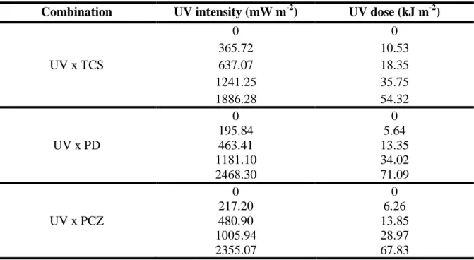
Combination of chemicals and UV radiation ... 41 Chemicals Analysis ... 41 Data analysis ... 42 3. Results ... 43 Chemicals Concentrations ... 43 Combined toxicity ... 43 4. Discussion ... 48 5. Conclusion ... 51 Supplementary data ... 59
Combined effects of Ultraviolet radiation and xenobiotics on zebrafish embryos - changes in bacterial communities ... 63
Abstract ... 63
1. Introduction ... 64
2. Materials and Methods ... 68
Test Organisms ... 68
Characterization of zebrafish embryos microbial diversity ... 68
Zebrafish embryo combined assay... 69
Data analysis ... 72
3. Results ... 72
Characterization of zebrafish embryos microbial diversity ... 72
Characterization of zebrafish embryos bacterial communities in combined exposure . 75 4. Discussion ... 83
Characterization of zebrafish embryos microflora ... 83
Characterization of zebrafish embryos microflora under UV and chemical exposure . 84 5. Conclusion ... 86
III
Figure Contentes
Figure 1- Xenobiotic exposure effects adapted fromScott and Sloman (2004) ...4
Figure 2 - Chemical structure of Triclosan (Dann & Hontela, 2011) ...5
Figure 3 – Chemical structure of Prochloraz (Ohlsson, Ullerås, & Oskarsson, 2009). ...7
Figure 4- Zebrafish egg ... 13
Figure 5- Zebrafish distribution marked with black dots (Spence et al., 2008) ... 14
Figure 6 – Male and Female zebrafish (Lab, 2009) ... 15
Figure 7 – PCR technique scheme (DS DNA= Double Strand DNA) (Tang & Stratton, 2012) ... 18
Figure 8 – Nested PCR scheme (Tang & Stratton, 2012) ... 19
Figure 9- Schematic electrophoresis gel (Gachet et al., 1998) ... 20
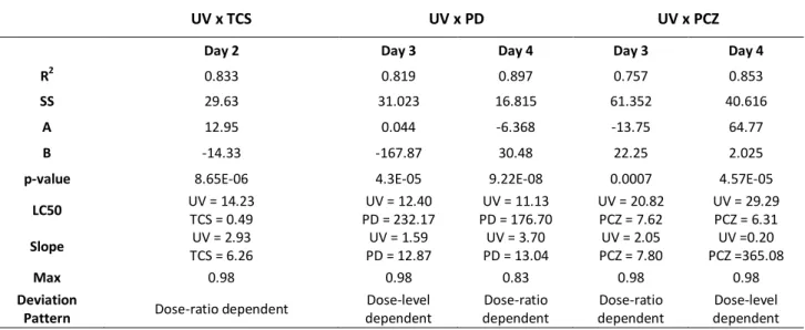
Figure 10 - Dose response relationship of survival rates of zebrafish embryos at day 2 exposed to the combination of UV radiation and TCS, showing a dose-ratio deviation from the IA conceptual model. ... 45
Figure 11 - Dose response relationship of survival rates of zebrafish embryos exposed to the combination of UV radiation and potassium dichromate, showing a dose-level dependency at day 3(A) and dose-ratio response at day 4 of exposure (B). ... 47
Figure 12 - Dose response relationship of survival rates of zebrafish embryos exposed to the combination of UV radiation and prochloraz, showing a dose-ratio at day 3 of exposure (A) and dose-level response at day 4 (B). ... 48
Figure 13 - DGGE image representing zebrafish embryos bacterial communities at 24, 48, 72 and 96 hours of development. M refers to marker. ... 73
Figure 14 - Ordination diagram (MDS) of bacterial community of zebrafish embryos at 24 ( ), 48 (▲), 72 (■) and 96 ( ) hours post fertilization. ... 74
Figure 15 - DGGE image of combined effect of UV radiation and triclosan. Units are in mg/L to TCS concentration and Kj.m-2 to UV dose. M refers to marker. ... 76
Figure 16 - Ordination diagram (MDS) of bacterial community by treatment in combined test of UV radiation with TCS. Triangles correspond to 0 Kj.m-2 UV dose; asterisks to 3.10 Kj.m-2 and diamonds to 6.53 Kj.m-2. Numbers above symbols refer to TCS concentration in mg/L. ... 77
Figure 17 - Shannon Index in combined exposure to UV radiation with TCS ... 77
Figure 18 - DGGE gel image of combined effect of UV radiation and potassium dichromate. Units are in mg/L to PD concentration and Kj.m-2 to UV dose. M refers to marker. ... 78
Figure 19 - Ordination diagram (MDS) of bacterial community by treatment. Triangle correspond to 0 Kj.m-2 UV dose; asterisks to 3.10 Kj.m-2 and diamonds to 12.22 Kj.m -2 . Numbers above symbols refer to PD concentration in mg/L. ... 79
Figure 21 - DGGE gel image of combined effect of UV radiation and prochloraz. Units are in mg/L to PCZ concentration and Kj.m-2 to UV dose. M refers to marker. ... 81 Figure 22 - Ordination diagram (MDS) of bacterial community by treatment. Triangles correspond to 0 Kj.m-2 UV dose; asterisks to 3.18 Kj.m-2 and diamonds to 6.47 Kj.m-2. Numbers above symbols refer to PCZ concentration in mg/L ... 82 Figure 23 - Shannon Index in combined exposure to UV radiation with PCZ ... 82
V
Table Contents
Table 1- Interaction studies between temperature increasing and metals (adapted from Holmstrup et al. (2010)) ...9 Table 2- Interaction studies between temperature increasing and pesticides (adapted from
Holmstrup et al. (2010)). ...9 Table 3 – Perspective of prokaryotes abundance in the world (Whitman et al., 1998) ... 10 Table 4 – Fish natural microflora ... 12 Table 5 - UV intensities (mW m-2) and correspondent doses for each combined exposure. ... 41 Table 6 - Interpretation of parameters that define the functional form of deviation pattern
from concentration addiction (CA) and independent action (AI) (adapted from Jonker at al. 2005) ... 44 Table 7 - Summary of MixTox analyses for the combination of UV radiation and triclosan,
potassium dichromate or prochloraz to zebrafish survival. ... 44 Table 8 - Chemical concentration and UV doses exposure ... 71 Table 9 - Shannon index calculated for bacterial communities of zebrafish embryos of
different ages. (S =total number of taxa) ... 74 Table 10 - Two-Way ANOVA analysis of microbial diversity (Shannon Index) in
1
1. INTRODUCTION
1.1. Climate changes
Climate can be expressed by the variation of temperature, precipitation and other physical properties, or in other words, by the typical meteorological conditions of a particular local for a certain period of time (Posas, 2011; Wuebbles, Jain, Edmonds, Harvey, & Hayhoe, 1999).
In the last two decades of the 20th century, an overall warming was becoming noticed with surface temperatures increasing 0,16 % per decade (Lean, 2010). This effect can be in part explained by anthropogenic influences, such as the emission of greenhouse gases, chlorofluorocarbons (CFCs) and other chlorinated and brominated halocarbons substances. While greenhouse gases absorb infrared radiation, turning the earth surface warmer, CFCs lead to ozone depletion. Given that ozone has an important role in filtering the ultraviolet (UV) radiation (Biales, Bencic, Villeneuve, Ankley, & Lattier) its depletion leads to increasing radiation reaching the earth’ surface (Lean, 2010; McKenzie et al., 2011; Wuebbles et al., 1999). Consequently, both gases contribute for changing the radiative balance of the atmosphere and modify the rate of warming and cooling of the earth. In fact, climate changes and ozone depletion are interconnected, variations in ozone layer can produce changes in climate and reciprocally. Nevertheless, UV radiation can be changed by climate changes without affecting ozone layer (McKenzie et al., 2011; Wuebbles et al., 1999).
Decreasing the greenhouse gases and ozone depleting substances (ODSs) emissions became essential to face the rise of temperature and UV radiation on earths’ surface. Therefore two international agreements were created to ban the use of CFCs (Montreal Protocol) and limit the emission of greenhouse gases (Kyoto protocol) (Holtsmark & Mæstad, 2002; Partridge, 1998).
2
Evidences show that Montreal Protocol was successful and the ozone layer is recovering. However, UV radiation reaching the surface of the earth is not decreasing (McKenzie et al., 2011; Woodcock, 2009). Indeed, solar activity tends to increase in the recent centuries and multiple measurements that have been made in the last years show an increase of all wavelengths of solar radiance, especially middle UV radiation (Lean, 2010; Lean & DeLand, 2012).
1.1.1. Ultraviolet radiation
Ultraviolet radiation is the radiation comprised between 100 and 400 nm being an important environmental stressor for both aquatic and terrestrial organisms (Nazari et al., 2010; Tartarotti & Torres, 2009). It can be divided in three categories: UV-A (400-315nm), UV-B (315-280 nm) and UV-C (280-100 nm) (Dong, Svoboda, Tiersch, & Monroe, 2007). The UV-C radiation is mostly absorbed by atmospheric ozone, and has not a significant environmental effect. On the other hand, UV-A and UV-B radiation reach the surface of the earth, having a higher impact in the environment (Dahms, Dobretsov, & Lee, 2011; Dahms & Lee, 2010).
Over the last years, many studies have revealed the harmful effects of UV-A and UV-B radiation. The main consequence of UV radiation is the generation of reactive oxygen species (ROS) that will damage cellular macromolecules (Behrendt, Jonsson, Goldstone, & Stegeman, 2010). Furthermore, proteins, lipids and nucleic acids are the first target of UV radiation, causing damages that can compromise common cellular process (Dahms et al., 2011).
In the aquatic ecosystems, the penetration of UV radiation depends on the dissolved organic carbon and colored non-living organic matter, being higher in cleaner water (Behrendt et al., 2010; Häkkinen et al., 2002; Sayed Ael, Ibrahim, Mekkawy, & Mahmoud, 2007). Decreases in productivity of phytoplankton and zooplankton by UV influence have been reported in literature. This variation can affect community composition by altering their diversity and productivity (Dahms et al., 2011).
3 In fishes, studies have shown a decrease of both growth and metabolic rate and even an effect on swimming performance (Sayed Ael et al., 2007). In several species egg mortality increases when exposed to UV radiation. In the adults, UV radiation acts like an immunosuppressive agent, destroying the immune systems including blood cells (Häkkinen et al., 2002; Sayed Ael et al., 2007).
On the other hand, some organisms that are subjected to high intensities of UV radiation have developed protection mechanisms. Sommaruga (2001) showed some strategies of alpine organisms, such as the melanization of dorsal portion in Daphnia spp., the accumulation of carotenoids in some copepods, which can minimize ROS production, the production of scytonemin pigment, which is present only in cyanobacteria, in particular those that live under intense solar radiation condition, and mycosporine-like amino acids. However, these strategies are not present in all organisms, being aquatic organisms from environments at lower altitudes more sensitivity to UV radiation (Sommaruga, 2001).
1.2. Xenobiotic compounds
Xenobiotics are compounds prevenient from diverse human activities that reach the environment posing serious risks to organisms and ecosystems. These compounds, such as pesticides, pharmaceuticals and nanoparticles do not occur in the environment naturally. Natural elements, such as compounds released from a forest fire or mining activities, which would usually be present in the environment only in vestigial concentrations, are also considered xenobiotics (Chong & Huang, 2012; Scott & Sloman, 2004). Xenobiotics have a wide range of physico-chemical properties and derive from numerous applications. Actually, much time and money is spent in developing new substances with the appropriate feature for a particular use, mostly because society is highly dependent on supplies and activities that involve the use of artificial synthetized compounds or naturals elements that are not so abundant on the surface of the earth (Donner et al., 2010).
4
In general, organisms can degrade xenobiotics after they enter in the body (biotransformation) converting them to less toxic and more easily to excrete products. However, in some cases this process can produce metabolites with higher toxicity than the original compound (Rivière, Bach, & Grolleau, 1985; Scott & Sloman, 2004; Sturm, Cravedi, Perdu, Baradat, & Segner, 2001). Further, the increase of reactive oxygen species levels is a common cause of xenobiotic exposure, leading to an oxidative stress, cellular damage and tissue injury. Furthermore, behavioral disruption (i.g. reproducibility and predator avoidance) and physiological response (i.g. neurobiological, endocrine and hormonal dysfunction) are some of the effects observed in fish after chemical exposure (Figure 1) (V. I. Lushchak, 2011; Scott & Sloman, 2004).
losan
Triclosan [5-chloro-2-[2,4-dichloro-phenoxy]-phenol; TCS] is a broad spectrum antimicrobial compound created 40 years ago (Fig. 2) (Dann & Hontela, 2011; Orvos et al., 2002). In the last 25 years, their use has been increasing once it is becoming part of many contemporary products such as: hand soaps, shower gels, deodorant soaps, screen cream, toothpaste, plastics and some textiles (Dann & Hontela, 2011; Liu, Ying, Yang, & Zhou, 2009; Lubarsky et al., 2012). Xenobiotic exposure Physiological response - Neurobiological; - Endocrine; - Hormonal. Behavioral disruption - Reproducibility; - Predator avoidance. Enhance of Reactive oxygen levels - Oxidative stress; - Cellular damage; - Tissue injury.
5 Figure 2 - Chemical structure of Triclosan (Dann & Hontela, 2011)
TCS is deposed with domestic wastewater, arriving later to treatment station, however, studies show that wastewater treatment plant are not able to remove completely this compound. Thus, TCS has already been detected in surface water, sediments, soils and in aquatic species (Dann & Hontela, 2011; Orvos et al., 2002; Sankoda et al., 2011). In fact, this may be the primary route for TCS enter in environment. Some predictions indicate, an increasing detection of the TCS in waterways once antimicrobial products are largely used (Dann & Hontela, 2011; Liu et al., 2009).
Some studies have shown that TCS affects negatively a large variety of aquatic organisms. Algae are an important pathway of accumulation of this compound once they are the primary food source for many species and can affect the whole aquatic ecosystem. Moreover, literature revealed bioaccumulation of TCS on fish (Dann & Hontela, 2011; R. Oliveira, Domingues, Grisolia, & Soares, 2009). Effects induced by TCS on adult fishes are loss of equilibrium, blockage of the mouth, quietness and irregular swimming movements (Orvos et al., 2002). Acute toxicity was also showed by Oliveira et al (2009) on zebrafish early-life stages as spinal malformations, hatching delayed, abnormal pigmentation of the eye and body and undersized larvae.
1.2.1. Potassium Dichromate
Chromium is abundant heavy metal widely distributed in terrestrial and aquatic environments (Farag et al., 2006; O. V. Lushchak, Kubrak, Nykorak, Storey, & Lushchak, 2008; Mohan & Pittman Jr, 2006). Similarly to the others metals, this compound is not
6
biodegradable and represents an important environmental threat once it remains in ecosystems and can be accumulated through the food chain (El Nemr, El-Sikaily, Khaled, & Abdelwahab; O. V. Lushchak et al., 2008).
This compound occurs in 2+ [bivalent form, Cr(II)], 3+ [trivalent form, Cr (Erickson Iii, Zepp, & Atlas)] and 6+ [hexavalent form, Cr(VI)] oxidative forms. The 2+ form is not abundant in nature, is unstable and little is known about its hydrolysis. Cr 3+(Erickson Iii et al.) is a hard acid, insoluble, immobile in ambient circumstances and can form strong complexes with oxygen and other donor ligands (Barnhart, 1997; Mohan & Pittman Jr, 2006). The hexavalent form has higher water solubility and mobility what makes it more toxic than the others oxidative forms of Cr (Erickson Iii et al.). Indeed, due to their properties, Cr(VI) crosses readily the cell membrane and, once inside of the cell, it is reduced to Cr (Erickson Iii et al.) which form complexes with intracellular macromolecules as well as with genetic material. Chromate and dichromate are the most toxic forms of hexavalent chromium (Farag et al., 2006; Mishra & Mohanty, 2009; Mohan & Pittman Jr, 2006).
Chromium compounds are used by several industries such as textile, wood preservations, corrosion control, electroplating, metal finishing industries, stainless steel productions, etc. Frequently, hexavalent chromium is discarded with other untreated industrial effluents reaching the water surface. Actually, global discharge in water of chromium is 142000 metric tonnes by year (Mishra & Mohanty, 2009; Mohan & Pittman Jr, 2006).
In the last decades, several studies have been showing the toxicity of chromium, to aquatic organisms, especially fishes (Mishra & Mohanty, 2009). In the mudskipper
Perophthalmus dipes, some enzymes of gills, kidney and intestine were inhibited by
chromium. (O. V. Lushchak et al., 2008) Moreover, metabolic changes in the Indian major carp Labeo rohita, and alterations of growth and health parameters in Chinook salmon,
Oncorhynchys tshawytscha, and mummichog, Fundulus heteroclitus were also described
as effects of chromium (Farag et al., 2006; Roling, Bain, Gardea-Torresdey, Bader, & Baldwin, 2006; Vutukuru, 2003). Moreover, immune response and resistance to disease of tilapia, Oreochromis mossambicus, was suppressed by chromium exposure (Prabakaran, Binuramesh, Steinhagen, & Michael, 2006).
7
1.2.2. Prochloraz
Procloraz (N-propyl-N-[2-(2,4,6-trichlorophenoxy)ethyl]-1H-imidazole-1-carboxamide) (Fig. 3) is a broad spectrum fungicide that pertains at imidazole family. The base of its action is the inhibition of cytochrome P450, essential in the biosynthesis of ergosterol, a fundamental component in the fungal membrane. (Biales et al., 2011; Kinnberg, Holbech, Petersen, & Bjerregaard, 2007)
Figure 3 – Chemical structure of Prochloraz (Ohlsson, Ullerås, & Oskarsson, 2009).
Since the early 1970s, prochloraz has been widely used in horticulture and agriculture. Its main application is to reduce plant disease, inhibiting fungi metabolism. Nevertheless, due to its broad spectrum properties, it affects also non target organisms, and may decrease microbial diversity and soil fertility (Ohlsson et al., 2009; Tejada, Gómez, García-Martínez, Osta, & Parrado, 2011)
Effects of this compound have been documented by a number of researches. Indeed, prochloraz affects steroid hormone levels in fetuses and/or adults of Wister dams, interfere with the receptors of androgen (Ohlsson et al., 2009; Vinggaard et al., 2006) and also alter hepatic and microsomal monooxygenases activity of Japanese quail (Rivière et al., 1985). In fishes, it interferes with metabolic pathway (e.g. biotransformation process), and produces behavioral disorders such as changes on swimming patterns, loss of equilibrium and reduced activity (Domingues et al., 2011; Saglio, Bretaud, Rivot, & Olsén, 2003).
8
1.3. Stressors interactions in natural ecosystems
In their natural environment, organisms are subjected to a multiplicity of stressors, which can interact with each other (Beketov, Speranza, & Liess, 2011; Tu et al., 2012). Moreover, these interactions are unpredictable, and can produce synergistic effects (when the interaction produces higher effect than the sum of the individual), or an antagonistic effect (when the interaction produces smaller effect than the sum of the individual) (Szöcs, Kefford, & Schäfer, 2012).
Several studies have been made in order to understand the effects of singular chemicals in aquatic environments, however, few is known about possible interactions with environmental parameters. Climate changes, and specially, the increase of UVR, has highlighted the importance of including environmental factors as additional stressors in ecotoxicological tests (Beketov et al., 2011).
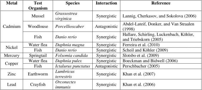
In the last two decades, some studies showed significant interactions of temperature, salinity and toxicants in grass shrimp (Palaemonetes pugio), in zooplankton, in rotifer (Brachionus rotundiformis) and in black tiger shrimp (Penaeus monodon) and worms (Enchytraeus albidus) (Gama-Flores, Sarma, & Nandini, 2005; Howard & Hacker, 1990; Silva, Holmstrup, & Amorim, 2013; Tu et al., 2012). The toxicity of metals and pesticides with the increasing of temperature was also showed by several studies (Table 1 and 2).
Moreover, the combined toxicity of metals and dissolved oxygen was showed by Ferreira et al. (2008) and Ferreira et al. (2010) revealing the enhancing of cadmium and nickel toxicity respectively at low dissolved oxygen levels in Daphnia magna.
9
Table 1- Interaction studies between temperature increasing and metals (adapted from Holmstrup
et al. (2010))
Metal Test
Organism
Species Interaction Reference
Cadmium
Mussel Grassostrea
virginica Synergistic Lannig, Cherkasov, and Sokolova (2006)
Woodlouse Porcellioscaber Antagonistic Abdel‐Lateif, Donker, and Van Straalen (1998)
Fish Danio rerio Synergistic Hallare, Schirling, Luckenbach, Köhler, and Triebskorn (2005)
Nickel Water flea Daphnia magna Synergistic Ferreira et al. (2010) Fish Danio rerio Synergistic Scheil and Köhler (2009) Mercury Springtail Folsomia candida Synergistic Slotsbo et al. (2009)
Copper Water flea Daphnia pulex Synergistic Boeckman and Bidwell (2006) Fish Ictalurus punctatus Antagonistic Perschbacher (2005)
Zinc Earthworm Lumbricus
terrestris Synergistic Khan et al. (2007)
Lead Crayfish Orconectes
immunis Synergistic Khan et al. (2006)
Table 2- Interaction studies between temperature increasing and pesticides (adapted from
Holmstrup et al. (2010)). Pesticides Test
Organism
Species Interaction Reference
Fenarimol Amphipod Monoporeia
affinis Synergistic
Jacobson, Prevodnik, and Sundelin (2008)
Diazinon Fish Danio rerio Synergistic Osterauer and Köhler (2008) Chlorpyrifos Insect Chironomus
tentans Synergistic Lydy, Belden, and Ternes (1999)
Imidachloprid Fish Danio rerio None Scheil and Köhler (2009) Terbufos Fish Cyprinodon
variegatus Synergistic
Brecken‐Folse, Mayer, Pedigo, and Marking (1994)
Trichlorfon Shrimp Palemonetes sp. Synergistic
Brecken‐Folse et al. (1994)Brecken‐ Folse, Mayer, Pedigo, and Marking (1994)
M-parathion Insect Chironomus
tentans Synergistic Lydy et al. (1999)
Pentachlorobenzene Insect Chironomus
tentans Synergistic Lydy et al. (1999)
Profenofos Fish Pimephales
promelas Synergistic Baer, Olivier, and Pope (2002)
Pyrethrine Lizard Anolis
10
1.3.1. Ultraviolet radiation and toxicants interaction
It would be expected that interactions between UVR and toxicants led to an additive effect or synergistic effect, once UVR can produce chemical changes by photoactivation, resulting in an increase of toxicity (Beketov et al., 2011).
Indeed, this enhanced toxicity was demonstrated by Huovien et al. (2001) with polycyclic aromatic hydrocarbons and by Jung et al. (2008) with sulfonamide antibiotics in
Daphnia magna. Moreover, synergism was also showed by several studies on these
organisms. Ribeiro et al (2011) observed synergism patterns on exposure at UV radiation and carbendazim, when the toxicity of combination was produced mainly by UVR. Kim et al. (2009) showed an increase of oxidative stress when D.magna were exposure to a different UV-B levels and sulfathiazole and, Nikillä et al. (1999) observed also an enhanced toxicity of pyrene under UV-B radiation.
1.4. Natural Microflora
Microorganisms are the most abundant form of life on the planet playing a critical rule, providing essential services that make our planet habitable (Table 3) (Li, Yu, Feng, Yan, & Gong, 2012; Prakash, Shouche, Jangid, & Kostka, 2013; Whitman, Coleman, & Wiebe, 1998).
Table 3 – Perspective of prokaryotes abundance in the world (Whitman et al., 1998)
Environment No. of prokaryotes cells (x1028)
Aquatic habitats 12
Ocean surface 355
Soil 26
11 Over their life, animals have living in a microbial rich environment where they constitute intimal associations with microbial communities. (Yan, van der Gast, & Yu, 2012) Every single organism have its own microflora, in other words, is colonized by several bacteria types that live in a symbiotic association with the host. This bacterial composition may consist on aerobic, facultative anaerobic and obligate anaerobic microorganisms. (Fraune & Bosch, 2010; Ringo, Olsen, Mayhew, & Myklebust, 2003)
The majority of these microorganisms is situated in the digestive tract and plays an important rule on hosts’ life (Kanther & Rawls, 2010; Yan et al., 2012). Natural microflora contributes to the host immune system, being the “first line defense” against pathogens, further, they furnish some nutrients that host could not access alone, such as vitamins, minerals and enzymes. Cell renewal in the intestine epithelium is also stimulated by these microrganisms (G. H. Hansen & Olafsen, 1999; Ozaktas, Taskin, & Gozen, 2012; Yan et al., 2012). Some authors have already shown the benefits of microflora, such as the preventing of infection by pathogenic fungi in crustacean eggs by bacteria producing an antifungal compound (Gil-Turnes & Fenical, 1992), the weight regulation by gut microbiota in mice and humans (Fraune & Bosch, 2010; Serino, Luche, Chabo, Amar, & Burcelin, 2009) and behavioral patterns monitoring in mammals (e.g. individuals, group members and parents recognition by odor and mate choice by producing secondary sexual traits) (Archie & Theis, 2011).
The natural microflora is specific of each organism, being different between species and inside of the same species. In fishes, microbiota is constituted mainly by four phyla:
Proteobacteria, Fusobacterium, Actinobacteria and Cyanobacteria (Li et al., 2012;
Roeselers et al., 2011a). Several studies have shown the differences between fish species microflora (Table 4).
12
Natural microflora can change with age, nutritional status and environment conditions (Ringo et al., 2003). Some environmental stressors such as temperature, oxygen levels, pH or pollutants can have a negative influence weakening these “first line defense” of the organism. Moreover, fitness of organisms may also be compromised which can also can change bacteria virulence (G. H. Hansen & Olafsen, 1999; Li et al., 2012).
1.4.1. Early life stages microflora
Vertebrates are born free of microrganisms, being colonized just in few hours (Yan et al., 2012). Indeed, the surface of eggs and larvae skin are good substrate for bacteria adhesion (Geir Høvik Hansen & Olafsen, 1989). This initial colonization is highly influenced by the surrounding environment and it is made by non-opportunistic bacteria that constitute the established microflora and will protect the host from pathogenic bacteria (Huys et al., 2001).
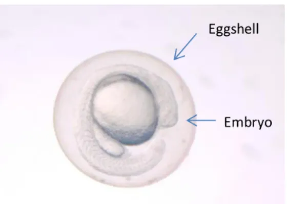
At egg stage of development, the eggshell is constituted by zona radiata and chorion (Fig. 4). This structure is mainly constituted by glycoproteins which make this place suitable for bacterial adhesion. Further, the embryo is able to secret some organic and
Table 4 – Fish natural microflora
Organism Specie Microflora Reference
Coho salmon Oncorhynchus kisutch
Pseudomonas sp.; Aeromonas sp.; Vibrio sp.
Romero and Navarrete (2006)
Salmon Salmo salar L.
Pseudomonas fluorescences;
Acinetobacter sp. Navarrete, Espejo, and Romero (2009)
Rainbow
trout Oncorhynchus mykiss
Proteobacteria
Kim, Brunt, and Austin (2007) Halibut Hippoglossus hippoglossus L. Pseudoalteromonas sp. Marinomonas sp. Vibrio sp. Bjornsdottir et al. (2010)
13 inorganic compound, producing a gradient which will act as a chemo-attractant for specific bacteria (G. H. Hansen & Olafsen, 1999). Some receptors present in the egg surface also act as bacteria selective (Olafsen, 2001).
Figure 4- Zebrafish egg
The egg adherent epiflora is highly diverse and act mostly has a barrier against pathogens present in the surrounding environment (G. H. Hansen & Olafsen, 1999). Some studies have shown the importance of natural microflora where these symbiotic interactions contribute to the survival of larvae (Brunvold et al., 2007). Moreover, various fish eggs are colonized by pigmented bacteria that constitute a protection against UV-B radiation (e.g. cod and halibut) (G. H. Hansen & Olafsen, 1999).
After hatching, fish larvae acquire its own/indigenous microflora when they start drinking and consume the suspended bacteria in the water. This happens before the yolk sac is consumed and establishes the primary intestinal microbiota (G. H. Hansen & Olafsen, 1999; Jensen, Øvreås, Bergh, & Torsvik, 2004; Olafsen, 2001). However, the main diversity and quantity of bacteria is uptake when the larvae start feeding (Fjellheim, Playfoot, Skjermo, & Vadstein, 2012).
Eggshell
14
1.5. Zebrafish
The zebrafish, Danio rerio, was firstly described by Hamilton-Buchanan in 1822 and belongs to the Cyprinidae freshwater fish family (Spence, Gerlach, Lawrence, & Smith, 2008).
These fish are native of South Asia and are distributed along India, Bangladesh, Nepal, Myanmar, and Pakistan (Fig. 5) (Lawrence, 2007; Scholz et al., 2008). They inhabit rivers, canals, ditches and lakes (Froese & Capuli, 2011; Spence et al., 2008).
Figure 5- Zebrafish distribution marked with black dots (Spence et al., 2008)
The zebrafish have a small size (maximum is 4 cm), its body is fusiform and laterally flattened (Gómez-Laplaza & Gerlai, 2010; Spence et al., 2008). This species is characterized by 5-7 dark blue longitudinal lines from operculum into the caudal fin and anal fin striped (Froese & Capuli, 2011; Spence et al., 2008).
15 The females have more rounded body than males and although both have similar coloration, the males have an anal fin more yellow (and larger) than females (Figure 6) (Spence et al., 2008). They have an external fertilization, where in every spawn, each female can produce hundred eggs that are after fertilized by sperm released in water by males (Scholz et al., 2008; Spence et al., 2008).
Figure 6 – Male and Female zebrafish (Lab, 2009)
The eggs are demersal, transparent and have a rapid development, being all the body formed at 24 hours post fertilization (hpf). The hatching occurs between 48 and 72 hpf and larvae reach maturity in 3-4months (Kimmel, Ballard, Kimmel, Ullmann, & Schilling, 1995; Scholz et al., 2008; Spence et al., 2008).
1.5.1. Zebrafish as a model test
Over the last decades, zebrafish has been widely used as a model test organism. Indeed, it is a powerful model being used in several areas of research, such as developmental biology (de Esch, Slieker, Wolterbeek, Woutersen, & de Groot, 2012), genetic and neurobiology (Sullivan & Kim, 2008), host-microbiota interactions (Roeselers et al., 2011b), learning and memory (Gómez-Laplaza & Gerlai, 2010), biomedical and human/animal disease (Giorgini et al., 2010) and (eco)toxicology (Scholz et al., 2008)
Actually, exceptional characteristics have led to its attraction. Firstly, their small size allow numerous organisms to be kept easily in a laboratory, it has a high reproductive rate, having each female hundred eggs in one spawn and rapid development (Chao et al.,
16
2010; Spence et al., 2008). In addition, both fertilization and development are external, being the eggs and larvae transparent which allows the monitoring of organs and mutant/transgenic lines development, the real-time observation of host-pathogen interaction and the response of immune system (Chao et al., 2010; de Esch et al., 2012). The adults are similar to mammals in relation of immune system response (Chao et al., 2010) and organs (Guo, 2009).
Moreover, its genome is completely sequenced and share about 60-80% of homolog genes with humans (de Esch et al., 2012; Howe et al., 2013; Meijer & Spaink, 2011). It is one of the best described organisms in developmental biology (Kimmel et al.) and have well described and broad molecular tools (de Esch et al., 2012).
1.6. Molecular Tools
Since the 30’s, with the origin of molecular biology, the life sciences have suffering a revolution (Thakur et al., 2008). Indeed, new molecular techniques brought new areas of investigation and re-arranged others (Burton, 1996).
The base of these tools are the macromolecules such as nucleic acids and progressed in two areas, the PCR-based techniques and non-PCR-based techniques, being, nowadays, the first one broadly used in studies relating the diversity of microbial communities (Dorigo, Volatier, & Humbert, 2005; R. B. Gasser, 2006).
These tools are very sensitive, specific, reliable and faster, which made them widely used in microbiology (Giraffa & Neviani, 2001; Justé, Thomma, & Lievens, 2008), parasitology (R. B. Gasser, 2006) and marine ecology (Burton, 1996). Actually, the traditional culture methods are not able to show/understand all the complexity of the microbial communities, once it is estimated that less than 1% of bacteria in the environment are cultivable (Amann & Ludwig, 2000; Dorigo et al., 2005; Fontana, Vignolo, & Cocconcelli, 2005). Moreover, it solved problems related with time
17 consumption in traditional methods with slow growing bacteria, and demanding cultured organisms (Drancourt et al., 2000).
1.6.1. Polymerase Chain Reaction (PCR)
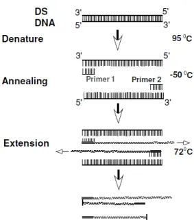
Polymerase Chain Reaction was discovered by Mullis in 1983 (Mullis, 1990). The main principle of PCR is the amplification of a specific DNA fragment from a complex DNA sample (Burton, 1996; Gachet, Martin, Vigneau, & Meyer, 1998). This technique is based on the properties of double strands of the DNA, consisting in three steps: denaturing, annealing and extension (Fig. 7) (Gachet et al., 1998; R. B. Gasser, 2006; Tang & Stratton, 2012).
In denaturation step, the two strands of the DNA are separated by the increasing of temperature to 93/95ºC resulting in two single strands of DNA. This permits to endonucleases to cut specific regions of the DNA strand (Gachet et al., 1998; Tang & Stratton, 2012).
Later, the temperature is reduced to 50/60ºC which permits the primers to contact with the complementary target sequence. The primers are small artificial oligonucleotides (20 – 30 nucleotids), designed to recognize specific sequences of the DNA target. These annealing of the primers with the DNA will allow the DNA polymerase to bind and initiate the DNA synthesis (Gachet et al., 1998; R. B. Gasser, 2006; Tang & Stratton, 2012).
The extension step occurs at 72ºC, where the DNA polymerase synthetize the new strand (Gachet et al., 1998; R. B. Gasser, 2006; Tang & Stratton, 2012).
18
Figure 7 – PCR technique scheme (DS DNA= Double Strand DNA) (Tang & Stratton, 2012)
After each cycling, the double strand of DNA is doubled in two new double strands, which means that in a standard PCR with 20 to 50 cycles, millions of DNA strands could be obtainable for analysis (Gachet et al., 1998; R. B. Gasser, 2006; Mock, Richardson, & Wolf).
The primer target is the 16S ribosomal DNA, which is widely distributed among bacteria. The gene sequences of 16S rDNA is great conserved within the same species and different between species (Drancourt et al., 2000; Woo, Lau, Teng, Tse, & Yuen, 2008). This difference in gene composition will allow the separation of fragment and results theoretically in a band pattern characteristic of the bacterial population, their fingerprinting (Fontana et al., 2005).
19
Nested PCR
The Nested PCR consists in the amplification of the product of the fist PCR with a second one (Fig. 8) (Bayley and Scott's, 2007; Tang & Stratton, 2012).
In the first PCR is amplified a specific sequence of DNA target, next, in the second PCR is amplified a shorter sequence inside of the first amplicon (Bayley and Scott's, 2007; Tang & Stratton, 2012).
This technique increases the sensitivity and specificity of the reaction (Bayley and Scott's, 2007; Tang & Stratton, 2012).
20
1.6.2. Electrophoresis
The electrophoresis technique consists on the migration of charged molecules through a matrix gel (Bayley and Scott's, 2007). The molecules, usually DNA or RNA, are charged negatively and when exposed to an electrical field, are forced to migrate from negative pole to the positive one (Bayley and Scott's, 2007; T. M. d. S. Oliveira, 2010). Due to their different sizes, the fragments will migrate differently over the gel porous, being the smaller/lighter fragments on the bottom of the gel (Fig. 9) (Gachet et al., 1998).
Figure 9- Schematic electrophoresis gel (Gachet et al., 1998)
The molecules are stained with a fluorescent dye, that binds DNA in order to the band pattern formed may be visualized under UV light (Bayley and Scott's, 2007; Gachet et al., 1998).
The electrophoresis technique is used after the PCR to determine/analyze if the previously technique worked and it was obtained the correct PCR product (Bayley and Scott's, 2007; Gachet et al., 1998).
21
1.6.3. Denaturing Gradient Gel Electrophoresis (DGGE)
Denaturing gradient gel electrophoresis is a technique based on an electrophoresis made on an acrylamide gel with a linear denaturing gradient (Gillan, 2004; Justé et al., 2008). The small DNA fragments are obtained by PCR and have the same size but different composition. These characteristic allows the physical separations by their melting properties, which depend on nucleotide sequence and GC content (Boutte, Grubisic, Balthasart, & Wilmotte, 2006; R. Gasser, Nansen, & Guldberg, 1996).
This technique is very used to get inter and intraspecific differentiation. Besides, the numbers of bands and their intensity can give an estimation of the community diversity and abundance (Dorigo et al., 2005; Manzano, Cocolin, Longo, & Comi, 2004).
1.7. Objectives
With the increasing of UV radiation reaching the earth’s surface, and a variety of pollutants threatening the aquatic ecosystems, there is a need to understand how these stressors interact and what effects can be produced on organisms’ survival and in their natural microflora, which plays an important role in animals’ health.
In order to answer this question, the aim of this work is to evaluate the effects of combined toxicity of UV radiation and three xenobiotics on zebrafish embryos.
To achieve the main objective, the work was divided into the following steps:
Evaluation of combined effects of UV radiation and xenobiotics triclosan, potassium dichromate and prochloraz, on zebrafish embryos;
Characterization of zebrafish embryos natural bacterial communities at 24, 48 72 and 96 hours post fertilization;
Characterization of zebrafish embryos natural bacterial communities in combined exposure of UV radiation and xenobiotics triclosan, potassium dichromate and prochloraz at 48 hours post fertilization.
22
1.8. Structure of the thesis
The current thesis is structured in the next chapters: Chapter 1: Introduction;
Chapter 2: Interactive effects of Ultraviolet radiation and xenobiotic compounds in zebrafish embryos.
Chapter 3: Combined effects of Ultraviolet radiation and xenobiotics on zebrafish embryos – changes in bacterial communities.
23
References
Abdel‐Lateif, H., Donker, M., & Van Straalen, N. (1998). Interaction between temperature and cadmium toxicity in the isopod Porcellio scaber. Functional Ecology, 12(4), 521-527. Amann, R., & Ludwig, W. (2000). Ribosomal RNA-targeted nucleic acid probes for studies in
microbial ecology. FEMS Microbiology Reviews, 24(5), 555-565.
Archie, E. A., & Theis, K. R. (2011). Animal behaviour meets microbial ecology. Animal Behaviour,
82(3), 425-436.
Baer, K., Olivier, K., & Pope, C. (2002). Influence of temperature and dissolved oxygen on the acute toxicity of profenofos to fathead minnows (Pimephales promelas). Drug and
chemical toxicology, 25(3), 231-245.
Barnhart, J. (1997). Occurrences, Uses, and Properties of Chromium. Regulatory Toxicology and
Pharmacology, 26(1), S3-S7.
Bayley and Scott's. (2007). Diagnostic Microbiology (twelve ed.): Mosby Elsivier.
Behrendt, L., Jonsson, M. E., Goldstone, J. V., & Stegeman, J. J. (2010). Induction of cytochrome P450 1 genes and stress response genes in developing zebrafish exposed to ultraviolet radiation. [Research Support, N.I.H., Extramural Research Support, Non-U.S. Gov't]. Aquat
Toxicol, 98(1), 74-82.
Beketov, M., Speranza, A., & Liess, M. (2011). Ultraviolet Radiation Increases Sensitivity to Pesticides: Synergistic Effects on Population Growth Rate of Daphnia magna at Low Concentrations. Bulletin of Environmental Contamination and Toxicology, 87(3), 231-237. Biales, A. D., Bencic, D. C., Villeneuve, D. L., Ankley, G. T., & Lattier, D. L. (2011). Proteomic
analysis of zebrafish brain tissue following exposure to the pesticide prochloraz. Aquatic
Toxicology, 105(3–4), 618-628.
Bjornsdottir, R., Karadottir, E. G., Johannsdottir, J., Thorarinsdottir, E. E., Smaradottir, H., Sigurgisladottir, S., & Gudmundsdottir, B. K. (2010). Selection of bacteria and the effects of bacterial treatment of Atlantic halibut (Hippoglossus hippoglossus L.) eggs and larvae. [Article]. Aquaculture, 302(3-4), 219-227.
Boeckman, C. J., & Bidwell, J. R. (2006). The effects of temperature, suspended solids, and organic carbon on copper toxicity to two aquatic invertebrates. Water, Air, & Soil Pollution, 171(1-4), 185-202.
Boutte, C., Grubisic, S., Balthasart, P., & Wilmotte, A. (2006). Testing of primers for the study of cyanobacterial molecular diversity by DGGE. Journal of Microbiological Methods, 65(3), 542-550.
Brecken‐Folse, J. A., Mayer, F. L., Pedigo, L. E., & Marking, L. L. (1994). Acute toxicity of 4‐ nitrophenol, 2, 4‐dinitrophenol, terbufos and trichlorfon to grass shrimp (Palaemonetes spp.) and sheepshead minnows (Cyprinodon variegatus) as affected by salinity and temperature. Environmental Toxicology and Chemistry, 13(1), 67-77.
Brunvold, L., Sandaa, R.-A., Mikkelsen, H., Welde, E., Bleie, H., & Bergh, Ø. (2007). Characterisation of bacterial communities associated with early stages of intensively
24
reared cod (Gadus morhua) using Denaturing Gradient Gel Electrophoresis (DGGE).
Aquaculture, 272(1–4), 319-327.
Burton, R. S. (1996). Molecular tools in marine ecology. J Exp Mar Bio Ecol, 200(1–2), 85-101. Chao, C.-C., Hsu, P.-C., Jen, C.-F., Chen, I.-H., Wang, C.-H., Chan, H.-C., . . . Lan, C.-Y. (2010).
Zebrafish as a model host for Candida albicans infection. Infection and Immunity, 78(6), 2512-2521.
Chong, N.-M., & Huang, J.-C. (2012). Production of xenobiotic degrader for potential application in bioaugmentation. Bioresource Technology, 125(0), 305-311.
Dahms, H. U., Dobretsov, S., & Lee, J. S. (2011). Effects of UV radiation on marine ectotherms in polar regions. [Research Support, Non-U.S. Gov't Review]. Comp Biochem Physiol C Toxicol
Pharmacol, 153(4), 363-371.
Dahms, H. U., & Lee, J. S. (2010). UV radiation in marine ectotherms: molecular effects and responses. [Research Support, Non-U.S. Gov't Review]. Aquat Toxicol, 97(1), 3-14.
Dann, A. B., & Hontela, A. (2011). Triclosan: environmental exposure, toxicity and mechanisms of action. [Review]. Journal of Applied Toxicology, 31(4), 285-311.
de Esch, C., Slieker, R., Wolterbeek, A., Woutersen, R., & de Groot, D. (2012). Zebrafish as potential model for developmental neurotoxicity testing: A mini review. Neurotoxicology
and Teratology, 34(6), 545-553.
Domingues, I., Oliveira, R., Musso, C., Cardoso, M., Soares, A. M. V. M., & Loureiro, S. (2011). Prochloraz effects on biomarkers activity in zebrafish early life stages and adults.
Environmental Toxicology, n/a-n/a.
Dong, Q., Svoboda, K., Tiersch, T. R., & Monroe, W. T. (2007). Photobiological effects of UVA and UVB light in zebrafish embryos: evidence for a competent photorepair system. [Comparative Study Research Support, Non-U.S. Gov't]. J Photochem Photobiol B, 88(2-3), 137-146.
Donner, E., Eriksson, E., Holten Lutzhoft, H.-C., Scholes, L. N. L., Revitt, D. M., & Ledin, A. (2010). Identifying and classifying the sources and uses of xenobiotics in urban environments: Springer Science + Business Media B.V.
Dorigo, U., Volatier, L., & Humbert, J.-F. (2005). Molecular approaches to the assessment of biodiversity in aquatic microbial communities. Water Research, 39(11), 2207-2218. Drancourt, M., Bollet, C., Carlioz, A., Martelin, R., Gayral, J.-P., & Raoult, D. (2000). 16S ribosomal
DNA sequence analysis of a large collection of environmental and clinical unidentifiable bacterial isolates. Journal of clinical microbiology, 38(10), 3623-3630.
El Nemr, A., El-Sikaily, A., Khaled, A., & Abdelwahab, O. Removal of toxic chromium from aqueous solution, wastewater and saline water by marine red alga Pterocladia capillacea and its activated carbon. Arabian Journal of Chemistry(0).
Erickson Iii, D. J., Zepp, R. G., & Atlas, E. (2000). Ozone depletion and the air–sea exchange of greenhouse and chemically reactive trace gases. Chemosphere - Global Change Science,
2(2), 137-149.
Farag, A. M., May, T., Marty, G. D., Easton, M., Harper, D. D., Little, E. E., & Cleveland, L. (2006). The effect of chronic chromium exposure on the health of Chinook salmon (Oncorhynchus tshawytscha). Aquatic Toxicology, 76(3–4), 246-257.
25 Ferreira, A. G., Serra, P., Soares, A. V. M., & Loureiro, S. (2010). The influence of natural stressors on the toxicity of nickel to Daphnia magna. Environmental Science and Pollution Research, 17(6), 1217-1229.
Ferreira, A. L. G., Loureiro, S., & Soares, A. M. V. M. (2008). Toxicity prediction of binary combinations of cadmium, carbendazim and low dissolved oxygen on Daphnia magna. Aquatic Toxicology, 89(1), 28-39.
Fjellheim, A. J., Playfoot, K. J., Skjermo, J., & Vadstein, O. (2012). Inter-individual variation in the dominant intestinal microbiota of reared Atlantic cod (Gadus morhua L.) larvae. [Article].
Aquaculture Research, 43(10), 1499-1508.
Fontana, C., Vignolo, G., & Cocconcelli, P. S. (2005). PCR–DGGE analysis for the identification of microbial populations from Argentinean dry fermented sausages. Journal of
Microbiological Methods, 63(3), 254-263.
Fraune, S., & Bosch, T. C. G. (2010). Why bacteria matter in animal development and evolution.
BioEssays, 32(7), 571-580.
Froese, R., & Capuli, E. (2011). FishBase, 2013, from www.fishbase.org
Gachet, E., Martin, G., Vigneau, F., & Meyer, G. (1998). Detection of genetically modified organisms (GMOs) by PCR: a brief review of methodologies available. Trends in food
science & Technology, 9(11), 380-388.
Gama-Flores, J., Sarma, S. S. S., & Nandini, S. (2005). Interaction among copper toxicity, temperature and salinity on the population dynamics of Brachionus rotundiformis (Rotifera). In A. Herzig, R. Gulati, C. Jersabek & L. May (Eds.), Rotifera X (Vol. 181, pp. 559-568): Springer Netherlands.
Gasser, R., Nansen, P., & Guldberg, P. (1996). Fingerprinting sequence variation in ribosomal DNA of parasites by DGGE. Molecular and Cellular Probes, 10(2), 99-105.
Gasser, R. B. (2006). Molecular tools—advances, opportunities and prospects. Veterinary
Parasitology, 136(2), 69-89.
Gil-Turnes, M. S., & Fenical, W. (1992). Embryos of Homarus americanus are Protected by Epibiotic Bacteria. The Biological Bulletin, 182(1), 105-108.
Gillan, D. C. (2004). The effect of an acute copper exposure on the diversity of a microbial community in North Sea sediments as revealed by DGGE analysis––the importance of the protocol. Marine Pollution Bulletin, 49(5–6), 504-513.
Giorgini, E., Conti, C., Ferraris, P., Sabbatini, S., Tosi, G., Rubini, C., . . . Carnevali, O. (2010). Effects of Lactobacillus rhamnosus on zebrafish oocyte maturation: an FTIR imaging and biochemical analysis. Analytical and Bioanalytical Chemistry, 398(7-8), 3063-3072.
Giraffa, G., & Neviani, E. (2001). DNA-based, culture-independent strategies for evaluating microbial communities in food-associated ecosystems. International Journal of Food
Microbiology, 67(1–2), 19-34.
Gómez-Laplaza, L. M., & Gerlai, R. (2010). Latent learning in zebrafish (Danio rerio). Behavioural
Brain Research, 208(2), 509-515.
26
Häkkinen, J., Vehniäinen, E., Ylönen, O., Heikkilä, J., Soimasuo, M., Kaurola, J., . . . Karjalainen, J. (2002). The Effects of Increasing UV-B Radiation on Pigmentation, Growth and Survival of Coregonid Embryos and Larvae. Environmental Biology of Fishes, 64(4), 451-459.
Hallare, A. V., Schirling, M., Luckenbach, T., Köhler, H. R., & Triebskorn, R. (2005). Combined effects of temperature and cadmium on developmental parameters and biomarker responses in zebrafish (Danio rerio) embryos. Journal of Thermal Biology, 30(1), 7-17. Hansen, G. H., & Olafsen, J. A. (1989). Bacterial colonization of cod (Gadus morhua L.) and halibut
(Hippoglossus hippoglossus) eggs in marine aquaculture. Applied and Environmental
Microbiology, 55(6), 1435-1446.
Hansen, G. H., & Olafsen, J. A. (1999). Bacterial Interactions in Early Life Stages of Marine Cold Water Fish. Microbial Ecology, 38(1), 1-26.
Holmstrup, M., Bindesbøl, A.-M., Oostingh, G. J., Duschl, A., Scheil, V., Köhler, H.-R., . . . Spurgeon, D. J. (2010). Interactions between effects of environmental chemicals and natural stressors: A review. Science of The Total Environment, 408(18), 3746-3762.
Holtsmark, B., & Mæstad, O. (2002). Emission trading under the Kyoto Protocol—effects on fossil fuel markets under alternative regimes. Energy Policy, 30(3), 207-218.
Howard, C., & Hacker, C. (1990). Effects of salinity, temperature, and cadmium on cadmium-binding protein in the grass shrimp,Palaemonetes pugio. Archives Of Environmental
Contamination And Toxicology, 19(3), 341-347.
Howe, K., Clark, M. D., Torroja, C. F., Torrance, J., Berthelot, C., Muffato, M., . . . Matthews, L. (2013). The zebrafish reference genome sequence and its relationship to the human genome. Nature.
Huys, L., Dhert, P., Robles, R., Ollevier, F., Sorgeloos, P., & Swings, J. (2001). Search for beneficial bacterial strains for turbot (Scophthalmus maximus L.) larviculture. Aquaculture, 193(1– 2), 25-37.
Jacobson, T., Prevodnik, A., & Sundelin, B. (2008). Combined effects of temperature and a pesticide on the Baltic amphipod Monoporeia affinis. Aquatic Biology, 1(3), 269-276. Jensen, S., Øvreås, L., Bergh, Ø., & Torsvik, V. (2004). Phylogenetic Analysis of Bacterial
Communities Associated with Larvae of the Atlantic Halibut Propose Succession from a Uniform Normal Flora. Systematic and Applied Microbiology, 27(6), 728-736.
Jung, J., Kim, Y., Kim, J., Jeong, D.-H., & Choi, K. (2008). Environmental levels of ultraviolet light potentiate the toxicity of sulfonamide antibiotics in Daphnia magna. Ecotoxicology, 17(1), 37-45.
Justé, A., Thomma, B. P. H. J., & Lievens, B. (2008). Recent advances in molecular techniques to study microbial communities in food-associated matrices and processes. Food
Microbiology, 25(6), 745-761.
Kanther, M., & Rawls, J. F. (2010). Host–microbe interactions in the developing zebrafish. Current
Opinion in Immunology, 22(1), 10-19.
Khan, M., Ahmed, S., Catalin, B., Khodadoust, A., Ajayi, O., & Vaughn, M. (2006). Effect of temperature on heavy metal toxicity to juvenile crayfish, Orconectes immunis (Hagen).
27 Khan, M., Ahmed, S., Salazar, A., Gurumendi, J., Khan, A., Vargas, M., & von Catalin, B. (2007).
Effect of temperature on heavy metal toxicity to earthworm Lumbricus terrestris (Annelida: Oligochaeta). Environmental Toxicology, 22(5), 487-494.
Kim, D. H., Brunt, J., & Austin, B. (2007). Microbial diversity of intestinal contents and mucus in rainbow trout (Oncorhynchus mykiss). Journal Of Applied Microbiology, 102(6), 1654-1664.
Kimmel, C. B., Ballard, W. W., Kimmel, S. R., Ullmann, B., & Schilling, T. F. (1995). Stages of embryonic development of the zebrafish. Developmental Dynamics, 203(3), 253-310. Kinnberg, K., Holbech, H., Petersen, G. I., & Bjerregaard, P. (2007). Effects of the fungicide
prochloraz on the sexual development of zebrafish (Danio rerio). Comparative
Biochemistry and Physiology Part C: Toxicology & Pharmacology, 145(2), 165-170.
Lab, M. (2009). Collecting zebrafish eggs, 2013, from
https://wiki.med.harvard.edu/SysBio/Megason/CollectingEggs
Lannig, G., Cherkasov, A. S., & Sokolova, I. M. (2006). Temperature-dependent effects of cadmium on mitochondrial and whole-organism bioenergetics of oysters (Crassostrea virginica).
Marine Environmental Research, 62, Supplement 1(0), S79-S82.
Lawrence, C. (2007). The husbandry of zebrafish (Danio rerio): A review. Aquaculture, 269(1–4), 1-20.
Lean, J. L. (2010). Cycles and trends in solar irradiance and climate. Wiley Interdisciplinary
Reviews: Climate Change, 1(1), 111-122.
Lean, J. L., & DeLand, M. T. (2012). How Does the Sun’s Spectrum Vary? Journal of Climate, 25(7), 2555-2560.
Li, X., Yu, Y., Feng, W., Yan, Q., & Gong, Y. (2012). Host species as a strong determinant of the intestinal microbiota of fish larvae. The Journal of Microbiology, 50(1), 29-37.
Liu, F., Ying, G.-G., Yang, L.-H., & Zhou, Q.-X. (2009). Terrestrial ecotoxicological effects of the antimicrobial agent triclosan. Ecotoxicology and Environmental Safety, 72(1), 86-92. Lubarsky, H. V., Gerbersdorf, S. U., Hubas, C., Behrens, S., Ricciardi, F., & Paterson, D. M. (2012).
Impairment of the Bacterial Biofilm Stability by Triclosan. [Article]. Plos One, 7(4).
Lushchak, O. V., Kubrak, O. I., Nykorak, M. Z., Storey, K. B., & Lushchak, V. I. (2008). The effect of potassium dichromate on free radical processes in goldfish: Possible protective role of glutathione. Aquatic Toxicology, 87(2), 108-114.
Lushchak, V. I. (2011). Environmentally induced oxidative stress in aquatic animals. Aquatic
Toxicology, 101(1), 13-30.
Lydy, M. J., Belden, J., & Ternes, M. (1999). Effects of temperature on the toxicity of M-parathion, chlorpyrifos, and pentachlorobenzene to Chironomus tentans. Archives Of Environmental
Contamination And Toxicology, 37(4), 542-547.
Manzano, M., Cocolin, L., Longo, B., & Comi, G. (2004). PCR–DGGE differentiation of strains of Saccharomyces sensu stricto. Antonie van Leeuwenhoek, 85(1), 23-27.
McKenzie, R. L., Aucamp, P. J., Bais, A. F., Bjorn, L. O., Ilyas, M., & Madronich, S. (2011). Ozone depletion and climate change: impacts on UV radiation. [Review]. Photochem Photobiol
28
Meijer, A. H., & Spaink, H. P. (2011). Host-pathogen interactions made transparent with the zebrafish model. Current drug targets, 12(7), 1000.
Mishra, A. K., & Mohanty, B. (2009). Effect of hexavalent chromium exposure on the pituitary– interrenal axis of a teleost, Channa punctatus (Bloch). Chemosphere, 76(7), 982-988. Mock, K. E., Richardson, B. A., & Wolf, P. G. Molecular tools and aspen management: A primer and
prospectus. Forest Ecology and Management(0).
Mohan, D., & Pittman Jr, C. U. (2006). Activated carbons and low cost adsorbents for remediation of tri- and hexavalent chromium from water. Journal of Hazardous Materials, 137(2), 762-811.
Mullis, K. B. (1990). The unusual origin of the polymerase chain reaction. Scientific American,
262(4), 56-61.
Navarrete, P., Espejo, R. T., & Romero, J. (2009). Molecular Analysis of Microbiota Along the Digestive Tract of Juvenile Atlantic Salmon (Salmo salar L.). Microbial Ecology, 57(3), 550-561.
Nazari, E. M., Ammar, D., de Bem, A. F., Latini, A., Muller, Y. M., & Allodi, S. (2010). Effects of environmental and artificial UV-B radiation on freshwater prawn Macrobrachium olfersi embryos. Aquat Toxicol, 98(1), 25-33.
Ohlsson, Å., Ullerås, E., & Oskarsson, A. (2009). A biphasic effect of the fungicide prochloraz on aldosterone, but not cortisol, secretion in human adrenal H295R cells—Underlying mechanisms. Toxicology Letters, 191(2–3), 174-180.
Olafsen, J. A. (2001). Interactions between fish larvae and bacteria in marine aquaculture.
Aquaculture, 200(1–2), 223-247. doi: http://dx.doi.org/10.1016/S0044-8486(01)00702-5
Oliveira, R., Domingues, I., Grisolia, C. K., & Soares, A. (2009). Effects of triclosan on zebrafish early-life stages and adults. [Article]. Environmental Science and Pollution Research, 16(6), 679-688.
Oliveira, T. M. d. S. (2010). PCR em tempo real: métodos e aplicações. Master, Universidade de Aveiro.
Orvos, D. R., Versteeg, D. J., Inauen, J., Capdevielle, M., Rothenstein, A., & Cunningham, V. (2002). Aquatic toxicity of triclosan. [Article]. Environmental Toxicology and Chemistry, 21(7), 1338-1349.
Osterauer, R., & Köhler, H.-R. (2008). Temperature-dependent effects of the pesticides thiacloprid and diazinon on the embryonic development of zebrafish (Danio rerio). Aquatic
Toxicology, 86(4), 485-494.
Ozaktas, T., Taskin, B., & Gozen, A. G. (2012). High level multiple antibiotic resistance among fish surface associated bacterial populations in non-aquaculture freshwater environment.
Water Research, 46(19), 6382-6390.
Partridge, M. R. (1998). The Montreal protocol. What does transition to CFC free metered dose inhalers mean for the health professional now? Revue Française d'Allergologie et
d'Immunologie Clinique, 38(7, Supplement 1), S94-S96.
Perschbacher, P. W. (2005). Temperature effects on acute copper toxicity to juvenile channel catfish Ictalurus punctatus. Aquaculture, 243(1–4), 225-228.