Universidade de Lisboa
Faculdade de Ciências
Departamento de Biologia Animal
Towards the characterization of
plasmatocyte heterogeneity in
Drosophila melanogaster
Alexandre Castanho Barata Leitão
Dissertação de mestrado orientada por Doutor Élio Sucena
1,2Instituto Gulbenkian de Ciêade de Ciê ncias da Universidade de Lisboa
Mestrado em Biologia Evolutiva e do Desenvolvimento
2009
Faculdade de Ciências
Departamento de Biologia Animal
Towards the characterization of
plasmatocyte heterogeneity in
Drosophila melanogaster
Alexandre Castanho Barata Leitão
Dissertação de mestrado orientada por: Doutor Élio Sucena
1,21Instituto Gulbenkian de Ciência
2Faculdade de Ciências da Universidade de Lisboa
Mestrado em Biologia Evolutiva e do Desenvolvimento
i
Acknowledgements
This work is my first contribution to science. I want to acknowledge everyone that made this possible:
- A special gratitude to my grandfather José Castanho for all the passion for facts that he transmitted me.
- To my parents for encouraging me to look and search for facts. - To Élio Sucena for teaching me how to do questions.
- To Vitor Faria for all the discussions, help, and friendship. Without him all this work would be very different.
- To my amazing brainstorming team: Thiago Carvalho, Jocelyne Demengeot, António Jacinto and Luís Teixeira.
- To my special discussion guests: Marc Dionne and David Scnheider.
- To the persons that eared all my music in loop mode during this months and never complained: Sophie Dias and Catarina Moreira.
- To my companions that share the same passion for science: Pedro Lima, Sara Esteves, Inês Trancoso and Alexis Hazbun.
- To everyone that makes the IGC an awesome place to work and have fun.
- To Patrícia Basto for giving me the opportunity to forget all about questions and facts.
ii
Resumo
Virtualmente todas as espécies de animais interagem com microrganismos ao longo da sua vida. Estas interacções podem evoluir em relações benéficas (comensalismo e mutualismo) ou antagonistas (parasitismo). As infecções de parasitas têm um alto custo de fitness nos hospedeiros uma vez que diminuem a sua viabilidade. Para combater as infecções os animais possuem um notável sistema de reconhecimento e resposta contra patogénios, o sistema imunitário. Nos insectos, tal como na maioria dos metazoários, o sistema imunitário inato pode ser particionado em mecanismos de resposta humorais e celulares. Entre os exemplos melhor caracterizados de respostas humorais encontra-se a produção de péptidos antimicrobiais, pequenas proteínas que quando se encontram em circulação eliminam bactérias e fungos. A fagocitose é um mecanismo celular que não depende da produção de proteínas, sendo muito eficaz na eliminação de patogénios. No entanto, os dois ramos do sistema imunitário estão interligados, operando sinergeticamente. O estudo destas interligações é um passo importante para compreender a reposta imunitária como um sistema integrado.
No género Drosophila, 95% das células do sistema imunitário (hemócitos) são plasmatócitos. Estas células participam em variados processos durante o desenvolvimento e durante a resposta imunitária. Ao longo da embriogénese e estádio de pupa os plasmatócitos desempenham um papel importante na remodelação dos tecidos, fagocitando células mortas e sintetizando matriz extracelular. Durante uma infecção de bactérias ou fungos os plasmatócitos produzem péptidos antimicrobiais, fagocitam e agregam os patogénios. No estado larvar parte dos plasmatócitos encontram-se fixos a diferentes tecidos ou em circulação, desempenhando um papel de “vigilância” de feridas e infecções. Os plasmatócitos que circulam na hemolinfa agregam-se nos locais de ferida formando um coágulo para impedir a perda de hemolinfa e entrada de patogénios. Estas células desempenham igualmente um papel importante no reconhecimento dos ovos de vespas parisitárias e possivelmente induzem a diferenciação de outro tipo de hemócito na glândula linfática.
Podem ser encontrados outros dois tipos de hemócitos na hemolinfa de Drosophila, as células cristal e os lamelócitos, ambos com funções muito especializadas na resposta imunitária. As células cristal constituem 5% dos hemócitos encontrados em larvas de Drosophila e os lamelócitos só são encontrados em larvas após infecção de parasitas de grandes tamanhos, como um ovo de vespa. As células cristal são cruciais no processo de melanização, uma resposta imunitária presente em artrópodes que consiste na formação e deposição de melanina nos locais de infecção. Esta
iii acumulação de melanina restringe o acesso de nutrientes por parte do patogénio. Por outro lado a cascata de formação de melanina produz muitos radicais de oxigénio que causam diversos danos nos patogénios. Os lamelócitos são grandes células alongadas que se agregam à volta dos ovos de artrópodes parasitas formando uma cápsula. Esta cápsula sofre posteriormente um processo de melanização que leva à morte do embrião parasita.
A classificação dos três tipos de hemócitos descritos em cima é baseada em critérios morfológicos e bioquímicos. No entanto, alguns novos estudos começaram a caracterizar a expressão de RNA e proteína nas diferentes classes de hemócitos. Estes novos estudos mostram que algumas proteínas são expressas apenas numa parte da população de plasmatócitos. Neste trabalho pretendemos continuar a caracterização genética dos plasmatócitos de Drosophila com o propósito de perguntar se existem diferentes subpopulações destas células com diferentes funções durante uma resposta imunitária.
Para analisar indirectamente a expressão génica em plasmatócitos recorremos ao sistema GAL4/UAS-GFP, sendo a expressão de GAL4 dependente dos promotores dos nossos genes de interesse. Analisámos a expressão GFP com 5 promotores de genes que se sabem estar expressos em plasmatócitos de larvas de Drosophila:
hemolectin, peroxidasin, croquemort, serpent e hemese. Utilizando a técnica de
citometria de fluxo foi possível determinar a percentagem de plasmatócitos que expressam cada um dos genes.
Os nossos resultados mostram que o gene hemolectin é expresso em ~99% dos plasmatócitos. Como este gene não é expresso noutro tecido de Drosophila é um bom gene repórter para estudos que pretendam analisar a totalidade de plasmatócitos. Por outro lado, não encontrámos expressão de croquemort nos plasmatócitos do 3º estádio larval e apenas 5% dos plasmatócitos expressam serpent. Como ambos os genes são altamente transcritos durante a fase de embrião isto indica-nos que existe uma diferenciação destas células durante o desenvolvimento larvar. Os plasmatócitos apresentam assim uma complexa dinâmica de expressão génica durante as diferentes fases do ciclo da Drosophila, o que pode estar relacionado com diferentes funções destas células durante diferentes fases.
Os genes peroxidasin e hemese são expressos no 3º estádio larval mas apenas em subpopulações de plasmatócitos. O gene peroxidasin é expresso em ~50% dos plasmatócitos e o gene hemese em ~80 %. Estes dois genes tornam-se assim bons candidatos para marcadores de subpopulações funcionais de plasmatócitos. Utilizando a tecnologia de separação de células por fluorescência (Fluorescence Activated Cell
iv Sorting) foi possível isolar diferentes populações de células vivas para realizar ensaios
in vitro.
A caracterização morfológica das células hemese positivas e hemese negativas mostra uma diferença significativa na extensão do corpo celular na lâmina de vidro e no número de lisosossomas secundários. Os plasmatócitos hemese positivos têm um eixo maior e apresentam mais lisossomas secundários. Com o intuito de compreender se alguma destas subpopulações de plasmatócitos desempenha um papel na regulação da resposta imunitária testámos a capacidade destas células em inibir a reacção de melanização, uma resposta humoral. Os nossos resultados dos ensaios in
vitro não apoiam esta hipótese.
Este trabalho mostra que não podemos considerar os plasmatócitos como uma população homogénea de células. Futuros estudos na resposta imunitária celular têm que ter em conta esta observação pois diferentes subpopulações de plasmatócitos podem estar a desempenhar funções diferentes. Os resultados apresentados abrem novas perspectivas de estudo em outras áreas como o estudo da diferenciação celular. Será necessário conduzir novas investigações neste sistema para perceber qual o papel destas subpopulações na resposta imunitária ou durante o desenvolvimento.
v
Abstract
Insect innate immune system can be partitioned into humoral and cellular defense mechanisms. However, both branches of immune system are interconnected, acting in a synergistic way. The study of these interconnections is an important step to comprehend the immune response as an integrated system. In Drosophila genus 95% of “blood cells” (hemocytes) are plasmatocytes. These cells participate in numerous processes during development and immune response. Throughout embryogenesis and pupal stage plasmatocytes play an important role in tissue remodeling, phagocytizing dead cells and synthesizing extracellular matrix. During an immune response they are responsible for production of antimicrobial peptides, phagocytosis and aggregation of pathogens. In addition, it is possible to find two other types of hemocytes in
Drosophila’s hemolymph, crystal cells and lamellocytes, both with very specialized
functions in immune response. Crystal cells constitute 5% of hemocytes in Drosophila and lamellocytes are only found in larvae upon infection with large pathogens, such as wasp eggs. Classification of these three hemocyte types is based on morphological and biochemical criteria. However, some new studies have begun to characterize RNA and protein expression in Drosophila’s hemocyte classes. Here, we want to go further in the genetic characterization of plasmatocytes with the purpose off asking if there are different subpopulations of plasmatocytes performing different functions during immune response. For this propose we used flow cytometry technique to analyze gene expression in Drosophila larvae plasmatocytes. Our results show that two out of the five GAL4 lines analyzed drive expression of GFP in subpopulations of Drosophila larval plasmatocytes. This observation indicates that plasmatocytes do not form a homogeneous population of cells in Drosophila’s larvae hemolymph. We then used Fluorescence Activated Cell Sorting (FACS) to sort and perform in vitro experiments with hemese positive and hemese negative plasmatocytes independently. In vitro analysis confirmed that both subpopulations correspond to previous plasmatocyte descriptions. We hypothesized that one of these two different subpopulations of plasmatocytes is responsible for modulation of melanization, an immune response of insects. However, the results obtained in our specific in vitro setting did not support this hypothesis but further work is needed to ascertain this matter in a definitive way. Key words: innate immune system, Drosophila, plasmatocytes, melanization
vi
Index
Acknowledgements ... i Resumo ... ii Abstract ... v Introduction ... 1Cellular immunity meets humoral immunity: a brief historical perspective ... 1
Drosophila epithelial and humoral defense mechanisms ... 2
Hemocytes classes in Drosophila and hemocyte mediated immune responses ... 3
Plasmatocytes, surveillance and phagocytosis ... 3
Lamellocytes and encapsulation... 5
Crystal cells and melanization ... 5
Classes of hemocytes in other insects... 5
Plasmatocytes: a genetically and functional heterogeneous population of cells? ... 6
Objectives ... 6
Methods ... 7
Fly stocks ... 7
Fly crosses ... 7
Hemocyte collection ... 8
Hemocytes staining for FACS analysis ... 8
Morphological characterization of plasmatocytes ... 8
PO inhibition assay... 9
Phagocytosis assay ... 9
Statistical analysis ... 9
Results... 10
Confirmation of GFP expression in hemocytes using GAL4/UASGFP system ... 10
FACS protocol ... 11
Analysis of candidate genes with heterogeneous expression in plasmatocytes ... 13
Morphological analysis of hemese positive and hemese negative cells ... 13
Phagocytic activity of hemese positive and hemese negative cells ... 15
Phenoloxidase inhibition assay... 15
Discussion ... 17
1
Introduction
Cellular immunity meets humoral immunity: a brief historical perspective
Virtually all animal species interact with microorganisms during their lifetime. These interactions can evolve into beneficial relations for one or both species as commensalism or mutualism. However, numerous times microorganisms become pathogenic for the host, reducing its viability. To fight pathogenic infections animals possess a notable set of recognition and defense mechanisms, the immune system. Although we can trace back references of immunologic observations to ancient Greek civilization, it’s not until the end of 19th century that immunology was formed as a
scientific discipline1. At that time two different mechanisms were proposed to explain
how hosts fight pathogens: cellular immunity and humoral immunity.
Ilya Metchnikoff, during his experiments with starfishes to study comparative embryology, discovered phagocytosis and later proposed a theory that linked macrophages and macrophages-like-cells with organism “equilibrium”, performing functions in tissue remodeling and immunity2. According to Metchnikoff, phagocytes in
starfish and macrophages in vertebrates were the cells responsible to phagocyte pathogens and this way to fight infections. This theory would find some resistance, especially in German scientific community, where a humoral theory of immunity was giving its first steps. According to the humoral theory of immunity, clearance of pathogens in the host was possible due to “factors” present in the serum. Antibodies, which are present in vertebrate humoral immune system, would turn out to be a hallmark of immunology studies during the 20th century. But Metchnikoff ideas of
cellular immunity would be recognized and reconciled with humoral immunity theory when, in 1908, he shared a Nobel Prize with Paul Ehrlich, one of the first scientists to theorize about antibody functions3.
The development of scientific research in immunology during the twentieth century confirmed the importance of cellular and humoral processes in host defense. Most noticeably, several investigations indicate that the two “branches” of immune system are interconnected and act in synergy. With few exceptions, animals rely on both mechanisms to fight pathogens. This way, when we investigate cells of immune system it’s always important to test the relation of the two systems.
As in several other domains of biology, Drosophila became a model of excellence for the study of innate immune system due to the great variety and versatility of its genetic tools. Nevertheless, while great effort has been made in identifying signaling and response mechanism of Drosophila humoral response, much less is known regarding
2 the genetic mechanisms of cellular immune responses4. In the next sections we will
briefly resume what is known about the two branches of Drosophila’s immune system to fully understand our working hypothesis.
Drosophila epithelial and humoral defense mechanisms
Epithelia are the first line of defense in all metazoans, working mainly as a physical barrier. In insects, some epithelia, as the respiratory tract, constitutively produce Antimicrobial Peptides (AMPs) to avoid infection of the body cavity by pathogens. This kind of defenses are not dependent on recognition of the pathogen, however, a full set of defenses is activated upon this recognition. One of the first activated responses of
Drosophila immune system when a pathogen is able to infect the gut lumen is to
produce Reactive Oxygen Species (ROS). This production of ROS is dependent of
duox proteins family and it has been shown to limit the proliferation of Erwinia carotovora in the gut5.
Probably the immune mechanism best characterized in Drosophila is the “systemic immune response”. In case of an infection in hemolymph several AMPs are produced and released into the hemolymph. Production of AMPs is an extremely conserved immune response in animals, probably common to all metazoan. These small peptides are produced mostly in the fat body, a mesoderm derived tissue that is localized in insects body cavity, in contact with the hemolymph6. During the first larval stage fat
body becomes immuno-competent and remains functional throughout Drosophila’s life. Production of AMPs is dependent on activation of three described genetic pathways: Toll, IMD, and JAK/STAT6.
A rapid response of Drosophila immune system upon tissue damage or infection is melanization: neo-synthesis and deposition of melanin. After an activation cascade proPhenoloxidase (proPO) is cleaved into Phenoloxidase (PO), an enzyme that catalyzes oxidation of phenols to orthoquinones6. Quinones are thought to be toxic to
microbes and they polymerize melanin non-enzymatically7. Melanin physically
encapsulates pathogens limiting their access to nutrient acquisition. In larvae, proPo is synthesized by a specific “blood cell” type (crystal cells in Drosophila) but other components of proPO activation are produced by other cells including fat body cells8.
Several proteins of this cascade are present in hemolymph leading to a rapid response upon activation9.
3 Hemocytes classes in Drosophila and hemocyte mediated immune responses
Hemocytes constitute the cellular immune branch of invertebrates. Similarly to vertebrates, Drosophila has two hematopoietic waves throughout development. The first one occurs during embryogenesis where it is possible to discriminate mature hemocytes as early as stage 10 10. The earliest hematopoietic gene marker described
so far is Serpent, a GATA family protein expressed in head mesoderm 11. Hemocytes
produced during embryonic hematopoietic wave constitute the big portion of larvae hemocytes and they will persist in hemolymph during all life stages of Drosophila12.
Hematopoiesis continues during larval stage in the lymph gland, a dorsal organ adjacent to dorsal vessel13. Just after the beginning of pupation mature hemocytes
produced in the lymph gland are released into the hemolymph and the lymph gland disrupts. This way, hemocytes in pupa and adult stages are a heterogeneous population of cells derived from embryonic and larval hematopoietic waves.
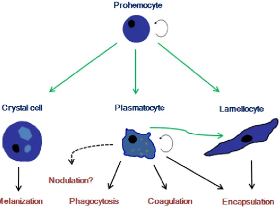
In Drosophila’s larvae hemolymph it’s possible to distinguish three classes of differentiated hemocytes in circulation: Plasmatocytes, Lamellocytes and Crystal cells (Fig. 1). Some authors refer the presence of pro-hemocytes in circulation but there is no consensual morphological description or genetic marker to distinguish these cells. The major constituent of blood cells are plasmatocytes that can reach 95% of total blood cells14. Crystal cells constitute about 5% of immune cells. Lamellocytes aren’t
present in adult or pupa stage and are rarely found in non infected larvae. However, when a large body like a wasp egg enters the larval body cavity, lamellocytes are found in large number. These three classes of hemocytes are involved in different immune mechanisms to fight infections as discussed below.
Plasmatocytes, surveillance and phagocytosis
Phagocytosis is a rapid immune response because it is not dependent on protein production to limit microorganisms growth. In Drosophila only plasmatocytes are capable to phagocytize foreign elements, among others, bacteria, yeast, Sephadex beads and ink particles. Recognition of bacteria and funguses mediated by a series of Pattern Recognition Receptors (PRRs) that upon ligation activate signaling cascades that will regulate formation of a phagosome15. Effector molecules are introduced in the
phagosome that will mature to a phagolysosome, where bacteria are killed. How plasmatocytes recognize particles that have never been present in their environment is a challenge to our knowledge on pattern recognition.
When in circulation, plasmatocytes are spherical cells with 5-8 µm diameter16. In
4 philapodia. Golgi apparatus and rough endoplasmic reticulum are well developed in these cells and they contain several phagolysosome-like inclusions. In larva, plasmatocytes can be found in circulation or adherent to tissues. The role of adherent plasmatocytes is not well established yet. Circulating plasmatocytes are thought to work as a surveillance system detecting cuticle wounds and infections in the hemolymph. Plasmatocytes form aggregates at sites of tissue injuries that works a physical barrier, preventing microorganism infections. These cells aggregates are reinforced by fibers to form a clot. The clot formation is dependent of plasmatocyte activity and humoral factors (Lemaitre). We don’t have many evidence so far, but probably plasmatocytes signal to fat body upon infection to produce AMPs17.
Plasmatocytes are also the first cells to adhere to wasp eggs in the hemolymph. In short, plasmatocytes mediate several responses in drosophila’s immune system.
Another cellular immune response observed in insects is the entrapment of large numbers of bacteria by multilayer aggregate of hemocytes in a poorly understood process named nodulation18. In Galleria mellonella nodulation is mediated by
plasmatocytes-like cells19. To our knowledge, nodulation is not studied in Drosophila.
Figure 1 - Hemocyte classes present in Drosophila larvae and their functions during an immune response: prohemocytes are present in embryo hematopoietic tissue and in lymph
gland. This cell type is mitotically active and have the potential to differentiate into all three classes of hemocytes (green arrows). Plasmatocytes have also the potential to differentiate into lamellocytes.
5 Lamellocytes and encapsulation
When infected with a large body, as a wasp egg, Drosophila’s larvae mount a dramatic immune response: encapsulation. After recognition of Leptopilina boulardi egg in the body cavity, a mechanism that is under discussion, lamellocytes are differentiated, enter circulation and form a multilayer structure (capsule) around the intruder. Lamellocytes are differentiated in two different localizations, in lymph gland and in a population of sessile plasmatocytes in posterior zone of larvae20,21.
Lamellocytes are larger than plasmatocytes and don’t have phagolisosome-like granules. After encapsulation the formed body becomes melanized and eventually the parasite egg is killed. The exact cause of death is unknown but ROS produced in melanization cascade may be implicated6.
Crystal cells and melanization
Cristal cells are large cells present in the embryo and in circulation in larvae. Mature crystal cells produce a great amount of pro-phenoloxidase (proPO) that they store in crystallized form. They were named crystal cells due to their crystal like inclusions. However, in other drosophila species these inclusions do not present crystal like structure16. Upon activation crystal-cells release these structures into hemolymph
activating this way the melanization cascade. This can happen after encapsulation, nodulation or cuticle injury. Adult flies lack crystal cells but melanization cascade is still activated upon infection or cuticle injury. We still don’t know what is source of pre-PO in adult flies.
Classes of hemocytes in other insects
During his detailed description of insect anatomy and physiology, Jan Swammerdam described for the first time insect hemocytes. In his seminal scientific work “Bybel der Nature of Historie der Insecten”, 1737, Swammerdam clearly describes head louse (Pediculus humanus) hemocytes22. With development of microscopy and histological
techniques it was possible to distinguish several classes of hemocytes in insects. As stated before, in Drosophila it’s possible to distinguish three types of hemocytes according to their morphology4. However in some other insect classes it’s possible to
distinguish different numbers of hemocytes types16. In Aedes aegypti, Hillyer and
Christenses described only two types of hemocytes in circulation, granulocytes and oenocytoids23. Functionally, granulocytes are equivalent to Drosophila’s plasmatocytes
6 and oenocytoids to crystal cells. Evidences suggest that mosquitoes do not have a specialized cell type for capsule formation as lamellocytes in Drosophila. This observation suggests that mosquitoes do not have natural parasitoids (insects that parasites arthropods). Lepidoptera usually possess four types of hemocytes. For example in Pseudoplusia includens mature hemocytes found in hemolymph are separated in spherule cells, oenocytoids, granular cells and plasmatocytes16. Once
again the nomenclature does not correspond to Drosophila classification. Drosophila’s plasmatocytes correspond to Lepidopteran granular cells, crystal cells to oenocytoids, lamellocytes to Lepidopetra’s plasmatocytes and there is no morphological equivalent to spherule cells in Drosophila. This cell type is present in every Lepidopteran species studied so far but its function in immune system remains unknown.
Unfortunately there are not so much studies in other insect’s classes to have a clear picture of how different the numbers hemocyte types evolved. But evidences described here suggest that number of hemocytes types and functions evolved in different insect lineages.
Plasmatocytes: a genetically and functional heterogeneous population of cells? Recent studies indentified some monoclonal antibodies that bind specifically to hemocytes. Kurucz and colleagues identified antibodies that bind to all hemocytes classes (H1) or specifically to plasmatocytes (P1a and P1b), lamellocytes (L1) and crystal cells (C2, C3, C4 and C5)24. Interestingly one of the antibodies, H2, binds to
plasmatocytes but only to a fraction of plasmatocytes. In a more detailed study Kurucz and colleagues reported that P1a and P1b recognize two different epitopes of the same molecule, Nimrod C125. Some plasmatocytes do not express this protein but we don’t
know to each extent. hemolectin antibody also binds to plasmatocytes but not all plasmatocytes express this protein26.
These few detailed studies of plasmatocyte gene expression led us think that plasmatocyte do not form a homogeneous population regarding gene expression. An interesting question immediately arises with this observation: are plasmatocytes divided into different functional classes?
Objectives
The aim of this work is to go further in the characterization of Drosophila’s plasmatocytes. We focused on heterogeneity of gene expression in circulating plasmatocytes with the ultimate aim of ascribing different functions to these
7 plasmatocytes subpopulations. The first goal of our work is to establish a Fluorescence Activated Cell Sorting (FACS) protocol to analyze gene expression in live plasmatocytes and separate putative plasmatocyte subpopulations. After this analysis we wish to test an in vitro for putative roles of plasmatocyte subpopulations in modulation of melanization response.
Methods
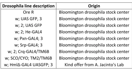
Fly stocksFly stocks used in this work are described in table 1. Flies were fed with standard food and maintained at 25º C and 70% humidity.
Drosophila line description Origin
Ore R Bloomington drosophila stock center w; UAS GFP, 3 Bloomington drosophila stock center w; 2; UAS GFP Bloomington drosophila stock center w; 2; He-GAL4 Bloomington drosophila stock center w; Pxn-GAL4; 3 Bloomington drosophila stock center w; Srp-GAL4; 3 Bloomington drosophila stock center w; 2; Crq-GAL4/TM6B Bloomington drosophila stock center w; SCO/CYO; TM2/TM6B Bloomington drosophila stock center w; HmlΔ-GAL4 UASGFP; 3 Kind offer from A. Jacinto's Lab
Fly crosses
In order to achieve GFP expression in hemocytes we generated flies with two constructions: promoter-GAL4 and UAS-GFP. Crosses were carried with virgin females and young males in food tubes supplemented with fresh yeast. w; pxnGAL4 UAS GFP line was obtained previously in our lab.
A generalist scheme of our crosses is presented below (x represents one of the hemocyte promoters):
Cross 1.1:
8 Cross 1.2: Cross 2: Cross 3:
With this final cross we end up with final genotype: w; xGAL4; UASGFP.
Hemocyte collection
Hemocytes were collected by rupturing abdominal larval cuticle in ice cooled Schneider’s medium containing 1% sodium azide for analysis or without sodium azide when cells were sorted for tests. For each FACS analysis 50-60 larvae were bled in 800 µl medium.
Hemocytes staining for FACS analysis
Hemocytes were stained with a modified protocol from Tirouvanziam et al.27. 200 µl of Schneider’s medium with 100 µM Monochlorobimane was added to 800 µl hemocytes suspension and incubated at 25º C for 20 min. Reaction was stopped by adding 3ml of ice cooled Schneider’s medium. Hemocytes were pelleted by centrifugation at 430g for 5 min at 4ºC and ressuspended in 400 µl Schneider’s medium with 2µg/ml Propidium Iodide (PI) just before FACS analysis.
Morphological characterization of plasmatocytes
Hemocytes were sorted to Ringer solution and transferred to glass slides. Slides were incubated at room temperature in a humid chamber for 15 min before analysis. Images were taken with a Leica DMIRE microscope coupled with a Hamamatsu CCD camera using 100x objective. Cells counts and measurements were done in Image J software28.
9 PO inhibition assay
To test PO inhibition a modified protocol from Gregorio et al. was used29. 50.000 He
positive and He negative cells were sorted to 200µl Ringer’s solution and kept at 4º C until assayed. About 50 larvae were bled in 150 µl Ringer’s solution to collect hemolymph. This suspension was mixed and 15 x 10µl was placed in individual wells of a 96 well plate. 20µl of hemese positive or hemese negative was added to a well (five replicates) and for control 20µl of Ringer’s solution was added to five wells. Mixture was incubated at RT for 30min for PO activation. After incubation 270µl of phosphate buffer pH 6.0 and 30µl of 100mM methilcatechol was added to each well. 4-methilcatechol is a substrate of PO. After 2 min incubation at 30ºC Optic Density (OD) was continuously measured at 405nm for 3 min. Last time point (5min of reaction) was used to test for statistically differences in means of OD.
Phagocytosis assay
50 HeGAL4 line larvae were bled in 100µl Schneider’s medium to collect hemocytes. Samples were mixed and divided in two replicates of 50 µl each. Hemocytes were pelleted by centrifugation at 430g for 5 min at 4ºC. Supernatant was discarded and hemocytes were resuspended by adding 2x10⁶ heat-killed Alexa-594
conjugated E.coli (Invitrogene) in a total volume of 20 µl Schneider’s medium. Samples were incubated at 25ºC for 15 min in a humid chamber. Fluorescence of extracellular E.coli was quenched by adding 5 µl trypan blue solution (20mM Sodium phosphate dibasic, 150mM Sodium Chloride, 1.5mM Potassium Chloride, and 0.04% trypan blue, pH5.3). Plasmatocytes with fluorescence particles were analyzed under a fluorescence microscope.
Statistical analysis
D’ Agostino and Pearson normality test was applied to groups to check for normal distribution of data. Groups that passed normality test were compared using Student t-test with α=0,05. Means of groups that deviated from normality were analyzed with Wilcoxon test also with α=0,05. For means comparison of three or more groups a one way ANOVA was used followed by a Tukey’s test with α=0,01 . All data was analyzed using GraphPad Prism version 5.02 for windows (GraphPad Software, San Diego, California, USA).
10
Results
Confirmation of GFP expression in hemocytes using GAL4/UASGFP system Gal4/UASGFP system allows us to follow the expression of a gene in vivo. GAL4 is an 881 amino acid protein with transcriptional activity first identified in Saccharomyces
cerevisiae. GAL4 recognizes 4 related 17bp Upstream Activating Sites (UAS) to drive
expression of GAL10 and GAL130. Under the control of an endogenous gene promoter,
GAL4 is capable to drive expression of a report gene under control of UAS sequences in Drosophila (Fig.2). GAL4/UAS system became widely used to follow gene expression. Using this system it’s possible to analyze the percentage of Drosophila’s hemocytes that are using a determinant promoter to express a protein.
Here we used GAL4 lines under control of 5 different gene promoters: peroxidasin,
hemese, croquemort, serpent and hemolectin. All these lines were produced by
insertion of a genetic construct fusing the gene promoter to Gal4 coding region. This way we can’t be sure if cells that do not express GFP aren’t expressing the gene using a different region of gene promoter not present in construct. This problem would be
Figure 2 - GAL4/UAS system: the scheme represents an putative cross with its 3 possible
phenotypes. Flies possessing only GAL4 construct express the protein in hemocytes. When flies have both constructs GAL4 protein recognizes UAS sequences and drive expression of GFP in hemocytes.
11 avoided if we had used monoclonal antibodies to analyze gene expression. However, this system allows us to infer cell differences since we can test if all cells use a certain promoter (the construct) to express a gene. Moreover, using GAL4/UAS system we can have live cells after analysis of gene expression to use for in vitro or in vivo experiences.
All Gal4 lines used in this study drive expression of GFP in hemocytes of last stage embryos (Fig. 3A-E). Though, in some lines, expression of GFP is not restricted to hemocytes (Table 2).
FACS protocol
One aim of this work was to establish a Fluorescence Activated Cell Sorting (FACS) protocol to separate plasmatocytes from other cell types so we could use these cells in
in vitro and in vivo experiments. Monochlorobimane (MCB) binds to glutathione
creating a fluorescent molecule, glutathione-S-bimane (GSB). Glutathione is a conserved tripeptide that is crucial in cellular redox reactions31. In FACS analysis it is
possible to gate human leukocytes from whole blood as PI negative/GSB positive cells. Using e33c-GAL4 line Tirouvaziam and colleagues showed that monochlorobimane staining of hemolymph cells specifically stained hemocytes (plasmatocytes and
Fig 3 – Expression of GFP in plasmatocytes of L1 stage larvae: (A) peroxidasin (B) serpent
(C) hemolectin (D) hemese (E) croquemort, it is possible to see expression of GFP in gut epithelium.
12 lamellocytes)27. However, e33c-GAL4 line drives expression of GFP in several tissues,
not specifically in hemocytes32. Here we used the same protocol of MCB staining in
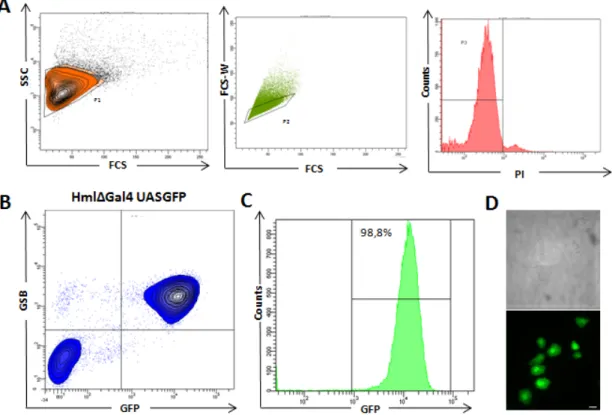
HmlΔGal4 line that only expresses GFP in plasmatocytes and crystal cells. HmlΔGal4 line is thought to drive expression of GFP in total population of plasmatocytes in circulation and tissue- bonded. When we sorted PI negative/GSB positive cells to a glass slide we only identified plasmatocytes (Fig. 4D). No crystal cells were observed in sorted cells during our experiments. If we plot GSB expression and GFP expression in the same graphic it’s possible to see that over 98% of cells events positive for GSB are also GFP positive (Fig. 4C). This result confirms that we can reproduce protocol of Tirouvaziam et al.27 .With this FACS protocol we were able to analyze expression of different genes in larval plasmatocytes and sort live cells at the end of analysis.
Figure 4 – FACS analysis of circulating plasmatocytes: (A) Chosen criteria to select live
single cells with a correspondence to plasmatocyte morphology, Side Scatter (SSC), Forward Scatter (FCS), Forward Scatter Width (FCS-W), Propidium Iodide (PI). (B) Analysis of GFP
expression in HmlΔGal4-UASGFP line (C) GFP expression of GSB-positive/PI-negative cells
(D) GSB-positive/PI-negative sorted cells. 100% of sorted cells correspond to plasmatocyte
13 Analysis of candidate genes with heterogeneous expression in plasmatocytes Expression of four genes was analyzed in larval plasmatocytes. Croquemort (Crq) is a receptor of apoptotic cells and its expression is essential during embryogenesis33. In 3th
stage larvae croquemort is not expressed in plasmatocytes (Fig. 5A). GFP protein that was detected in the first stage larvae (Fig. 3E) is probably a non-degraded protein that was produced during embryogenesis.
Serpent (Srp) is also essential during embryogenesis, playing an essential role in
hematopoiesis34. In late larval stage serpent is expressed in lymph gland cells but only
5% of circulating plasmatocytes express serpent (Fig. 5A). These serpent positive cells can be a small population of circulating plasmatocytes (with cell lineage relations) that did not repress expression of serpent or cells derived from lymph gland.
Peroxidasin (Pxn) is an extracellular protein expressed in plasmatocytes with functions
in phagocytosis and immune defense35. In 3th stage larva about 50% of cells express
peroxidasin (Fig. 5A,B). Expression of peroxidasin is variable not forming two clear populations. We did not continue the studies in this GAL4 line but for future analyses we have to consider the hypothesis of dividing positive population in peroxidasinhi and
peroxidasinlow to better describe cell variation.
Hemese (He) is a mediator of immune response36. In our analysis about 80% of cells
are positive for Hemese. In this reporter GFP expression is more discrete with two clear populations of positive and negative cells (Fig. 5A,B). This result was more appealing to us to continue in vitro assays.
Morphological analysis of hemese positive and hemese negative cells
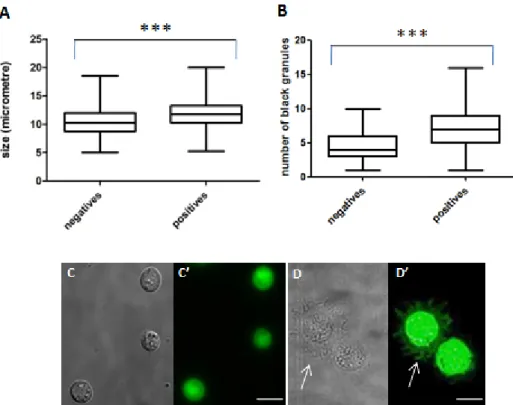
When sorted to a glass slide both hemese positive and hemese negative cells present the same type of morphology that correspond to previously descriptions of plasmatocytes16. These cells spread in slide with pseudopods and lamellopods (Fig.
6D). There is no size class that separates the two cell types, however, when we measure the longer axis of several cells (n=280), He positive cells are statistically larger than He negative cells (Fig. 6A). This happens because He positive cells spread more in glass. When in suspension, He positive cells are rounder and smaller (Fig. 6C). Other characteristic of plasmatocytes is the presence of phagolysosome–like dark inclusions. These dark inclusions are present both in hemese positive and negative cells. However positive cells tend to have a larger number of this inclusions ( ̴ 7) than negative cells ( ̴ 4) (Fig. 6B).
14
Figure 5- Analysis of 4 diferent GFP drivers in circulating plasmatocytes: (A) expression of
GFP in plasmatocytes (GSB positive cells) in Crq, Srp, Pxn and He Gal4 lines. (B) percentage
of plasmatocytes expressing GFP in Pxn and He lines in 5 independent assays.
15 Phagocytic activity of hemese positive and hemese negative cells
One important function of plasmatocytes in immune response is to phagocytize foreign bodies as pathogenic bacteria. Thus, we checked for phagocytic activity in both plasmatocytes subpopulations. It is possible to quench the fluorescence of heat-killed Alexa-594 conjugated bacteria with trypan blue. This molecule is excluded by live cells, therefore, phagocytized E.coli particles retain their fluorescence and is possible to count how many cells were phagocytized. In our short analysis of phagocytosis both
hemese positive and He negative cells phagocyte E.coli (Fig. 7A-C).
Phenoloxidase inhibition assay
Phenoloxidase activity is an important immune response to fight infections with the sub-products generated like ROS37. These products have an essential role in killing
bacteria but they can also harm the host. This way phenoloxidase activity is tightly controlled in Drosophila. Several proteins were identified that inhibit proteases involved
Figure 6 - Morphological analysis of sorted He positive and He negative plasmatocytes:
(A) size of the longer axis measured in each cell (n=280). (B) number of black granules
(secondary lysosomes) inside each plasmatocyte. (C) just after acquisition He positive
plasmatocytes present a round shape. (D) after 20 min incubation plasmatocytes spread in glass
16 in PO cascade. One appealing hypothesis for a function of plasmatocytes subpopulation is the regulation of an immune response such as melanization. Using a modified protocol from De Gregorio et al. we tested if hemese positive or hemese negative cells were responsible for Phenoloxidase activity inhibition or enhancement29.
When we collected hemolymph from larvae and incubated in buffer solution we saw an increase of PO activity (Fig.5). This was assessed with addition of 4-methilcatechol, a substrate of PO. 4-methylcatechol is catalyzed by PO to methyl-o-quinone, with higher OD. Neither when we added an excess of hemese positive cells or hemese negative cells activity of PO did neither increase nor diminished (Fig. 8A, B).
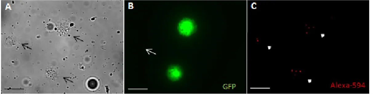
Figure 7 - Phagocytosis of fluorescence E.coli by w;HeGAL4;UASGFP line plasmatocytes: (A) three plasmatocytes were analyzed in this Bright Field area (black
arrows). (B) One plasmatocyte do not express GFP under control of He promoter (white
arrow). (C) All hemocytes in this field phagocytized E.coli (arrow heads). Scale bar 10μm
Figure 8 - Melanization inhibition assay: (A) means of five replicated measurements of Optic
Density (OD) during last 3 minutes of reaction. Methilcatechol with hemolymph (circles), 4-Methilcatechol with hemolymph and He positive plasmatocytes added (squares), 4-4-Methilcatechol with hemolymph and He negative plasmatocytes added (triangles) and 4-Methilcatechol without hemolymph (inverted triangles). (B) analysis of 5 replicates in time point 10 (5 min of reaction).
Each condition is significantly different from blank control meaning that we can detect phenoloxidase activity in hemolymph solution. Adding of hemese positive or hemese negative plasmatocytes do not increase nor diminish phenoloxidase activity significantly.
17
Discussion
Results presented here indicate that circulating plasmatocytes do not constitute a homogeneous population of cells in Drosophila melanogaster larvae regarding gene expression. Some hypotheses are proposed in this discussion to direct future research on plasmatocytes subpopulations function. Further studies are needed to complement our analysis and better describe plasmatocyte heterogeneity.
Serpent expression is restricted to a small population of plasmatocytes in 3th larvae
hemolymph. We did not proceed with analysis of this subpopulation; however, an interesting hypothesis rises. This small population of cells could be a potential pool of undifferentiated cells ready to proliferate upon infection since serpent is a marker of undifferentiated hemocytes. It is also possible that serpent positive circulating cells in 3th stage larvae are plasmatocytes derived from lymph gland, where hemocytes are
expressing serpent. This hypothesis is supported by the fact that after pupation a big number of serpent positive cells are found in hemolymph, just right after lymph gland disruption (António Jacinto, personal communication). If plasmatocytes are “leaking” in the lymph gland it would be interesting to investigate if these cells have a physiologic important role in immune system. Other possibility is that these cells come from lymph gland as an artifact of our protocol, when we are collecting hemolymph. This way, when we collect Drosophila’s hemocytes in larval stages we can be collecting a small fraction of cells derived from lymph gland hematopoiesis. Nevertheless, the number of
serpent positive cells in 3th stage larvae is too reduced to have a big impact in the
interpretation of our data.
In our FACS analysis we found that hemolectin is expressed in ̴ 99% of plasmatocytes. HmlΔGAL4 line is, this way, the best described reporter to track plasmatocytes. We never saw crystal cells after sorting. This probably occurs because crystal cells are very reactive and just after hemolymph collection they disrupt their cell membrane38. It is important to notice that about 1% of plasmatocytes do not express
hemolectin reporter. This could explain the small number of plasmatocytes found in
larvae hemolymph by Charroux et al. after genetic ablation of cells expressing Hml17.
Plasmatocytes present in embryo do not express hemolectin. Only before hatching it is possible to distinguish a population of hemolectin positive cells that increase in percentage during larva development26. Switch on of hemolectin expression seems to
18 embryogenesis. Hematopoiesis seems to be a continuous process in embryo and larva. Other evidence for this is croquemort expression pattern. In larva, croquemort is highly up-regulated33. In 3th stage larvae we didn’t find any expression of croquemort in
plasmatocytes. This switch of gene expression in embryo to larva transition is an interesting developmental problem per se because hemocytes constitute a non connected tissue and still they continue the same developmental program. In our discussion it is important to keep this observation in mind because we will only focus on 3th stage larvae plasmatocytes. It will be important to extend this analysis to other
stages in Drosophila life cycle to fully understand heterogeneity of plasmatocytes.
Peroxidasin expression is not homogeneous in larva’s circulating plasmatocytes. In
FACS analysis it is possible to distinguish a GFP negative population and a population expressing different levels of GFP. We can separate plasmatocytes in peroxidasin positive and peroxidasin negative but it would be a better description if we separate the positive population in high-expression and low-expression. In cuticle adjacent plasmatocytes it is also possible to find positive and negative plasmatocytes populations for peroxidasin marker (António Jacinto, personal communication). Therefore, peroxidasin positive and negative subpopulations are not exclusive of circulating plasmatocytes.
Hemese is expressed in 80% of circulating plasmatocytes and is absent in 20%. In
this case it is possible to clear distinguish a positive and a negative population. It will be important to understand if these subpopulations of cells are lineage specific or are defined as a response mechanism. We tend to favor the first hypothesis since subpopulations maintained their relative percentage in several independent analyses. Nevertheless additional tests are needed to justify this view. Hemese positive cells spread more in glass slides ending up with a longer axis than hemese negative cells. Some controversy is still found in literature about morphological classification of plasmatocytes. Although most of the times they are considered a homogeneous population some authors consider the split of “classical plasmatocyte cells” in podocytes and plasmatocytes39. Their argument is that some plasmatocytes have more
and longer philapodia. Probably this is only noticeably with higher amplification has it is given by electron microscopy. This would be an important future analysis to characterize the two plasmatocytes subpopulations and see if this classification fits with our data.
One possibility to explain the existence of two different plasmatocytes types is that positive cells are mature plasmatocytes while negative ones are immature cells. Nevertheless, we have one observation that goes against this hypothesis. In the in vitro phagocytosis assay it was possible to confirm that both plasmatocytes subpopulations
19 are capable to phagocytize E. coli cells. Thus, He negative plasmatocytes constitute a subpopulation of mature cells capable to recognize and phagocytize foreign cells. This assay only confirmed that both subpopulations are capable to phagocytize bacteria cells in vitro, not the phagocytic rate of the two subpopulations, nor the phagocytic competence in vivo, something that has to be addressed in the future. It will be also important to test if both subpopulations are cable to phagocytize others microorganisms as yeasts and different species of Gram positive and Gram negative bacteria. We know that activation of AMP production in fat body is somehow specific to pathogen species. Toll mediated response is mainly activated when host is infected with a Gram positive bacteria or yeast and Imd (immune deficiency) pathway when infected with a Gram negative bacteria. Each subpopulation of plasmatocytes could be more readily to respond depending on type of infection.
The main hypothesis that motivated this research can be stated like this: is there a subpopulation of plasmatocytes responsible for modulation of immune response? One crucial process after pathogen clearance is the turn off immune response40. The
constitutively activation of an immune response can have deleterious effects. We asked if a subpopulation of plasmatocytes is responsible for immune response turn off. To test this broad hypothesis we had to choose a specific immune response for functional assays. We chose melanization response taking into account theoretical and practical reasons. In a practical point of view phenoloxidase inhibition assay was established in community, and so, it was possible for us to compare results. On the other hand, melanization is, theoretically, a response that needs a tightly regulation. Without negative regulation of melanization infected larvae die upon infection with overmelanized bodies29. Several studies found proteins important in inhibition of
phenoloxidase activity29,41,42. However, we still don’t know where those proteins are
expressed or how they are regulated. Thus, for us it was an appealing hypothesis to consider a subpopulation of plasmatocytes to be responsible for regulation of phenoloxidase activity. Our in vitro assay did not support this hypothesis. Nevertheless, this is not a clear rejection of our hypothesis. Activation of phenoloxidase is artificial in this assay and we don’t know to each extend is activated. It will be crucial to test melanization response in vivo with larvae that lacks hemese positive cells or hemese negative cells. To achieve this goal we have to develop a genetic construct to eliminate subpopulation plasmatocytes or improve cell transfer in Drosophila larvae.
Several other hypotheses were raised during our experiments. For example, if one of the subpopulation is responsible for signaling to the fat-body. Other open possibility is a role of different subpopulations in development since plasmatocytes are crucial in embryo development.
20 In conclusion, findings reported here alert us to the fact that plasmatocytes aren’t a homogeneous population of cells. We think that this observation must be taken into account in future studies of Drosophila immune system. Different plasmatocytes subpopulations may be responding differently in immune responses. The next crucial step is to investigate what are the functions of these subpopulations. It is possible that some of the plasmatocytes functions are functionally divided in subpopulations. On the other hand, subpopulations of plasmatocytes may be responsible for functions that we currently don’t know. Moreover our observations may generate question in other research fields. For example, if our different subpopulations of cells are lineage specific it would be interesting to investigate how are they formed.
21
References
1. Kuby, J. Immunology, xx, 660 p., [8] p. of plates (W.H. Freeman, New York, 1994).
2. Tauber, A.I. Metchnikoff and the phagocytosis theory. Nat Rev Mol Cell Biol 4, 897-901 (2003).
3. Kaufmann, S.H. Paul Ehrlich: founder of chemotherapy. Nat Rev Drug Discov 7, 373 (2008).
4. Lavine, M.D. & Strand, M.R. Insect hemocytes and their role in immunity. Insect
Biochem Mol Biol 32, 1295-309 (2002).
5. Ha, E.M., Oh, C.T., Bae, Y.S. & Lee, W.J. A direct role for dual oxidase in Drosophila gut immunity. Science 310, 847-50 (2005).
6. Lemaitre, B. & Hoffmann, J. The host defense of Drosophila melanogaster.
Annu Rev Immunol 25, 697-743 (2007).
7. Leclerc, V. et al. Prophenoloxidase activation is not required for survival to microbial infections in Drosophila. EMBO Rep 7, 231-5 (2006).
8. Jiang, H., Wang, Y. & Kanost, M.R. Pro-phenol oxidase activating proteinase from an insect, Manduca sexta: a bacteria-inducible protein similar to Drosophila easter. Proc Natl Acad Sci U S A 95, 12220-5 (1998).
9. Cerenius, L., Lee, B.L. & Soderhall, K. The proPO-system: pros and cons for its role in invertebrate immunity. Trends Immunol 29, 263-71 (2008).
10. Tepass, U., Fessler, L.I., Aziz, A. & Hartenstein, V. Embryonic origin of hemocytes and their relationship to cell death in Drosophila. Development 120, 1829-37 (1994).
11. Rehorn, K.P., Thelen, H., Michelson, A.M. & Reuter, R. A molecular aspect of hematopoiesis and endoderm development common to vertebrates and Drosophila. Development 122, 4023-31 (1996).
12. Holz, A., Bossinger, B., Strasser, T., Janning, W. & Klapper, R. The two origins of hemocytes in Drosophila. Development 130, 4955-62 (2003).
13. Lanot, R., Zachary, D., Holder, F. & Meister, M. Postembryonic hematopoiesis in Drosophila. Dev Biol 230, 243-57 (2001).
14. Beckage, N. Insect Immunology, 384 (Academic Press, 2007).
15. Kocks, C. et al. Eater, a transmembrane protein mediating phagocytosis of bacterial pathogens in Drosophila. Cell 123, 335-46 (2005).
16. Ribeiro, C. & Brehelin, M. Insect haemocytes: what type of cell is that? J Insect
Physiol 52, 417-29 (2006).
17. Charroux, B. & Royet, J. Elimination of plasmatocytes by targeted apoptosis reveals their role in multiple aspects of the Drosophila immune response. Proc
Natl Acad Sci U S A (2009).
18. Marmaras, V.J. & Lampropoulou, M. Regulators and signalling in insect haemocyte immunity. Cell Signal 21, 186-95 (2009).
19. Buyukguzel, E., Tunaz, H., Stanley, D. & Buyukguzel, K. Eicosanoids mediate Galleria mellonella cellular immune response to viral infection. J Insect Physiol 53, 99-105 (2007).
20. Markus, R. et al. Sessile hemocytes as a hematopoietic compartment in Drosophila melanogaster. Proc Natl Acad Sci U S A 106, 4805-9 (2009).
21. Sorrentino, R.P., Carton, Y. & Govind, S. Cellular immune response to parasite infection in the Drosophila lymph gland is developmentally regulated. Dev Biol 243, 65-80 (2002).
22. Pathak, J.P.N. Insect immunity, 169 (Kluwer Academic Publishers, 1993). 23. Hillyer, J.F. & Christensen, B.M. Characterization of hemocytes from the yellow
22 24. Kurucz, E. et al. Definition of Drosophila hemocyte subsets by cell-type specific
antigens. Acta Biol Hung 58 Suppl, 95-111 (2007).
25. Kurucz, E. et al. Nimrod, a putative phagocytosis receptor with EGF repeats in Drosophila plasmatocytes. Curr Biol 17, 649-54 (2007).
26. Goto, A., Kadowaki, T. & Kitagawa, Y. Drosophila hemolectin gene is expressed in embryonic and larval hemocytes and its knock down causes bleeding defects. Developmental Biology 264, 582-591 (2003).
27. Tirouvanziam, R., Davidson, C.J., Lipsick, J.S. & Herzenberg, L.A. Fluorescence-activated cell sorting (FACS) of Drosophila hemocytes reveals important functional similarities to mammalian leukocytes. Proc Natl Acad Sci U
S A 101, 2912-7 (2004).
28. Abramoff, M.D., Magelhaes, P.J., Ram, S.J. Image Processing with ImageJ.
Biophotonics International 11, 36-42 (2004).
29. De Gregorio, E. et al. An immune-responsive Serpin regulates the melanization cascade in Drosophila. Dev Cell 3, 581-92 (2002).
30. Duffy, J.B. GAL4 system in Drosophila: a fly geneticist's Swiss army knife.
Genesis 34, 1-15 (2002).
31. Sies, H. Glutathione and its role in cellular functions. Free Radic Biol Med 27, 916-21 (1999).
32. Harrison, D.A., Binari, R., Nahreini, T.S., Gilman, M. & Perrimon, N. Activation of a Drosophila Janus kinase (JAK) causes hematopoietic neoplasia and developmental defects. EMBO J 14, 2857-65 (1995).
33. Franc, N.C., Heitzler, P., Ezekowitz, R.A. & White, K. Requirement for croquemort in phagocytosis of apoptotic cells in Drosophila. Science 284, 1991-4 (1999).
34. Waltzer, L., Ferjoux, G., Bataille, L. & Haenlin, M. Cooperation between the GATA and RUNX factors Serpent and Lozenge during Drosophila hematopoiesis. EMBO J 22, 6516-25 (2003).
35. Nelson, R.E. et al. Peroxidasin: a novel enzyme-matrix protein of Drosophila development. EMBO J 13, 3438-47 (1994).
36. Kurucz, E. et al. Hemese, a hemocyte-specific transmembrane protein, affects the cellular immune response in Drosophila. Proc Natl Acad Sci U S A 100, 2622-7 (2003).
37. Tang, H., Kambris, Z., Lemaitre, B. & Hashimoto, C. Two proteases defining a melanization cascade in the immune system of Drosophila. J Biol Chem 281, 28097-104 (2006).
38. Rizki, M.T. & Rizki, R.M. Functional significance of the crystal cells in the larva of Drosophila melanogaster. J Biophys Biochem Cytol 5, 235-40 (1959).
39. Shrestha, R. & Gateff, E. Utrastructure and cytochemistry of the cell types in the larval hematopoietic organs and hemolymph of Drosophila melanogaster.
Development Growth and Differentiation 24, 65-82 (1982).
40. Schneider, D.S. How and why does a fly turn its immune system off? PLoS Biol 5, e247 (2007).
41. Nappi, A.J., Frey, F. & Carton, Y. Drosophila serpin 27A is a likely target for immune suppression of the blood cell-mediated melanotic encapsulation response. J Insect Physiol 51, 197-205 (2005).
42. Tang, H., Kambris, Z., Lemaitre, B. & Hashimoto, C. A serpin that regulates immune melanization in the respiratory system of Drosophila. Dev Cell 15, 617-26 (2008).