Ricardo André Baptista Pereira da Costa
Mestre em Física
The role of succinate dehydrogenase
in the
Drosophila melanogaster
intestinal stem
cells and tissue homeostasis
Dissertação para obtenção do Grau de Doutor em Bioengenharia (MIT-Portugal)
Orientador: António Coelho Jacinto, Professor Catedrático Convidado, CEDOC – FCM/UNL
Co-orientador: Manuel Nunes da Ponte,
Professor Catedrático, FCT/UNL
Júri:
Presidente: Prof. Doutor José Paulo Barbosa Mota
Arguentes: Doutora Catarina de Cértima Fernandes Homem Doutora Zita Carvalho dos Santos
Vogais: Prof. Doutor António Alfredo Coelho Jacinto Prof. Doutor Cláudio Emanuel Moreira Gomes Doutor Miguel Jorge Zuzarte de Mendonça Godinho Ferreira
Doutora Rita Oliveira Teodoro
Ricardo André Baptista Pereira da Costa
The role of succinate dehydrogenase in the
Drosophila melanogaster
intestinal stem
cells and tissue homeostasis
Submitted to the graduate degree program in Bioengineering and the Graduate Faculty of the Nova University of Lisbon
in partial fulfillment of the requirements for the degree of Doctor of Philosophy.
Orientador: Prof. António Coelho Jacinto, CEDOC/NMS-UNL Co-orientador: Prof. Manuel Nunes da Ponte, FCT-UNL
September, 2017
iv
The role of succinate dehydrogenase in the Drosophila melanogaster intestinal stem cells
and tissue homeostasis
Copyright © Ricardo Pereira da Costa, Faculdade de Ciências e Tecnologia, Universidade Nova de Lisboa.
v In 2011, as a recent physics postgraduate with no background in biology, I wanted to engage in a biotechnological field and joined the MIT-Portugal Program in Bioengineering. During the first semester of classes, I developed a strong interest in fundamental biology and decided I wanted to work with stem cells in Drosophila. Later in 2012 I have decided to join the Laboratory of Dr. Leanne Jones (UCLA) and while waiting for my visa approval, I was kindly invited by a member of my thesis committee, Dr. Miguel Godinho Ferreira, to rotate in his laboratory working on a project with zebrafish to gain more experience in a biology laboratory setting.
I finally joined the Dr. Jones Lab at UCLA in April 2013, on a 2-year term, where I started a project to study micro-RNAs in adult stem cells, which after 7 months we have decided to abandon due to unforeseen events, and I then started the project described in this thesis. After the end of my stay at UCLA, in April 2015, my adviser Professor António Jacinto accepted me in his lab at CEDOC/NMS to continue my project. During that period, as my scholarship ended, I had the pleasure to tutor school students in mathematics and physics until recently. In the meanwhile, in 2016, I initiated a fruitful collaboration with the Environmental and Biological Mass Spectrometry Group at Faculdade de Ciências da Universidade de Lisboa, where I learned to use gas chromatography—mass spectrometry to quantify metabolites in my biological samples, and finalized my project.
Declaration:
The experiments described in this thesis were performed in the Jones Laboratory at University of California, Los Angeles; the Tissue Morphogenesis and Repair Lab at CEDOC/NMS, Universidade Nova de Lisboa; and at the Environmental and Biological Mass Spectrometry Group at the Departamento de Química e Bioquímica, DQB, of Faculdade de Ciências da Universidade de Lisboa. All the experimental work was performed by me, with the exception of 2 groups of samples in the screen presented in part 1 of the Results and Discussion chapter — the lines 106700 KK and 106095 KK — that were dissected and
immuno-stained by R. Demarco at the Jones Lab, UCLA.
vii I would like to express my strong gratitude to Professor Leanne Jones for accepting me in her laboratory at UCLA. Dr. Jones took many chances with me, I was a student with a limited background in biology and as we learned that I could only stay at UCLA for 2 years, Dr. Jones maintained her support and showed extraordinary commitment. I am also very grateful to have worked with the other members of the Jones laboratory, from whom I have learned so much. Particularly, I would like to express my special gratitude to Rafael Demarco for enthusiastic mentorship, support and impromptu biology lessons; and also to Pedro Resende for his support and introducing me to the lab and the city of Los Angeles. I also feel thankful to have had the opportunity to learn from the other talented members of the MCDB department, namely the scientists at the Walker Lab, who I thank for valuable scientific and technical feedback and reagents.
I am greatly indebted to Prof. António Jacinto for accepting me in his laboratory and providing me with everything I needed to complete my doctoral work. I also need to express my heart-warmed appreciation to the other past and present members of the AJ Lab for a warm welcome, kindness and valuable feedback.
I am also thankful to the GEMAB group at FCUL namely Dr. Ana Marques, for teaching me how to use the GC-MS equipment and particularly grateful to Professor Carlos Borges for his openness in accepting me in his laboratory and trusting me with the equipment.
I would also like to extend my gratitude to all the coordinators and members of the MIT-Portugal Program, namely to José Silva Lopes and Fátima Silva Lopes for being very helpful with all the administrative needs, and to all the different universities, institutes, laboratories, and companies that we have interacted with along the way. I am also very grateful to have had the company and support of my colleagues in the doctoral program, as well as peers from other programs, namely the BEB PhD programme in Coimbra.
I would like to thank the members of my thesis committee, Prof. Manel Nunes da Ponte, Dr. Rita Teodoro and Dr. Miguel Godinho Ferreira for spending their valuable time in the process, and the enthusiastic support from Miguel in giving me my first insights into the biology of aging. I would also like to acknowledge the financial support from Fundação para a Ciência e a Tecnologia.
And finally, a very special gratitude to my family and all my friends and loved ones for support and encouragement during these years, to whom I dedicate this thesis.
ix Adult stem cells maintain tissue homeostasis and act in response to challenges such as infection or mechanical damage. The fruit fly Drosophila melanogaster has emerged as a powerful model to study adult stem cells due to its conserved pathways and the existence of different stem cell systems, particularly a stem cell niche in the intestine. Intestinal stem cells (ISCs) have a major role in maintaining tissue homeostasis and improvements in their function result in refined tissue function in aged or damaged organs. Metabolism plays an important role in regulating stem cell activity, influencing cellular events such as differentiation and proliferation. However, several aspects remain unraveled, particularly the role of different subunits of the mitochondrial electron transport chain (ETC) — end players in the oxidative phosphorylation process.
Given the importance of metabolism in regulating stem cell activity, we hypothesized that ETC subunits also have a role in regulating ISC activity and tissue homeostasis. To test our hypothesis, we performed a candidate screen to knock down individual subunits of the ETC specifically in ISCs and their direct progeny — enteroblasts — in the adult fly and studied their requirements for normal cell division, differentiation, survival and impact in surrounding differentiated cells. Subunits of the ETC complex II, which converts succinate to fumarate in the Krebs cycle, particularly subunit D (SdhD), emerged from the screen as strong candidates required for normal ISC activity.
Knockdown of SdhD in ISCs resulted in inhibition of cell division, hypertrophy with polyploidy, and ultimately, cell death. At a tissue level, evidence of a differentiation bias towards secretory enteroendocrine cells in lieu of absorptive enterocytes was observed upon SdhD knockdown in progenitor cells. Further experiments showed that knockdown of SdhD causes succinate to accumulate, possibly due to a decrease in the function of succinate dehydrogenase activity. Succinate is a known ligand for the GPR91 receptor in mammals which is known to be involved in cellular hypertrophy and death via apoptosis. Further studies need to be performed to determine the existence of a succinate receptor in
Drosophila.
This work has unveiled a direct relationship between inhibition of complex II subunits and stem cell hypertrophy and death, with succinate as a possible intermediate, and provides a suitable model for the study of the molecular pathways underlying cellular
x
hypertrophy and death in metabolic-deficiency backgrounds. These insights may contribute to the understanding of hypertrophic pathologies, or proliferative diseases, such as cancer.
xi As células estaminais existem em organismos adultos onde possuem um importante papel na manutenção da homeostase e na resposta a danos no tecido. A mosca-da-fruta
Drosophila melanogaster destaca-se como um modelo animal no estudo de células estaminais, graças aos seus diferentes mecanismos moleculares conservados entre espécies e à presença de células estaminais adultas, nomeadamente de um nicho de células estaminais no intestino. As células estaminais intestinais (ISCs, na sigla inglesa) desempenham um papel crucial na regulação da homeostase no tecido, e vários estudos demonstram que melhorias na sua função celular resultam em melhorias na capacidade dos tecidos num contexto de envelhecimento ou detioração de tecidos. O metabolismo desempenha um papel crucial na regulação do comportamento e actividade de células estaminais através de moléculas metabólicas intermediárias e produtos de reacção, que controlam como estas se diferenciam ou dividem. No entanto, ainda existem vários aspectos a serem estudados, nomeadamente o papel de diferentes subunidades dos complexos da cadeia de respiração mitocondrial (ETC, na sigla inglesa).
Dada a importância do metabolismo na regulação da actividade de células estaminais, levantámos a hipótese de que subunidades da ETC desempenham um papel na regulação do comportamento das ISCs, com um impacto na homeostase no tecido. Para testar esta hipótese, induzimos subexpressão de diferentes subunidades da ETC em células progenitoras no intestino (incluindo ISCs) por vias de interferência de RNA, e avaliámos os efeitos no comportamento de células progenitoras e o consequente papel no tecido intestinal. Subunidades do complexo II, nomeadamente a succinato dehidrogenase, subunidade D (SdhD) destacou-se entre as várias, por ser requerida em ISCs.
Subexpressão de SdhD inibe a replicação de células estaminais, induz poliploidia, hipertrofia e por fim, morte celular. A nível de impacto no tecido intestinal, as células progenitoras sofrem uma mudança de comportamento, notando-se uma preferência na diferenciação em células enteroendócrinas em vez de enterócitos.
Análises quantitativas indicaram que subexpressão da SdhD causa uma acumulação de succinato no tecido. O succinato é um activador conhecido do receptor acoplado à proteína G 91 (GPR91), que foi em estudos prévios associado a hipertrofia e apoptose.
Os resultados obtidos neste trabalho contribuíram para um melhor entendimento relativamente aos mecanismos de regulação da actividade celular por processos
xii
metabólicos, demonstrando que a Drosophila melanogaster é um modelo importante para o seu estudo. Uma melhor compreensão acerca dos processos de regulação de hipertrofia e replicação celular poderá ter um impacto positivo no desenvolvimento de terapias para doenças relacionadas com hipertrofia, ou com replicação celular, como o cancro.
Termos chave: células estaminais intestinais, metabolismo, hipertrofia, morte celular,
xiii
Preface ... v
Acknowledgements ... vii
Abstract ... ix
Resumo ... xi
Table of contents ... xiii
List of Figures ... xv
List of Tables ... xvii
List of Abbreviations ... xix
Introduction ... 1
1- Stem cells: role in development and tissue repair ... 3
2-The intestine of Drosophila melanogaster ... 8
3- Role of metabolism in stem cell activity ... 15
3.1- Energy production in Metazoa ... 15
3.2- Metabolic regulation of stem cell activity ... 18
3.3- Metabolism in the Drosophila intestinal progenitor cells ... 21
3.4- Implications of the electron transport chain in cell maintenance and organismal longevity ... 23
4- Aims and outline of the thesis: ... 27
Materials and methods ... 30
Fly husbandry and stocks ... 31
Immuno-fluorescence ... 34
qRT-PCR ... 34
Assessment of oxidative state using the mito-roGFP probe ... 36
Imaging and cell counting ... 36
Apoptag kit for detection of apoptosis (TUNEL assay) ... 37
Quantification of succinate by GC-MS ... 37
Results and discussion ... 43
I - Screen for electron transport chain subunits with an impact in progenitor cell activity 45 1.1- Knockdown of succinate dehydrogenase subunit D in progenitor cells causes a decrease in its population ... 50
1.2- qRT-PCR quantification confirms downregulation of SdhD transcripts upon RNAi expression ... 55
II – Effects of SdhD knockdown in ISCs and EBs ... 57
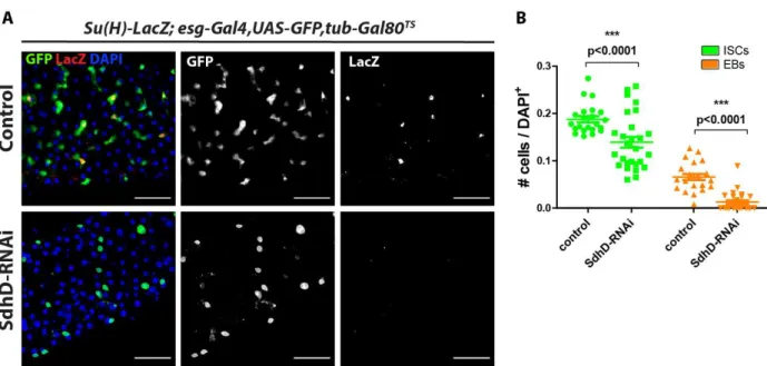
2.1- SdhD is required in intestinal stem cells, but not enteroblasts, to maintain progenitor cell population ... 57
2.2- SdhD is required for normal self-renewal rates in ISCs ... 61
2.3- SdhD is required for ISC survival ... 64
2.4- Loss of SdhD triggers hypertrophy in intestinal stem cells ... 69
III- Effects of progenitor cell-specific SdhD knockdown in differentiated cells ... 73
SdhD-RNAi in progenitor cells leads to an increased population of enteroendocrine cells ... 73
IV- Mechanisms of action ... 75
xiv
4.1- No evidence of ROS accumulation upon SdhD knockdown ... 75
4.2- Mitotic catastrophe ... 78
4.3- The importance of the enzymatic activity of succinate dehydrogenase ... 79
4.4– Succinate as a driver of hypertrophy and cellular death ... 81
General Discussion ... 87
Conclusion ... 93
Supplementary data ... 97
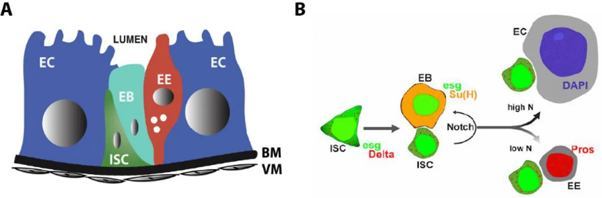
xv Figure 1: Stem cell niche morphology and cell lineage specification in the midgut of
Drosophila melanogaster ... 11
Figure 2: Krebs cycle and the electron transport chain ... 17
Figure 3: Derivatization of succinic acid ... 38
Figure 4: Candidate screen workflow diagram ... 46
Figure 5: Results of candidate screen for regulators of progenitor cell activity ... 49
Figure 6: Effects of SdhD knockdown in progenitor cells using the 5961GSdriver ... 50
Figure 7: Effects of SdhD knockdown in progenitor cells using the esg-gal4 driver ... 51
Figure 8: Progression of the SdhD knockdown phenotype using the esg-gal4 driver ... 53
Figure 9: Levels of SdhD and PTPMT1 mRNA upon RNA-interference against SdhD ... 56
Figure 10: Effects of ISC-specific SdhD-knockdown in progenitor cells ... 58
Figure 11: Effects of EB-specific SdhD-knockdown in the abundance of EBs ... 60
Figure 12: Effects of SdhD knockdown in the rates of ISC division ... 62
Figure 13: ISCs depleted of SdhD show hallmarks of cell death ... 67
Figure 14: Knockdown of SdhD causes hypertrophy in ISCs ... 70
Figure 15: Cell cycle abbreviations in different cell types ... 70
Figure 16: Effects of SdhD knockdown in differentiation choices ... 74
Figure 17: Analysis of the oxidative environment in mitochondria using the mito-roGFP probe ... 76
Figure 18: Effects of SOD2-RNAi in the abundance of intestinal progenitor cells ... 77
Figure 19: Effects of SdhA knockdown in intestinal progenitor cells ... 80
Figure 20: Levels of succinate in whole flies upon ubiquitous SdhD knockdown ... 85
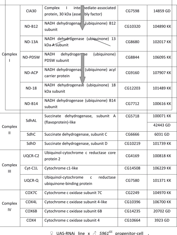
xvii Table 1: List of RNAi lines selected for the candidate screen ... 45 Supplemental Table 1: BLAST alignment results of human GPR91 and the Drosophila melanogaster proteome……….………...91
xix β-Gal β-galactosidase
DAPI 4',6-diamidino-2-phenylindole
EB Enteroblast
EC Enterocyte
EE Enteroendocrine cell
EGFR Epidermal growth factor receptor
EM Electron microscopy
ETC Electron transport chain
EtOH Ethanol
Esg Escargot
FAD Flavin adenine dinucleotide
FOV Field of view
GFP Green fluorescent protein
GTP Guanosine triphosphate
ISC Intestinal stem cell
JNK c-Jun N-terminal kinase
MAPKs Mitogen-activated protein kinases
MPCs Mitochondrial pyruvate carriers
NAD Nicotinamide adenine dinucleotide
OXPHOS Oxidative phosphorylation
qRT-PCR Quantitative real-time reverse transcription-polymerase chain reaction
Pros Prospero
Rpr Reaper
RNA Ribonucleic acid
RNAi RNA-interference
ROS Reactive oxigen species
SdhA Succinate dehydrogenase, subunit A
SdhC Succinate dehydrogenase, subunit C
SdhD Succinate dehydrogenase, subunit D
SEM Standard error of the mean
Su(H) Suppressor of hairless
TCA Tricarboxylic acid cycle
Tub Tubulin
UAS Upstream activating sequence
YFP Yellow fluorescent protein
1
3
Introductory remarks
In part 1 of his chapter, I introduce the different types of stem cells and their role in development and tissue maintenance. In the second part, I describe the intestine of the fruit-fly Drosophila melanogaster as an adult stem cell niche model and the conserved mechanisms that regulate homeostasis and stress responses. In part 3, I present the basis of metabolic regulation of stem cells, explore the existing gaps in the field, and describe the workplan to answer some of the pending questions.
1- Stem cells: role in development and tissue repair
Stem cells are characterized by their ability to divide through mitosis to produce more stem cells (self-renewal) and differentiate to a specific cell lineage. These traits allow them to build new tissue and organs during development where a great number of new cells is required, and to provide tissue homeostasis or regeneration during adulthood [1][2].
Stem cells are divided into three main categories: embryonic stem cells (ESCs), adult stem cells (ASCs; also referred to as somatic stem cells), and induced pluripotent stem cells (iPSCs). Early embryonic stem cells in the morula are totipotent, as they can give rise to all cell types in the body, including the extraembryonic, or placental cells. Upon formation of the blastocyst, the inner mass cells are considered to be pluripotent, as they generate the 3 types of embryonic germ layers: ectoderm, mesoderm and endoderm. During development, these cells become more restricted in their ability to differentiate into multiple lineages. In
adults, where stem cells are only capable of forming a limited number of cell types, they are considered to be multipotent or unipotent [3][4][5]. ASCs are found in different tissues of adult organisms across species. The most studied in mammals are hematopoietic stem cells (HSCs) and mesenchymal stem cells (MSCs), which have a lower self-renewal potential than ESCs [6]. Germline stem cells are usually considered to be ASCs. The third category of stem cells, iPSCs, is obtained from mature adult cells, normally fibroblasts, that were artificially reprogrammed in vitro to an ESC-like state mainly through overexpression of core stemness regulators [7], which are detailed below.
Embryonic stem cells
4
or intestinal lineages [13]. As such, ESCs have a high therapeutic potential to repopulate and repair damaged or degenerating tissues.
Induced pluripotent stem cells
iPSCs are generated from differentiated cells by expressing core transcription factors that are required for maintenance of pluripotency and proliferation of ESCs. In their 2006 groundbreaking study, Yamanaka and colleagues reprogrammed murine fibroblasts to iPSCs by retroviral-mediated expression of the ‘stemness’-associated factors OCT4 (octamer-binding transcription factor 4), SOX2 (SRY (sex-determining region Y)-box 2), KLF4
(Kruppel-like factor 4), and C-MYC [14]. In 2007, Yu and colleagues generated mice iPSCs by expressing
a different set of factors, OCT4, SOX2, NANOG, and LIN28, using a lentiviral system [15].
Following these accomplishments, researchers were also able to generate iPSCs derived from
human somatic cells [16]. iPSCs exhibit many traits similar to ESCs, such as morphology, cell-surface markers, telomerase activity and epigenetic marks, but they differ in global gene expression [7].
Adult stem cells
Most adult tissues contain reservoirs of stem cells used to replenish differentiated cells that are lost upon tissue injury, infection or normal cell turnover. Upon stress, differentiated cells send a variety of signals to stem cells to regulate their replication and differentiation dynamics [2][17]. In some organisms, upon injury, fully differentiated cells can also de-differentiate to stem cells in order to fulfill the requirements of stem cell
population to provide the tissue with newly formed cells [1]. In the absence of injury, ACSs continue to generate new cells to replace those that have worn out, in order to maintain tissue homeostasis. While many of the pathways responsible for ASC activation are shared with those in pluripotent stem cells during development, the dynamics of cell communication and proliferation are distinct, as are the types of cells involved. Studies in
mice and humans have shown the existence of different ASC types across the organism, including in the bone marrow [18], skeletal muscle [19], skin epithelia [20], intestine [21], heart [22][23], brain [24][25], and lungs [6][26]. These cells are used sparingly and are generally tucked away in protected niches [6]. They are also scarce, and with the relative exception of HSCs, are difficult to isolate.
HSCs produce all the different cells present in the mammalian blood, including platelets, red blood cells and white blood cells. These cells and their progenitors can be
5 including myocytes, chondroblasts, fibroblasts, osteoblasts and adipocytes. MSCs are usually isolated from bone marrow and umbilical cord blood and display a different expression
profile of cell surface markers than that of HSCs [7].
The stem cell nice
ASCs are often tucked away in niches, physically protected from potential damage, either mechanical or from pathogens. The stem cell niche also comprises a pool of intricate signals occurring between the stem cells and their environment, that are required for proper tissue homeostasis and response to damage. In the niche, ASCs are largely quiescent, i.e., they display low proliferation and differentiation rates. The germline of Drosophila was one of the first stem cell niches to be characterized [27]. In mammals, the bone marrow is an example of a niche, where HSCs reside [6]. The concept of the niche is better perceived in part 2 of this chapter in the context of the Drosophila intestinal epithelium.
Applications
Due to their potential to self-renew, possibility of in vivo growth and patient specificity, stem cells have a notable potential for regenerative medicine. Animal models and clinical studies have shown that transplantation of stem cells from diverse origins can successfully treat many acute and chronic diseases. Bone marrow transplantation is the most common example, used to treat aplastic anemia, leukemias, immune deficiency and
hemoglobin disorders. The potential for stem cell therapy extends to many disorders, including myocardial infarction, Parkinson disease, myelin disease and liver failure [6][28].
iPSCs present a high potential for transplantation treatments as they have the same genetic signature as other cells in the patient, greatly reducing chances of immune rejections, and
can be genetically corrected [16].
To fully explore the therapeutic potential of stem cells it is essential to understand the mechanisms that regulate their activity. Dissecting the mechanisms of proliferation is also of great importance for the development of targeted therapies against tumor progression, as cancer cells share many common replication pathways with stem cells [29][30]. The mechanisms responsible for stem cell division and differentiation are largely conserved among species, and as such, several animal models are used to study stem cell-mediated development, regeneration and homeostasis, as described below.
Non-mammalian models of development and regeneration
6
insights about morphological patterning during development and stem cell activation in adults. Amphibians such as urodeles (salamanders or axolotls) or anurans (frogs or toads) have high regeneration potential and have been studied as models for limb repair. As tadpoles, anurans are able to regenerate limbs and tails but this capacity to regenerate
rapidly declines during metamorphosis [5]. Zebrafish has emerged as a powerful model organism to study vertebrate development and regeneration [17][31]. Zebrafish is able to regenerate retina, heart and the fin, which has been extensively used a model for regeneration. ASCs have also been characterized in the intestine and bone marrow. Regeneration in zebrafish may rely on stem cell activation, or dedifferentiation of surrounding cells. Zebrafish offers several advantages such a short generation time, live imaging and fast regeneration of the caudal fin upon amputation [5][31]. The chicken embryo, possibly the oldest system used to study vertebrate development — dating back to Aristotle around the year 350 BCE — has contributed substantially to the understanding of human development and disease [32]. The chicken embryo is an attractive model due to being phylogenetically closer to mammals than zebrafish, having a convenient size, being stationary, having a short incubation time, and the egg being nutritionally self-sufficient [33][34].
The fruit-fly Drosophila melanogaster as a model for adult stem cells
The fruit-fly Drosophila melanogaster has been extensively used as a model to study development and ASCs [27]. It offers several advantages such as having a fully-sequenced genome, short reproductive cycle, relatively low maintenance and large availability of genetic tools such as collections of transgenes and mutated alleles, drivers (promoters) of gene expression that cell type — and temporal — specific, RNAi-interference (RNAi) constructs, and different types of fluorescent reporters [35]. Furthermore, Drosophila shares several conserved pathways with humans, including those involved in metabolism and also regulation of stem cells [36].
8
2-
The intestine of
Drosophila melanogaster
The intestine absorbs ingested water and food and provides a barrier against harmful agents, such as bacteria and toxins from the environment. To maintain these roles, it must be highly adaptive in response to both environmental cues and intrinsic signals.
Morphology
The intestine of Drosophila comprises three main morphologically distinct sections: the foregut, the midgut, and the hindgut [41]. The foregut is located at the most anterior part of the intestine and stores food and regulates its flow into the midgut for further processing. The midgut, of endodermal origin, corresponds to the small intestine in humans and is responsible for food digestion and nutrient absorption. In similarity with the mammalian small intestine, the midgut is highly compartmentalized and can be divided according to differences in gene expression, pH, metabolism, enzymes and immunity. It is surrounded by a network of tubules known as trachea that deliver oxygen to its cells and by longitudinal and circular muscle that mediate intestinal peristalsis. The midgut comprises a simple linear epithelium, in contrast to the small intestine of mammals that contains several invaginations named Crypts of Lieberkühn, that harbor stem cells at its bottom part. The hindgut, of ectodermal origin, is located at the posterior-most part of the intestine and corresponds to the large intestine in humans [41][42]. The focus of this work is on the midgut part of the intestine, where a stem cell nice exists and has been used as a model to dissect pathways involved in somatic stem cell behavior [37][38][39][40].
Composition
The midgut of Drosophila is composed of several absorptive enterocytes (ECs), which are terminally-differentiated, large polyploid cells. ECs contain a brush border consisting of microvilli on its apical side (depicted in Figure 1A, in blue), that increase its surface area to aid nutrient absorption, in similarity with mammalian ECs [41]. Enteroendocrine cells (EEs), present in both Drosophila and mammalian intestine, are fully differentiated diploid cells that are responsible for segregating regulatory peptides. These peptide hormones are responsible for regulation of appetite, gastrointestinal motility and secretion, and have
9 Intestinal stem cells (ISCs; Figure 1A, in green) are responsible for generation of new cells in the midgut. These diploid cells reside near the BM and commonly form pairs with their daughter cells, the enteroblasts (EBs; in cyan) which express the transcription factor Suppressor of hairless (Su(H)) [37][38]. ISCs and EBs are collectively referred to as
“progenitor cells”. ISCs are tucked away in their niche, where they are separated from the lumen where potential damaging factors exist. At this location, ISCs receive signals both from muscle cells below and from the other cell types around.
The mammalian small intestine, in contrast to the linear epithelium of the Drosophila
midgut, has several epithelial protrusions called villi that greatly increase its absorptive surface area. These are maintained by stem cells identified by their cell-surface markers Bmi1 (B lymphoma Mo-MLV insertion region 1 homolog) and Lgr5 (Leucine-rich repeat-containing G-protein coupled receptor 5) [41].
Homeostasis/differentiation
To maintain homeostasis upon loss of differentiated cells due to injury or normal cell turnover, ISCs divide to replenish tissue. ISCs can divide symmetrically to proliferate, generating a new identical ISC, or asymmetrically to generate an ISC and a newly formed EB. EBs are transient cells that do not divide, and eventually differentiate to either ECs or EEs [41]. EBs are formed as small, diploid cells and while differentiating into ECs may go through endoreduplication and appear larger [44].
The Notch pathway is a major regulator of progenitor cell maintenance and differentiation in different organisms and systems, including the fly and mammalian intestines. In Drosophila, ISCs express the Delta ligand which binds the cell-surface Notch receptor in EBs and activates the Su(H) factor that identifies them in the midgut (Figure 1 B). Both progenitor cell types express the transcription factor Escargot (Esg), a homologue of the mammalian Snail family, that acts as an inhibitor of differentiation [45]. The differentiation choice to either ECs or EEs is controlled by the intensity of Delta-Notch signaling between ISCs and EBs: higher levels of Notch signaling lead EBs to differentiate into ECs, while lower Notch activity causes them to differentiate into EEs [45]. It has also been observed the existence of a subset of ISCs that accumulate Pros in a polar manner inside the cell and, upon division, Pros remains in one of the new cells that will become an EE [42][46].
10
translocates into the nucleus, where it binds to different transcription factors (including Su(H) (the homologue of mammalian CBF1) to activate the expression of target genes [47][48][49].In the mammalian intestine, Notch is essential to maintain the stem cell crypt compartment in its undifferentiated, proliferative state and also controls absorptive versus secretory fate decisions. In Drosophila, Delta–Notch signaling also promotes the mitotic-to-endocycle switch during differentiation to ECs [50]. In EBs, Esg positively regulates Notch activity via its target Amun. Notch-RNAi in progenitor cells causes Esg-positivetumors and elevated numbers of EEs [45]. Loss of Notch function within ISCs may block differentiation in EBs, leading to over-proliferation of ISCs [38]. In contrast to the mammalian intestine, where Notch favors proliferation of stem cells, in Drosophila is required for differentiation. However, in both Drosophila and mammals, Notch activation favors differentiation towards an enterocyte fate at the expense of secretory cells [40][51].
11 Figure 1: Stem cell niche morphology and cell lineage specification in the midgut of Drosophila melanogaster A. Cross-sectional representation of the cellular organization in the mono-layered intestinal epithelium of
Drosophila. Intestinal stem cells (ISCs, in green) are located near the basal membrane (BM) and do not share an interface with the lumen. Enteroblasts (EBs, in cyan) are transitory daughter cells of ISCs that differentiate into the other cell types. EBs may assume different shapes and sizes while differentiating. Enteroendocrine cells (EEs, in red) are small diploid cells that segregate peptides and hormones to aid digestion. Enterocytes (ECs) are large polyploid cells responsible for nutrient absorption that have microvilli to increase the area of contact with the lumen. All cells lay on the BM that is surrounded by visceral muscle (VM) that regulates the movement of food. Image adapted from Dutta et al [53]. B. Lineage specification in the midgut. ISCs divide either asymmetrically to originate a new ISC and a daughter EB, or symmetrically to generate two identical ISCs. ISCs express Delta, which binds the Notch receptors in EBs (in orange), and activates the Suppressor of hairless (Su(H)) transcription factor in these cells. ISCs and EBs are commonly found in pairs and are collectively referred to as progenitor cells. They both express the transcription factor Escargot (Esg, in green). EBs differentiate into either ECs or EEs depending on the intensity of the Delta-Notch (N) signaling originating from the ISC-EB interface: higher levels of Notch signaling cause EBs to differentiate to ECs, while low Notch signaling promotes differentiation to EEs. The fluorescent stain DAPI marks the nuclei of all cells, which are considerably larger in ECs (in blue). EEs are marked by expression of the transcription factor Prospero (Pros). Image obtained from Loza-Coll et al. [45].
Aging in the intestine of Drosophila melanogaster
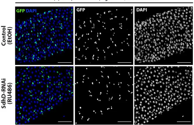
During aging, there is a loss of tissue homeostasis, causing ISCs to divide more often, and a widespread accumulation of cells that express both markers of progenitor (Esg+) and differentiated cells, suggesting a compromised ability for terminal differentiation. These cells form stacks of double and triple layers as opposed to the single layer found in healthy intestines and show reduced barrier permeability [54][55][56]. Examples of these intestines are found later in sections 1.1 and 2.2 of the Results and Discussion chapter.
Mechanisms of stem cell modulation
12
apoptotic program and are excised to the lumen, in similarity with the mammalian case. The rates of progenitor cell division and differentiation are adjusted in response to EC and EE
turnover through a combination of positive and negative feedback loops initiated by these cells [42]. Several conserved pathways are involved in autocrine or paracrine signaling occurring between progenitor and differentiated cells in the niche, which are described below.
JAK/STAT (Janus kinase/signal transducers and activators of transcription) is a pathway activated by a variety of ligands that is involved in proliferation, differentiation and apoptosis, during development, homeostasis or stress-responses. In the midgut of
Drosophila, Jak-Stat is activated by the cytokines of the Unpaired family Upd, Upd2 and Upd3. Upon stress, ECs release these cytokines to promote ISC proliferation and EB differentiation, via stimulation of Delta-Notch signaling [58]. Unpaired ligands bind the Domeless receptor, phosphorylating Hopscotch, a type of Janus kinase, promoting
translocation of the Stat92E transcription factor to the nucleus, to activate its target genes
[59].In the mammalian intestine JAK-STAT plays a similar role [58].
Wntsignaling (Wingless, or Wg, in Drosophila) is a conserved pathway that plays key roles both during development and in maintenance of different adult tissues [60]. Endogenous Wg ligands are expressed at low levels in ISCs of Drosophila and act to promote self-renewal in these cells [61]. Wg is also expressed in the visceral muscle [61] and EBs upon injury [62] to promote ISC division. Conversely, Wg signaling from the visceral muscle inhibits
Upd2 and Upd3 expression in ECs suppressing ISC activity via inhibition of the JAK-STAT pathway, suggesting a dual role part of a tightly regulated network [63]. In the mammalian
small intestine, Wnt is expressed in the crypt to promote stem cell renewal and control differentiation [64]. Wnt/Wg signaling works by binding of the Wnt/Wg ligand to its coreceptors, Frizzled2 and LRP (Arrow in Drosophila), to initiate a sequence of events that
leads to Dishevelled–mediated inactivation of a protein destruction complex that allows
stabilized β-catenin (Armadillo in Drosophila) to translocate to the nucleus, binding to the transcription factor T-cell factorthatpromotesthe activation or repression of genes related to proliferation, apoptosis and cell fate [65][60]. Notch also acts downstream of the Wg
pathway to control maintenance and differentiation of Drosophila intestinal progenitor cells [61].
13 including Foxo (Forkhead Box O transcription factor), inducing changes in gene expression related to growth, proliferation and differentiation. Upon stress, JNK mediates the transcription of damage-repair genes such as thor and small heat shock proteins. In the midgut, JNK is activated in ECs and EEs. ECs with dysfunctional tricellular junctions signal to ISCs to promote proliferation and regeneration [69]. JNK pathways have a dual-role in promoting apoptosis or inducing stem cell proliferation, depending on the type of activated transcription factors [54]. JNKs are also present in the mammalian intestine, where it modulates Wnt signaling [68][41][67].
The epidermal growth factor receptor (EGFR) is activated by ligands produced in the intestinal epithelium upon damage or stress, promoting ISC proliferation. The EGFR ligand Vein is expressed in the visceral muscle to promote ISC maintenance and proliferation. ISCs also express two different EGFR ligands, Spitz and Keren, that act as autocrine signals to maintain these cells [70]. Upon damage, the EGFR receptor induces activation of the RAS GTPase and MAPK, which phosphorylates the transcriptional repressor Cic (Capicua), activating expression of cell cycle regulator genes Pointed, Ets21C, Cdc25 and cycE (Cyclin E) [71]. Mammal ISCs also require EGFR signaling for proper function [41].
IGF (insulin-like growth factor; InR in Drosophila) signaling is a conserved pathway
involved in cell growth and proliferation. In the Drosophila midgut, muscle cells and EBs
express insulin-like peptides named Dilps that induce ISC proliferation [72]. Dilpsactivate InR thatsignals through Chico, a homolog of the mammalian insulin receptor substrates 1–4 (IRS
1–4), activating PI3K (phosphatidylinositol 3′-kinase), leading to phosphorylation of Akt (protein kinase B), which inhibits the transcription factor Foxo and activates TOR (target of rapamycin) protein kinases, that promote growth and proliferation [73][74]. Insulin signaling
may also stimulate cell proliferation via activation of the RAS/MAPK pathway [75].
Hedgehog (Hh) signaling is another conserved pathway involved in cell growth and
proliferation. In the midgut of Drosophila Hh is activated in injured EBs and promotes the expression of Upd2 to activate the JAK-STAT pathway that promotes ISC proliferation. Hh is
also active in the mammalian intestine where it promotes differentiation via inhibition of
Wnt signaling [76].
Hippo is a conserved pathway initially identified in Drosophila that controls organ size
co-14
activator Yorkie (YAP and TAZ in mammals). When active, Yorkie translocates to the nucleus to bind the transcription factor Scalloped (TEAD family in mammals) to induce expression of
a wide range of genes involved in autocrine or paracrine cell proliferation and survival responses, such as diap1 (Drosophila inhibitor of apoptosis), cycE, Upd1/2/3 and Vein
[77][78][79]. Stress-induced Hippo pathway inactivation in ECs of Drosophila results in increased stem cell proliferation via expression of Upd JAK/STAT ligands. In ISCs, Hippo inactivation or Yki overexpression results in increased proliferation [51]. In the mouse intestine, overexpression of YAP also leads to activation of progenitor cell proliferation [80]
The pathways here described act synergistically to maintain homeostasis in the
Drosophila midgut. As an example, inactivation of either EGFR, Wg or JAK/STAT results in progressive ISC loss over time, while simultaneous disruption of these 3 pathways leads to total loss of ISCs. However EGFR over-activation can partially replace Wg or JAKSTAT, and vice versa [70]. Upon stress, JNK and EGFR signaling act simultaneously to initiate ISC proliferation, via activation of Sox21a (a member of the conserved Sox family of transcription factors) in these cells [81].
15
3- Role of metabolism in stem cell activity
3.1- Energy production in Metazoa
Metabolism transforms nutrients into usable forms of energy and provides substrates for synthesis of new molecules. In the cytoplasm, glucose is broken down into pyruvate via glycolysis, reducing nicotinamide adenine dinucleotide (NAD+) to NADH and producing adenosine triphosphate (ATP) from adenosine diphosphate (ADP). Pyruvate obtained from glycolysis is then transported into the mitochondria via mitochondrial pyruvate carriers (MPCs), where it is converted to acetyl-CoA by pyruvate dehydrogenase that enters the Citric Acid Cycle (also Krebs Cycle, or TCA (tricarboxylic acid) cycle). Under limiting oxygen conditions, pyruvate from glycolysis is instead reduced to lactate by lactate dehydrogenase, regenerating NADH back to NAD+ to reenter glycolysis. This fermentation process is less efficient than aerobic respiration (described below), as for each molecule of glucose, aerobic respiration generates 36 molecules of ATP while anaerobic glycolysis generates only 2 molecules [82]. This process, however, can produce ATP at a faster rate than aerobic respiration [82][83].
Cells also produce ATP via protein catabolism. In this process, the α-amino group of amino acids is removed, and the resulting carbon skeleton is converted into different metabolic intermediates depending on the amino acid. These intermediates include pyruvate, acetyl-CoA, acetoacetyl-CoA, or the TCA cycle intermediates α -ketoglutarate, succinyl CoA, fumarate, and oxaloacetate and can be used to form fatty acids, ketone bodies, or glucose [86]. Amino acids that are converted to acetyl-CoA or
acetoacetyl-CoA are termed ketogenic amino acids since they can give rise to ketone bodies or fatty
acids. Amino acids that yield pyruvate, α-ketoglutarate, succinyl CoA, fumarate, or oxaloacetate are termed glucogenic amino acids [86][87].
16
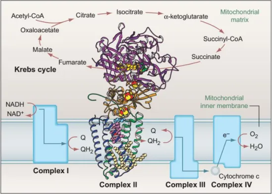
In aerobic respiration, acetyl-CoA generated from the different aforementioned processes enters the Krebs cycle in the mitochondrial matrix, where it undergoes several transformations to yield NADH and ATP (Figure 2).
The ETC occurs in the inner mitochondrial membrane and oxidizes NADH and succinate from the Krebs cycle, building up a proton gradient across the inner mitochondrial membrane, which is ultimately used to generate ATP from ADP (adenosine diphosphate) (Figure 2). This highly conserved process is termed oxidative phosphorylation (OXPHOS) [85].
The ETC comprises 5 complexes. Complexes I - IV are responsible for the transport of electrons to the corresponding reducing sites or the transfer of H+ protons across the membrane, while complex V (ATP synthase) uses the potential created by the proton gradient to generate ATP, channeling H+ back into the mitochondrial matrix. ATP is then transported to the cytosol to be used by the cell as energy [85].
Complex I (NADH:ubiquinone oxidoreductase) is the largest complex in the ETC and contains 44 subunits which are encoded by both mitochondrial and nuclear genome [89]. These subunits are assembled by 4 assembly factor proteins in humans, and at least 1 in
Drosophila [90]. Complex I oxidizes NADH from the Krebs cycle to NAD+ with removal of 2 electrons. These electrons are transported to a ubiquinone (coenzyme Q10, Q, Figure
2) binding site, where it is reduced to ubiquinol (QH2). The electron flow within the complex is thought to cause conformational changes in its proteins, causing them to pump the hydrogen protons originating from NADH to the inter-membrane space [91].
Complex II(succinate dehydrogenase, SDH) participates both in the Krebs cycle and in the ETC [85]. It is composed of 4 subunits—SdhA, SdhB, SdhC and SdhD. The subunit A is responsible for the enzymatic activity that leads to the conversion of succinate to fumarate via reduction of the flavin adenine dinucleotide (FAD) cofactor to FADH2 in the Krebs cycle. The remaining subunits—SdhB, SdhC and SdhD—channel the resulting electrons via iron-sulfur clusters to the ubiquinone binding site, where it is reduced to ubiquinol, as part of the ETC. While SdhA and SdhB are lipophobic components, SdhC and SdhD are lipophilic subunits that keep the complex attached to the mitochondrial inner membrane [92].
17 encoded. This complex oxidizes cytochrome c (reduced at complex III) and transfers the resulting electrons to reduce molecular oxygen (O2) in the mitochondrial matrix, generating water. Finally, complex V (ATP synthase) uses the electrochemical potential generated from the proton gradient to add an inorganic phosphate group to ADP to form ATP, through a rotational motor mechanism. This process requires that protons that built up in the intermembrane space are transported back to the matrix, completing the process [85].
Figure 2: Krebs cycle and the electron transport chain
The Krebs cycle takes place in the mitochondrial matrix and oxidizes acetyl-CoA in a series of steps to produce NADH and FADH2 that are used by the electron transport chain (ETC). The ETC takes place in the inner
mitochondrial membrane (IMM) and is composed of 5 complexes: complex I oxidizes NADH from the Krebs cycle to NAD+, coupled to a reduction of ubiquinone (Q) to ubiquinol (QH2); complex II is composed of 4
subunits (with their protein structure detailed in the picture) and participates in both the Krebs cycle and the ETC; it oxidizes succinate to fumarate in the Krebs cycle, and the resulting electrons reduce ubiquinone to ubiquinol in the IMM as part of the ETC. Ubiquinol is then oxidized in complex III, which is coupled to a reduction of cytochrome c. Cytochrome c is then oxidized in complex IV and the resulting electrons reduce molecular oxygen (O2) to water (H2O). Finally, complex V (ATP synthase; not shown) generates ATP using the
18
3.2- Metabolic regulation of stem cell activity
This section covers the differences in mitochondrial dynamics and metabolic requirements between stem cells and their differentiated counterparts, describing how changes in metabolic pathways may strongly influence stem cell behavior.
Besides playing a fundamental role in energy production through oxidative phosphorylation (OXPHOS) or amino acid or fatty acid catabolism, mitochondria play
important roles in cell signaling by metabolic intermediates, reactive oxygen species (ROS),
calcium homeostasis, and apoptosis [93].
Mitochondria structure in different cell types
Several studies have established correlations between cell potency (or stemness) and the structure of their mitochondria. Electron microscopy studies reveal that while somatic
cells such as fibroblasts show mature, elongated mitochondria, with numerous cristae and an electron-dense matrix, ESCs or iPSCs contained fewer, immature, and globular
mitochondria. [94][95][96][97]. Accordingly, during differentiation of ESCs and iPSCs, the mitochondria of these cells change to a more mature morphology and replicate to higher
numbers [95]. These data suggest that stem cells rely less on OXPHOS for obtaining their
energy.
Metabolic shifting between stemness states
In addition to the increase in mitochondrial biogenesis and structural differences, studies in different cell populations indicate that while differentiated cells mainly depend on OXPHOS as the primary source of energy production, stem cells traditionally rely largely on glycolysis as their main source of NADH and ATP. Accordingly, differentiation of ESCs and iPSCs is generally associated with a metabolic shift from a predominant glycolysis-based metabolism in these cells toward an increased OXPHOS-based metabolism in differentiated cells. The general preference of stem cells for glycolysis may be due to the hypoxic environment in the niche in which they normally exist [93][98][99].
19 other cells types in the bone marrow [102]. The preferential use of anaerobic glycolysis by HSCs is mediated by a pyruvate dehydrogenase kinase (PdK)-dependent mechanism, which in normal conditions suppresses the activity of pyruvate dehydrogenase in the mitochondria, and consequently inhibits OXPHOS. Importantly, PdK-mutant HSCs display lower lactate dehydrogenase activity and reduced self-renewal potential, while maintaining normal differentiation capacity, indicating that inhibition of OXPHOS is required for stem cell self-renewal [103].
Conversely, differentiated somatic cell lines display increased oxygen consumption rates and intracellular ATP content while secreting less lactate, suggesting a preference for OXPHOS [96]. Furthermore, differentiation of ESCs and iPSCs is associated with increased intracellular ATP levels and lower lactate production, while nuclear reprograming to iPSCs is coupled with a shift to glycolysis, and stimulation of glycolysis improves reprogramming
efficiency, while inhibition of glycolysis decreases it [104].
Stem cells not only have different metabolic requirements when compared to their differentiated progeny, but also exhibit strong shifts in behavior when changes in distinct metabolic pathways are induced. Stimulation of mitochondrial biogenesis with S-nitrosoacetylpenicillamine (SNAP) in ESCs reduces the expression of pluripotency markers and triggers differentiation [97][105][106]. Similarly, overexpression of PGC-1α (peroxisome proliferator-activated receptor (PPAR) gamma coactivator-1α) — an important endogenous promoter of mitochondrial biogenesis — in mouse iPSCs triggers their differentiation into adipocytes [107].
Conversely, inhibiting mitochondrial function promotes pluripotency and prevents
differentiation. Mitochondrial uncoupling by CCCP (carbonyl cyanide m-chlorophenylhydrazone) in ESCs increases the expression of the stemness factors NANOG, SOX2, and OCT4 and represses differentiation [108]. Schell and colleagues report that inhibition of MPCs (mitochondrial pyruvate carriers) in mouse intestinal stem cells and in vitro intestinal cell bodies, stimulates proliferation and population growth. These effects are coupled with decreased oxygen consumption and reduced citrate levels, confirming lower OXPHOS activity. The authors also show that endogenous MPC expression increases following differentiation of ISCs [109].
20
level. Upon OXPHOS inhibition, ESCs and iPSCs still show reduced ATP levels, suggesting that OXPHOS also contributes to energy production in these cells [101][110].
21
3.3- Metabolism in the
Drosophila
intestinal progenitor cells
In similarity with the stem cell types described in the previous section, metabolism also plays an important role in the maintenance of intestinal progenitor cells of Drosophila, with consequences at a tissue level and animal lifespan. These cells are dependent on metabolism in similar ways as the different types of mammalian stem cells and also present fewer mitochondria than differentiated cells in the same tissue [111].
Schell and colleagues, in the study cited in the previous section using mice intestinal stem cells [109], also explore the role of mitochondrial pyruvate carriers (MPCs) in ISCs of
Drosophila. The authors show that ISCs express lower levels of MPCs compared to the other differentiated cell types in the intestine, and that inhibition of MPCs causes ISCs to over-proliferate, while overexpression of MPC suppresses their ability to proliferate. These results reinforce the correlation between decreased OXPHOS and increased proliferation, while demonstrating a parallel between mammalian and Drosophila intestinal stem cells, in terms of metabolic-dependent behavior.
M. Rera and colleagues, exploring the role of the conserved mitochondrial biogenesis regulator dPGC-1 in progenitor cells of Drosophila, show that dPGC-1 overexpression raises mitochondrial content and increases the activity of the Krebs cycle enzyme citrate synthase and expression of ETC complexes I, III, IV and V. This is accompanied by delayed aging phenotypes in the intestine, such as restriction of progenitor cell over-proliferation and maintenance of intestinal barrier integrity, resulting in longer lifespan of the fly [112]. This work reinforces the correlation between increased aerobic respiration and lower stem cell activity, and reveals the importance of proper intestinal progenitor cell function to maintain tissue homeostasis, positively impacting the lifespan of the organism.
Singh et al. show that ISCs rely heavily on lipolysis–beta oxidation, while no evidence of activity of these pathways was found in differentiated cells in the intestine. Furthermore, attenuation of the lipolysis pathway leads to ISC death by necrosis [113]. Beta oxidation has previously been shown to be important for adult neural stem cell maintenance in mice [114][115].
The aforementioned reports indicate that intestinal progenitor cells in Drosophila, in similarity with the different mammalian stem cell types presented in the previous section,
rely less on OXPHOS than their differentiated counterparts. Despite that, they still depend upon mitochondria for maintenance. Accordingly, ablation of the mitochondrial autophagy
22
during aging [111]. Interestingly, while blocking pyruvate uptake by MPC inhibition induces ISC over-proliferation, blocking beta oxidation results in necrotic ISC death, indicating that
these cells might rely on lipolysis-beta oxidation as a energy source to some level, although probably not predominantly, since these cells present fewer mitochondria, and the products
of beta oxidation (acetyl-CoA, NADH and FADH2) require mitochondria to be oxidized in the
23
3.4- Implications of the electron transport chain in cell maintenance
and organismal longevity
As described in the previous sections, stem cells are sensitive to ablations of different OXPHOS components which commonly correlate with increased proliferation. Inhibition of ETC complexes or particular subunits commonly results in similar effects.
Reprogramming of somatic cells into iPSCs is accompanied by a downregulation of several subunits of ETC complexes I and II [104]. In ESCs, inhibition of complex III with antimycin A, results in increased expression of stemness genes such as NANOG and OCT4, and reduced expression of genes that promote differentiation [110]. In ESCs and iPSCs, inhibition of complex V with oligomycin, decreases ATP levels only by less than 5%, suggesting that these cells rely only marginally on OXPHOS, or that upon OXPHOS inhibition
they are able to shift their metabolism to keep up with energetic demands [101]. In mice, keratinocyte-specific knockout of mitochondrial transcriptional factor A (TFAM), a key activator of transcription in mitochondria, causes a reduction in the activity of complexes I,
III, IV, and V, but not complex II or citrate synthase, which are not encoded by mitochondrial DNA [116]. Accordingly, TFAM-deficient cells exhibit significantly lower oxygen consumption,
and lower levels of ROS [117]. At a functional level, these cells display impaired differentiation, increased proliferation, and no evidence of apoptosis, reinforcing the
connection between decreased OXPHOS activity and increased proliferation / impaired differentiation. Due to the broad number of genes affected by this knockout, it is not clear
whether the phenotypes arise from inhibition of a particular respiratory complex, or from a
general downregulation of the aerobic respiration machinery.
In intestinal progenitor cells of Drosophila, expression of NDI1 — the yeast equivalent of ETC complex I (NADH dehydrogenase) — results in increased non-endogenous NADH oxidation and inhibits ISC over-proliferation in older flies, while lessening the number of cells expressing both markers of differentiated and progenitor cells, which are normal aging phenotypes, causing an extension in the lifespan of the fly [56]. These phenotypes are coupled with a reduction in the levels of activated (phosphorylated) AMPK (5’ adenosine monophosphate-activated protein kinase, that senses high AMP/ATP ratios that occur in low energy conditions), independent of insulin signaling. This work shows that ETC activity, at least NADH dehydrogenase, also has an impact in ISCs, and reinforces the correlation between increased OXPHOS activity and decreased proliferation.
24
of the developing eye disc [118][119]. The authors show that the lower ATP are sufficient to sustain survival and differentiation but not to progress through the cell cycle. The cell cycle-arrest phenotype is caused AMPK phosphorylation due to a higher AMP/ATP ratio, that leads to activation of the tumor-suppressor p53 and subsequent degradation of the G1-S transition protein Cyclin E, ultimately causing arrest in G1. Furthermore, the authors show that mutations in the PDSW subunit of Complex I and mitochondrial ribosomal proteins mRpL4 and mRpL17 also cause cell cycle arrest in the same cells. These data oppose the reports previously described, by coupling downregulation of OXPHOS to inhibition of proliferation. This raises the question why inhibition of OXPHOS components in the cells of the Drosophila developing eye causes cell cycle arrest, while in other stem cells types it induces proliferation. It appears to be a cell type-dependent effect since inhibition of different components (CoVa, PDSW, mRpL4 and mRpL17) result in a similar phenotype. Since the authors describe an ATP-dependent mechanism, it does not appear to be any specific mechanism related to the mutations, but rather a special requirement in these cells for the wild type gene in ATP production. Perhaps cells in the developing eye of Drosophila are unable to counteract the low ATP levels by increasing anaerobic respiration, or do not receive enough nutrients to maintain their ATP levels from anaerobic respiration (since this process is less efficient) or lipolysis – beta oxidation. A different report shows that oxygen deprivation or cyanide treatment (which inhibits cytochrome c oxidase of complex IV in the ETC) coupled with a slight reduction in ATP levels cause cell cycle arrest in Drosophila
embryos [120]. However, the phenotype was not rescued by addition of ATP into the embryo, which suggests a different mechanism than the one in the eye disc.
Copeland and colleagues [121] unraveled the importance of individual subunits of the ETC for the lifespan of Drosophila. In a knockdown screen, the authors show that while ubiquitous knockdown of specific subunits of the ETC via RNAi (RNA-interference) results in lethality or premature death, knocking down different subunits of the same complexes increases longevity in the fruit fly. The ATP levels and fertility rates of these long-lived flies remain unchanged in some of the cases, indicating that the lifespan extension is not caused by a general slow-down of biological processes, and suggesting that their cells are able to counteract the specific subunit knockdown by shifting metabolism from OXPHOS. It remains to be determined what are the specific mechanisms underlying the observed lifespan extension.
27
4- Aims and outline of the thesis:
The reports presented in this chapter show that in general, a depletion of aerobic respiration, rather than deleterious, is essential for stem cell division. This has a few exceptions that appear to be specific of the cell type.
Despite the efforts in understanding the role of the ETC in stem cell maintenance, several questions related to how different complexes and subunits regulate distinct stem cell types still remain. Are different subunits/complexes required in a specific cell type in the same manner? Do different cell types have distinct requirements for a specific subunit/complex? Through which mechanisms?
In addition, due to a previous lack of tools with cell-type specificity for ISCs or EBs, some studies did not differentiate between these 2 cell types and as such they were treated equally [56][112]. Consequently, there is currently a gap in knowledge regarding how metabolism regulates these two cell types differently.
This work aimed at filling the gap in understanding the role of individual ETC complex subunits in intestinal progenitor cell maintenance — both in ISCs and EBs — and how it affects tissue homeostasis. Given the role of different subunits in the organism in mediating lifespan and the tight relationship between lifespan and intestinal stem cell regulation, we hypothesized that different ETC subunits may have distinct roles in regulating intestinal progenitor cells in Drosophila, with a consequent impact in tissue homeostasis.
28
Thesis outline:
Screen for the requirement of different ETC subunits in progenitor cells
To identify ETC subunits that are required for normal function in intestinal progenitor cells, we performed a candidate screen to knock down individual subunits specifically in these cells. Their abundance in the tissue was compared to controls as a readout of survival, self-renewal and differentiation rates. Succinate dehydrogenase subunit D (SdhD) of Complex II was identified as a strong candidate, that upon knockdown caused a significant reduction in the number of progenitor cells.
The requirements for SdhD in progenitor cell maintenance
In this section, I explore the requirements for SdhD in each type of progenitor cells (ISCs and EBs) by knocking down SdhD in these cell types individually and characterize its phenotypes in terms of survival and self-renewal potential, assessing differences between the two cell types.
The requirements for SdhD in progenitor cell differentiation
In this part, I explore how knocking down SdhD in progenitor cells affects the differentiation choice towards an enterocyte or enteroendocrine fate, by counting the number of differentiated cell types upon SdhD knockdown in progenitor cells, and performing lineage-tracing analysis.
Mechanisms originating from SdhD knockdown
30