UNIVERSIDADE DE LISBOA
Faculdade de Medicina de Lisboa
Instituto de Medicina Molecular
The role of alpha-synuclein phosphorylation in
synucleinopathies
Elisa Basso
Tese orientada por:
Prof. Doutor Tiago Fleming Outeiro
Doctoral degree in Biomedical Science, specialization in Neuroscience
Doutoramento em Ciencias Biomédica, Especialidade em Neurociencias
Todas as afirmações efectuadas no presente documento são da exclusiva
responsabilidade do seu autor, não cabendo qualquer responsabilidade à Faculdade de
Medicina de Lisboa pelos conteúdos nele apresentados
.
The experimental work presented was done at the Cellular and Molecular Neuroscience Unit, Instituto de Medicina Molecular, Faculdade de Medicina de Lisboa, Universidade de Lisboa and the Biomedicine Department, Faculty of Health, Aarhus Universitet. The financial support was given by EC Framework 7 Marie Curie Fellowship Training Network Grant (NEURASYNC).
O trabalho experimental constante de a presente tese foi realizado no Unidade de Neurociência Celular e Molecular, Instituto de Medicina Molecular e Faculdade de Medicina de Lisboa , Universidade de Lisboa e do Biomedicine Department, Faculty of Health, Aarhus Universitet. O apoio financeiro foi dado pela EC Framework 7 Marie Curie Fellowship Training Network Grant (NEURASYNC).
As opiniões expressas nesta publicação são da exclusiva responsabilidade do seu autor.
A impressão desta dissertação foi aprovada pelo Concelho Científico da
Faculdade de Medicina de Lisboa em reunião de 18 de Fevereiro de
2014.
i
Table of Contents
Acknowledgments ... iii Abstract ... v Resumo ... vii Abbreviations ... ix1.
Introduction ... 1 1.1 Synucleinopathies ... 1 1.2 Parkinson’s disease ... 21.2.1 Genetics of Parkinson’s disease ... 4
1.2.2 Pathophysiology of Parkinson’s disease ... 6
1.2.3 Animal models of Parkinson’s disease ... 10
1.2.3.1 Neurotoxin models ... 10
1.2.3.1.1 6-Hydroxydopamine (6-OHDA) ... 10
1.2.3.1.2 MPTP ... 11
1.2.3.1.3 Paraquat and Rotenone ... 11
1.2.3.2 Genetic models ... 12
1.2.3.2.1 Animal models ... 12
1.2.3.2.2 In vitro models ... 14
1.3 Multiple System Atrophy ... 16
1.3.1 Genetics of Multiple System Atrophy ... 19
1.3.2 Animal models of Multiple System Atrophy ... 19
1.3.3 An oligodendroglial cell model of Multiple System Atrophy ... 20
1.4 Alpha-synuclein ... 22
1.4.1 Alpha-synuclein structure ... 22
1.4.2 Alpha-synuclein function ... 23
1.4.3 Alpha-synuclein aggregation ... 25
1.5 Alpha-synuclein post-translational modifications ... 27
1.5.1 Alpha-synuclein phosphorylation ... 27
1.5.1.1 Phosphorylation at Serine residues ... 28
1.5.1.2 Phosphorylation at Tyrosine residues ... 31
1.5.2 Alpha-synuclein ubiquitination ... 32
1.5.3 Alpha-synuclein truncation ... 33
1.5.4 Alpha-synuclein oxidation ... 34
1.5.5 Alpha-synuclein acetylation ... 34
1.5.6 Alpha-synuclein sumoylation ... 34
ii
1.6 S. cerevisiae as a model for neurodegenerative diseases ... 36
1.6.1 S. cerevisiae as a model for Parkinson’s disease ... 38
2.
Aims of the study ... 413.
Materials and Methods ... 434.
Results ... 524.1 The role of PLK2 on alpha-synuclein aggregation in yeast ... 52
4.2 The role of PLK2 on alpha-synuclein aggregation in mammalian cell models of Parkinson’s disease ... 57
4.3 Yeast functional screening to identify novel kinases involved in alpha-synuclein pathobiology ... 61
4.4 The role of alpha-synuclein phosphorylation on microtubule retraction in oligodendroglial cells ... 69
4.5 Effect of tyrosine kinases inhibitors on microtubule retraction ... 79
5. Discussion ... 84
6. General conclusions and future perspectives ... 90
iii
Acknowledgements
During my PhD I met many amazing people who enriched my persona and encourage my personal growth. They step in during this harsh adventure as they were meant to be and I am very thankful to all of them.
I thank my supervisor Tiago Outeiro for all the scientific support, for his constant presence and for his wise advices. For always being honest and supportive, for his positive attitude, for sharing his personal experience during difficult moments and for always believe in my work. I thank my co-supervisor Sandra Tenreiro for her continuous scientific and personal support, for sharing the frustrations of my work and for guiding me during the most important steps of my personal growth.
I thank my co-mentor Poul Henning Jensen for the opportunity to work in his laboratory, for always looking after me while adapting into a new environment and for the lively scientific discussions. For helping me in my personal growth and let the Danes know a different side of Italy.
I have found really true friends in these years and I will never be thankful enough for them. I want to thank Ana, my first friend in Lisbon. She helped me finding a place to stay, she showed me the city, she offered me great dinners and the best support a person can desire when moving into a new city. Unfortunately, she moved to San Francisco for her doctoral studies, but she kept giving me her incredible support.
I want to thank Leonor, my laboratory buddy. We shared joyful and hard moments, long working hours and week-ends in the laboratory; we always support and advice each other’s. She listened to my never-ending problems and she always found the positive side of it. She never let me giving up.
Oldriska, I never found such a generous, honest and strong girl. You passed through harsh times and showed me that even the hardest condition can bring happiness and, most importantly, they are just a prelude to something marvelous.
Teresa, you have a great positive energy that you willingly share with your smile. You are an intelligent woman, full of passions and scientific questions who taught me to look at everything from a new prospective.
Thank you to Patricia for always making me smile and sharing long hours in the laboratory; to Sandra Jacinto for your support and wise words; to Hugo for being there every time I needed, for your scientific support and your constructive questions; to Rita, for all your happiness and Italian taste; to Federico, for being such an outspoken, clever and fun person. Thank you to Pedro, for sharing the hardest and latest months of my work, for being so optimistic, for supporting my crazy moments and for all the help while I was not in the lab. Thank you to
iv
Zrinka, for all the scientific teachings, for being there at the right time and moment and for always supporting me; to Filipa, for our unforgettable conversation in English/Portuguese and your kindness; to Sueli, Susana, Thomas, Dina and Rita Ramos for sharing fun, kind words and help.
Thank you to the lab technicians, Andreia for always being very kind and nice to me and Tiago Mendes for being such an understanding person and for all the laughs and jokes.
Thank you to my flat mates Sofia and Diana, for listening to all my worries, experiments and passion, and for always having a word of encouragement for me.
From Aarhus, I am thankful to Ali for helping me settling into a new environment, for all the laughs, long hours in the lab, frustrations, support, and Indian music. A special thanks to Laerke, for giving me real friendship, for being the sweetest person I have ever met, for sharing liters of coffee and tons of cakes; you are an amazing person and I hope you know that. Thank you to Cristine, for your scientific and personal support and for relieving my frustrations; to Jette, for her technical and scientific support and for her wise words; to Christine, for her positive energy and scientific support, to Jin for sharing frustrations and technical help. Thank you to Manuela, for sharing any kind of problems and being a true friend. Thank you to Carmela and my Danish Italian family, for never give up on me, for being always present, and for being an important model for my personal growth.
Special thanks to my family, for simply being what they are. They know when to listen, when to support and when to reproach. Grazie per il vostro sostegno incondizionato.
v
Abstract
Alpha-synuclein (aSyn) is a pre-synaptic protein linked to Parkinson’s disease (PD) both by genetic and pathological evidence. The gene encoding for aSyn was the first to be associated with familial forms of PD. In concert, aSyn was identified as the main component of Lewy Bodies (LBs), one of the pathological hallmarks of PD along with the loss of dopaminergic neurons from the substantia nigra. The discovery that the same protein –aSyn- and its aggregation propensity were involved in the pathogenesis of both sporadic and genetic PD cases propelled the aSyn research field. Remarkably, further findings characterize a range of diseases with parkinsonian features and presenting aSyn aggregates, the so-called synucleinopathies. These include amongst others PD, Parkinsons’s disease with dementia, dementia with Lewy Bodies (LBs), and Multiple System Athrophy (MSA). Nonetheless, the exact mechanisms responsible for aSyn aggregation and toxicity still remain unknown. Try to understand the molecular pathways behind aSyn misfolding properties is crucial to shed light into the neurodegeneration process, and in the search for therapeutic treatments that may alleviate the social burden of PD and related diseases.
This thesis focuses on the study of the role of aSyn phosphorylation, a post-translational modification (PTM) that was shown to be essential in modulating aSyn function, aggregation and toxicity. aSyn is indeed phosphorylated on serine 129 (Ser-129) in physiological conditions, phosphorylation that goes awry during the pathogenesis of the disease resulting in 90% of aSyn phosphorylated in the LBs of brain from patients and transgenic animals models of PD. The importance of this modification became soon specific since antibodies used against this residue were adopted as common staining procedure for LBs structures. Recently, other phosphorylation sites have received intense attention; in particular tyrosine 125 (Tyr-125) phosphorylation levels were shown to be significantly reduced in diseased brains.
Despite the great effort to discover the kinases mediating aSyn phosphorylation; the contribution of this modification to aSyn toxicity and aggregation remains elusive. This is primarily due to the usage of several different methodological approaches, the difficulty to study in vivo such a rapid and reversible modification and the absence of phosphorylation mutants’ forms that can properly mimic the effect of this PTM.
For these reasons, the purpose of this thesis was to investigate the pathways involved in aSyn phosphorylation using a simple, effective and well-established model for neurodegenerative diseases: the yeast Saccharomyces cerevisiae. This model has been extensively used to identify several pathways involved in PD and to generate a consistent model for aSyn aggregation that mimics the disease one. Therefore, we took advantage of the power of yeast to investigate the effects of aSyn phosphorylation on Ser-129 co-expressing human aSyn and
vi
known kinases able to phosphorylate it; namely the Polo-like kinases family (PLKs). We studied aSyn inclusions formation and toxicity and we then proceeded to validate the results in established mammalian cell model of PD. Our approach demonstrated a unique role for one of the member of the kinases family -PLK2- in aSyn inclusion formation. We showed that PLK2 expression and aSyn phosphorylation on Ser-129 are both required for aSyn inclusion formation.
The role of aSyn Tyr-125 phosphorylation was then studied in an established oligodendroglial model of MSA. We demonstrated that this modification can prevent aSyn aggregation propensity, but only when phosphorylation on Ser-129 occurred. We then identified novel tyrosine kinases inhibitors that may be involved in the disease, but further experimental data will elucidate these potential targets.
Furthermore, using yeast we implemented a functional screening and characterized novel kinases, but most importantly, new pathways involved in aSyn pathobiology. ATG1, a kinase involved in autophagy, represented one of the major modulator of aSyn aggregation and toxicity in yeast; however this novel player requires further validation in mammalian systems to establish its potential for therapeutic purposes.
Altogether, these results provide novel insights and implications for the function of aSyn phosphorylation in PD: a co-operative role for serine and tyrosine residue embodies a novel clue to unravel aSyn misfolding behavior. Our data represent a unique opportunity to tackle one side of aSyn phosphorylation function ad further validate the importance of this PTM in aSyn pathobiology.
vii
Resumo
A alfa-sinucleina (aSyn) é uma proteína sináptica que está associada à doença de Parkinson (DP) segundo evidências genéticas e patológicas. O gene da aSyn foi o primeiro a ser associado a casos familiares de DP e a proteína que é codificada foi a primeira a ser identificada e como maioritariamente presente nos corpos de Lewy (CL), uma das características patológicas da doença. A descoberta de que a aSyn e a sua propensão para agregar estão envolvidas não só na patogénese dos casos esporádicos como dos casos genéticos estimulou a investigação nesta área. Estudos posteriores identificaram uma série de doenças que apresentam várias características parkinsónicas, incluindo a presença de agregados de aSyn, e que por isso foram designadas sinucleinopatias. Neste grupo de doenças inclui-se a doença de Parkinson com demência, demência com Corpos de Lewy e Atrofia de Múltiplos Sistemas (AMS). No entanto, os mecanismos específicos responsáveis pela agregação e toxicidade da aSyn ainda estão por desvendar. É crucial entender as vias moleculares responsáveis que levam à perda de conformação da aSyn para aprofundar o conhecimento sobre o processo de neurodegeneração e encontrar novas terapias que possam atenuar o peso soció-económico da DP e doenças relacionadas.
Esta tese foca-se no estudo do papel da fosforilação da aSyn, uma modificação pós-traducional essencial na modulação da função da aSyn, na sua agregação e toxicidade. Em condições fisiológicas apenas 4% da aSyn é fosforilada na serina 129 (Ser-129), enquanto que na DP 90% da aSyn está fosforilada e deposita-se nos CL dos cérebros dos doentes, um mecanismo reprodutível em modelos de animais transgénicos da DP. A importância desta modificação tornou-se específica dado que têm sido usados como procedimento comum anticorpos contra este resíduo para corar os CL. Recentemente, outros locais de fosforilação têm sido alvo de estudo, em particular a tirosina 125 (Tir-125), cujos níveis de fosforilação estão particularmente reduzidos nos cérebros de doentes.
Apesar do grande esforço para descobrir as cinases que fosforilam a aSyn, a contribuição desta modificação para a sua agregação e toxicidade é ainda desconhecida por várias razões: a utilização de métodos experimentais diferentes, o facto de ser uma modificação difícil de estudar in vivo dada a rapidez e reversibilidade da fosforilação, e porque não existem mutantes que consigam mimetizar convenientemente o efeito desta modificação.
Por estas razões, o objectivo desta tese é estudar as potenciais vias envolvidas na fosforilação da aSyn, usando um modelo simples, eficaz e bem estabelecido para doenças neurodegenerativas: a levedura Saccharomyces cerevisiae. Este modelo tem sido usado com sucesso na identificação de várias vias envolvidas na DP, tais como a disfunção mitocondrial e do proteassoma e, surpreendentemente a formação de inclusões de aSyn que mimetizam as inclusões observadas na doença. Tirando partido deste modelo caracterizámos o efeito de
viii
cinases conhecidas na fosforilação da Ser-129, nomeadamente da família das cinases tipo-Polo. Caracterizámos a formação de inclusões de aSyn e a sua toxicidade em levedura, e posteriormente validámos os resultados em modelos celulares de mamíferos de DP. Demonstrámos um papel único da PLK2, um dos membros da família das cinases tipo Polo, quanto à formação de inclusões de aSyn. Verificámos que a expressão de PLK2 e a fosforilação de aSyn são necessários para a formação de inclusões de aSyn.
O papel da fosforilação da Tir-125 foi estudado num modelo celular já estabelecido de AMS em oligodendrócitos. Nós demonstrámos que esta modificação pode prevenir a propensão da aSyn para agregar, mas apenas quando a fosforilação na Ser-129 ocorre simultaneamente. Posteriormente identificámos novos inibidores de cinases de tirosinas que podem estar envolvidas na doença, mas mais estudos são necessários para identificar estes potenciais alvos.
Além disso, implementámos um screening funcional nas leveduras e caracterizámos novas cinases e novas vias envolvidas na patobiologia da aSyn. A cinase com maior efeito na modulação da agregação e toxicidade da aSyn foi a Atg1 que está envolvida na autofagia. É necessário validar o papel desta cinase em sistemas de mamíferos para estabelecer o seu potencial terapêutico.
Em resumo, estes resultados proporcionam novos conhecimentos e implicações para a função da fosforilação da aSyn na DP: um papel conjunto da fosforilação na serina e tirosina que aponta uma nova pista para desvendar a perda de conformação da aSyn. Os nossos resultados representam uma oportunidade única para estudar a função da fosforilação e por outro lado validar a importância desta modificação pos-traducional na patobiologia da aSyn.
ix
Abbreviations
6-OHDA – 6-Hydroxydopamine AD – Alzheimer’s disease aSyn – Alpha-synuclein
BAC – Bacterial artificial chromosome BBB – Blood brain barrier
BiFC – Bimolecular fluorescent complementation assay CHIP – Carboxyl terminus of Hsp70-interacting protein CKs – Casein kinases
CMA – Chaperone mediated autophagy CSF – Cerebro-spinal fluid
CSPalpha – Cysteine string protein alpha DA – Dopamine
DLB – Dementia with Lewy Bodies GBA – Glucocerebrosidase
GCIs – Glial cytoplasmic inclusions GFP – Green fluorescent protein GNIs – Glial nuclear inclusions GRKs – G-coupled-receptor kinases HDAC – Histone deacetylase
HSPs – Heat shock proteins KO – Knock-out
LBs – Lewy Bodies LNs – Lewy Neurites
LRRK2 – Leucine-rich repeat kinase 2 MAO-B – Monoamine oxidase B
MAPT – Microtubule associated protein Tau
MPPP – 1-methyl-4-phenyl-4-proprionoxy-piperidine MSA – Multiple System Atrophy
MT – Microtubule
NBIA – Neurodegeneration with brain iron accumulation type I NCIs – Neuronal cytoplasmic inclusions
NNIs – Neuronal nuclear inclusions ORF – Open reading frame
PD – Parkinson’s disease
PDD – Parkinson’s disease with dementia PLD – Phospholipase D
x
PLKs – Polo-like kinases PRKN – Parkin
Prp – Prion
PTMs – Post-translational modifications PUFAs – Polyunsaturated fatty acids
rAAV – Recombinant adeno-associated virus ROS – Reactive oxygen species
S129A – SA S129D – SD
Ser-129 – serine 129
SIAH – Seven in absentia homologue TH – Tyrosine hydroxylase
TOR – Target of rapamycin Tyr-125 – tyrosine 125
1
1. Introduction
1.1. Synucleinopathies
In 1997 alpha-synuclein (aSyn) was identified as one of the main proteins present in Lewy Bodies (LB), one of the pathological hallmarks of Parkinson’s disease (PD) 1. Abnormal
depositions of fibrillar aSyn characterized a group of diverse disorders collectively called synucleinopathies 2. The most common aSyn disorders include PD, Parkinsons’s disease with dementia (PDD), dementia with Lewy Bodies (DLB), Multiple System Athrophy (MSA) and Neurodegeneration with brain iron accumulation type I (NBIA) 3, 4.
Although synucleinopathies are characterized by the deposition of filamentous aSyn aggregates, what permits distinguishing among these disorders is the localization of aSyn depositions and their ultrastructure 5, 6. The three main aSyn pathological depositions found in these disorders are: LBs, Lewy neurites (LNs) and glial cytoplasmic inclusions (GCIs) (Fig .1). LBs mainly deposed in the substantia nigra of the brain, but as the disease progresses, they are also found in other areas such as the limbic system, the olfactory bulb and the neocortex. Some studies proposed that LBs pathology starts in the lower brainstem and olfactory bulb with the substantia nigra only affected in the middle stage of the disease, suggesting a staging progressive disease pattern with aSyn pathobiology sequentially spreading throughout the brain: the so-called Braak stages of the disease 7, 8. Inclusion bodies have a globular/spherical structure in the cytoplasm and perinuclear compartment (LBs), and spindle-like in the cellular processes (Lewy neurites) (Fig. 1). Ultrastructurally, LBs form eosinophilic inclusions, between 5-25 μm in size, composed of a halo of straight fibrils and a dense granular core 5. LBs mainly
consist of aSyn phosphorylated on Ser-129 1, 9, but numerous others proteins were described to deposit in LBs 10, such as ubiquitin 11, neurofilaments 12 and synphilin-1 13. Remarkably, LBs are present only in surviving neurons, thus their formation may represent a protective mechanism used by neuronal cells to disarmed harmful misfolded proteins 14.
Glial cytoplasmic inclusions (GCIs) can have triangular, sickle, half-moon, oval, or conical shape. Immunostaining analysis revealed a major protein component represented by aSyn. As for LBs aSyn is phosphorylated on Ser-129, and several other proteins co-deposited with it, such as ubiquitin, tubulin, and synphilin-115. Ultrastructurally, GCIs consist of randomly arranged, tightly packed, granule-coated filaments, similar to the LBs ones, which are 10-20 μm in diameter. GCIs are predominately found in oligodendrocytes with few neuronal inclusions that can be easily differentiated from LBs due to their intra-nuclear localization 15 (Fig. 1). GCIs are widespread in the nervous system, although a higher density can be seen in motor cortical area, caudate nucleus, putamen, globus pallidus, cerebellar white matter and in the olfactory
2
bulbs. aSyn deposits are also found in another set of disorders: sporadic and familial Alzheimer’s disease (AD), Down’s syndrome, taupathies such as Guam parkinsonian– dementia complex, Gaucher disease and other lysosomal storage disorders and motor neuron diseases 6. In these disorders aSyn pathobiology is considered secondary to the main pathological process. To date, some patients can present the same clinical features characterizing synucleinopathies but without aSyn deposition.
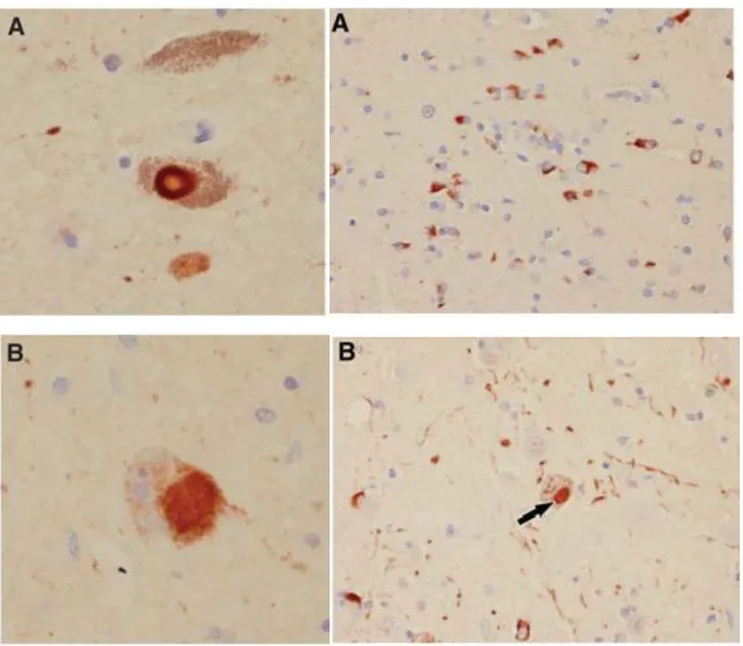
Figure 1. Microscopic findings in PD (on the left) and MSA (on the right) with aSyn immunohistochemistry. On the left panel: A. typical brainstem Lewy Body (LB), B. staining of a
cortical Lewy Body (LB). On the right panel: A. Glial cytoplasmic inclusions (GCIs) in the putamen, B. neuronal cytoplasmic inclusion (NCI) in pontine nuclei (arrow). Adapted from 16.
1.2.
Parkinson’s disease
Parkinson’s disease (PD) is the second most prevalent neurodegenerative disorder characterized by degeneration of dopaminergic neurons from the substantia nigra in the brain
3 and affecting approximately 1% of the population older than 60 years 17. The majority of PD cases are sporadic, while only about 5% of them are known to have dominantly or recessively inherited forms that arise from specific genes. Clinically patients display bradykinesia (slowness of the movement), rigidity, resting tremor and postural instability (Fig. 2). Two are the main pathological hallmarks of the disease: intracellular proteinaceous inclusions named LBs and loss of dopaminergic neurons in the substantia nigra pars compacta (Fig. 2) 18. LBs are characterized by the deposit of misfolded proteins and they are mainly composed of aSyn and ubiquitin. The biochemical nature of these deposits remained unknown until 1997 when aSyn was found to be their main component 1. The original idea came from a previous study, where a genetic mutation in aSyn gene (SNCA) caused a familiar form of the disease 19. These studies indicated for the first time that the same protein might be involved in both the sporadic and genetic cases of PD 1, 5, 20.
The symptoms appear in chronological order, with non-motor signs manifesting prior to motor ones. Subtle neuropsychological problems can be visible early in the course of PD, but there are not commonly assessed by clinicians. They involved problem-solving strategies, attention capacities and decision making. They manifest in difficulties with everyday activities such as organizing medications and paying bills. Memory deficits are mainly detected with further progression of the disease 21. Autonomic dysfunctions are then described: orthostatic hypotension, constipation, insomnia, urinary frequency and urgency and sweating abnormalities 22. Particularly, olfactory deficits may represent an early sign of PD, although research findings are not consistent 23, 24.
Neuropsychiatric symptoms are prevalent in the late stages of the disease. The most common observed are depression, anxiety, apathy and irritability, with the majority of patients displaying at least one of these symptoms 25. Motor dysfunction manifest only after extensive dopaminergic neuron loss, suggesting the presence of a compensatory mechanism in the early stages of the disease that can counteract the progressive neurodegeneration 26. An attributable cause for PD is still unknown but both environmental and genetics factors are believed to contribute to its pathogenesis. In PD sporadic cases several environmental risk factors have been identified: exposure to heavy metals such as manganese, copper, pesticides or brain injury might increase the risk to develop PD 17, 27, 28. A specific case referred back to 1982, when several drug users experienced Parkinsonism symptoms while using a drug: 1-methyl-4-phenyl-4-proprionoxy-piperidine (MPPP). A side product, MPTP, derived by its synthesis was shown to be converted into MPP+ by dopaminergic neurons using endogenous enzymes: monoamine oxidase-B (MAO-B) 29. This bio-product was able to impair the complex I of the mitochondrial chain, generating reactive oxygen species (ROS), implicated in PD pathogenesis
4
Figure 2. Clinical and pathological features of Parkinson’s disease. On the left panel the major
clinical manifestations in PD patients. On the right panel the selective death of dopaminergic neurons in the substantia nigra of PD patients (www.adam.com).
1.2.1. Genetics of Parkinson’s disease
It is firmly believed that PD is determined by both multiple genetic and environmental factors; nonetheless numerous gene mutations have been correlated to autosomal dominant or recessive PD cases, counting for 5% of the total 31, 32. Two of the genes responsible for autosomal dominant inherited cases of PD are the SNCA gene encoding aSyn 19 and Leucine-rich repeat kinase 2 (LRRK2) 33. Autosomal recessive cases involved genes for PRKN (Parkin) 34, PINK-135 and DJ-1 36 (for a complete list of genes associated with familial forms of PD see Table 1). Remarkably, all genetic forms present aSyn pathobiology; except for some cases carrying PRKN mutation and rarely LRRK2 mutation. Investigators also defined polymorphisms as risk factor for PD: carriers of a single Glucocerebrosidase (GBA) mutant allele have five time higher risk for PD 37 as well as at the SNCA gene promoter (detailed in paragraph 1.2.2). Recently, genome-wide studies have propelled the discovery of novel genes associated with PD; amongst them data showed consistent association with SNCA and microtubule-associated protein Tau (MAPT) loci 38, 39.
5 Table 1. Genes linked with familial forms of PD. (AD=autosomal dominat; AR= autosomal recessive; UR=unknown relevance).
Locus Gene Protein Inheritance
PARK1/PARK4 SNCA Alpha-synuclein AD
PARK2 PARK2 Parkin AR
PARK3 SPR(?) (?) AD (not
validated)
PARK6 PINK1 Pten-induced kinase 1 AR
PARK7 PARK7 DJ1 AR
PARK8 LRRK2 Leucine-rich repeat
kinase 2
AD
PARK9 ATP13A2 ATPase type 13A2 AR
PARK10 PARK10 (?) UR
PARK11 GIGYF2 (?) GRB10 interacting
GYF protein 2
AR
PARK12 PARK12 (?) UR
PARK13 Omi/HTRA2 HtrA serine peptidase
2
UR
PARK14 PLA2G6 Phospholipase A2,
group VI
AR
PARK15 FBX07 F-box protein 7 AR
PARK16 PARK16 (?) UR
PARK17 VPS35 Vacuolar protein
sorting 35 Homolog
AD
PARK18 EIF4G1 Eukaryotic translation
initiation factor 4 gamma, 1
AD (not validated)
aSyn (SNCA) was the first identified gene to cause dominantly inherited PD when, in 1996, a point mutation leading to the substitution A53T was described in Italian and Greek families. Subsequently, two other point mutations were discovered: A30P 40 and E46K 41. Moreover, duplication and triplication of the gene locus were reported 42-44, with 50% increase in gene dosage for duplication generating idiopathic PD, while 100% increase producing a more severe phenotype with dementia 45. aSyn protein expression correlated with the number of gene copies, demonstrating a link between aSyn expression levels, disease progression and severity 46. Polymorphisms in the promoter region of SNCA gene were also identified 47. An imperfect dinucleotide repeat called REP1 presents allele-length variability and shows association of individuals with 263 base pair allele-length with higher risk for PD. Interestingly, this polymorphism increases three times aSyn expression levels
48. aSyn coding exons, particularly 5 and 6, demonstrated also genetic variability correlated
6
1.2.2. Pathophysiology of Parkinson’s disease
The primary neuropathological deficit in PD is the loss of dopaminergic neurons in the substantia nigra pars compacta portion of the basal ganglia accompanied by LBs formation –inclusions enriched in aSyn and ubiquitin- in surviving neurons. The classic depigmentation of the basal ganglia area, visible in post-mortem PD brain, correlates with depletion of the neurotransmitter dopamine in the putamen (corpum striatum), primary site of substantia nigra neuronal projections. In a healthy individual dopamine is released from substantia nigra pre-synaptic terminals to the extracellular space where it will bind and activate two main dopamine transporters called D1 and D2. Neurodegeneration of the substantia nigra neurons impaired the dopamine release, decreased the nervous input to the striatum region, ultimately leading to the motor manifestations characteristic of PD. Remarkably, the neuronal changes occurring in PD, are also found in other brain systems such as serotonergic and cholinergic ones, as well as in the cortex, olfactory bulbs, and the autonomic nervous system.
7
Figure 3. Intracellular pathways implicated in Parkinson’s disease pathogenesis. Several
factors may play a role in aSyn pathobiology: missense mutations and genomic multiplications of SNCA (the gene encoding aSyn), oxidative stress and C-terminal phosphorylation. All these factors have the capacity of increasing aSyn levels and/or promoting aSyn oligomerization and fibrils formation. The cell can respond rapidly and degraded misfolded aSyn by ubiquitin–proteasome system (UPS) and/or endosomal–lysosomal pathways. Gene mutations that may alter the activity of the UPS such as parkin and UCH-L1 can lead to neuronal toxicity. On the other side formation of aSyn oligomers, not effectively cleared, can promote their accumulation and induced toxicity by inhibiting the proteasome. aSyn genetic mutations can instead impair its vesicular binding, altering the vesicle trafficking. Similarly, aSyn can disrupt dopamine production (inhibition of tyrosine hydroxylase), packaging and vesicle dynamics, thus impairing dopamine release and promoting its accumulation in the cytoplasm ultimately generating ROS. Parkin and DJ-1 mutants can alter the proteasome function impairing aSyn clearance. Furthermore, proteasome function requiring ATP synthesis by mitochondria, mutation of PTEN-induced kinase 1 (PINK1), DJ-1 and parkin can compromise normal mitochondrial function resulting in early-onset Parkinsonism. Leucine-rich repeat kinase 2 (LRRK2) instead can play a role in cellular trafficking and intracellular signaling modulating aSyn aggregation state 50.
.
It is generally believed that aSyn promotes the neurodegenerative process through a toxic gain-of-function. Triggering events involved: aSyn gene duplication/triplication or promoter polymorphisms that lead to increase protein levels 51; gene mutations or oxidative stress that promotes aSyn misfolding behavior with formation of toxic oligomeric species 52, 53; and cell degradation system malfunctioning, which enhances aSyn accumulation and ultimately LBs deposition 54 .
The effects produced by this toxic gain-of-function are expressed mainly at the synapses where neurotransmitters release is decreased. The SNARE protein complex (the machinery for vesicle release) is re-distributed and synaptic vesicles’ recycling is inhibited 55-58. It is believed that aSyn oligomers play an important role in this scenario, they may be able to generate membrane pores that can alters the calcium voltage-gated channels and disrupt calcium homeostasis 59-61 and/or induce leakage of dopamine at vesicles membranes, which is known to contribute to the production of ROS 62.
Oxidative stress in particular, can be triggered by several factors: mitochondrial aSyn over-expression and/or mutation can disrupt the complex I of the respiratory chain 63-65; aSyn oligomers can induce mitochondria malfunction or morphological changes 66-68; aSyn gene mutations or genes know to be associated with PD, such as DJ-1 69, 70 can alter mitochondria morphology and stimulate mitochondrial autophagy; or environmental mitochondrial toxins (a known risk factor) can induce mitochondria dysfunction.
8
Furthermore, mutations in parkin and PINK1 can cause mitochondrial deficit and accumulation of ROS and dopamine metabolites.
A crucial factor in aSyn mediated toxicity -discovered in yeast- is the ER-to-Golgi trafficking impairment, comprising in particular Rab1, a protein involved in this transition 71. The delay in the ER to Golgi transition arises from aSyn re-distribution at the SNARE complex 72, corroborating aSyn function and interaction with the synaptic vesicular system 73.
The formation of aSyn aggregates can be modulated by other gene mutations also associated with PD, such as LRRK2. LRRK2 co-expression with aSyn promotes aggregate formation, phosphorylation, and aSyn release into the extracellular media 74.
aSyn genetic mutants are also able to induce proteasome dysfunction in several cell cultures and in vivo systems 75-78, and it is thought that oligomeric aSyn species exert this effect 79, 80. aSyn mutants can also impair chaperone mediated autophagy (CMA) 81-83, although this is still controversial. However, impairment of the degradation system could reinforce the formation of aSyn oligomeric species, aggregates formation and LBs formation.
aSyn can interact with chaperones protein such as: Hsp27, Hsp70, and Hsp90 84. Overexpression of Hsp27 can reduce aSyn mediated toxicity; Hsp70 was shown to inhibit aSyn fibrils binding to aSyn oligomers 85; and carboxyl terminus of Hsp70-interacting protein (CHIP) can co-localize with aSyn in LB and decrease aSyn fibrillization 86. Furthermore, CHIP can ubiquitinate aSyn 87 and direct it for degradation via the proteasome and the lysosome pathways 86.
A recent finding has generated great attention within the PD research field. aSyn, mostly considered an intracytoplasmic protein, it was detected in plasma, saliva and cerebro-spinal fluid (CSF) 88, 89, offering the evidence for aSyn potential secretion. It is not clear how aSyn secretion occurs, but it seems to take place through a non-classical secretory pathway. In SH-SY5Ycells aSyn can be released via exosomal vesicles whose function is dependent on calcium influx 90. Secretion of both aSyn monomers and fibrils was also observed in cell cultures as a result of proteasome impairment or mitochondrial dysfunction 89. aSyn release through exosomes was further confirmed in SH-SY5Y cells that present, interestingly, lysosomal dysfunction. This perturbation also produced an increase in aSyn cell to cell transmission and formation of insoluble aggregates 91. Remarkably, extracellular secreted aSyn, especially oligomeric forms, can influence the surrounding environment leading to cell death 90, 92. aSyn can be uptaken by neighboring cells via endocytosis, here aSyn aggregates, but not monomers, are then directed to the lysososmal compartment for degradation, reflecting a possible protective mechanism 93. Conversely, other studies demonstrated that aSyn oligomeric species, after being uptaken, can seed fibrillization of
9 endogenous aSyn 60, 94-97. Significant in vivo studies validated aSyn cell to cell transmission and spreading hypothesis: progenitor cells implanted in the hippocampus of aSyn transgenic mice were able to incorporate aSyn 98, 99; and fetal dopaminergic grafts in humans showed LB pathology years after implantation 100101. A recent study using WT mice has demonstrated that a single striatal injection of aSyn fibrils produced aSyn intracellular transmission throughout the brain, reducing dopamine levels and generating motor symptoms PD-like 102. Remarkably, LB-like inclusions with aSyn highly phosphorylated were detected and a spreading mechanism dependent on neuronal connectivity was demonstrated.
This phenomenon represents a potential explanation of aSyn propagation, according to Braak’s staging hypothesis 8 and the possibility that aSyn may indeed act as a prion-like
protein. aSyn staining in a series of PD patients and age-matched controls suggested that Lewy pathology evolves in a sequential and recognizable manner, starting in the olfactory system, peripheral autonomic nervous system, and dorsal motor nucleus of the vagus; then proceeding to the dopaminergic neurons of the substantia nigra only in the middle stage of the disease and only at later stage to the cortex layers 7 (Fig 4). These sequential processes firmly address the possibility that Lewy pathology can spread from affected to unaffected regions in a prion-like manner.
It is clear that many different factors can play a prominent role in aSyn toxicity, particularly which one initiates the pathological cascade; nonetheless each cellular process needs to be properly evaluated to unravel aSyn abberant behavior.
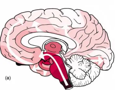
Figure 4. Distribution and progression of Lewy Body pathology according to 7, 103. The
gradual decrease in red shading intensity represents the topographical expansion of the lesions during the course of the disease. Pathology initially occurs in the dorsal motor nucleus and frequently in the anterior olfactory nucleus, then expands gradually into related areas as the brain stem (white arrows) taking an upward direction (white arrows).
10
1.2.3. Animal models of Parkinson’s disease
Animal models are a necessary tool in the study of PD pathogenesis and progression, since research on neurodegenerative diseases requires the presence of a complex nervous system to unravel the affected biological processes. However, an animal model that fully recapitulates all the key features of the disease has still to be developed. The ideal model should be age-dependent, progressive and accompanied by dopaminergic loss 104, in addition to present motor symptoms, neurodegeneration dopamine reversible and LBs and LNs 105.
1.2.3.1. Neurotoxin models
1.2.3.1.1.
6-Hydroxydopamine (6-OHDA)6-OHDA is a hydroxylated analogous of dopamine (DA) that uses DA transporter to reach the cytosol where it is oxidized, generates ROS and ultimately leads to oxidative stress 106, 107. Here, it also inhibits the complex I and IV of the mitochondrial respiratory chain 108, 109.
As a well-established model of PD, it is injected stereotactically in the rat brain 110, because it cannot pass the blood-brain-barrier (BBB). Unilateral injection is preferred because bilateral injection often results in adipsia, aphagia, and death. Several studies inject 6-OHDA into the striatum to see retrograde degeneration, because the progressive and less extensive lesion is more relevant to PD, and furthermore, synaptic terminals have been shown to die prior to subtantia nigra neurons 111. 6-OHDA treatment produces the loss of more than 80% of nigrostriatal neurons 109, 112 and motor impairment 113, 114. Notably, it has been shown to produce non-motor symptoms of PD, including cognitive and gastrointestinal dysfunction 115,
116. The major drawbacks are the absence of LB inclusions and no phenotype
age-dependecy.
The 6-OHDA model does not involve aSyn pathobiology, although it was shown to interact with aSyn 117, therefore it is primarily used as preclinical model to assess pharmacological therapies 118, 119.
11 1.2.3.1.2. MPTP
The 1-methyl-4-phenyl-1,2,3,6-tetrahydropyridine (MPTP) model dates back to the early 1980s when several Californian drug users showed symptoms similar to PD 29,
120. These patients had self-administered synthetic desmethylprodine (MPPP)
contaminated with MPTP 29. Successful treatment with L-DOPA and post- mortem analysis demonstrating loss of nigrostratial connections, improved the knowledge on PD and the molecular mechanisms behind the disease 120, 121.
MPTP is lipophilic thus can easily cross the BBB. It is subsequently uptaken by astrocytes and metabolized to MPP+ by monoamine oxidase-B (MAO-B). MPP+ is a toxic molecule uptaken by dopaminergic neurons through dopamine transporter 122,
123
where interacts with the complex I of the mitochondria respiratory chain generating ROS 124. MPTP primarily damages the nigrostriatal dopaminergic neurons 122, 125, 126.
The toxin is administered to animals by stereotactical, intravenous or intraperitoneal injection 109. Non-human primates treated with MPTP showed aSyn positive LB-like structures and presented similar clinical, pathological and pharmacological response as idiopathic PD 127, 128. LBs formation remains controversial, although described in monkeys, it is not present in mice unless chronically exposed over 30 days at low doses, while rats are resistant to the toxin. MPTP monkey model is mainly used to distinguish behavioral and symptomatic components of PD, whereas mouse to study the neuronal death process, and as an initial screening tool to test potential treatments for PD 129. Anyway, MPTP represent the most used toxin based animal model of PD due to its ability to produce PD-like effects in humans and non-human primates, its reproducible nigrostriatal system lesions, and its easy routes of administration.
1.2.3.1.3. Paraquat and Rotenone
Once MPTP was described as a potential molecule to model PD pathophysiology, similar compounds started to be screened. The herbicide paraquat (N,N’-dimethyl-4-4’-bipiridinium) was identified in 1985 130; paraquat seems able to pass the BBB
using the neutral amino acid transporter 131, 132, accumulates in the brain age-dependently, but it is still unknown how enters the dopaminergic neurons.
12
Toxicity is mediated by generation of superoxide 133, although the damage induced in the dopaminergic system has not been consistently observed 134, 135. Systemic injection can be used to induce motor deficits and loss of dopaminergic neurons in a dose- and age-dependent manner 136-138. Its treatment generates aSyn aggregates that resemble LBs in PD 139, 140. Although epidemiological studies suggested an increased risk for PD after paraquat exposure 141-143, the lack of significant effect on striatal dopamine depletion limits the use of this model for neuroprotection studies. Rotenone is a widely used pesticide, highly lipophilic that can cross the BBB and act on the complex I of the mitochondrial respiratory chain. The rotenone model was only implemented in 2000 developing a chronic low-dose treatment 144. Infused continuously, rotenone produced selective nigrostriatal neurodegeneration and aSyn cytoplasmic inclusions. This study suggests that a chronic low-dose treatment may be required to produce LBs, a feature difficult to reproduce in other toxin-based animal models 145. Rotenone can also be inhaled, subcutaneously injected or orally delivered 146. Remarkably, in rats, motor dysfunctions and dopaminergic cell loss were reversed by L-DOPA treatment 144, 147, 148 and toxicity affected other neuronal systems as described in PD 149. The rotenone infusion model, despite its positive features, has not been generally adopted; this is mainly due to the inability to reproduce consistently the parkinsonian phenotype 150-152.
1.2.3.2. Genetic models
1.2.3.2.1. Animal models
The number of genetic models based on PD associated genes is enormous and far from the purpose of this thesis, therefore only the principal findings will be summarized.
aSyn genetic models are much influenced by the promoter used to induce aSyn expression 104. Under Thy-1 promoter, aSyn is highly expressed in the substantia nigra; in some mice motor impairment is present as well as inclusion bodies, the latter not resembling LBs 153. The Prion (Prp) promoter induced instead a widespread aSyn expression but, mostly, not consistent with PD pathology 154. Only the transgenic for aSyn A53T genetic mutation showed aSyn aggregation and age-dependent neurodegeneration 155, 156. Tyrosine hydroxylase promoter (TH) directs aSyn expression exclusively in the dopaminergic neurons, but only double mutants for A53T and A30P aSyn genetic mutants showed loss of dopaminergic neurons,
13 motor impairment and decreased in dopamine levels age-dependent without protein inclusions 157. One reason of the failure of these models may reside in Syn fibrillization propensity; indeed little amounts of mouse aSyn are able to inhibit human aSyn fibrillizzation in vitro 158. A conditional transgenic mouse was recently generated using aSyn under a tetracycline promoter, although it failed to show aSyn aggregation, displayed dopaminergic cell loss and progressive motor impairment 159. Interestingly, mice knocked-out (KO) for aSyn did not show alterations in the dopaminergic neurons development or maintenance, however they present a reduction in striatal dopamine and an attenuation of dopamine-dependent motor response to amphetamine 160. Remarkably, these mice are also resistant to MPTP and other mitochondrial toxins 125, 161.
Rat models of lentiviruses and adeno-associated viruses are also used to model PD pathology. This method allows stereotactical injection of aSyn constructs directly in the substantia nigra, even in older animals. Anyway, they require proper handling and they may have reproducibility issues. Rats overexpressing aSyn WT or mutants showed selective loss of nigral dopaminergic neurons 162 that in adult animals was accompanied by aSyn positive inclusions 162-164. Recently, human aSyn bacterial artificial chromosome constructs (BAC) have been used in rats, here aSyn expression showed prominent coversion to C-terminally truncated form. In old animals aSyn was converted into insoluble fibrils, resembling PD ones, and the animals suffered severe loss of the dopaminergic projections 165.
In Drosophila models, over-expression of aSyn WT, A53T and A30P mutations develop motor deficits, loss of dopaminergic neurons age-dependent and inclusions formation 166. C. elegans models over-expressing aSyn showed dopaminergic neuron loss but no LB-like inclusions 167, 168. The effect of aSyn mutations on dopaminergic neurons did not show major differences compared to aSyn WT. Although flies and nematode models lack of endogenous aSyn, their use allowed the discovery of numerous genes and pharmacological modifiers of aSyn toxicity. Both fly, nematode and mouse models demonstrated an increase in aSyn toxicity in presence of rotenone and 6-OHDA 169, corroborating environmental factors as risk for PD.
Saccharomyces cerevisiae - another powerful model to study aSyn pathobiology - for the purpose of this thesis will be described in details in section 1.6.
LRRK2 mutations are associated with dominant inherited form of PD: overexpression of LRRK2 in Drosophila and C. elegans leads to degeneration of dopaminergic neurons 170-172. Mice KO for LRRK2 did not display any toxicity related
14
to neuronal dopaminergic development and maintenance 173, while several LRRK2 mouse models showed only low levels of neurodegeneration 174.
Mutations to parkin, DJ1 and PINK1 cause autosomal recessive forms of PD. Drosophila models displayed age-dependent loss of dopaminergic neurons and locomotors abnormalities, whereas the mice models failed to replicate many key features of PD. KO rodent models of these genes did not show nigrostriatal degeneration, or cytoplasmic inclusions that resembles PD cases, except for PINK1 KO mice showing reduced dopamine release in the striatum 175. However, recently KO parkin in old mice generated neurodegeneration in the substantia nigra 176.
1.2.3.2.2. In vitro models
The number of in vitro models of PD is broad, therefore, for the purpose of this thesis, only the principal mammalian cell models will be described.
The expression of solely aSyn in several cell models does not induce cytoplasmic inclusions or strong cytotoxicity, thus other methods started to be employed, such as protein overexpression or toxin-based. Neuronal cells over-expressing aSyn were shown to form cytoplasmic inclusions and presented mitochondrial deficits 177. Human neuroblastoma cells expressing aSyn A53T or A30P mutants, exposed to hydrogen peroxide or the neurotoxin MPP+ were more vulnerable to oxidative stress, particularly the A53T one 178; similar results were obtained using menadione-induced oxidative stress 179. In another model, neuroblastoma cells over-expressing aSyn WT, A53T and A30P mutants showed increased ROS production, more pronounced in the mutants. Remarkably, exposure to dopamine decreased cell viability, phenomenon further enhanced in the mutants 180. Likewise, only accumulation of aSyn in cultured dopaminergic neurons leaded to dopamine dependent apoptosis and ROS production 181.
aSyn A53T or A30P inducible expression in differentiated PC12 showed increased sensitivity to apoptotic cell death (while treated with proteasome inhibitors), mitochondrial depolarization and elevation of caspase 3 and 9 76.
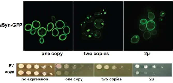
Further methods took advantage of known aSyn binding protein detected in LBs. Co-expression of the protein synphilin-1 and aSyn promotes the formation of cytoplasmic inclusions 13, 182. The fusion of green fluorescent protein (GFP) to the C-terminal of aSyn was shown to undergo truncation within the GFP domain and to induce aSyn aggregation 183, 184; this aggreagation propensity was further enhanced by co-expressing synphilin-1 (Fig. 5).
15 The bimolecular fluorescent complementation assay (BiFC) has demonstrated great impact in the study of aSyn oligomers formation. This assay involves the fusion of two non-fluorescent fragments of GFP to the proteins of interest. In case an interaction occurs between the two proteins of interest, the reporter fragments can combine together and reconstitute the activity of GFP 185. Stabilization of aSyn oligomers via BiFC in human neuroglioma cell line, increased intracellular toxicity, which could only be rescued reducing oligomers formation 186 (Fig. 5).
Other cellular models relied on aSyn extracellular uptake as seed for the fibrillization of the intracellular aSyn 96, 97, 187, 188. Unfortunately cell culture studies showed some discrepancies on the effect of aSyn expression and/or mutations on citotoxicity; in some, aSyn expression made the cells more sensitive to cellular stress 189, 190, while in others was shown to be protective 191, 192. These discrepancies probably arise from the use of different cell lines, different promoters or protein tags 193.
Although the scientific community is still looking for the perfect PD model, the studies hereby described have demonstrated to be essential to identify the molecular mechanisms (yeast, worms and flies), the biological pathways (cellular models) and the behavioral features (animal models) involved in PD pathogenesis and aSyn pathobiology.
Figure 5. Schematic representation of aSyn aggregation models. Misfolded aSyn
monomers may promote the formation of dimers and oligomers that, if failed to be degraded by the intracellular protein degradation machinery, can seed aggregation of other aSyn monomers leading to fibrils formation and ultimately to LBs proteinaceous inclusions.
16
Several assays are used to characterize which species can be formed at cellular levels: the bimolecular fluorescence complementation (BiFC) assay, which depends on formation of a fluorescent complex, allows visualization of aSyn dimers and oligomers (green); aggregates formation can instead be modelled co-expressing aSyn and synphillin-1 (red), which positively react for ThioflavinS staining (green) 194.
1.3. Multiple Systems Atrophy
The term Multiple Systems Atrophy (MSA) was initially used to describe three different disorders that had as common feature neuronal atrophy: olivopontocerebellar atrophy 195, striatonigral degeneration 196 and Shy-Drager syndrome 197, 198. MSA was included in the synucleinopathies group when Papp and colleagues described that glial cytoplasmic inclusions (GCIs) –one of the neuropathological hallmarks of these disorders- contained aSyn as main component 199.
MSA is a progressive, sporadic disease, with adult onset (between 55 and 58 years old). Clinical hallmarks comprise variable combination of parkinsonism, cerebellar ataxia, autonomic dysfunction and/or corticospinal dysfunction 200. MSA motor phenotype is primarily divided into patients with predominant parkinsonism (MSA-P) or cerebellar ataxia (MSA-C) 201. The heterogeneity of clinical features increases the chances of wrong diagnosis as well as the absence of specific biomarkers, thus a definitive validation necessitates of post-mortem histopathological analysis.
The main pathological features comprise: neuronal loss, gliosis, myelin pallor and severe axonal degeneration. In addition, between 10 to 32% of MSA patients suffer from cognitive dysfunction 202. Reactive astrocytes, containing swollen and intense GCIs, and activated microglia are common histological findings in MSA white matter, with astrogliosis paralleling the severity of neurodegeneration 203, 204 (Fig. 6).
17
Figure 6. Common histological features of Multiple System Atrophy (MSA). A. Neuronal loss
(arrowheads) B. gliosis (hypertrophic astrocytes) C. myelin pallor (blue staining). D-E Putamen immunohistochemistry using antibodies specific to aSyn shows glial cytoplasmic inclusions (GCIs), the hallmark lesions of MSA (arrows show intra-nuclear inclusions). F. Intra-nuclear inclusions (arrow). 205.
The characteristic neuropathological hallmarks of MSA are cytoplasmic inclusions in oligodendroglial cells called glial cytoplasmic inclusions (GCIs). aSyn is the main component and antibodies against it represent the most specific methods to identify GCIs; anyway, other proteins are found in GCIs, namely ubiquitin and p62. GCIs have variable diameter, 5 to 20 µm, and are located close to the nucleus. They present several morphologies such as triangle, half-moon, oval, conical and flame-shape, remarkably, the nucleus of GCIs containing cells is larger than the lacking one. Ultrastructurally, GCIs are randomly arranged, loosely packed filaments coated with a dense granular material 206. aSyn fibrils can be organized as twisted filaments (5-18 nm diameter) and straight filaments (10 nm diameter) 3, similarly to aSyn ones in PD and DLB. The widespread presence of GCIs aSyn positive classifies GCIs as unique and sufficient markers to diagnose MSA, even without a clinical history 207. Interestingly, aSyn can accumulate in other cell types: in the nuclei of oligodendrocytes to form glial nuclear inclusions (GNIs) 199, in the cytoplasm and nuclei of neurons as neuronal cytoplasmic inclusions (NCIs) and neuronal nuclear
18
inclusions (NNIs) 208, and in cell processes and neuritis (Fig. 6). While GNIs are difficult to observe, NCIs and NIIs are consistently found in MSA 209.
GCIs present another common component: p25, also known as tubulin polymerization-promoting protein (TPPP) 210, 211. p25 has a significant role in microtubules stabilization, oligodendrocytes projections, and ciliary structures212. A prominent function involved oligodendrocytes differentiation since p25 is expressed while the myelination process occurs 213, 214. Remarkably, during the neurodegeneration process, p25 delocalizes from myelin to the oligodendrocytes cell soma, increasing cell body size, promoting GCIs formation and suggesting that p25 redistribution may be an early event in the generation of GCIs 215. It was indeed shown that p25 presence within the cellular body favors aSyn aggregation, contributing to oligodendroglial dysfunction and neuronal degeneration 216. It is still unknown the reason why aSyn is found in oligodendrocytes since it is a protein of neuronal origin; furthermore studies addressing its role in non-neuronal cells have led to contradictory results. The analysis of aSyn mRNA levels in brain regions known to be affected in MSA, resulted either invariant or down-regulated in MSA brain compared to controls 217, 218. Differential expression of mRNA aSyn splicing variants was detected in MSA and PD brain 219, 220, while in situ hybridization for aSyn and proteolipid protein, an oligodendrocyte marker, failed to characterize aSyn expression in either MSA or control brains 221. Conversely, rat oligodendrocytes brain cultures expressed aSyn but in a developmental manner 222. Novel hypothesis are considering that aSyn expression and/or degradation may be up-regulated during specific steps of the neurodegenerative process or uptaken by neighboring neurons. The former hypothesis gained ground when detergent soluble aSyn was primarily found in MSA brains, while only a small amount was SDS insoluble, suggesting that altered solubility may precede GCIs formation 223.
Similarly to LBs, aSyn in GCIs is found post-translationally modified: nitration 30, 224 and phosphorylation at Ser-129 are the major modifications 9, indicating oxidative stress and aSyn aggregation as principal factors involved in MSA pathogenesis. The foremost mechanism of pathogenesis cascade predict that early aSyn overexpression and/or delocalization may influence p25 distribution from the myelin to the oligodendrocytes cell soma, generating myelin dysfunction and increasing the cell soma size 215. This mechanism will initiate the neurodegeneration process, followed by microglia activation 225, aSyn fibrils formation and deposition in GCIs 226. This process is further enhanced by p25 delocalization and incorporation into the same inclusions 211. The oligodendrocytes soma gets then larger, the nucleus becomes pale and myelin degenerates 215. Ultimately, the formation of GCIs disrupts cellular functions and leads to cell death 227. Cell death may be responsible for aSyn aggregates release into the extracellular that may be uptaken by nearby neurons: a potential source for the formation of NCIs. Thus, persistent neuroinflammation 228,
19 oligodendroglial and neuronal dysfunction, and aSyn inclusions formation could altogether contribute to the neurodegenerative process and potentially spread it into other regions of the brain 229. Although there are still missing pieces in this process, this hypothesis is contributing to investigate the potential cellular mechanisms involved in MSA pathogenesis, particularly the ones that trigger aSyn presence in the GCIs.
1.3.1. Genetics of Multiple Systems Atrophy
Genetic variations can modulate susceptibility to develop sporadic forms of neurodegenerative diseases, recent studies have suggested that genetic factors may play a role also in MSA 230. Studies on few families suggested rare Mendelian inherited forms of MSA. In a German family with autosomal dominant inheritance, the mother and one daughter presented classical features of MSA whose diagnosis was confirmed histopathologically in the mother 231, and another family with two members affected having potential autosomal dominant inherited form 232. Autosomal recessive inheritance was described in multiple Japanese families 233, and in UK were found ten proven cases of MSA
234. Nonetheless, these studied failed to identify a causative gene, mainly due to the limited
number of affected individuals.
To improve the identification of MSA related genes, screens were performed among genes known to be involved in other neurodegenerative diseases with overlapping pathology. aSyn gene, SNCA, was scanned with no positive results, although variation within the locus were associated with a risk of MSA 235-237. To date, SNCA is the most robust genetic association so far identified.
The gene MAPT encoding for Tau protein, was also associated in pathological validated cases of MSA 238 and interestingly, few MSA cases present Tau localization within the GCIs
39, 239.
1.3.2. Animal models of Multiple Systems Atrophy
Animal models of MSA are based on three different approaches: toxin-based lesion of striatonigral structures; overexpression of human aSyn in oligodendrocytes; and the two methods combined 240.
Toxin-based models (6-OHDA, MPTP, quinolinic acid and 3-nitropropionic acid) failed to reproduce the characteristic pathological hallmark of MSA, GCIs, thus limiting their practice.
20
Transgenic mice model overexpressing aSyn under protein lipid promoter demonstrated GCIs formation and biochemical composition similar to MSA ones, although not argyrophilic. Mice also showed age-related loss of dopaminergic cells in the substantia nigra and subtle motor impairment 226, 240. Mice overexpressing aSyn under 2′, 3′-cyclic nucleotide 3′-phosphodiesterase (CNP) promoter presented also GCI-like inclusions in oligodendrocytes, degraded myelin, cortical atrophy together with age-related motor impairment 241. These models support evidence of neurodegeneration as a result of human aSyn overexpression in oligodendrocytes; nevertheless, the mild motor deficits could not qualify them as absolute model for MSA.
For these reasons researchers started to use a combined approach. 3-nitropropionic acid, a mitochondrial inhibitor was administered to the PLP-aSyn overexpressing mice 240. This model showed GCI-like inclusion formations, neuronal loss in the striatum, and severe motor and cerebellar impairment, thus replicating many of the clinical features of MSA and suggesting that aSyn is the key element that renders oligodendroglial cells vulnerable to oxidative stress.
Remarkably, the MSA mice models described above showed consistently microgliosis 240, as described in the pathological MSA cases. A recent study using aSyn overexpression under PLP promoter indicates that microglial activation may be an early event in the pathogenesis of the disease underscoring neuronal loss 242.
1.3.3. An oligodendroglial cell model of Multiple Systems Atrophy
aSyn overexpression in glioblastoma, astrocytoma cell line or rat primary mixed glial cultures leads to the formation of intracellular fibrils. In the same models expression of a C-terminally truncated form of aSyn promotes further the formation of aSyn aggregates 243. In oligodendroglial cells derived from primary Wistar rat brain glial cultures (OLN-93 WT) aSyn WT and A53T mutant expression can positively model MSA, inducing aSyn aggregation, increasing its insolubility and promoting oxidative stress 244. This model was used in the study of this thesis and will receive a further description.
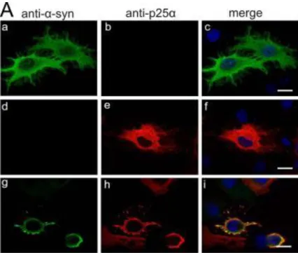
In rat oligodendroglial OLN WT cells co-expression of aSyn and p25 induces aSyn-dependent toxicity and progressive cellular degeneration aSyn-dependent on the ability of p25 to promote aSyn aggregation 216. p25 is primarily expressed in rat nervous tissue 214 and localizes specifically to the cytoplasm and myelin sheaths of oligodendrocytes in adult rats
245
. p25 can play a pathogenic role in MSA since can stimulates aSyn aggregation of α-syn in vitro 216 and it was shown to redistribute from the myelin to oligodendrolial cell soma prior to accumulation and aggregation of αSyn in GCIs 215
21 The co-expression of aSyn and p25 initiates a cellular response characterized by accumulation of soluble aSyn oligomers phosphorylated at Ser-129, activation of caspase-3, retraction of the microtubules (MT) to the perinuclear region and apoptotic cell death (Fig. 7). Apoptosis involves activation of caspase 3, phosphatidylserine externalization, nuclear condensation and fragmentation. The toxicity is dependent on aSyn aggregation since treatment with amyloid aggregation inhibitors (ASI1D peptide, baicalein and CongoRed) abrogates MT retraction. Most importantly, the toxicity depends on aSyn phosphorylation on Ser-129 because the aSyn SA mutant reduced the toxicity as well as addition of serine kinase inhibitors (DMAT, DRB, and emodin). Moreover, treatment with DMAT was able to reduce aSyn soluble oligomeric high molecular weight species 246. The same results were subsequently reproduced showing that oligodendroglial apoptotic cell death was partially rescued by sirtuin-2 inhibition 227.
Recently, it was demonstrated that the cytotoxicity caused by co-expressing aSyn and p25 relies on stimulation of the death domain receptor FAS and caspase-8 activation. Primary oligodendrocytes derived from aSyn transgenic mice express pro-apoptotic FAS receptors, which make them sensitive to FAS ligand-mediated apoptosis. These results were corroborate by an increase FAS expression found in brain extracts from MSA cases 247, suggesting FAS expression as an early hallmark of oligodendroglial degeneration.
A novel study using differentiated PC12 catecholaminergic nerve cells showed that expression of p25 is also able to induce aSyn delocalization into autophagosomes. Remarkably, p25 prevented autophagosomes fusion with lysosomes, causing an increase in aSyn secretion to the medium. aSyn secretion was shown to be dependent on autophagy, suggesting p25 involvement in aSyn extracellular release and potentially its spread during the disease process 248.