UNIVERSIDADE DE LISBOA
FACULDADE DE CIÊNCIAS
DEPARTAMENTO DE BIOLOGIA ANIMAL
Urinary Excretion of Pyrrole Compounds in Rats
Exposed to Hexanedione and Co-exposed to
2,5-Hexanedione and N- Acetylcysteine
Sara Bonucci Alves Borges da Costa
Dissertação
Mestrado em Biologia Humana e do Ambiente
UNIVERSIDADE DE LISBOA
FACULDADE DE CIÊNCIAS
DEPARTAMENTO DE BIOLOGIA ANIMAL
Urinary Excretion of Pyrrole Compounds in Rats
Exposed to Hexanedione and Co-exposed to
2,5-Hexanedione and N- Acetylcysteine
Sara Bonucci Alves Borges da Costa
Dissertação orientada pela Professora Doutora Maria Luísa Mateus
(DTB/FFUL) e Professora Doutora Deodália Dias (DBA/FCUL)
Mestrado em Biologia Humana e do Ambiente
“Take up one idea. Make that one idea your life – think of it, dream of it, live on that idea. Let the brain, muscles, nerves, every part of your body, be full of that idea, and just leave every other idea alone. This is the way to success.”
The work presented in this Dissertation is inserted under the research project on exposure to n-hexane, developed by Professora Doutora Maria Luísa Mateus.
i
Acknowledgments
First, I would like to thank my supervisor Professora Doutora Maria Luísa Mateus, for the opportunity that she gave me to work in the area that I chose; for the availability, dedication and mainly the flexibility so I could finish on time and for the careful review of the present dissertation.
To Professora Doutora Deodália Dias, as supervisor/ coordinator, for all the time and resources and for letting me work in her lab so I did not delayed my dissertation. Thank-you for all the advices, support, fast answers and help to all my questions.
To Professora Doutora Isabel Rivera, for all the help with the experimental part, time and resources spent. Thank-you for all the willingness every day that gave me a lot of motivation to continue the work.
A special thank-you to Dr. Erin Tranfield, for the flexibility on my schedule. I really appreciate all the advice, availability, patience and friendship.
To my work-colleagues. André Barros, for his huge patience in helping me to solve some “existential crisis” with this dissertation. Ana Sousa and Catarina Correia for the support and help in work in the days that I went to college and even the days that I was at work.
To my family, mom, grandmother, brother and aunt. If I am here, was not only by myself. I had a lot of obstacles until now, sometimes life was not easy, but I did not overcome them alone because they were there. Mom, I would like to thank her for all the efforts she always done for me. I know all of them gave me the best they could.
To André Pereira for all the patience and support every day. I was not easy to deal with, talking about the dissertation every single day. Thank-you for all the motivation and encouragement. Thank-you for being the amazing person you are.
Last, but not least, I would like to thank all my friends. There is a point in life that we need to start seeing the ones that never left our side, and for that I can say that I am a lucky person, because I have “few but good”. Bruno Santos, José Ramos, Patrícia Santos, Raquel Simões, Samantha Rodrigues, Sara Correia, Sofia Ribeiro, Susana Janota, Tomás Silva. Thank-you for understanding that really friendship do not means talking every day, or being together every day. Thank-you for everything!
iii
Abstract
n-Hexane is a solvent that has many uses, either in pure form or as a component of the commercial mixture hexane. Highly purified n-hexane is primarily used as a reagent, frequently used in the chemical and food industries, in the formulation of glues and paints and as a degreasing agent and extract solvent.
It is well known that this solvent presents neurotoxic effects, thus, it is very important to study biomarkers, of exposure and/ or effect, as tools of human biomonitoring, acting as indicators of exposure, as well as predictive biomarkers to prevent the occurrence of neurotoxic effects. In this context, it is imperative to understand the mechanism of n-hexane toxicity and identify endpoints that may be selected as predictive biomarkers of neurotoxicity.
The principal aim of this work was to develop accurate procedures to quantify biomarkers in urine of rats exposed to 2,5-hexanedione (2,5-HD), the main metabolite responsible for n-hexane neurotoxicity. This γ-diketone reacts with primary amines of lysine in protein neurofilaments, yielding the formation of pyrrole compounds. However, the formed pyrroles may oxidize and react to other protein nucleophiles, inducing the cross-linking between proteins and causing damage to cellular proteins. The most sensitive proteins to this damage are neurofilaments and other cytoskeletal proteins. In fact, the altered cross-linked proteins aggregate in the distal axon, often just proximal to a node of Ranvier, disrupting the normal physiological cellular activities and causing the neurotoxic effect.
To accomplish the goal of this dissertation, 2,5-HD was administrated in rats during 12 dosages and the pyrrole concentrations were measured, to assess if there were any difference between the control group and exposed rats.
Simultaneously, was studied the role of N-acetylcysteine (NAC) as a possible protective agent of neurotoxic effects evaluating a group of co-exposed rats (2,5-HD+NAC) and a NAC exposed group.
v
Sumário
A Toxicologia é uma ciência que estuda, entre outros factores, os mecanismos de ação e possíveis efeitos que podem advir da exposição humana a agentes químicos (tóxicos), provocando alterações biológicas no organismo. Esta exposição pode ocorrer através do ar, água, comida, objectos, interferindo diretamente com o ambiente e com o Homem. Neste sentido, o ramo da Saúde Pública aumentou a sua intervenção nesta área, tornando a avaliação da exposição a esses agentes num aspeto de alta importância e prioridade na sua ação, com o intuito de prevenir e/ou minimizar os possíveis riscos/ efeitos na saúde humana, através da criação de protocolos de monitorização biológica ambiental e do estabelecimento de limites (mínimos e máximos) de exposição. O objetivo destes dois tipos de protocolo é aumentar a área de atuação, conjugando a identificação e quantificação dos agentes presentes no local de exposição com a quantificação em diferentes amostras biológicas, para que a avaliação do risco de exposição seja o mais correta possível.
Um dos grupos alvo desse estudo são os solventes orgânicos, principalmente devido às características volatilidade e lipofilicidade que intervêm no mecanismo de absorção e deposição destes solventes no organismo humano. Após a exposição, ocorre a absorção destes químicos que são, imediatamente, transportados pelo sangue, até aos órgãos onde ocorre a sua metabolização (principalmente o fígado), dando origem a metabolitos que, posteriormente, serão degradados e excretados do organismo, provocando o aparecimento de alguns sintomas físicos, tais como dormência, perda de sensibilidade. A gravidade destes sintomas/ efeitos está principalmente associada à via de absorção, sendo as alterações neurológicas as mais frequentes (como por exemplo neuropatias, axonopatias, mielinopatias).
O n-Hexano é um solvente orgânico, altamente volátil e lipofílico, que tem várias aplicações, seja sob a forma pura ou enquanto componente de uma mistura comercial de hexano. A sua forma altamente purificada é primeiramente usada como reagente, sendo as misturas utilizadas nas indústrias químicas e alimentares, na formação de colas e tintas, como desengordurante e solvente de extração.
Da literatura e de estudos anteriores, sabe-se que este solvente é responsável pelo aparecimento de efeitos neurotóxicos (maioritariamente alterações neurológicas), principalmente devido à sua capacidade de acumulação no organismo, sendo, por isso, de grande importância a ação da Saúde Pública na criação/ parametrização de
vi
protocolos de controlo e monitorização. Dentro destes protocolos, surgem os estudos realizados com biomarcadores, de exposição e/ ou de efeito, estando os biomarcadores de exposição associados à quantificação do agente químico e respetivos metabolitos e os biomarcadores de efeito associados à avaliação do potencial dos efeitos resultantes da exposição. Ambos poderão ser utilizados como ferramentas de monitorização humana, que atuem tanto como indicadores de exposição ou como biomarcadores preditivos da ocorrência desses efeitos neurotóxicos. O grau de severidade dos efeitos causados está relacionado com a via de exposição, tempo e grau de exposição, podendo afetar várias partes do corpo, como pele, mucosas das membranas, sistema respiratório, fígado, sangue, sistema reprodutivo e sistema nervoso. Assim, com base no referido anteriormente, percebe-se que é fundamental o conhecimento do mecanismo de toxicidade dos agentes e identificação de endpoints que possam ser escolhidos para utilização enquanto biomarcadores de previsão da neurotoxicidade desses agentes.
No caso concreto desta dissertação, o agente em causa é o n-hexano que, após ser metabolizado no fígado dos organismos, origina vários metabolitos, sendo a 2,5-Hexanodiona (2,5-HD) um deles e o principal responsável pelos efeitos adversos que decorrem da exposição ao n-hexano. Posteriormente, esta γ-dicetona é distribuída por vários órgãos/ zonas no organismo, reagindo com os vários componentes lá existentes, dos quais se destacam as proteínas associadas ao funcionamento do sistema nervoso. Esta interação é feita através das aminas primárias do aminoácido lisina nas proteínas (dos neurofilamentos dos neurónios), conduzindo à formação dos aductos pirrólicos, que irão desnaturar a proteína o que, consequentemente, a fará perder a sua função, provocando alterações neurológicas e electrofisiológicas. Por outro lado, os pirróis formados podem também oxidar e reagir com outras proteínas nucleofílicas, induzindo a ligação cruzada entre agregados de proteínas no axónio distal, normalmente próximo de um nódulo de Ranvier, o que também irá interferir com o normal funcionamento das atividades celulares fisiológicas.
Todas as alterações anteriormente referidas estão associadas à acumulação da γ-dicetona em várias partes do organismo que, por métodos analíticos, pode ser quantificada através de uma reação química entre o 4-Dimetilaminobenzaldeído (componente do reagente de Ehrlich) e o anel pirrólico que se forma após contacto da dicetona com as proteínas.
Face a todas estas alterações pode-se ainda falar em possíveis agentes que possam atuar na diminuição e/ ou reversão dos efeitos causados pela dicetona. De entre
vii
essas substâncias está a N-Acetilcisteína que, devido às suas propriedades antioxidantes, tem a capacidade de manter os níveis intracelulares de Glutationo (GSH), que ajuda a reduzir a concentração das Espécies Reativas de Oxigénio (ROS) responsáveis tanto por destabilizações celulares e como pela inibição/ atraso na morte celular. No caso concreto dos compostos pirrólicos, parece atuar reduzindo/ impedindo a oxidação do anel pirrólico, que é o passo determinante na formação dos pirróis, na medida em que provoca rutura e alteração das biomoléculas e células do organismo humano.
Assim, com o intuito de perceber a extensão das alterações causadas pela exposição do organismo à 2,5-HD, através da quantificação dos pirróis, foram estipulados quatro objetivos para esta dissertação: i) desenvolvimento de procedimentos analíticos que permitissem determinar qual o reagente de Ehrlich e respetivas condições de reação (nomeadamente a temperatura de reação) que apresentassem maior sensibilidade na determinação e quantificação dos pirróis; ii) validação do método previamente escolhido como o mais sensível/ adequado, de acordo com normas e parâmetros já definidos na literatura; iii) determinação da influência da administração (por injeção) de 2,5-HD na concentração dos pirróis, em amostra de urina de ratos Wistar e iv) teste do efeito de proteção da NAC face à formação dos aductos pirrólicos, enquanto agente antioxidante que atua na redução e/ou eliminação dos efeitos resultantes da exposição ao metabolito 2,5-HD.
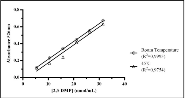
Para o primeiro objetivo, utilizou-se um método espectrofotométrico para quantificação dos pirróis, com base numa reação colorimétrica entre soluções-padrão, de concentrações conhecidas, e o reagente de EH. Experimentalmente foram comparados dois reagentes diferentes, um preparado com trifluoreto de boro e outro com ácido clorídrico, à temperatura ambiente e a 45ºC. Estes estudos foram realizados com o intuito de perceber qual o reagente de EH e a temperatura que permitia uma maior sensibilidade do método e, consequentemente, uma melhor aproximação da verdadeira concentração dos pirróis nas amostras de urina analisadas. Dos resultados obtidos, concluiu-se que o melhor método a utilizar é o reagente de EH com ácido clorídrico, à temperatura ambiente, devido à maior sensibilidade, simplicidade e menor toxicidade, estando esta última característica associada à ausência de trifluoreto de boro, uma substância bastante tóxica.
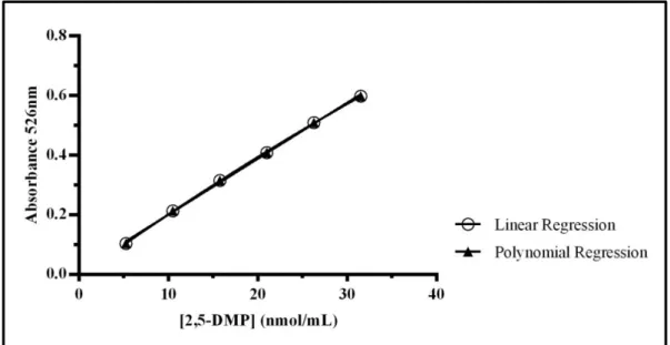
Após a escolha do método a utilizar, procedeu-se à validação, seguindo parâmetros definidos para métodos internos de ensaio em análise química, tais como: linearidade, gama de trabalho, limites de deteção e quantificação, sensibilidade, precisão
viii
e exatidão. Nesta parte foi avaliada a curva de calibração determinada para o reagente de EH com ácido clorídrico à temperatura ambiente. Tanto a linearidade como a gama de trabalho são parâmetros que foram analisados estatisticamente, comparando um valor calculado utilizando os resultados obtidos com um valor já definido na literatura e, em ambos os casos, os resultados estavam bem ajustados. De acordo com os resultados obtidos, 2,4966 nmol/ mL é a menor concentração que pode ser detetada nas amostras (limite de deteção) e 3,3810 nmol/ mL a menor concentração possível de ser quantificada utilizando a curva previamente determinada. 0,01876 é um valor que está associado à capacidade do método em distinguir pequenas diferenças entre as concentrações dos analitos. Por fim, dentro da precisão temos a repetibilidade e a precisão intermédia que permitem avaliar a reprodutibilidade do método em condições de variabilidade, tais como laboratórios, analistas, equipamento, tipos de reagentes e duração. A exatidão foi o único parâmetro que não foi avaliado neste trabalho experimental por não haver nenhum valor teórico que se pudesse utilizar para comparação.
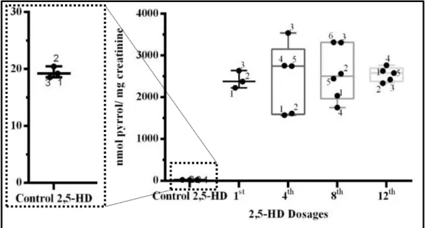
O cumprimento do terceiro objetivo foi feito através da comparação da concentração dos compostos pirrólicos entre grupos de ratos expostos a diferentes doses de 2,5-HD. As doses foram injetadas por via intraperitoneal, em dias alternados, durante um total de 12 administrações (doses), das quais foram avaliadas as doses 1,4,8 e 12, que estão associadas aos dias de recolha de urina. Da comparação dos resultados obtidos, para o grupo controlo (injeção intraperitoneal de soro fisiológico) concluiu-se que a primeira dose resultou na distribuição da 2,5-HD pelos tecidos, pois os níveis deste composto na urina dos ratos expostos são bastante superiores aos níveis apresentados pelos ratos do grupo controlo. Quanto às outras doses, não foi possível observar se houve diferença significativa, pois i) existe uma grande variabilidade entre os animais, responsável por grandes desvios na análise estatística e ii) devido ao possível surgimento do estado estacionário, em que a determinada altura, a taxa de absorção se torna igual à taxa de eliminação, tornando a concentração dos pirróis constante.
Para o quarto e último objetivo desta dissertação, os ratos foram co-expostos à 2,5-HD (injeção intraperitoneal) e NAC (adicionada à água de beber), com o intuito de comparar as concentrações nos dois grupos para testar o possível efeito protetor da NAC, face à injeção de 2,5-HD. Para a primeira dose administrada, foi possível observar que existe um fator protetor quando se adiciona NAC na água de beber que é
ix
dada aos ratos, pois a concentração de compostos pirrólicos do grupo co-exposto é inferior à concentração destes mesmos compostos na exposição única a 2,5-HD. Com a continuação da exposição para as restantes doses, o efeito da NAC foi-se tornando menos evidente, o que poderá estar associado ao facto da dose de NAC administrada nos ratos não ser suficiente para reduzir o efeito da exposição repetida à 2,5-HD.
Palavras-chave: n-hexano, 2,5-hexanodiona, compostos pirrólicos, neurotoxicidade,
xi
Contents
Acknowledgments ... i Abstract ... iii Sumário ... v Contents ... xiList of Figures ... xiii
List of Tables ... xv
List of Abbreviations ... xvii
Chapter 1 – Introduction ... 3
1 General Introduction ... 3
2 Control and exposure assessment ... 4
2.1 Organic Solvents ... 6
3 Neurotoxicity ... 8
3.1. Evaluation of neurotoxic effects ... 11
4 n-Hexane ... 12
4.1 n-Hexane biotransformation ... 14
4.2 Mechanism of action and pathology ... 16
4.3 Neuroprotective strategies ... 18
5 Biomarkers of exposure and of effect of n-hexane... 20
5.1 Characteristics of 2,5-HD biomarker ... 20
5.2 Analytical detection of pyrrole derivatives ... 21
Chapter 2 – Evaluation of the Ehrlich sensibility in the determination of pyrroles adducts and method validation ... 25
1 Introduction ... 25
2 Material and methods ... 28
3 Results ... 29
3.1 Study of the reaction time ... 29
3.2 Study of the influence of reagents and temperature for pyrrole detection ... 30
3.3 Method validation ... 32
xii
Chapter 3 – Study of the pyrrole concentration in urine of rats exposed to 2,5-HD
and co-exposed to 2,5-HD+NAC ... 41
1 Introduction ... 41
2 Material and methods ... 41
3 Results ... 43
3.1 Guanidine stability ... 43
3.2 Results of exposition to 2,5-HD ... 44
3.3 Results of co-exposition to 2,5-HD+NAC ... 48
3.4 Comparison between exposition to 2,5-HD and co-exposition to 2,5-HD + NAC 51 4 Discussion ... 52
Chapter 4 – Final Considerations and Future Perspectives ... 57
1 Final Considerations ... 57
2 Future Perspectives ... 60
References... 63
xiii
List of Figures
1.1: Control and exposure assessment through biomonitoring. ... 5
1.2: Schematic representation of the morphological structure of neuron... 9
1.3: Neuronal patterns resulting from the chemicals action. ... 10
1.4: Biotransformation of n-hexane. ... ..15
1.5: Schematic illustration of reaction between the pyrrole ring and the Ehrlich reagent. ... 21
2.1: Calibration curve for the EH-A, at room temperature and at 45ºC. ... 30
2.2: Calibration curve for the EH-B, at room temperature and at 45ºC. ... 31
2.3: Calibration curve for the EH-B and the EH-A, at room temperature. ... 31
2.4: Calibration curve for the EH-B and the EH-A, at 45ºC. ... 32
2.5: Regressions for the validation of the method. ... 33
3.1: Boxplot of pyrroles concentration from the control group and 2,5-HD exposed groups. ... 45
3.2: Evolution of the concentration of pyrroles during the 12 dosages of 2,5-HD. ... 47
... 49
3.3: Boxplot of pyrroles concentration from the control group and 2,5-HD+NAC exposed groups. ... 49
3.4: Evolution of the concentration of pyrroles during the 12 dosages of 2,5HD+NAC. ... 51
3.5: Comparison between the exposition to 2,5-HD and co-exposition to 2,5-HD+NAC. ... 52
xv
List of Tables
2.1: Influence of time in the reaction between pyrroles adducts and DMAB. ... 29 2.2: Linearity and working range.. ... 33 2.3: Limit of detection, quantification and sensibility... 33 2.4: Repeatability, reproducibility and intermediate precision. ... 34 3.1: Experimental conditions per group. ... 42 3.2: Amount of 2,5-HD and NAC. ... 42 3.3: Samples stability for pyrrole detection... 44 3.4: Pyrrole concentration for the control and all dosages, after 2,5-HD exposure.. ... 44 3.5: Pyrrole concentration for the 3 samples per group of control and exposed to
2,5-HD. ... 45
3.6: Results from the statistical analysis of the difference between the control and the
dosages for 2,5-HD exposure.. ... 46
3.7: Results from the statistical analysis of the difference between all 2,5-HD dosages..
... 46
3.8: Pyrrole compounds concentration for the groups of rats exposed to 2,5-HD, after 12
dosages. ... 47
3.9: Pyrrole concentration for the control and all dosages, after co-exposition to
2,5-HD+NAC. ... 48
3.10: Pyrrole concentration for the 3 samples per group of control and co-exposed to
2,5-HD+NAC. ... 49
3.11: Results from the statistical analysis of the difference between the control and all
dosages for 2,5-HD+NAC co-exposure. ... 50
3.12: Results from the statistical analysis of the difference between all 2,5-HD+NAC
dosages.. ... 50
3.13: Pyrrole compounds concentration for the groups of rats co-exposed to
2,5-HD+NAC, during 12 dosages. ... 51
3.14: Summary of the results from exposure to HD and co-exposure to
xvi
A-1: Spectrophotometric results for the control group, in 2,5-HD exposure.. ... 71 A-2: Spectrophotometric results for the 1st dosage group, in 2,5-HD exposure.. ... 71
A-3: Spectrophotometric results for the 4th dosage group, in 2,5-HD exposure. ... 71
A-4: Spectrophotometric results for the 8th dosage group, in 2,5-HD exposure.. ... 71
A-5: Spectrophotometric results for the 12th dosage group, in 2,5-HD exposure. ... 72
A-6: Spectrophotometric results for the control group, in 2,5-HD+NAC co-exposure.. 72 A-7: Spectrophotometric results for the 1st dosage group, in 2,5-HD+NAC co-exposure. ... 72
A-8: Spectrophotometric results for the 4th dosage group, in 2,5-HD+NAC co-exposure. ... 73
A-9: Spectrophotometric results for the 8th dosage group, in 2,5-HD+NAC co-exposure.. ... 73
A-10: Spectrophotometric results for the 12th dosage group, in 2,5-HD+NAC co-exposure. ... 73
xvii
List of Abbreviations
2,5-DMP – 2,5-Dimethylpyrrol 2,5-HD - 2,5-Hexanedione BEI – Biological Exposure Index CNS – Central Nervous System CV – Coefficient of Variation CYP450 – Cytochrome P450
DMAB – 4-Dimethylaminobenzaldehyde EH – Ehrlich’s Reagent
EH-A – Ehrlich’s reagent with boron trifluoride EH-B – Ehrlich’s reagent with hydrochloric acid F – Fisher-Snedecor test GSH – Glutathione LD – Limit of Detection LQ – Limit of Quantification NAC – N-acetylcysteine NF – Neurofilaments NF-H – Neurofilaments-High NF-L – Neurofilaments-Low NF-M – Neurofilaments-Medium
OLAARP – Oxidized Lipid/ Amino Acid Reaction Products PG – Test-value
PNS – Peripheral Nervous System ROS – Reactive Oxygen Species RT – Room Temperature
xviii
C
HAPTER
1
CHAPTER 1–INTRODUCTION
3
C
HAPTER1
–
I
NTRODUCTION1 G
ENERALI
NTRODUCTIONToxicology, as a science, is the study of adverse effects, caused by xenobiotic, having evolved since old medicines/ poisons (Old Toxicology) for the study of Molecular Biology (Modern Toxicology), studying the mechanisms of action and the possible effects that could come from human exposure to toxins, resulting in an acute effect (exposure to high concentrations, yet occasional) or chronic effect (repeated exposures, yet at low concentrations) (Casarett and Klaassen, 2008). Simultaneously through evolution and development of Toxicology, appears the evolution of Man that, while a living being, and due to the development of even more complex activities, with the goal to guarantee his survival, is subjected to various risks arising from the environmental factors involved, such as psychological, accidental, physical and/ or chemical. The exposure to any of these factors can occur in many ways, interfering directly with environment and with Man, principally the chemical agents, at which a population is exposed on a day-to-day basis, through the air, the water and the food (Amorim, 2003).
Thereby, the assessment of exposure to these chemical agents has turned into an important aspect of Public Health, in an attempt to prevent and/ or minimize the possible risks/ effects on human health, arising from the interaction between chemical agents and human organism, before an illness (intoxication) installs (Amorim, 2003). This exposure assessment was to draw up plans/ protocols for human monitoring, from exposure to chemicals, based on a routine evaluation and interpretation of biological and/ or environmental parameters (Amorim, 2003). The identification and quantification of those effects are parameters that integrate the new concept of Toxicology, with effect since 1975, associated to security assessment and possible risks resulting from the exposure to chemical agents (Casarett and Klaassen, 2008).
CHAPTER 1–INTRODUCTION
4
2 C
ONTROL AND EXPOSURE ASSESSMENTThe toxic effects of a chemical agent are related with its ability to generate a certain biological effect, that only occurs when the agent, or its metabolites, are on the minimal necessary conditions (concentration and period of time) to cause toxic manifestations (Casarett and Klaassen, 2008). The risk assessment resulting from exposure in the workplace is the main objective of Occupational Toxicology, to protect the workers exposed to potentially hazardous chemicals (Winder and Stacey, 2005).
The development of this branch of Toxicology has been increasing due to the rise in the production of organic chemicals, with an annual production of hundreds of millions of tons, responsible for a huge part of the effects that appear in the population exposed to them (Winder and Stacey, 2005). This rise led to the need to establish permissible exposure levels (exposure limits), characterized by the concentration at which almost all the workers may be exposed, day after day, without adverse effects on their health (Amorim, 2003; Casarett and Klaassen, 2008). These exposure limits may be open to possible alterations, being the control of those variations made by two types of monitoring: the Environmental Monitoring and the Biological Monitoring. The Environmental Monitoring consists of the identification and quantification of the agents (chemical) present in the air at the location of exposure, being their exposure limits called Threshold Limit Values (TLV). Biological Monitoring is associated to the quantification of the agent concentration, or its metabolite, in several biological samples like blood and urine, through the comparison between obtained value and the referenced values in literature, being these limits designated by the Biological Exposure Index (BEI) (Casarett and Klaassen, 2008).
Both monitoring have a mutual objective that consists in the risk assessment of exposure in health, fulfilled through different evaluation parameters. However, in the achievement of this purpose the Biological Monitoring presents an advantage over the Environmental, based on an evaluation of the chemical in the certain target organ, allowing a more effective determination of protection measures in the exposure to chemicals (Casarett and Klaassen, 2008).
The biological indicators or biomarkers that are used to characterize biological/ biochemical/ molecular markers (Costa, 1996), are defined as all the substances, or
CHAPTER 1–INTRODUCTION
5 subproducts of their metabolism, as well as any early biochemical change, which can be measured in biological fluids, tissue or exhaled air allowing the evaluation on the exposure intensity and health risk. Among the objectives of their use are i) exposure evaluation, absorbed quantity or internal dose, ii) evaluation of chemicals effects and iii) evaluation of individual susceptibility. This classification divides the biomarkers (Figure 1.1) into three classes, exposure, effect and susceptibility, respectively (Amorim, 2003).
Figure 1.1: Control and exposure assessment through biomonitoring. Schematic representation of the
steps from biomonitoring, since exposure to chemical agents (C.A.) until the onset of the disease. (Adapted from Amorim, 2003).
From the classes of biomarkers referred, those whose function, in respect to the risk assessment, is best defined are biomarkers of exposure and effect (Lowry, 1995).
The biomarkers of exposure allow the quantitative measurement of both a chemical or its metabolites, such as reversible biochemical change, in biological fluids that present signs of exposure to the agent (Amorim, 2003; Lowry, 1995). Must be specific, detectable in vestigial quantities, available for non-invasive techniques and quantitatively related with previous exposures (Costa, 1996). They also present a great importance in the determination of the nature of exposure to the chemical, its bioavailability and, in some cases, the potential presented by the chemical for production of adverse effects (Lowry, 1995).
The biomarkers of effect reflect an evaluation of the potential of adverse effects caused by a chemical, through the evidence of the interaction between the chemical and the biological receptors. These biomarkers must still reflect early biochemical modifications that precede structural or functional changes (Amorim, 2003; Costa, 1996; Lowry, 1995).
CHAPTER 1–INTRODUCTION
6
The role of biomarkers of susceptibility, despite not very well defined, also contributes to the risk assessment, identifying, in one population, the individuals that have a difference, genetic or acquired, in the susceptibility due to the exposure to the chemicals. This allows the knowledge about which external factors can raise, or decrease, the individual risk in the development of the organism response, during chemical exposure. This is because, besides similar exposure, genetic differences in metabolism may produce different doses in the target organ, interfering with the responsiveness of the organism to the agent upon exposure (Amorim, 2003; Costa, 1996).
For the quantitative determination of biomarkers be possible, it is necessary to know two main characteristics used in the characterization of the chemical agent: toxicokinetics and toxicodynamics. The toxicokinetics is the study of the metabolizing process of the chemical (absorption, distribution, accumulation, metabolism and excretion), being its study associated to the determination of the chemical substance, or any of its metabolites, in a biological medium. The toxicodynamics is related with the mechanism of action (in the target organ) of the chemical, being its study applied in cases of measurement of biological changes in the organism caused by the chemical exposure, to identify what are the effects resulting from that change (Amorim, 2003).
2.1 O
RGANICS
OLVENTSOrganic solvents are chemical substances that have the ability to dissolve, dilute or disperse one or more substances that are insoluble in water. They constitute a big and diverse chemical group, still in development (Casarett and Klaassen, 2008), quite relevant in industry (Williams et al., 2000), for example as constituents of paints, varnishes, lacquer, aerosol products (sprays), adhesives, intermediates in chemical syntheses, fuel and fuel additives (Casarett and Klaassen, 2008).
Besides specific properties of each compound, based on a set of characteristics, such as number of carbon atoms, number and kind of chemical bonds, chemical configuration and the presence of functional groups (Winder and Stacey, 2005), this group of compounds presents two common characteristics: volatility and lipophilicity (Casarett and Klaassen, 2008).
CHAPTER 1–INTRODUCTION
7 Both properties are responsible for the absorption and deposition of the solvent in the organism, varying according to molecular weight. In general, the lipophilicity of solvents increases with increasing numbers of carbon and/or halogen atoms, while volatility decreases. These properties also vary according with the charge of the molecule, particularly its absence, that, coupled with low molecular weight, make inhalation the major route of solvent exposure and providing a ready absorption across lung (inhalation route), gastrointestinal tract (ingestion) and skin (dermal exposure) (Casarett and Klaassen, 2008).
Once absorbed, solvents may be transported by the blood to the organs where biotransformation may occur, resulting in the formation of metabolites that will be degraded and excreted from the organism. In the case of absorption following ingestion or dermal exposure, organic solvents are absorbed into the venous circulation, making the transport to the liver faster, resulting in faster metabolism, degradation and excretion. Absorption following inhalation route happens via the alveoli in the lungs. Due to the lipophilic characteristics of the solvents, the solvents cross to the blood, spread in the organism, making the transport to the liver slower and consequently decreasing the speed of metabolism, degradation and excretion of absorbed chemicals (Williams et al., 2000). Thus, it can be stated that there is a relationship between the absorption route and the severity of the adverse effects, resulting from the exposure to solvents, depending on the effects from factors like solvent toxicity, exposure route, frequency and volume of inhaled air, individual susceptibility, interaction with other chemicals (Casarett and Klaassen, 2008; Williams et al., 2000).
Among the factors defining the severity of effects, the frequency of exposure is one of the most important because of the distinction between acute or chronic effects. Occasional acute exposures lead to effects very different from those caused by more extended exposures, yet at similar concentrations (Casarett and Klaassen, 2008; Williams et al., 2000). Of the main effects, the one of particular importance is neurotoxicity which is associated to alterations in the central and peripheral nervous system (Williams et al., 2000).
CHAPTER 1–INTRODUCTION
8
3 N
EUROTOXICITYNeurotoxicology is an area that connects neurosciences and toxicology and plays a very important role in the understanding of the functions of the nervous system. The field of neurotoxicity is important in our understanding of how the nervous system work, how environmental factors may play a role in system disorders and how to intervene to prevent damage and restore affected/ loss functions (Costa, 1996; Williams et al., 2000).
By definition, neurotoxicity is defined as any permanent or reversible effect on the structure or function of the central and/ or peripheral nervous systems by a biological, chemical or physical agent (Costa, 1996; Spencer, 1990; Winder and Stacey, 2005). However, its evaluation could have some limits due to the complexity of nervous system function, the multiple nature of neurotoxic events, the variability and inaccessibility of cellular and molecular locals that compose this system as well as the later expression of neurotoxic effects, after prolonged exposures or even after a latency period (Amorim, 2003).
Nervous system, one of the main human systems, is responsible for the control, coordination and regulation of corporal activities, through reception, transmission and integration of the information that allows the reaction and adaptation to the surrounding environment (Kulig et al., 1996). It could be divided into two subsystems: the Central Nervous System (CNS), comprising mainly the brain and spinal cord, and the Peripheral Nervous System (PNS), comprising all other components, including sensory and motor nerves (LoPachin and DeCaprio, 2004; Winder and Stacey, 2005). This distinction is important because some toxins appear to target only the central or the peripheral nervous system, but not both (Williams et al., 2000).
The main structural unit of the nervous system is the neuron, also called nervous cell, which connects with other neurons and is supported by auxiliary cells. The neuron exists in several shapes and sizes and with different functions, however they present a common morphological structure-based, composed by dendrites, cellular body and axon, shown in Figure 1.2 (LoPachin and DeCaprio, 2004; Williams et al., 2000).
CHAPTER 1–INTRODUCTION
9
Figure 1.2: Schematic representation of the morphological structure of neuron. The oranges
rectangles are highlighting the common structures in neurons. (Adapted from Winder and Stacey, 2005).
Due to the importance of neuron functions to the correct development and organism regulation, any factor that cause changes in its normal functioning, will induce functional and/ or morphological changes, that could reflect, mainly, in the decreasing of the initials capabilities of the system (LoPachin and DeCaprio, 2004). Following this idea, and considering that neurotoxins could affect separate parts of the nervous system, the study about the mechanism of action of neurotoxins, namely the target, becomes quite relevant and important. A more specific understanding allows a more specific treatment with the aim of decreasing and/ or reversing the changes caused by the chemicals (Casarett and Klaassen, 2008).
The response of the nervous system to chemical agents is based on a set of aspects, including i) the maintenance of a biochemical barrier between the brain and the blood, ii) the importance of the high energy requirements of the brain, iii) the maintenance of an environment rich in lipids, iv) the transmission of information across extracellular space at the synapse, v) the distances over which electrical impulses must be transmitted, coordinated and integrated and vi) the development and regenerative patterns of the nervous system (Casarett and Klaassen, 2008).
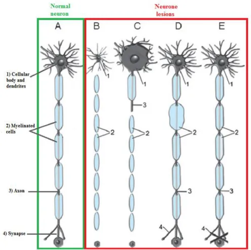
From the interaction between a certain chemical and a certain target can result many pathologies, also designated by pathologic responses. The four main targets of nervous system are: the neuron, the axon, myelinating cells and the neurotransmission system. The respective pathologies are designated by neuropathies, axonopathies, myelinopathies and toxicity associated to neurotransmission (Figure 1.3) (Casarett and Klaassen, 2008).
CHAPTER 1–INTRODUCTION
10
Figure 1.3: Neuronal patterns resulting from the chemicals action. A. Normal neuron; B. Neuropathy;
C. Axonopathy; D. Myelinopathy; E. Toxicity in transmission (Adapted from Casarett and Klaassen, 2008).
The neuropathies (Figure 1.3-B) are caused by the specificity of a given toxin to the neurons, or to any of its groups, producing changes that, in this case, are responsible for the loss of cellular body and all its associated processes, with no potential for regeneration. In cases of severe exposure, the neurons may die (Casarett and Klaassen, 2008).
When the target is the axon, the pathologies are called axonopathies (Figure 1.3-C) and the toxins are responsible for axon degeneration and consequent myelin sheath damage, maintaining the cellular body intact. In this setting, there is potential for regeneration and recovery from the toxic injury as the axonal stump sprouts and regenerates (Casarett and Klaassen, 2008).
Myelinopathies (Figure 1.3-D) are morphologic changes that occur on the myelin sheath that coats the axons. Myelin function is based on a proper electrical isolation of neuronal processes and, in its absence, the transmission of impulses along
CHAPTER 1–INTRODUCTION
11 axons may change, for example reducing the rate of transmission, which can affect the normal functioning of nerve cells, leading to the development of myelinopathies (Casarett and Klaassen, 2008).
The last figure (Figure 1.3-E) illustrates a change that occurs in the transmission of nerve impulses at synapses. In this case, the changes are taking place in the intracellular signaling mechanisms, keeping the structure of the neuron intact. They also may lead to a decrease in the capabilities/ functions of the nervous system, mainly through behavioral changes, since the transmission does not occur properly (Casarett and Klaassen, 2008).
3.1. E
VALUATION OF NEUROTOXIC EFFECTSThe nervous system is one of the most complex systems that exist in the human organism, due to its many constituents, that after certain stimuli produce different responses. This complexity makes it difficult to study the toxic effects that appear after chemical exposure, because in different targets, the same chemical could cause different responses, making necessary the choice of an adequate method of study. Kulig (Kulig et al., 1996) argued that the choice of the approach to be followed and the methods to use in a particular investigation depend on the scientific question that underlies the study.
Following the guiding thread of Kulig’s idea, Tilson (Tilson, 2000) stated that, for the neurotoxic studies in humans, behavioral studies in animals should be used, because they allow a good evaluation of neurobiological functions (sensory, motor, autonomous and cognitive functions) that are affected in humans, during chemical exposure.
Behavioral studies are techniques generally used in a tiered-testing scheme, based on decision points including in each stage of evaluation that allows the investigator to know whether or not available information is sufficient for concluding if a chemical should be considered a neurotoxin or not (Tilson, 1993). If, on the first tier, a chemical was observed to be a neurotoxin, the second tier is necessarily required, corresponding to the chemical characterization (Tilson, 2000). Characterization studies might be based on results from the first tier, already existing published data, or on new toxicological data suggesting that the chemical may pose a human neurotoxic risk (Tilson, 2000). The high degree of recurrence of this type of research is justified by its
CHAPTER 1–INTRODUCTION
12
greater sensitivity to other indicators of neurotoxicity and its early observation, allowing an observation of effects during exposure.
Based on this, Tilson (Tilson, 1993) presented one of the possible models to assess toxicity, which comprises two stages: i) toxic identification and ii) characterization of resulting effects from the toxic exposure. First stage, which consists of the initial evaluation of the neurotoxic potential of the chemical (Becking et al., 1993), is based on less expensive, simple and rapid methods, such as sensorimotor functions, locomotor activities and neurological signs. In the second stage, the methods chosen are more complex and specific, because they are used to specifically assess sensory and motor dysfunctions and to quantitatively measure chemical changes, which may be induced mainly in cognitive functions, allowing a better characterization of the nature and the mechanisms of the effects induced by the toxins (Becking et al., 1993; Tilson, 1993).
Later, Kulig (Kulig et al., 1996) justified the importance in using behavioral studies, stating that direct observation of an animal behavior, during chemical exposure, is one of the simplest methods to document clinical signs of neurological and behavioral impairment and a logical starting point for investigating the potential neurotoxic effects of a chemical whose data about neurotoxicity are incomplete or nonexistent.
4
N-H
EXANEn-Hexane is an organic solvent, which is very volatile, colorless and with an unpleasant odor, fat soluble, easy evaporation, highly inflammable and is possible to accumulate in the body (cumulative effect) (Cheng et al., 2012; Song et al., 2012; U.S. Department of Health and Human Services, 1999; Zhang et al., 2013). It can be used in pure form, particularly as a laboratory reagent or as component in a mixture, being its applications, in this last case, almost all associated with the industry, as a solvent or a thinner (Jorgensen and Cohr, 1981). Among several industrial applications are the production of tires and impregnation of materials in the rubber industry, production of tablets in the pharmaceutical industry and applications for the perfume industry. n-Hexane can still be used in product adhesives, cleaners, textiles, furniture manufacturing, printing industry, shoe-making, solvent cements and thinners and, in minor amount, in crude oil and natural gas (Fedtke and Bolt, 1987a; Integrated Risk
CHAPTER 1–INTRODUCTION
13 Information System, 2005; Jorgensen and Cohr, 1981; U.S. Department of Health and Human Services, 1999).
n-Hexane can be present in the air, in food and water and in objects/ materials that could contact directly with humans, which means the absorption of this solvent could occur in many ways. If it is in the air that we breathe, it can enter the body through the lungs; if it is in food or drinking water, it enters by ingestion through the stomach and intestines; if we come into contact with it, it enters through the skin. How much n-hexane enters the body depends on the exposure time, the amount at which humans are exposed and the exposure route (U.S. Department of Health and Human Services, 1999). From the possible exposure routes, inhalation is the most studied, because it is the most likely form of contamination. It occurs in the lungs, through the process of passive diffusion in membranes of epithelial cells. After, it enters into circulation, being transported to the liver, where it is converted, by metabolic processes, into its metabolites, which are distributed to various body organs, mainly the brain, liver and kidney (Integrated Risk Information System, 2005).
Depending on the route of exposure, as well as the time and amount of exposure, the effects that n-hexane cause are various, affecting many parts of the organism, such as skin, mucous membrane, respiratory system, liver, blood, reproductive system and nervous system (central and peripheral) (Jorgensen and Cohr, 1981). From these neurotoxic effects are, specifically, the progressive sensorimotor neuropathy beginning with prickling, burning and loss of sensation, first in the feet, moving gradually up the legs and eventually involving hands (Genter St Clair et al., 1988).
The first neurotoxic effects associated with n-hexane exposure were noticed in Japan, in the mid-60, in workers from the shoes industry. Workers of this industry were exposed to several chemicals, beyond n-hexane, complicating the identification of symptoms deriving from n-hexane exposure. It was only in 1969 that Yamamura (Integrated Risk Information System, 2005; Yamamura, 1969) began a study that allowed the determination of specific symptoms resulting from the n-hexane exposure.
Following this idea, n-hexane was characterized as a neurotoxic compound, making necessary the development of protection measures that could reduce exposure and, consequently, the neurotoxic risk. To this end, the toxicokinetics and
CHAPTER 1–INTRODUCTION
14
toxicodynamics of n-hexane were characterized to try to determine the target and mode of action of n-hexane and its metabolites (Casarett and Klaassen, 2008).
4.1
N-H
EXANE BIOTRANSFORMATIONn-Hexane shares the common property of organic solvents, high volatility, causing exposure occurrence mostly in the form of vapor, via inhalation (U.S. Department of Health and Human Services, 1999). It is absorbed by the lungs, being rapidly transported to the other organs of the body.
The metabolism of n-hexane occurs, mainly, in the liver, where the toxin undergoes a set of reactions of hydroxylation and hydrogenation, shown in Figure 1.4. It starts with a hydroxylation of n-hexane, by the action of mixed function oxidases to form either 1- or 3-hexanol, in a detoxification pathway, or 2-hexanol in a bioactivation pathway. Through the bioactivation pathway, 2-hexanol is converted to 2-hexanone and 2,5-hexanodiol. Both of these metabolites are then further metabolized to 5-hydroxy-2-hexanone, through a new oxidation reaction, that, by a hydroxylation reaction, generates 4,5-dihydroxy-2-hexanone. By oxidation of this metabolite, through acid hydrolysis of urine, or oxidation of 5-hydroxy-2-hexanone, 2,5-HD is formed. 2,5-HD is the final compound of n-hexane biotransformation, being considered the major toxic metabolite produced in humans, responsible for the production of effects associated to n-hexane exposure. After this, it is important to clarify that the difference between bioactivation and detoxification pathways is associated with the toxicity of the metabolite that is being studied, i.e., if the metabolite is the most toxic that results from the metabolizing, the pathway that will end in its formation will be called bioactivation. If, in reverse, the metabolite is not the most toxic, it will be formed through a detoxification process. That is why here (for the n-hexane) was studied the metabolism through the bioactivation pathway.
At the end of metabolizing, the excretion/ elimination of the resulting products of n-hexane exposure can occur in different ways, being the urine the main form of elimination from the body (Integrated Risk Information System, 2005; U.S. Department of Health and Human Services, 1999) and, consequently, the most studied.
CHAPTER 1–INTRODUCTION
15
Figure 1.4: Biotransformation of n-hexane. Compounds marked with orange rectangles indicate the
route of metabolism of n-hexane-associated neurotoxicity. With red color is marked the major metabolite responsible for n-hexane associated neurotoxicity. (Adapted from Integrated Risk Information System, 2005).
The metabolism of n-hexane to 1-, 2- and 3-hexanol is carried out by, at least, four enzymes, whose identification was not possible by kinetic data collected in some studies. However, from the observation of requirements from the reactions catalyzed by these enzymes, mainly the presence of NADPH, some authors may suggest that the enzymes responsible for the metabolizing of n-hexane could be Cytochrome P450 (CYP450) isozymes (Crosbie et al., 1997; Soriano et al., 1996). Further studies reinforced this idea, admitting that CYP450 catalyzes the initial steps of n-hexane metabolism, the detoxification and bioactivation pathways, being considered CYP450 dependent reactions. So, these enzymes are the ones that, acting directly on n-hexane, are going to metabolize the toxic, through bioactivation pathways, yielding, among others metabolites, 2,5-HD, responsible for the neurotoxicity associated to n-hexane exposure (Integrated Risk Information System, 2005).
2,5-HD is also considered a consequence of exposure to methyl-n-butyl-ketone, another industrial solvent whose exposure results in similar effects to those produced by
CHAPTER 1–INTRODUCTION
16
n-hexane exposure (Abou-Donia et al., 1982; Perbellini et al., 1993; Pyle et al., 1992; Tshala-Katumbay et al., 2009). The similarity in the resulting neurotoxic effects indicates a common mechanism of toxicity, essentially related with oxidation and production of the toxic metabolite, the 2,5-HD (Pyle et al., 1992). This neurotoxic effect is thought to be attributed to the reaction between this metabolite and functional (NH2
)-groups of axonal proteins under formation of substituted pyrroles adducts (Fedtke and Bolt, 1987b), point that will be discussed later.
4.2 M
ECHANISM OF ACTION AND PATHOLOGYNeurons are cells of the human body that, when exposed to 2,5-HD, have a high degree of structural alteration, mostly due to the degradation of their constituent proteins, at axon level (Song et al., 2012). These cells, under physiological conditions, are capable of maintaining a low level of autophagic activity that consists of the elimination of some damaged organelles and proteins. This cellular process is important to maintain the homeostasis of axons (Song et al., 2012). In the presence of 2,5-HD, autophagy levels are substantially decreased, leading to the gradual accumulation of proteins, organelles and aberrant structures in the membrane of the axon terminals. This accumulation is confirmed by observation of axonal swellings (Song et al., 2012) which, together with axonal atrophy, retraction of the myelin sheath of the nodes of Ranvier and segmental demyelination (Wang et al., 2008), characterize the most frequent pathologies designated axonopathies.
The emergence of these effects in workers exposed to n-hexane led to the development of several studies, to try to discover the mode of action of n-hexane, specifically the mechanism by which 2,5-HD causes axonopathies.
Initially, many authors reported that the structural changes observed in axons, atrophy or degeneration, were associated with the reactivity of γ-diketones, stating that the 2,5-HD was, clearly, the diketone responsible for axonopathies (Lopachin and Decaprio, 2005; LoPachin and DeCaprio, 2004; Tshala-Katumbay et al., 2009). Other studies revealed that axonopathies may be caused by several factors, such as changes in cytoskeletal proteins of axons that occur in the neurofilaments (NF), which can achieve a reduction in the diameter of the axons and consequent decrease in conduction velocity
CHAPTER 1–INTRODUCTION
17 of the nerve, causing neurological and electrophysiological modifications (Lopachin and Decaprio, 2005).
For the case of NF, since they are the primary cytoskeletal components in the large and myelinated fibers which are highly vulnerable to n-hexane and related neurotoxins (Decaprio and Fowke, 1992), there are many studies that prove that the change in its structure or its normal functioning can be reflected in the pathologies described above. The NF, described as the intermediate filaments in mature neurons (Wang et al., 2011), are the most abundant components of the neurons (Wang et al., 2011) that have an essential role in the establishment and maintenance of the maturation of the axons caliber (Chiu et al., 2000). Structurally, the filament itself is composed by three subunits proteins (NF-Low ( NF-L), NF-Medium (NF-M) and NF-High (NF-H)), that copolymerize via coiled-coil interactions of the rod domains. According to an immunohistochemical analysis, NF-L represents the core and the tail domains of NF-M and NF-H form the peripherally oriented filament arms (Muma and Hoffman, 1994). These latter structures mediate NF-NF interactions (Decaprio and Fowke, 1992). Each of the subunits has its own molecular weight: NF-L has 68kDa, NF-M 95kDa and NF-H 115kDa (Chiu et al., 2000; Muma and Hoffman, 1994; Wang et al., 2011).
The contribution of NF to the development of axonopathies can occur in two ways: through the change of axon caliber or through the change of NF protein constituents, both after exposure to the diketone 2,5-HD (Chiu et al., 2000; LoPachin and DeCaprio, 2004).
The decrease in axon caliber of NF is a mechanism that is not well understood, but there are studies that claim that this mechanism is based on a decrease in the interfilamentar space through dephosphorylation processes, associated with some amino acids residues (lysine, serine and proline), which, when phosphorylated, are responsible for the maintenance of that interfilamentar space (Chiu et al., 2000) and respective thickness of axon caliber. Subsequent studies have established a model, stating the axon caliber is regulated by domains of NF-M and NF-H, which form long bridges, enabling determination of interfilamentar space, defining the various spaces and the specific size of the axons.
Changes at the level of NF protein constituents pass through adduct formation, which are irreversible covalent bonds formed by linking a chemical and a biological
CHAPTER 1–INTRODUCTION
18
molecule (Lopachin and Decaprio, 2005; Tornqvist et al., 2002; Wang et al., 2011). The toxicity of the chemical agent is associated only with the time that the formation of adducts “breaks” the structure in question or destroys the function of the macromolecule (Cheng et al., 2012). In the specific case of n-hexane, the reaction of its metabolite, γ-diketone 2,5-HD, with neurofilamental proteins (only about 5%) (LoPachin and DeCaprio, 2004), leads to the formation of pyrrole adducts of 2,5-dimethylpyrrol (2,5-DMP) (LoPachin et al., 2005), through its interaction with the ε-amino groups of lysine residues. This leads to the formation of misfolded proteins that, besides contributing to the decrease of axon caliber, may affect normal growth and development of axons, by reducing the handling capacity of the proteins that interact with the polymeric cytoskeleton (LoPachin and DeCaprio, 2004; LoPachin et al., 2005; Wang et al., 2011). The determination of proteins and/ or pyrroles formed in biological samples, such as serum, in study cases of n-hexane exposure, may allow inference of a possible presence and degree of injury in neurons (Song et al., 2008).
A third hypothesis used to try to justify the development of axonopathies focuses on the role of cytoskeletal proteins. According to the authors, this hypothesis is based on the “uncoupling” ability of 2,5-HD. The axon caliber is established by a three-dimensional assembly, comprising axon specific proteins that allow crossing between NF, actin and microtubules (Chiu et al., 2000). After exposure to n-hexane, due to the action of 2,5-HD, the connections between the components of this set are destroyed, because this metabolite acts in the cross-links between axon proteins, decreasing the likelihood of their occurrence (Chiu et al., 2000). More recent studies reveal that the axon atrophy involves multiple proteins of cytoskeleton, which presents the main target of the metabolite 2,5-HD (Zhang et al., 2010). They also stated that for this theory to be “accepted” and the consequences of adduct formation of cytoskeletal proteins be concretely defined, relevant targets need to be found in the amino acids residues of cytoskeletal proteins (Zhang et al., 2010).
4.3 N
EUROPROTECTIVE STRATEGIESBased on the proposed mechanisms of action of 2,5-HD and all the effects that they could cause in human health, some authors tried to propose studies focused on body detoxification, related with n-hexane exposure.
CHAPTER 1–INTRODUCTION
19 2,5-HD, as the major metabolite responsible for the neurotoxicity resulting from n-hexane exposure, can cause alterations in the body, in cells or biological processes, that could result in an overconsumption of oxygen, leading to the production of free radicals, also known as Reactive Oxygen Species (ROS). These molecules are considered highly reactive radicals that frequently attack biological molecules, by abstraction of a hydrogen atom. Among these biological molecules are lipids, whose reaction with free radicals is called lipid peroxidation (Halliwell, 1993; Kerksick and Willoughby, 2005; Mittler, 2002). This process has been associated with important pathophysiological events in a variety of diseases, drug toxicities and traumatic or ischemic injuries (Hidalgo et al., 1998). Its toxicity is based on the ability to modify protein reactive groups, producing modified proteins, called Oxidized Lipid/Amino Acid Reaction Products (OLAARPs) that could be determined. Among all the OLAARPs, only a few can be chemically determined, from which the pyrroles (Hidalgo et al., 1998), mainly compounds that result from the mechanism of action of 2,5-HD.
Following this reasoning, some authors decided to study some neuroprotective strategies to decrease or reverse the neurotoxic effects caused by 2,5-HD, which is the last metabolite of n-hexane and is responsible for the neurotoxic effects (Terenghi et al., 2011). Among several agents the N-acetylcysteine (NAC), an acetylated cysteine residue, is studied in the present work, for two reasons: its ability to maintain or increase intracellular levels of Glutathione (GSH) or to inhibit or delay cell (neuron) death (Aruoma et al., 1989; Moschou et al., 2008).
GSH is an essential compound for the attenuation of oxidative stress in cells and organs and its intracellular concentration (Sagara et al., 2010), is endogenously synthesized all throughout the body and it is basically found in all cells (Kerksick and Willoughby, 2005).
NAC has a major function through the depletion of free radicals and is a powerful antioxidant that, experimentally, has been shown to increase the GSH content in mammalian cells. Its administration to the cysteine-free medium maintains the intracellular contents of cysteine, GSH and NAC, fact that supports the possibility that NAC, after permeation into neuronal cells, is hydrolyzed to release cysteine in the cell, which is used for GSH synthesis (Sagara et al., 2010). However, in the presence of
CHAPTER 1–INTRODUCTION
20
cysteine, NAC has a deleterious effect on the cells, due to its low stability in the complete medium as compared with the cysteine-free medium (Sagara et al., 2010).
NAC also acts as a cysteine donor, being involved in the inhibition or delay of cells death, according to the concentrations administered (Moschou et al., 2008). According to some studies, some authors observed that, in small concentrations, NAC inhibits some of the ROS, which led the scientific community to ponder the increase of that concentration, in order to completely eliminate ROS (Moschou et al., 2008).
The inclusion of NAC in this study is related with the statement of some authors about the oxidation of the pyrrole ring being a critical step in n-hexane neurotoxicity. When oxidation occurs, also occurs formation of electrophilic compounds, which can react with unconjugated pyrroles, originating dimers or, otherwise, react with NH or SH groups present in proteins (Casarett and Klaassen, 2008). So, the role of NAC as an antioxidant could be important in order to reverse the formation of dimers, which originated modified proteins, responsible for the neurotoxic effects.
5 B
IOMARKERS OF EXPOSURE AND OF EFFECT OF N-
HEXANE5.1 C
HARACTERISTICS OF2,5-HD
BIOMARKERIn the analysis of the toxicity of n-hexane metabolites there are several biomarkers, being the most common the ones related to 2,5-HD, in blood and urine. The biomarker in urine is the most common mainly due to its way of obtaining, an easy, rapid and non-invasive method (Perbellini et al., 1993).
For the urine biomarker is important to pay attention to the form in which it is presented: free form or total form, i.e., just as the metabolite responsible for the neurotoxicity of n-hexane or as a transformation of other compounds, to reduce issues in quantitative determination. This is due to the fact that 2,5-HD has already been detected in urine samples from humans that were not exposed to n-hexane. These studies allow scientists to deduce the existence of a metabolic process, consisting in transforming the metabolite 4,5-dihydroxy-2-hexanone into 2,5-HD, by acid hydrolysis, due to a pH modification in urine (Perbellini et al., 1993).
Thus, the 2,5-HD formed in normal metabolism of n-hexane joins to 2,5-HD formed in urine by acid hydrolysis, leading to artefacts in the analysis, because the amount of 2,5-HD quantified is not proportional to the concentration of n-hexane to
CHAPTER 1–INTRODUCTION
21 which an individual has been exposed. This leads to inaccurate determinations, not fulfilling the objectives outlined in biomonitoring of n-hexane (Wang et al., 2008).
5.2 A
NALYTICAL DETECTION OF PYRROLE DERIVATIVESTo have the development of the neurotoxic effects caused by exposure to n-hexane, is stated in literature that the formation of pyrrole adducts, resulting in cross-linking of proteins, through the binding between 2,5-HD and biomolecules, is an essential step for the induction of n-hexane neurotoxicity. This fact supports the importance of studying the formation of these complexes in various biological samples, besides blood and urine such as the sciatic nerve, liver, kidney, brain, etc., to try to understand the relationship between the amount of 2,5-HD and pyrrole adducts, in vivo (Yin et al., 2013).
These complexes not only occur between 2,5-HD and proteins, but also between any chemical that can interact with a biomolecule, making fairly wide this class of compounds. Thereby, its specific determination becomes complicated, because there are no specific tests to determine the exact type of complex formed.
The most common used method is the reaction between the pyrrole adducts and the 4-dimethyilaminobenzaldehyde (DMAB), main component of Ehrlich’s reagent (EH). This reaction occurs under acid conditions (Hidalgo et al., 1998), through an electrophilic attack from the carbon atom of DMAB to the pyrrole ring. This attack forms a cation (Figure 1.5) highly conjugated that absorbs light in the visible spectrum, with a higher or lower intensity (higher or lower pink intensity) according with the pyrroles adducts concentration in the biological medium analyzed (Campbell et al., 2010; Glowaz et al., 1992; Kessler et al., 1990).
Figure 1.5: Schematic illustration of reaction between the pyrrole ring and the Ehrlich reagent.
Pyrroles detection happens due to the absorption in the visible spectrum of the positive charged compound that is formed during the reaction (Brady and Robins, 2002).
CHAPTER 1–INTRODUCTION
22
O
BJECTIVES
To develop this dissertation four objectives were defined:
A. Development of analytical procedures to choose the higher sensitive Ehrlich
reagent and reaction conditions (namely temperature of reaction) to use in pyrrole determination;
B. Determination of all the parameters for the validation of the method used for
pyrroles detection;
C. Using an in vivo assay, with Wistar rats repeatedly exposed to 2,5-HD, the
elimination of pyrroles was studied in urine samples;
D. The protective effect of N-Acetylcysteine, as an antioxidant agent, was studied