BIAL – PORTELA & COMPANHIA – S. A.
Departamento de Investigação e Desenvolvimento
Development and implementation of in vivo models
for evaluation of drugs with potential analgesic activity
Rita Silvério de Magalhães Machado
Licenciada em Bioquímica pela Universidade do Porto
Dissertação submetida para satisfação parcial dos requisitos do grau de mestre
em
Engenharia Biomédica
Dissertação realizada sob a supervisão de Professor Doutor Patrício Soares da Silva do Instituto de Farmacologia e Terapêutica da Faculdade de Medicina da Universidade do Porto
To Bruno.
"Knowing is not enough, we must apply. Willing is not enough, we must do."
ABSTRACT
A large number of behavioural methods have been developed to evaluate the analgesic effect of drugs in animals, but many authors have been neglecting the importance of the assessment of drug induced neurotoxicity or drug induced motor impairment, sedation or stimulation, prior to the nociception tests.In the present work, two in vivo models for evaluation of drugs with potential analgesic activity were developed and fully implemented, using NMRi mice: one model was constituted by the formalin paw test associated with the rotarod test and the open-field test (FRO) and the other model was constituted by the writhing test, also associated with the rotarod test and the open-field test (WRO).
The results obtained in the present study demonstrate that eslicarbazepine acetate (1 - 300 mg/kg) orally administered to NMRi mice 60 minutes before the tests produced dose-dependent antinociceptive activity in the two models tested (FRO and WRO). This effect was significant against chemically (formalin and acetic acid) induced nociception. The doses of the eslicarbazepine acetate, carbamazepine and oxcarbazepine that produced analgesia in the formalin paw and writhing tests were smaller in magnitude than those required to produce effects in both the rotarod and open-field tests. The ED50 value obtained for eslicarbazepine acetate in
the formalin paw test was 41.1 (38.9 - 44.0) mg/kg and in the writhing test was 108.1 (97.4 - 118.9) mg/kg. Moreover the results obtained for protective index (PI) of eslicarbazepine acetate, when compared with the protective indices for carbamazepine and oxcarbazepine show that eslicarbazepine acetate is just about 3-fold more protective against formalin induced-pain than carbamazepine and oxcarbazepine. In the writhing test, eslicarbazepine acetate has shown a PI between carbamazepine and oxcarbazepine.
In conclusion, one can state that both models FRO and WRO are sensitive tools of great importance for the evaluation of the analgesic activity of both old and new drugs and the rotarod and the open-field tests together play an important role in the developed models.
RESUMO
Um grande número de métodos comportamentais para avaliação do efeito analgésico de fármacos em animais têm vindo a ser desenvolvidos, mas muitos autores têm vindo a negligenciar a importância da avaliação da neurotoxicidade induzida por fármacos ou da deficiência motora, sedação ou estimulação induzidas por fármacos, antes dos testes de nocicepção. No presente trabalho foram desenvolvidos e totalmente implementados dois modelos
in vivo para avaliação de fármacos com potencial actividade analgésica, usando ratinhos NMRi:
um modelo foi constituído pelo teste da formalina na pata, associado ao teste do “rotarod” e ao teste de campo aberto (FRO) e outro modelo foi constituído pelo teste das contorções abdominais, também associado ao teste do “rotarod” e ao teste de campo aberto (WRO).
Os resultados obtidos no presente estudo demonstram que o acetato de eslicarbazepina (1 - 300 mg/kg) administrada oralmente a ratinhos NMRi 60 minutos antes dos testes produziu actividade antinociceptiva dependente da dose nos dois modelos testados (FRO e WRO). Este efeito foi significativo após nocicepção induzida por agentes químicos (formalina e ácido acético). As doses de acetato de eslicarbazepina, carbamazepina e oxcarbazepina que produziram analgesia nos testes da formalina na pata e das contorções abdominais foram mais reduzidas do que as necessárias para produzir efeito em ambos os testes de “rotarod” e de campo aberto. O valor de ED50 obtido para o acetato de eslicarbazepina no teste de formalina na pata foi 41,1
(38,9 - 44,0) mg/kg e no teste das contorções abdominais foi 108,1 (97,4 - 118,9) mg/kg. Os valores obtidos para o índice de protecção (PI) do acetato de eslicarbazepina, quando comparados com os índices de protecção para a carbamazepina e oxcarbazepina, mostram que o acetato de eslicarbazepina é cerca de 3 vezes mais protector contra a dor induzida pela injecção de formalina do que a carbamazepina ou a oxcarbazepina. No teste das contorções abdominais, o acetato de eslicarbazepina apresentou um PI entre o da carbamazepina e o da oxcarbazepina.
Em conclusão, pode constatar-se que ambos os modelos FRO e WRO são ferramentas sensíveis de grande importância para a avaliação da actividade analgésica de fármacos clássicos e contemporâneos e que os testes de “rotarod” e de campo aberto em conjunto, desempenham um papel importante nos modelos desenvolvidos.
ACKNOWLEDGEMENTS
It is a pleasure to thank the many people who made this dissertation possible.
First of all I would like to thank the CEO of BIAL – Portela & Companhia S. A., Doctor Luís Portela, for consenting the use of the laboratory facilities and all the necessary time for this dissertation to be concluded.
I would like to thank the help of my supervisor Professor Patrício Soares da Silva for his support, advice and interest over the course of this project.
I also would like to acknowledge the interest and availability of Doctor Lyndon Wright during the course of this project.
I ought to express my sincere appreciation to my colleagues, especially to Leonel Torrão (recent Master in Biomedical Engineering) and to Carlos Lopes (Honours Bachelor of Science), for all the scientific discussions and to Mrs. Lurdes Ferreira for all the care and concern with the animals used in this study.
I would like to thank my dear friends for all their support and relaxing moments.
I would like to express gratitude to my parents Lourdes and Manuel and to my brother
Ricardo, who began my education and whose guidance is with me in whatever I pursue.
My final words go to my dear husband Bruno whose love, encouragement, support and infinite patience made possible this project to reach its conclusion in due time.
TABLE OF CONTENTS
LIST OF FIGURES 9
LIST OF TABLES 11
LIST OF ABBREVIATIONS 12
CHAPTER I 14
INTRODUCTION AND OBJECTIVE 14
1 Pain 15
2 Animal Models of Pain 19
2.1 Formalin Paw Test 20
2.2 Writhing Test 22
3 Other Behavioural Models 24
3.1 Rotarod Test 24
3.2 Open-field Test 25
4 Antiepileptics as Adjuvant Analgesics 27
4.1 Carbamazepine 27 4.2 Oxcarbazepine 29 4.3 Eslicarbazepine Acetate 30 5 Objectives 32 5.1 General Objective 32 5.2 Specific Objectives 32 CHAPTER II 33
MATERIAL AND METHODS 33
1 Reagents 34
2 Drugs, Solutions and Treatment Groups 35
3 Laboratory Equipment 36
4 Softwares 37
5 Animals 37
5.1 Species Used 37
5.3 Ethical Aspects 38 5.4 Justification for the Choice of Species, Route of Administration and Dose
Levels 38
6 Rotarod Test 39
6.1 Habituation to Test Environment 39
6.2 Experimental Procedure 40
6.3 Data Presentation 41
7 Open-Field Test 42
7.1 Habituation to Test Environment 42
7.2 Experimental Procedure 43
7.3 Data Presentation 43
8 Formalin Paw Test 44
8.1 Habituation to Test Environment 44
8.2 Experimental Procedure 45
8.3 Data Presentation 47
9 Writhing Test 48
9.1 Habituation to Test Environment 48
9.2 Experimental Procedure 49
9.3 Data Presentation 51
10 Statistical Analysis 52
CHAPTER III 53
RESULTS 53
1 Rotarod Test and Open-Field Test 54
1.1 The Effect of Eslicarbazepine Acetate, Carbamazepine and
Oxcarbazepine in the Rotarod Test in Mice 54
1.2 The Effect of Eslicarbazepine Acetate, Carbamazepine and
Oxcarbazepine in the Open-Field Test in Mice 57
2 Formalin Paw Test 60
2.1 The Effect of Eslicarbazepine Acetate, Carbamazepine and
Oxcarbazepine in the Formalin Paw Test in Mice 60
3.1 The Effect of Eslicarbazepine Acetate, Carbamazepine and
Oxcarbazepine in the Writhing Test in Mice 66
4 Determination of the Protective Index of Eslicarbazepine Acetate,
Carbamazepine and Oxcarbazepine 71
CHAPTER IV 74
DISCUSSION AND CONCLUSION 74
CHAPTER V 81
LIST OF FIGURES
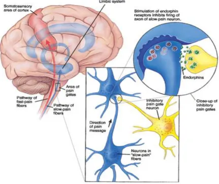
Figure 1: Illustration of the Gate Control Theory proposed Melzack and Wall in the
1960's. ... 18
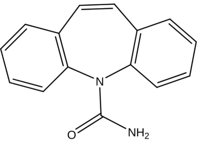
Figure 2: Chemical structure of carbamazepine. ... 28
Figure 3: Chemical structure of oxcarbazepine. ... 29
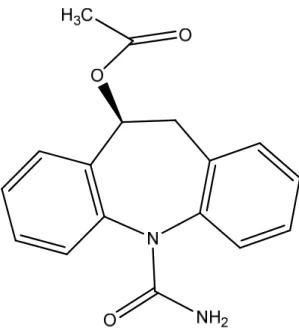
Figure 4: Chemical structure of eslicarbazepine acetate. ... 30
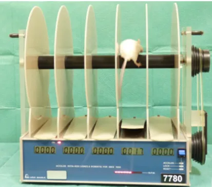
Figure 5: Rotarod apparatus for testing motor coordination. ... 40
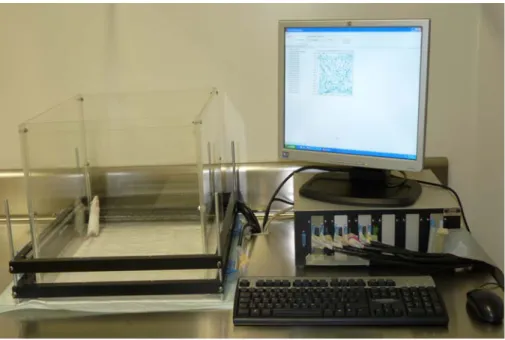
Figure 6: Photobeam Activity System – Open Field. ... 42
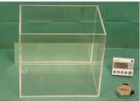
Figure 7: Observation chamber, chronometer and counter. ... 44
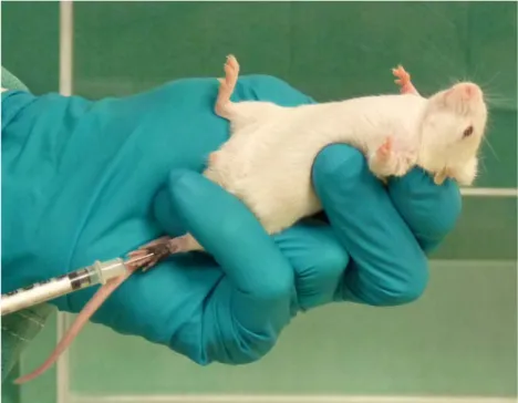
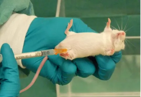
Figure 8: Subcutaneous injection of formalin solution in the dorsal part of the left hindpaw of the mouse. ... 45
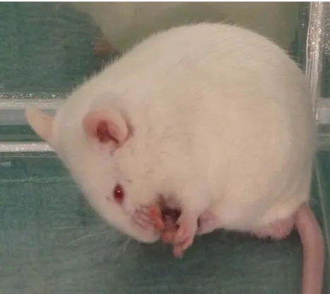
Figure 9: Mouse licking formalin injected paw after subcutaneous injection of formalin solution in the dorsal part of the left hindpaw. ... 46
Figure 10: Intraperitoneal injection of acetic acid solution. ... 49
Figure 11: Mouse performing a writhe in the writhing test. ... 50
Figure 12: Effect of eslicarbazepine acetate, carbamazepine and oxcarbazepine orally administered doses on motor performance of mice expressed as time spent on rotarod (seconds). ... 55
Figure 13: The median toxicity dose (TD50) value for eslicarbazepine acetate-induced motor impairment in mice. ... 56
Figure 14: The ambulation frequency in the open field test after oral administration of eslicarbazepine acetate (1 – 450 mg/kg). ... 57
Figure 15: The rearing frequency in the open field test after oral administration of eslicarbazepine acetate (1 – 450 mg/kg). ... 58
Figure 16: The open field test for carbamazepine (100 and 300 mg/kg) and oxcarbazepine (100 and 300 mg/kg) orally administered. ... 59
Figure 17: Effect of eslicarbazepine acetate orally administered in the late phase of
the formalin paw test (15 – 30 min). ... 62 Figure 18: Protective effect of oral administration of vehicle (V), eslicarbazepine
acetate (ESL) 300 mg/kg, carbamazepine (CBZ) 100 mg/kg, oxcarbazepine (OXC) 100 mg/kg and morphine (M) 64 mg/kg on the nociceptive reaction to subcutaneous injection of formalin in the hindpaw
of mice. ... 63 Figure 19: ED50 value for eslicarbazepine acetate licking/biting response in the late
phase of the formalin paw test. ... 64 Figure 20: ED50 value for carbamazepine licking/biting response in the late phase of
the formalin paw test. ... 65 Figure 21: Effect of eslicarbazepine acetate in writhing test, expressed as number of
writhes induced by p.o. injection of acetic acid in mice.. ... 68 Figure 22: Protective effect of oral administration of vehicle (V), eslicarbazepine
acetate (ESL) 300 mg/kg, carbamazepine (CBZ) 100 mg/kg, oxcarbazepine (OXC) 100 mg/kg and morphine (M) 64 mg/kg on the
nociceptive reaction to intraperitoneal injection of acetic acid in mice. ... 69 Figure 23: ED50 value for eslicarbazepine acetate acetic acid-pain evoked response in
LIST OF TABLES
Table 1: The effects of different doses of eslicarbazepine acetate and single doses of carbamazepine, oxcarbazepine and morphine in the late phase of the formalin paw test (evaluated between 15 and 30 minutes after injection of
formalin) in mouse. ... 61 Table 2: The effects of different doses of eslicarbazepine acetate and single doses of
carbamazepine, oxcarbazepine and morphine in the writhing test (evaluated
for 10 minutes, starting 5 minutes after injection of acetic acid) in mouse.. ... 67 Table 3: ED50, TD50 values and the corresponding Protective index (PI), with 95%
confidence limits (95% CL) and correlation coefficient (R2) in inducing
antinociception in the formalin paw test and motor impairment in mice. ... 72 Table 4: ED50, TD50 values and the corresponding Protective index (PI), with 95%
confidence limits (95% CL) and correlation coefficient (R2) in inducing
LIST OF ABBREVIATIONS
ANOVA - Analysis of variance CBZ – Carbamazepine CL – Confidence interval cm - Centimetre CMC – Carboxymethyl cellulose CYP – Cytochrome EC – European Commission
ED50 - The dose that was antinociceptive in 50 % of animals tested
ESL - Eslicarbazepine Acetate
FELASA - Federation of European Laboratory Animal Science Associations FRO - The formalin paw test associated with the rotarod test and the open-field test kg – Kilogram M – Morphine mg - Milligram min - Minute ml – Milliliter mm – Millimeter N - Sample size NMDA - N-methyl-D-aspartate
NSAID - Nonsteroidal anti-inflammatory drug OXC – Oxcarbazepine
PI – Protective Index p.o. - Oral administration
R2 – Coefficient of determination sec - Second
S.E.M. - Standard error of the mean
tmax - The time that it takes to reach the maximum concentration of drug
TD50 - The dose that induces motor impairment in 50% of animals tested
V - Vehicle
v/v - Volume in volume w/v - Weight in volume
WRO - The writhing test associated with the rotarod test and the open-field test µl - Microliter
CHAPTER
I
1 Pain
Pain has been part of the human experience for more than 50,000 years. Pain has been a very real and immediate concern in all ages, but the attitudes and responses of people to pain have been shaped by magical, theological, demonological, philosophical, and practical influences in varying degrees with shifting emphasis (Kucharski and Todd, 2008).
Pain is the most frequent reason patients seek medical advice. Pain is the body's mechanism of self-preservation. There is no consensual definition for pain, but it was described by the International Association for the Study of Pain, in November 2007, as “An unpleasant sensory and emotional experience associated with actual or potential tissue damage, or described in terms of such damage”. Pain tells you when your finger is touching a hot surface or when a fall has resulted in an injury that requires your attention. In this way, pain acts as a warning sign to alert you when damage to your body is occurring or may occur. Also, pain promotes the healing process as we take great care to protect an injured body part from further damage to minimize the experience of more pain. In fact, the inability to experience pain is a dangerous condition, because injury can occur and go unnoticed. Those individuals who are born with conditions in which they lack nociceptors (receptors for noxious stimuli), tend to live short lives due to their inability to recognize the pain stimuli as a signal for potential or ongoing tissue damage.
Pain can be classified in different ways. From the point of view of its aetiology, pain can be nociceptive induced or it can be neuropathic. Nociceptive pain refers to pain arising from direct exogenous (mechanical, chemical, thermical, and electrical) or endogenous (inflammation, and tissue ischemia) stimulation of nociceptors, such as occurs with trauma or inflammation. Neuropathic pain refers to pain arising from damage to the nervous system. Pain can also be spontaneous, associated with psychological factors and so called psychogenic (Dubner and Hargreaves, 1989). Some types of mental or emotional problems can cause, increase, or prolong pain.
From the point of view of its duration, pain can be classified in acute pain and chronic pain. Acute pain occurs for brief periods of time and it is always an alarm signal that something may be wrong. Chronic pain (also referred to as “persistent pain”) is continuous and recurrent and is one of the symptoms of chronic diseases. The boundaries between acute and chronic pain are
not defined. Acute pain can range from momentary pain (may last a few seconds or as long as the
stimulus is applied) to several hours or days after an injury (also referred to as “clinical pain”)
while chronic pain is defined as pain that lasts more than three months, whether it is due to a continuous inflammatory process or a neuropathy (a disease or abnormality of the nervous system) (Purves et al., 2008) .
Scientific evidence gathered over the years made it clear that many different mechanisms modulate the transmission of nociceptive information at every level of the nervous system. The first evidence of the existence of a pain modulation system was the observation that electrical stimulation of discrete areas of the brain inhibited responses to painful stimuli (Fields and Adams, 1974). At about the same time, the discovery of opiate receptors and of endorphins provided insight into the manner in which narcotic analgesics relieve pain. It is now generally accepted that pain modulation is achieved through a series of complex interactions between inputs from the periphery, interneurons in the spinal cord and descending control systems from the brain (Fields, 1984).
In the last twenty years, extraordinary progress has been made on understanding the physiology of pain (Purves et al., 2008). The discovery of a complex nervous system network for the modulation of pain, and the isolation and description of endorphins had a great contribute to our understanding of pain transmission (Steeds, 2009). Nevertheless there are still many areas of the physiology of pain that remain unknown. The physiology of pain involves nociceptors, combined with an intricate system of afferent and efferent neuronal connections. Nociceptors are relatively unspecialized nerve cell endings that initiate the sensation of pain (noci- is derived from the Latin for “hurt”) (Purves et al., 2008). All nociceptors are free nerve endings that are sensitive to painful mechanical stimuli, extreme heat or cold, and chemical stimuli. Most pain originates when nociceptors are stimulated and nerve impulses are transmitted to the brain through the pain pathways. It is important to note that almost all body tissues are equipped with nociceptors, an important fact considering pain has primary warning functions. Nociceptors can be classified in two types: unimodal receptors and polymodal receptors (Nathan, 1976; Bonica, 1990). Unimodal receptors, also known as mecanothermal nociceptors, are mainly present in the skin and respond to strong pressure applied to the skin and strong stimuli as pinprick or sudden heat (greater than 45°C). This type of receptor is mainly associated with small myelinated primary afferent neurons designated Aδ type that transmits impulses rapidly. Stimulation of this
type of receptor results in pain that occurs early after injury and is usually sharp, well-localized, and pricking (Nathan, 1976; Bonica, 1990). Polymodal receptors are the free nerve endings of unmyelinated primary afferent neurons of the C type which are widely distributed throughout most tissues and respond to tissue damage (Nathan, 1976; Bonica, 1990). The best known theory describing how painful stimuli may be altered at the spinal level is the Gate Control Theory (Melzack and Wall, 1965). The Gate Control Theory is illustrated in Figure 1. This theory postulates that painful stimuli have to pass through a gate in order to be communicated to the central nervous system, lacking one-to-one correspondence between the severity of the stimulus and the severity on pain experience. According to this theory, the peripheral nervous system communicates with the central nervous system through a complex interplay: upon injury, pain messages originated in the nociceptors associated with the damaged tissue flow along the peripheral nerves to the spinal cord where they encounter “nerve gates” that open or close depending upon a number of factors, after which the modulated pain messages reach the brain. So, the modulation of the pain messages occurs at the spinal cord level. When the gates are open, pain messages reach the brain more or less easily and pain can be intense. When the gates are closed, pain messages are prevented from reaching the brain without even being experienced. The spinal cord receives inputs from 2 types of nerve fibers: Aδ fibers and the C-fibers (Steeds, 2009). A typical example of the Gate Control Theory is that after bumping one’s knee, rubbing the area seems to provide some relief because this action activates other sensory nerve fibers that are even “faster” than Aδ fibers which send information about pressure and touch that reach the spinal cord and brain, overwriting some of the pain messages carried by the Aδ fibers and the C-fibers (Steeds, 2009).
One of the most critical aspects in pain research is its assessment; pain is a highly complex phenomenon that by its nature presents assessment difficulties. Pain is a subjective experience which is difficult to express and quantify, and is not directly measurable. Further difficulties occur since individual patients react to similar painful stimuli in many different ways. In addition many factors are known to influence pain response. They include ethnicity, sex, age, culture, personality, the type, duration and intensity of pain, and other psychological variables such as fear, anxiety and stress. However, the measurement of pain is essential for the study of its mechanisms and for the evaluation of pain control methods.
Figure 1: Illustration of the Gate Control Theory proposed
Melzack and Wall in the 1960's. Available from http://www.garysturt.free-online.co.uk/pain.htm
2 Animal Models of Pain
Throughout history, scientists have performed experiments on animals to understand animal and human biological structure and function. Practical, economical and scientific reasons make preliminary studies in animals the best solution for studies of a human biological phenomenon. A laboratory animal model describes a biological observable fact that the species has in common with the target species, being “analogy” the key word for understanding the concept of animal models (Rose and Woodbury, 2001). What is generally understood by the term animal model is modelling humans. The focus of research is not the used animal but the analogy of physiological behaviour of this animal to our own species. The practice of studying biological phenomena and diseases in laboratory animal models is well established in biomedical sciences (Rose and Woodbury, 2001). The valid “extrapolatability” of results generated in an animal model depends on the selection of a suitable animal model. Induced animal models involve healthy animals in which the condition to be investigated is experimentally induced.
Animal models of pain have been invaluable tools for understanding pain mechanisms. To study pain transmission, identify new pain targets and characterize the potential analgesic profile of novel compounds, an array of experimental animal pain models has been developed (mainly in rodents) attempting to replicate the many human pain conditions, including inflammatory, neuropathic, visceral and cancer pain states (Walker et al., 1999; Joshi and Honore, 2006). The experience of pain is necessary for survival, so it is reasonable to assume that the mechanisms of pain perception are highly conserved across species (Liebeskind, 1991). This assumption has led to a vast array of important findings concerning pain via experiments conducted in rodents (Liebeskind, 1991). Animal models of pain are mostly models of nociceptive pain (pain arising from an identifiable lesion causing tissue damage, accompanied by stimulation of nociceptors in somatic or visceral structures) (Steeds, 2009). In animals, as well as in humans, in order to study pain of any kind, the appropriate stimuli must be applied in order to provoke the desired pain sensation. Different nociceptive pain tests use different stimuli with different characteristics: (i)
stimulus may be thermal, mechanical or chemical; (ii) may or may not produce tissue damage;
(iii) and may produce a short or a long lasting pain response (Franklin and Abbott, 1989).
Animal models of nociceptive pain have been developed as assays of pain behaviour and fall mainly into two categories: (i) those that employ an acute nociceptive stimulus and that are
concerned with short-term nociceptive responses; (ii) and those that are concerned with the long-term responses (Walker et al., 1999; Rose and Woodbury, 2001).
There are two main types of methods for assessing pain, based on the duration of the applied stimulus: (i) the response to the stimulation is fixed and stimulation intensity increases until a defined standard response occurs; (ii) the stimulus is standardized, and the strength and duration of response is measured (e.g. the formalin paw test and the writhing test) (Meyer and Svendsen, 2003). The formalin paw test and the writhing test constitute methods for the measurement of pain by behavioural responses to stimulation of nociceptors by injection of irritant substances and are amongst the most frequently used contemporary models of pain (Rose and Woodbury, 2001).
2.1 Formalin Paw Test
The formalin paw test represents a model of moderate continuous pain, suitable for the evaluation of mild analgesics (Hunskaar et al., 1985). The chemical stimulation involving the administration of formalin is a progressive stimulation, with a duration in the order of minutes and with an inescapable character once it has been applied (Le Bars et al., 2001). In 1939, Lewis and Kellgren injected small volumes of hypertonic saline to produce experimental pain in humans (Tjolsen et al., 1992). This kind of work preceded the use of formalin as a noxious agent for animals (Tjolsen et al., 1992). Dubuisson and Dennis (1977) introduced the formalin test, administering 50 µl of 5 % formalin under the dorsal surface of a forepaw of rats and cats.
Following subcutaneous injection of formalin into the hindpaw of an animal, it displays spontaneous pain behaviour that is increased hindpaw licking/biting. The formalin paw induced-pain response is difficult to assess in the forepaws of rodents, because normal grooming behaviour is typically based on animals licking the forepaws. The response to licking the hindpaws is assumed to be nociceptive (Tjolsen et al., 1992). The formalin-induced pain has been reported to be quantified in many different ways. Dubuisson and Dennis originally created behavioural categories that were used to assess formalin-induced pain by the weighted score method of behavioural rating (Coderre et al., 1993). Others used cumulative score methods to
assess pain-evoked responses, using a single parameter, such as scoring the total amount of time spent licking, biting or shaking the injected paw (Hunskaar and Hole, 1987).
Characteristically the formalin paw test produces a biphasic response, with the two phases being distinguished pharmacologically. An early acute phase (first phase) occurring immediately after the injection of the diluted formalin solution lasts for about 3 - 5 minutes, which is thought to be caused by direct chemical stimulation of nociceptors (Abbott et al., 1995). A 10 - 15 minutes gap between the two phases occurs until the second phase begins (Tjolsen et al., 1992). The first phase is considered by some authors to be the early acute phase together with the gap between the two phases lasting for about 15 minutes (Abbott et al., 1995). A later tonic phase (second phase) starting 15 minutes after the formalin injection and lasting for no more than 40 minutes, is believed to occur due to a peripheral inflammatory process mediated by prostaglandins (Tjolsen et al., 1992).
Formalin is the most common substance used as noxious stimuli for intradermal injections in several animal species. (Wheeler-Aceto and Cowan, 1991). Formalin is an aqueous solution of 37 % (w/v) formaldehyde. A range of 0.02 to 5 % formalin solution concentrations have been reported in mice, with the most often injected volumes ranging from 20 to 25 µl (Tjolsen et al., 1992). It has been demonstrated that the subcutaneous injection of different concentrations of formalin may induce the first phase or both phases; formalin concentrations of 0.05 to 0.2 % induce high licking/biting activity only in the first phase and formalin concentrations of 1 % or higher induce high licking/biting activity on both the first and the second phases (Rosland et al., 1990).
It has been reported that there are sex differences in the pain–related responses. Female mice show lower pain–related responses compared to males in the formalin paw test (Capone and Aloisi, 2004). Experimental results suggest that estrogen reduces the efficacy of endogenous pain modulation mechanisms, triggering an increase in spinal nociceptive neuronal activity (Spooner et al., 2007).
Many publications refer to the mouse paw formalin test being done using mice habituated and not habituated to the testing environment and handling (Capone and Aloisi, 2004). Exposure to handling-induced stress or to a new environment strongly influences the formalin-induced
behaviour in mice (Tjolsen et al., 1992). Animals should be well adapted to the test observation chamber because the occurrence of the typical exploratory activity of animals in a new environment could distract them from the formalin-induced pain response. They should also be well habituated to handling and restraining because any abnormal discomfort may provoke stress in the animals which can mask the formalin-induced pain results.
2.2 Writhing Test
The writhing test consists of intraperitoneal injection of agents that irritate serous membranes, inducing a very typical behaviour in the mouse and rat which is characterized by abdominal contractions, movements of the hindpaws and twisting of dorsoabdominal muscles (Hendershot and Forsaith, 1959). Some authors consider these behaviours as resulting from visceral pain and others reject this hypothesis and consider the pain as arising from peritoneal pain (Le Bars et al., 2001). Several compounds are used, the most common being the acetic acid. Several modifications to this method have been made over the years, mainly concerning the chemical agent which causes the stimulus, its concentration and the volume of the injected solution.
Acetic acid is an algogenic substance that directly activates peripheral nociceptors on the sensory nerve fibers by inducing capillary permeability and releasing endogenous substances that excite pain nerve endings (pro-inflammatory substances) producing inflammation of visceral (subdiaphragmatic) and subcutaneous (muscle wall) tissues (Cervero, 1995). This produces the characteristic ‘writhing response’: lengthwise stretches of the torso with a concomitant concave arching of the back and extensions of the hind limbs (Collier et al., 1968; Bentley et al., 1981). Mice and rats present a very stereotyped behaviour when administered intraperitoneally with agents that irritate the serous membranes (Rose and Woodbury, 2001).
The acetic acid-induced writhing is a pain model widely used for detecting peripheral analgesia (Du et al., 2007). The original test was described in 1957 by Siegmund et al. and used phenylbenzoquinone as irritating agent but since then many modifications have been made to the method mainly concerning the chemical agent that, in turn, determines the duration of the effect:
acetylcholine, dilute hydrochloric acid, bradykinin, adrenaline, potassium chloride, tryptamine, ocytocin and acetic acid have all been used (Le Bars et al., 2001). Modifications to the concentration, temperature, and volume of the injected solution, the experimental conditions, and ways of monitoring behavioural changes have also been made in order to simplify the test and increase its sensitivity (Le Bars et al., 2001). This method has the advantage of allowing evidence to be obtained for effects produced by weak analgesics (Le Bars et al., 2001). Because all analgesics inhibit abdominal cramps, this method is useful for sorting through molecules whose pharmacodynamic properties are unknown (Le Bars et al., 2001).
3 Other Behavioural Models
3.1 Rotarod Test
The loss of motor coordination is one of the most promptly observed effects of drug intoxication. The rotarod test is widely used in biomedical research as one of the most commonly used tests of motor incoordination in rodents. Dunham and Miya (1957) were the first to describe a fixed speed rotating rod, called by these authors “rolling roller apparatus” for detecting neurological deficits in rats and mice. It is frequently used in early stages of drug development to screen-out drugs that might later cause impairment in human motor behaviour. Ataxia (loss of motor coordination) is a frequent endpoint for studies of drug intoxication. One of the most commonly used behavioural models of ataxia is the rotarod test (Rustay et al., 2003). The rotarod test assesses the effect of drugs on an animal’s balance and coordination or fatigue resistance on mice and rats, by measuring the amount of time that an animal is able to remain on a longitudinally rotating rod. Animals have to keep their balance on a rotating textured rod. When an animal drops onto the individual sensing platforms below, test results are recorded. Performance is measured by the time that an animal stays on the rod as a function of drum speed.
Many different rotarod systems are available from a number of companies, but almost all apparatus will automatically record the time at which the animal falls, and allow several animals to be tested at the same time (Carter et al., 2001). Many significant modifications have been made to the basic design of the first described rotarod test, like accelerating the rod continuously until the mouse or rat can no longer balance and fall, changing rod diameters from 3 - 8 cm, using fixed-speed rates of 3 - 31 rpm and acceleration rates of 3.5 - 60 rpm/min (Rustay et al., 2003). There are two basic versions of the rotarod test that are widely used: the fixed-speed and the accelerating-speed rotarod (Rustay et al., 2003; Monville et al., 2006). There is still little information on their equivalence or the relative power, reliability and sensitivity of the two protocols. The fixed-speed rotarod system allows changing parameters as rod diameter, maximum time limit for performance and rate of rotation, and requires extensive training of the animals. The accelerating-speed rotarod system eliminated the existence of a maximal time limit for performance, providing a model that starts at a slow speed allowing all animals to stay on and
accelerating at a constant rate until it becomes difficult enough so that all animals would eventually fall off (Rustay et al., 2003). This model allows for the measurement of drug-induced increased in performance, which is difficult to observe using a low rotation rate in the fixed-speed model.
Some strains of rodents are not suitable for usage in the rotarod test due to their propensity to jump from the rod instead of running on top. Also some strains have a propensity to hold onto the rod and instead of actively completing the task, they passively rotate around. Rustay et al. (2003) engineered a way to circumvent this confounding factor by using a rod of 6.3 cm of diameter, which was large enough to prevent most mice from being able to hold to the rod, avoiding passive rotation. These authors also recommend that, independently of the usage of a fixed-speed or accelerating-speed rotarod system, mice should be trained to a stable level of performance before drug administration so that changes seen after drug administration are the result of drug action and not due to effects on learning on the apparatus (Rustay et al., 2003).
3.2 Open-field Test
Open-field test is a common measure of general locomotion activity and exploratory behaviour in rodents (Walsh and Cummins, 1976). It was first developed by Hall in 1934 (Choleris et al., 2001) and it is commonly used to assess the sedative, toxic, or stimulant effects of compounds (Gould et al., 2009). The open-field test was defined by Choleris et al. (2001) as “an enclosed open area where an animal is placed and some form of behaviour, usually activity, is measured”. The open-field test was widely used in psychology and it has gradually spreaded out to neurosciences and psychopharmacology (Tvrdeic and Kocevski, 2008). The original studies were in rats, but this test has also been extensively used in mice (Gould et al., 2009).
This test allows the measurement of both qualitative and quantitative activity. But, although sometimes the open-field test is considered a “standard test”, many factors can vary among studies like size of the open field arena, shape, level of illumination, single exposure versus repeated exposure to the open-field arena, duration of testing, time of day of testing, food and water deprivation, sex of the animals, predator odour in the arena (Choleris et al., 2001). Most of the automated open-field test systems allow the measurement of the locomotor activity
by determining the amount of distance travelled as well as the various horizontal, vertical and stereotyped behaviours like time spent along the walls compared to time spent in the centre, distance moved over different time periods, and rearing. Typically, in psychopharmacological studies, animals are naive to the open-field arena and the effects of the pharmacological agents on the duration and frequency of some behaviour like locomotion and rearing are assessed (Choleris et al., 2001).
4 Antiepileptics as Adjuvant Analgesics
Pain can be treated in several different ways. Analgesics are drugs that relieve pain without blocking the conduction of nerve impulses or markedly altering the function of the sensory apparatus (Álamo and López-Muñoz, 2007). Analgesics are taken both to control acute and chronic pain. Analgesics are classified is three categories: (i) opioid analgesics, also called narcotics; (ii) non-opioid analgesics, also called non steroidal anti-inflammatory drugs – NSAIDs; and (iii) adjuvant analgesics, which are drugs with a primary indication other than pain but that have analgesic properties (Lussier et al., 2004; Becker and Phero, 2005).
Throughout the clinical usage of antiepileptic drugs, it has been observed that these drugs can be used in many diseases other than epilepsy. Antiepileptic drugs are already used in the treatment of bipolar disorder (Dilsaver et al., 1996), impulse control disorder (Berlin, 2008), eating disorders (Kaplan et al., 1983) and specially to manage pain (McQuay et al., 1995). Antiepileptic drugs have been used in the management of pain since the 1960s (McQuay et al., 1995). The presence of common clinical and physiopathological characteristics between epilepsy and several pain types made possible the usage of different antiepileptic drugs as they would appear in the market (Álamo and López-Muñoz, 2007). In 1912, phenobarbital, the first antiepileptic drug was marketed, but the appearance of more antiepileptic drugs has occurred at a very slow step (Álamo and López-Muñoz, 2007). One group of antiepileptic drugs containing carbamazepine and that is called nowadays classic antiepileptic drugs, was commercialized just before 1970 (Álamo and López-Muñoz, 2007). In 1990 more antiepileptics such as oxcarbazepine were marketed, being more effective, presenting less adverse effects and showing better pharmacokinetic and pharmacodynamic characteristics than the previously marketed antiepileptic drugs (Álamo and López-Muñoz, 2007). In 2009, about twenty years later, eslicarbazepine acetate was developed as a new antiepileptic drug which is structurally related to carbamazepine and oxcarbazepine, but with improved tolerability profiles and efficacy (Dulsat et al., 2009).
4.1 Carbamazepine
Kingdom for clinical treatment of epileptic seizures and has become the most frequently prescribed first-line drug for the treatment of partial and generalized tonic-clonic epileptic seizures all over the world (Araujo et al., 2004).
Although it is very effective in the majority of cases, soon it started showing a number of unwanted side effects. In clinical trials, about 10 to 25% of patients administered with carbamazepine showed adverse effects, with a higher incidence in the elderly (Sillanpaa et al., 2009). Carbamazepine active epoxide metabolite is the responsible for a number of adverse effects associated with carbamazepine therapy (Grant and Faulds, 1992). The main metabolic pathway is epoxidation which is catalysed primarily by the cytochrome P450 and results in the formation of a pharmacologically active carbamazepine epoxide metabolite, contributing to both therapeutic and adverse effects (Sillanpaa et al., 2009). Carbamazepine clinical application is complicated by the potent induction of hepatic oxidative metabolism, the potential for adverse reactions and its slow and variable absorption (Hainzl et al., 2002).
N
NH
2O
Figure 2: Chemical structure of carbamazepine.
Carbamazepine has been used for the treatment of neuropathic pain in humans. Recently, several studies have reported that carbamazepine has antinociceptive activity, for it has shown to be effective in different animal models of acute pain induced by inflammatory, thermal and mechanical nociceptive stimuli (Aoki et al., 2006).
4.2 Oxcarbazepine
Oxcarbazepine (Figure 3) was introduced commercially in Denmark in 1990, has been available throughout the European Union since 1999 and was launched in 2000 in the USA for the management of seizures (Faught and Limidi, 2009). Oxcarbazepine is an antiepileptic drug derived from carbamazepine.
Oxcarbazepine is the 10 - keto analogue of carbamazepine with a differing metabolic profile that manages to circumvent the carbamazepine adverse effects, retaining similar therapeutic utility (Grant and Faulds, 1992). After administration, oxcarbazepine is rapidly reduced to an active metabolite that is present in human plasma at much higher concentrations than the parent drug and that is the principal active agent (Faught and Limidi, 2009). Oxcarbazepine and its metabolite are neutral lipophylic agents that might be expected to easily pass through the blood-brain barrier and are widely distributed in the body without any significant affinity to a particular site (Álamo and López-Muñoz, 2007). The elimination of the double bond between C-10 and C-11 prevents oxidative attack by an epoxidase, which is what occurs with carbamazepine. It has a better side-effect profile and many fewer metabolic drug-drug interactions than carbamazepine, because it is metabolized by a non cytochrome P450 pathway (Grant and Faulds, 1992).
Oxcarbazepine exerted analgesic effects in animal models of neuropathic, inflammatory, somatic, and visceral pain (Tomic et al., 2010). The mechanisms of analgesia of oxcarbazepine are not completely understood but it was demonstrated that it suppresses peripheral sensory nerve firing, probably by blockade of sodium currents, and also affects some receptors involved in pain modulation to produce antinociception (Tomic et al., 2010).
4.3 Eslicarbazepine Acetate
Eslicarbazepine acetate (Figure 4) is a drug clinically developed for the treatment of epilepsy, with EMEA recently granting marketing authorization to BIAL (Dulsat et al., 2009). It is currently under clinical development for the treatment of neuropathic pain (Almeida et al., 2008; Almeida et al., 2009).
Eslicarbazepine acetate is a novel voltage-gated sodium channel blocker, designed to share with carbamazepine and oxcarbazepine the dibenzazepine nucleus bearing the 5-carboxamide substitute, but it is structurally different at the 10,11-position (Benes et al., 1999; Hainzl et al., 2001; Almeida and Soares-da-Silva, 2007). This molecular variation results in differences in metabolism which, among other things, prevents the formation of toxic epoxide metabolites (Almeida and Soares-da-Silva, 2003). Eslicarbazepine acetate mainly undergoes metabolic hydrolysis followed by glucoronidation with minimal CYP-mediated metabolism (Almeida et al., 2009). Oral administration of eslicarbazepine acetate to humans results in very low plasma concentrations of parent compound which is extensively biotransformed to S-licarbazepine by first-pass hydrolytic metabolism, being clearly the major circulating drug (Almeida et al., 2008).
There have been no concerning findings for human use of eslicarbazepine acetate, based on conventional preclinical studies of safety, pharmacology, toxicology, genotoxicity, reprotoxicity and carcinogenity, which makes believe that its use will undoubtedly increase in the future (Almeida et al., 2009).
5 Objectives
5.1 General Objective
Development and implementation of animal models for evaluation of drugs with potential analgesic activity, in NMRi mice.
5.2 Specific Objectives
• Development and implementation of the formalin paw test in mice; • Development and implementation of the writhing test in mice; • Development and implementation of the rotarod test in mice; • Development and implementation of the open-field test in mice;
• Examination of the dose-dependent effect of eslicarbazepine acetate on antinociception using the formalin paw and the writhing tests, on motor function using the rotarod test and on spontaneous exploratory behaviour and general activity using the open-field test; • Determination of the protective index of eslicarbazepine acetate.
CHAPTER
II
1 Reagents
All reagents and solutions used in all experimental procedures were of at least standard laboratory grade. The water used was obtained from an ultra-pure water producing system.
The reagents used and their respective suppliers are presented next: - Acetic Acid (Sigma-Aldrich®);
- Carbamazepine (Sigma-Aldrich®);
- Carboxymethyl cellulose (Sigma-Aldrich®);
- Eslicarbazepine Acetate (BIAL-Portela & Cª, S.A.); - Ethanol (Sigma-Aldrich®);
- Formalin solution 10 % (Sigma-Aldrich®); - Morphine (Sigma-Aldrich®);
- Oxcarbazepine (ChemPacific); - Sodium Chloride (Sigma-Aldrich®).
2 Drugs, Solutions and Treatment Groups
Administered vehicle was carboxymethyl cellulose (CMC) 0.5 % in ultra-pure water. The test drug eslicarbazepine acetate, the comparison drugs carbamazepine and oxcarbazepine and the reference drug morphine were dispersed in CMC 0.5 %.
Formalin (5 %) was obtained by dilution of the formalin solution 10 % in sodium chloride 0.9 % in ultra-pure water.
Acetic acid (0.8 %) was obtained by dilution of acetic acid reagent in sodium chloride 0.9 % in ultra-pure water.
Animals administered with the vehicle substance were considered to be the control group. Animals administered with the different concentrations of the test drug eslicarbazepine acetate (1 - 450 mg/kg), the comparison drugs carbamazepine (100 and 300 mg/kg) and oxcarbazepine (100 and 300 mg/kg), and the reference drug morphine (64 mg/kg) were considered to be the treated groups.
3 Laboratory Equipment
The laboratory equipment used is presented next:
- 26 gauge needle attached to a micro syringe by PE10 tubing; - 29 gauge needle attached to a micro syringe by PE10 tubing; - Automatic Pipettes Gilson PIPETMAN®;
- Balance Kern® ABJ;
- Balance Mettler Toledo® XP26; - Chronometer;
- Magnetic Stirrer IKA®; - Manual counter;
- MilliQ water system (Millipore ®); - Mirror;
- Plexiglass observation chambers (20 x 26 x 26 cm);
- Potentiometer WTW® pH 3000 with coupled electrode SenTix® 21;
- Rotarod apparatus with a rotating cylinder with a 6.3 cm diameter rod (Ugo Basile Biological Research Apparatus);
- San Diego Instruments Photobeam Activity System – Open Field composed by: o Clear acrylic enclosure (40 cm wide x 40 cm deep x 38 cm high);
o Photobeam mounding frame (with 16 photobeams in each direction, spaced by 2.5 cm);
o Rear frame; o Control unit; - Ultrasonic bath;
4 Softwares
The softwares used for data collection and data analysis are the ones described next: - GraphPad Prism® 5.02 (GraphPad Software, Inc.);
- Microsoft® Office Excel 2007;
- Photobeam Activity System Control software for Windows 2000/XP (version 2.0).
5 Animals
5.1 Species Used
Male NMRi mice, 30 - 40 g body weight range were used. They were supplied by Harlan Interfauna Ibérica, S. L., Spain.
5.2 Animal Housing
Animals were delivered to the laboratory (Laboratory of Pharmacological Research of the Department of Research and Development at BIAL - Portela & Cª S. A.) 2 weeks before the experiment, during which time they were acclimatized to the laboratory conditions. They were housed in groups of 5 in macrolon cages (25 x 19 x 13 cm) on wood litter (Litalabo - SPPS, 95100 Argenteuil, France) with free access to food (food certified by Harlan Teklab Global Diet®) and water until the day before being tested. The animal house was maintained under
artificial lighting (12 hours) between 8 am and 8 pm, at controlled ambient temperature of 22 ± 2 ºC, and relative humidity of 60 ± 10 %.
5.3 Ethical Aspects
The EC directive states that an animal should not be kept alive after an experiment if pain is experienced. At the end of the experiment, animals were sacrificed by the mechanical-physical method of dislocation of the cervical vertebrae, by stretching the animal and rotating the neck, away from the presence of other animals, which constitutes a painless method (Hellebrekers et al., 2001). These procedures were executed in accordance with the recommendations of the Federation of European Laboratory Animal Science Associations (FELASA), which is in accordance with the European Directive no. 86/609 and with the Portuguese legislation (Decreto-Lei 129/92, Portarias 1005/92 and 1131/97). The number of animals used was the minimum possible in compliance with current regulations and scientific integrity.
5.4 Justification for the Choice of Species, Route of Administration and Dose Levels
The choice of the species for a chosen animal model depends on the homology and evolutionary similarity between morphological structures and physiological processes between animals and humans (Beynen and Hau, 2001). Although mice and rats have many concurrent biological characteristics, they do not necessarily serve equally well as models of human malady. Mouse was chosen as animal model because of economical advantages and also because mice have shown the most similar metabolism to humans after administration of eslicarbazepine acetate (Alves et al., 2008).
The route of administration of compounds and vehicle were chosen based on the intent of oral administration of the compounds to humans. Mice animals were deprived of food for 18 hours prior to the experiment to avoid any possible food-drug interaction.
The study included several doses for the assessment of pain. The chosen doses were
based on the envisage determination of the effective doses for analgesic effect on the developed models.
To avoid any possible effect of the circadian rhythm in the animal models of pain developed during this experimental work, half of the tested animal for each group were tested in the morning and half in the afternoon.
6 Rotarod Test
The rotarod test allows the assessment of motor coordination and balance in rodents. Using rotating rods allows one to evaluate the duration that mice and rats can maintain balance at a defined rotating speed. Dunham and Miya (1957) described the first rotating rod for detecting neurological deficit in rats and mice.
6.1 Habituation to Test Environment
The newly acquired mice were housed for one week prior to being brought to the testing room for habituation. Adaptation was carried out in the testing room by transferring the animals, in their home cages, from the housing room to the testing room 2 hours before the training trials or before the test (Carter et al., 2001). For habituation to the rotating rod (Figure 5), two days and on the day before the experiment, each mouse was placed individually on the rotating rod and allowed to habituate to the forced motor activity for up to 60 seconds. Upon fall, each animal was returned to its home cage and allowed an inter-trial interval of 5 minutes. Mice were trained until they were able to remain on the rod for 2 consecutive trials (Carter et al., 2001). On the day of the experiment all animals were tested for their ability to stay on the rod for at least 60 seconds, to obtain the pre-drug time spent on the rod. Habituation to restraining and handling was performed between 8.00 am and 5.00 pm on the day before the rotarod test.
6.2 Experimental Procedure
The rotarod apparatus had a 6.3 cm diameter rod, suitably machined to provide grip and a fall high of 17 cm (Rustay et al., 2003). In this apparatus, a motor sets the rotor in motion via the gear belt at a selected speed. When the mouse falls off its cylinder section, the plate below trips and the corresponding counter disconnects, thereby recording the animal’s endurance time in seconds. The machine was set at a revolving consistent speed of 15 rpm; always before training the animals or testing them, the rotarod apparatus was manually calibrated so that it was revolving at a consistent speed of 15 rpm (Stepanovic-Petrovic et al., 2008).
Figure 5: Rotarod apparatus for testing motor coordination.
During the training sessions and at the end of testing each animal, the apparatus was thoroughly cleaned with 70 % (v/v) ethanol. The reason for this last described procedure is that animals defecate and urinate while on the rod and urine and faeces on the rod can impair the performance of the animals.
After adaptation of the animals to the testing room, the animals were separated in groups and each group was constituted by 10 animals under study, being each animal tested only once. Mice were placed on the rod while it was moving. The vehicle, test, comparison or reference drugs were administered orally 59 minutes before the test. All groups were administered by oral gavage at dosing volume of 8 ml/kg of body weight. Motor impairment was assessed for 60 seconds.
The time that the animal remained walking on the rod was scored in seconds, with the task ending when the animal fell off the rotarod or completed the 60 seconds trial. Many authors are concerned about how to score passive rotation when the animal grips on to the rotarod. That problem was circumvented here by using a 6.3 cm diameter rod, as proposed by Rustay et al. (2003). This author observed that using rods with 6.3 cm diameter or greater, the number of animals gripping on to the rotarod was neglectable.
6.3 Data Presentation
The results are expressed as mean ± S.E.M. (standard error of the mean) of the time spent on the rotarod in seconds. MS Excel was used to calculate mean and S.E.M..
The TD50 (the dose that induces motor impairment in 50 % of animals tested) was
calculated from a corresponding quantal dose–response curve, with 95 % confidence limits. The Boltzmann sigmoidal equation was used to derive the TD50 value. GraphPad Prism was used to
7 Open-Field Test
The open-field test was introduced in the 1930s by Calvin Hall (Leroy et al., 2009). The open-field test is a common measure of spontaneous exploratory behaviour and general activity in rodents that is often used to assess the sedative, toxic and stimulant effects of drugs. Recent open-field test systems allow registering general motor activity, locomotion, rearing and the speed of locomotion.
7.1 Habituation to Test Environment
The newly acquired mice were housed for one week prior to being brought to the testing room for habituation. Adaptation was carried out in the testing room by transferring the animals, in their home cages, from the housing room to the testing room 2 hours before the test and by transferring the animals from the rotarod to the open-field arena. Habituation to handling was performed between 8.00 am and 5.00 pm. The open-field test system is shown in Figure 6.
7.2 Experimental Procedure
The open-field arena consisted of an empty and transparent square arena, surrounded by walls that prevent the animal from escaping. A setup frame was mounted to fit a clear acrylic enclosure (40 cm wide x 40 cm deep x 38 cm high). The frame contains 2 rows of photobeams (16 photobeams mounted in each direction), spaced by 2.5 cm. Above the photo beam frame a rear frame was mounted to allow quantification of the number of rears that the animal performed. The photobeam frame was placed 30 mm above the top of the bench and the rear frame placed 80 mm above the top of the bench. Interruptions of the photobeams were collected by a computer by means of Photobeam Activity System Control software. Two parameters were evaluated: ambulation and rearing. Ambulation allows the assessment of normal motor activity and rearing allows the assessment of exploratory behaviour. The software identifies these two parameters as:
• Ambulation - all interruption of the photobeams in the lower rows; • Rearing - interruption of the photobeams in the upper rows (rear frame).
After adaptation of the animals to the testing room, the animals were separated in groups of 10 mice with each animal being tested only once. The vehicle, test, comparison or reference drugs, were administered orally 60 minutes before the test by oral gavage at dosing volume of 8 ml/kg of body weight. The same grouped mice tested for the rotarod test were tested in the open-field test. So, immediately after the rotarod test, mice were placed in the centre of the arena and allowed to explore for 15 minutes.
7.3 Data Presentation
The results are expressed as mean ± S.E.M. of the ambulation frequency and of the rearing frequency. Frequencies were defined as the number of occurrences per 15 minutes test time. MS Excel was used to calculate mean and S.E.M..
8 Formalin Paw Test
The formalin paw test was used to assess the antinociceptive effect of the administered drugs. The test described here is a modification of the one proposed by Hunskaar et al. (1985).
8.1 Habituation to Test Environment
Newly acquired mice were housed for one week prior to being brought to the testing room for habituation. Adaptation was carried out in the testing room on the day before the experiment by placing the animals singly in the observation chambers for 2 hours and on the day of the experiment by transferring the animals, in their home cages, from the housing room to the testing room 2 hours before the formalin paw test. The mouse observation chambers were clear plexiglass chambers with a mirror placed behind the chamber wall to allow an unobstructed view of the tested paw. Habituation to restraining and handling was performed between 8.00 am and 5.00 pm on the day before the formalin paw test. The observation chamber is shown in Figure 7, as well as a counter and a chronometer.
8.2 Experimental Procedure
After adaptation of the animals to the testing room, testing chamber, handling and restraining, using a 26 gauge needle attached to a micro syringe by PE10 tubing, 25 µl of the 5 % formalin was injected subcutaneously into the left dorsal part of the hindpaw of the mouse (Figure 8). Each animal was manually gently restrained in such a way that allowed the referred injection to occur. After the injection of the nociceptive stimulus, the animals were allowed to move freely during the observation period.
Figure 8: Subcutaneous injection of formalin solution in the
dorsal part of the left hindpaw of the mouse.
The vehicle, test, comparison or reference drugs, were administered orally 60 minutes before the test (i.e. 45 minutes before formalin) by oral gavage at a dosing volume of 8 ml/kg of body weight. Each group was composed of 10 animals under study. Each animal was tested only once and the tested paw was marked with a colour marker prior to the formalin injection, to allow
a clear observation of the formalin pain-evoked behaviour.
The mice were observed from 15 to 30 minutes following formalin injection. During the first 15 minutes post formalin injection, pain behaviour reflects the early phase of the formalin paw test and readings were neglected. The late phase of the formalin paw test is observed via pain behaviour in the 15 to 30 minutes time period following formalin injection and the pain-evoked behaviour was scored for this phase. The amount of time that each animal spent licking or biting the injected paw (pain-evoked behaviour) was timed with a chronometer. No food or water was available during the test. Figure 9 shows a mouse executing the typical pain-evoked behaviour caused by subcutaneous injection of formalin in the dorsal part of the left hindpaw.
Figure 9: Mouse licking formalin injected paw after
subcutaneous injection of formalin solution in the dorsal part of the left hindpaw.
8.3 Data Presentation
The results are expressed as mean ± S.E.M. of the amount of time that each animal spent licking or biting the injected paw after subcutaneous injection of formalin into the left dorsal part of the hindpaw of the mouse or as % protection conferred by orally administered drugs versus the vehicle administered group. The % protection was calculated as follows:
100 FCmean FTmean -FCmean Protection % = × (1)
“FCmean” represents the mean result for the amount of time that the control group mice spent licking/biting the injected paw following formalin injection.
“FTmean” represents the mean result for the amount of time that the treated group mice spent licking/biting the injected paw following formalin injection.
MS Excel was used to calculate mean, S.E.M. and % protection values.
The values of ED50 (the dose that was antinociceptive in 50 % of animals tested) were
estimated from the corresponding log dose-response curve, with 95 % confidence limits. GraphPad Prism was used to calculate the ED50 values.
9 Writhing Test
The antinociceptive effect of the administered drugs was assessed using the acetic acid-induced writhing test. The test described here is a modification of the one proposed by Collier et al. (1968).
9.1 Habituation to Test Environment
As for the formalin test, newly acquired mice were housed for one week prior to being brought to the testing room for habituation. Adaptation was carried out in the testing room on the day before the experiment by placing the animals singly in the observation chambers (Figure 7) for 2 hours and on the day of the experiment by transferring the animals, in their home cages, from the housing room to the testing room 2 hours before the writhing test. A mirror was placed behind the observation chamber wall to allow an unobstructed view of the acetic acid pain-evoked behaviour revealed by the animal. Habituation to restraining and handling was performed between 8.00 am and 5.00 pm on the day before the writhing test.
9.2 Experimental Procedure
After adaptation of the animals to the testing room, testing chamber, handling and restraining, using a 29 gauge needle attached to a micro syringe by PE10 tubing, mice were injected intraperitoneally with a 0.8 % (v/v) acetic acid solution (10 ml/kg of body weight) as an irritant stimulus and placed in the observation chamber for observation. Each animal was manually gently restrained in such a way that allowed the referred injection to occur. After the injection of the nociceptive stimulus, the animals were allowed to move freely during the observation period. Figure 10 shows the intraperitoneal injection of acetic acid solution to the mouse.
Figure 10: Intraperitoneal injection of acetic acid solution.
For the assessment of antinociception using the acetic acid-induced writhing test, the animals were separated in groups of 10 animals and each animal was tested only once. The vehicle, test, comparison or reference drugs were administered orally 60 minutes before the test and their ability to reduce the acetic acid pain-evoked behaviour was determined. All groups were administered by oral gavage at dosing volume of 8 ml/kg of body weight.
After the intraperitoneal injection of the acetic acid solution, the acetic acid pain-evoked behaviour was assessed. Five minutes post acetic acid injection, the number of accumulated writhes was recorded for 10 minutes, using a manual counter. A writhe consists of the contraction of the abdominal muscles accompanied by stretching of the hind limbs (Collier et al., 1968; Bentley et al., 1981). Figure 11 shows a characteristic writhe of a mouse injected intraperitoneally with acetic acid.
9.3 Data Presentation
The results are expressed as mean ± S.E.M. of the number of writhes induced by the intraperitoneal injection of acetic acid or as % protection conferred by orally administered drugs versus the vehicle administered group. The % protection was calculated as follows:
100 WCmean WTmean -WCmean Protection % = × (2)
“WCmean” represents the mean result for the number of writhes performed by the control group mice following acetic acid injection.
“WTmean” represents the mean result for the number of writhes performed by the treated group mice following acetic acid injection.
MS Excel was used to calculate mean, S.E.M. and % protection values.
The value of ED50 was estimated from the corresponding log dose-response curve, with
10 Statistical Analysis
Statistical analysis was done using software GraphPad Prism.
Statistical analysis of the data was evaluated by means of one-way ANOVA, applied to pairs of data for unpaired samples. For comparisons made between the treated and control groups, one-way ANOVA was followed by Dunnett’s test. For comparisons made between all pairs of data, the post hoc Bonferroni test was applied.
All differences between pairs of data were considered significant when the null hypothesis could be rejected at a risk α of less than 0.05 (p < 0.05).
CHAPTER
III
1 Rotarod Test and Open-Field Test
1.1 The Effect of Eslicarbazepine Acetate, Carbamazepine and Oxcarbazepine in the Rotarod Test in Mice
Mice were examined for motor incoordination in the rotarod test. All mice were trained to a stable level of performance before drug administration so that changes seen after drug administration are the result of drug action and not due to effects on learning on the apparatus.
In the rotarod test in mice, eslicarbazepine acetate (1 - 450 mg/kg) did not cause a reduction of the time spent on the rotarod, compared with the control group, except at the highest administered dose that caused a significant reduction. Carbamazepine (100 and 300 mg/kg) and oxcarbazepine (100 and 300 mg/kg) produced significant toxic effects on motor performance in mice compared with the control group but only at the highest tested dose (300 mg/kg). The effect on motor performance of mice orally administered with eslicarbazepine acetate (1 - 450 mg/kg.), carbamazepine (100 and 300 mg/kg.) and oxcarbazepine (100 and 300 mg/kg) are presented in Figure 12.
0 100 200 300 400 500 0 10 20 30 40 50 60 Eslicarbazepine Acetate Oxcarbazepine Carbamazepine *** *** * Dose (mg/k g) Ti m e s p en t o n ro ta ro d ( se c)
Figure 12: Effect of eslicarbazepine acetate, carbamazepine and oxcarbazepine orally
administered doses on motor performance of mice expressed as time spent on rotarod (seconds). Each point represents the mean ± S.E.M. of time spent on the rotarod obtained for 10 animals. Significant differences between vehicle and all doses administered for each treated group are indicated by * (p < 0.05) and *** (p < 0.001) (Dunnett’s test subsequent to one-way ANOVA).
The median toxicity dose (TD50) value for eslicarbazepine acetate-induced motor
impairment is shown in Figure 13. The TD50 in mice for eslicarbazepine acetate was
342.5 mg/kg, a value that is superior to that of carbamazepine (91.0 mg/kg) and of oxcarbazepine (239.4 mg/kg), accordingly with previously published data (Stepanovic-Petrovic et al., 2008). These values indicate that the motor incoordination caused by eslicarbazepine acetate is much lower than that caused by carbamazepine or oxcarbazepine.
0 100 200 300 400 500 0 10 20 30 40 50 60 TD50= 342.5 ± 6.4 mg/kg Eslicarbazepine acetate (mg/Kg) L a te nc y to fa ll (s ec )
Figure 13: The median toxicity dose (TD50) value for eslicarbazepine acetate-induced motor impairment in mice. Data was fitted using nonlinear regression with goodness of fit (R2) of
0.980. The Boltzmann sigmoidal equation was used to derive the TD50 value of
342.5 ± 6.4 mg/kg (TD50 value ± standard error) with 95% confidence intervals of 329.8 to 355.3. The number of mice for each dose group was ten.