Development of
new antimicrobial
peptides against
Mycobacterium
tuberculosis
Sara Cristina Pinto Silva
Molecular and Cellular Biology Master’s
Dissertation presented to Faculty of Sciences, University of Porto
2017
Supervisor
Prof. Dr. Nuno Filipe de Sousa Vale,
FCT Researcher, UCIBIO/REQUIMTE
Invited Professor of Pharmacology,
Faculty of Pharmacy, University of Porto
Laboratório de acolhimento:
Laboratório nacional de referência de mycobacterias, Porto
Instituto Nacional de saúde Ricardo Jorge
Todas as correções determinadas
pelo júri, e só essas, foram efetuadas.
O Presidente do Júri,
Declaração
Eu, Sara Cristina Pinto Silva, aluno/a com o número 201502152 do mestrado de Biologia Celular e Molecular da edição de 2016/2017, declaro por minha honra que sou a autora da totalidade do texto apresentado, não apresento texto
plagiado, e tomei conhecimento das consequências de uma situação de plágio.
Faculdade de Ciências da Universidade do Porto 13-09-2017
ACKNOWLEDGEMENTS
I want to acknowledge Professor Nuno Vale, for accepting to supervise and reviewing my thesis and for providing all the support, facilities and contacts necessary to perform the work. I also want to thank Dr. Anabela Santos Silva, Cristina Ferro and Professor José Manuel Correia da Costa, for all the support, care and access to the Laboratório nacional de referência de Mycobacterias in order to performed experiments with
Mycobacterium tuberculosis. Thanks also for Alexandra Fraga from University of Minho
for cytotoxicity study.
In addition, I express my gratitude to Ana Cristina Silva and Fernando Silva, my mother and father, without them none of this would be possible, I am truly blessed.
Finally, I acknowledge, Rui Ferreira and all my friends for their insatiable support and love.
Abstract
Tuberculosis (TB) is known as one of the 10 causes of death by infectious agent, worldwide. It affects around 10.4 million people per year and is still associated with a high number of deaths. Although the incidence of TB has declined in recent years, with HIV coinfection and the increasing appearance of multiple antibiotic resistance (MDR-TB) or cases of extensive resistance to antibiotics (XDR-(MDR-TB), it’s necessary the development of new effective TB therapy. Cationic antimicrobial peptides (CAMPs) emerge in the research as safe and effective against a variable range of bacterial and fungi pathogens, including Mycobacterium tuberculosis (Mtb). In addition, cinnamic acid had shown an interesting antimicrobial activity, particularly against TB. In the present project we synthesized, purified and characterized 10 peptides CAMP, which five present an N-terminal modification with addition of a cinnamic acid derivate, and also studied the antimicrobial activity against clinical isolates of Mtb and MDR-TB. In order to determine the cytotoxicity of AMPs towards eukaryotic cells, we performed a MTS assay in TPH-1 cells as the macrophage model. Almost all modified CAMP presented a enhance activity against both Mtb strains and were capable to disrupt heavily clumping of mycobacteria in culture. Moreover, all modified CAMPs were able to substantial inhibit the intra cellular growth of both strain at low concentrations.
Resumo
A Tuberculose (TB) caracteriza-se por ser uma doença infeciosa grave que, a nível global, afeta por ano cerca de 10,4 milhões de pessoas, estando ainda associada a um grande número de mortes. Apesar de nos últimos anos a taxa de incidência de TB ter diminuído, com a coinfecção com HIV e o aparecimento crescente de casos com resistência múltipla a antibióticos (MDR-TB) ou casos de resistência extensa a antibióticos (XDR-TB), tornaram urgente o desenvolvimento de terapias efetivas contra a TB. Os péptidos catiónicos antimicrobianos (CAMPs) surgem na investigação como sendo seguros e eficazes contra diversos patogénicos, incluído Mycobacterium tuberculosis (Mtb). Para além disso, os ácidos cinâmicos têm demonstrado um potencial antimicrobiano, também contra a TB. No presente projeto, foram sintetizados, purificados e caracterizados 10 péptidos CAMPs, nos quais 5 apresentavam modificação N-terminal com adição de um derivado ácido cinâmico, e foi igualmente estudada a atividade antimicrobiana contra Mtb e MDR-TB. O grau de citotoxicidade provocado por estes 10 CAMPs foi calculado por MTS em células TPH-1 previamente diferenciadas em macrófagos. Quase todos os CAMPs modificados apresentaram um aumento da atividade contra Mtb e MDR. Todos os CAMPs modificados foram capazes, em cultura, de quebrar agregados de Mtb e inibir substancialmente, em concentrações baixas, o crescimento celular de ambas as estirpes estudadas.
Index
Declaração ... 3
ACKNOWLEDGEMENTS ... 4
Abstract ... 5
Resumo ... 6
List of figures ... 9
List of tables ... 13
LIST OF ABBREVIATIONS ... 14
Objectives ... 16
1. Introduction ... 17
1.1.History ... 17
1.2.
Mycobacterium tuberculosis ... 17
1.3.
Transmission and Pathogenesis of Mtb ... 18
1.4.
Current treatment ... 20
1.5.
Development of resistant strains ... 23
1.6.
New drugs ... 24
1.7.
The impact of Mtb in the world ... 25
1.8.
CAMPs ... 27
1.9.
Natural occurring CAMPs ... 27
1.10.
Mechanism of action ... 28
1.11.
Antimicrobacterial potencial ... 30
1.12.
CAMPS against Mtb. ... 31
1.13.
Cinnamic acids ... 36
1.14.
Cinnamic acids agaisnt Mtb ... 36
1.15.
Solid Phase Peptide Synthesis (SPPS) ... 38
2. Materials and Methods ... 40
2.1.Reagents, Solvents and Equipment ... 40
2.2.
Peptide Synthesis by SPPS ... 40
2.2.1.
Manual Synthesis developed in this Project ... 40
2.2.2.
Preparation of the resin ... 41
2.2.3.
Kaiser Test ... 42
2.2.4.
Coupling of Amino Acids and Deprotection Cycles ... 43
2.2.5.
Structural modification of CAMPs ... 43
2.2.6.
Cleavage and deprotection of side chairs of the peptide ... 44
2.3.
Purification of the conjugates ... 44
2.3.1.
Antimicrobial peptides ... 44
2.4.
In vitro assays ... 46
2.4.1.
Mycobacterial strains, growth conditions and inoculum preparation ... 46
2.4.2.
Preparation of Resazurin ... 47
2.4.3.
Anti-Mycobacterium tuberculosis assays ... 47
2.5.
Cell culture ... 48
2.6.
Cytotoxicity assay ... 49
2.7.
Statistical analysis ... 49
3. Results ... 49
3.1.Peptide synthesis and characterization ... 49
3.1.1.
CAMP1 ... 49
3.1.2.
CAMP2 ... 50
3.1.3.
CAMP3 ... 51
3.1.4.
CAMP5 ... 52
3.1.5.
CAMP7 ... 53
3.2.
Structural modification of CAMP with cinnamic derivates ... 55
3.2.1.
Cin+CAMP1 ... 55
3.2.2.
Cin+CAMP2 ... 56
3.2.3.
Cin+CAMP3 ... 58
3.2.4.
Cin+CAMP5 ... 59
3.2.5.
Cin+CAMP7 ... 60
3.3.
Peptide Purification values. ... 62
3.4.
In vitro anti-tuberculosis assay ... 63
3.5.
Determination of peptide cytotoxicity ... 69
4. Discussion ... 70
5. Conclusions and Future perspectives ... 72
6. Suplementary Information ... 73
6.1.
Work Plan ... 73
6.2.
Peptides purification analysis. ... 75
7. References ... 80
List of figures
Figure 1- Cell wall structure of: a) Mycobacteria consist of thin layers of peptidoglycan
covalently bond with arabinogalactan, and a thick layer of mycolic acids. b) Gram-positive bacteria have a single lipid membrane surrounded by a thick layer of
peptidoglycan and lipoteichoic acid. (Adapted from Brown et al. 2015) ... 18
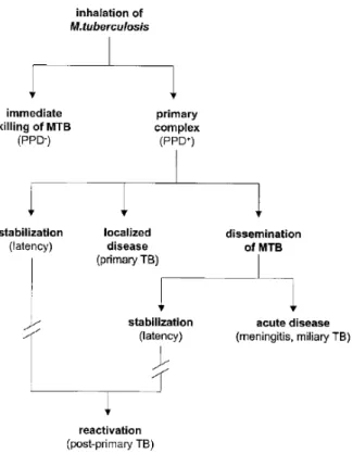
Figure 2 - Chronological events after inhalation of M. tuberculosis. Schematized the
different possible scenarios of Tuberculosis disease. (Adapted from van Crevel et
al. 2002) ... 20
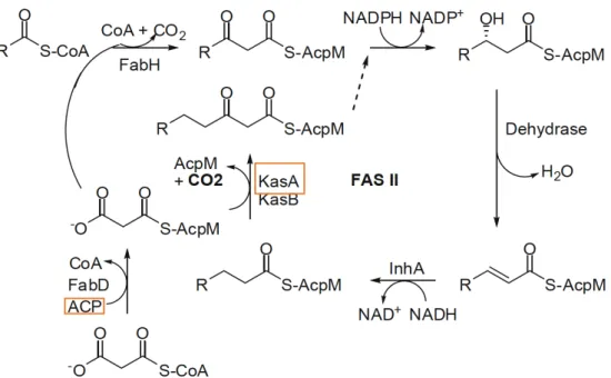
Figure 3 - General scheme of FAS-II system involved in mycolic acids biosynthesis
Orange frame enzyme targets of INH. (adapted from Yoya 2009). ... 22
Figure 4 - The molecular strutures of first line drugs used agaisnt TB (A - Rifampin; B -
Isoniazid; C - Pirazinamide; D – Etambutol). ... 23
Figure 5 - Molecular structures of (A) Bedaquiline and (B) Delamanid. ... 25
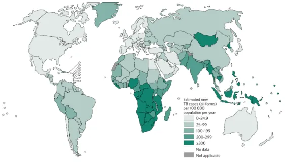
Figure 6 - Estimated TB incidence rates, 2015. Adapted from (WHO 2016) ... 25
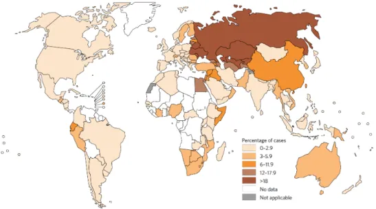
Figure 7 - Percentage of new TB cases with MDR-TB. Adapted from (WHO 2016) ... 26
Figure 8 - Schematic models of mechanism of action of CAMPs. (A) The carpet
mechanism (B) The barrel-stave mechanism (c) The toroid pore. (adapted from
Brogden 2005). ... 29
Figure 9 - Molecular structure of trans-cinnamic acid. ... 36
Figure 10 - General scheme of SPPS. X=O,NH; AA= amino acid; PG=protecting group;
TPG=temporary group; P - resin. ... 39
Figure 11 - Manual SPPS development in this project. ... 41
Figure 12 - Ninhydrin reaction with primary amines, resulting in the formation of
chromophore (deep blue). ... 42
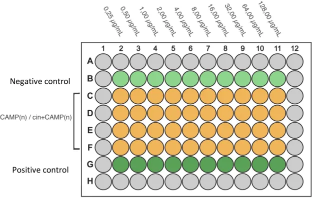
Figure 13 - schematized the 96 well-plate, was performed serial two-fold dilutions
(0,25-128 µg/mL); CAMP(n): CAMP without modification N-terminal;
Cin+CAMP(n), CAMP with modification in N-terminal coupling cinnamic acid;
Positive control (180µL medium + 20µL bacterial suspension + 1.04 µg/mL). ... 48
Figure 14 – Chromatogram of the product of the manual synthesis of the peptide
CAMP1, acquired with a HPLC system, with a C18 column, using ACN in water with
0.05% TFA as eluent, in gradient mode (0 – 100%), for 30 minutes, at a flow of 1
mL/min and detection at λ = 220 nm. ... 50
Figure 15 - Mass spectrum (LC-ESI/Orbitrap MS, positive mode) of the peptide CAMP1
(manual synthesis). ... 50
Figure 16 - Chromatogram of the product of the manual synthesis of the peptide
CAMP2, acquired with a HPLC system, with a C18 column, using ACN in water with
0.05% TFA as eluent, in gradient mode (0 – 100%), for 30 minutes, at a flow of 1
mL/min and detection at λ = 220 nm. ... 51
Figure 17 - Mass spectrum (LC-ESI/Orbitrap MS, positive mode) of the peptide CAM2
(manual synthesis). ... 51
Figure 18 - Chromatogram of the product of the manual synthesis of the peptide
CAMP3, acquired with a HPLC system, with a C18 column, using ACN in water with
0.05% TFA as eluent, in gradient mode (0 – 100%), for 30 minutes, at a flow of 1
mL/min and detection at λ = 220 nm. ... 52
Figure 19 - Mass spectrum (LC-ESI/Orbitrap MS, positive mode) of the peptide CAMP3
(manual synthesis). ... 52
Figure 20 - Chromatogram of the product of the manual synthesis of the peptide
CAMP5, acquired with a HPLC system, with a C18 column, using ACN in water with
0.05% TFA as eluent, in gradient mode (0 – 100%), for 30 minutes, at a flow of 1
mL/min and detection at λ = 220 nm. ... 53
Figure 21 - Mass spectrum (LC-ESI/Orbitrap MS, positive mode) of the peptide CAMP5
(manual synthesis). ... 53
Figure 22 - Chromatogram of the product of the manual synthesis of the peptide
CAMP7, acquired with a HPLC system, with a C18 column, using ACN in water with
0.05% TFA as eluent, in gradient mode (0 – 100%), for 30 minutes, at a flow of 1
mL/min and detection at λ = 220 nm. ... 54
Figure 23 - Mass spectrum (LC-ESI/Orbitrap MS, positive mode) of the peptide CAMP7
(manual synthesis). ... 54
Figure 24 - Chromatogram of the product of the manual synthesis of the peptide
Cin+CAMP1, acquired with a HPLC system, with a C18 column, using ACN in water
with 0.05% TFA as eluent, in gradient mode (0 – 100%), for 30 minutes, at a flow
of 1 mL/min and detection at λ = 220 nm. ... 56
Figure 25 - Mass spectrum (LC-ESI/Orbitrap MS, positive mode) of the peptide
Cin+CAMP1 (manual synthesis). ... 56
Figure 26 - Chromatogram of the product of the manual synthesis of the peptide
Cin+CAMP2, acquired with a HPLC system, with a C18 column, using ACN in water
with 0.05% TFA as eluent, in gradient mode (0 – 100%), for 30 minutes, at a flow
of 1 mL/min and detection at λ = 220 nm. ... 57
Figure 27 - Mass spectrum (LC-ESI/Orbitrap MS, positive mode) of the peptide
Cin+CAMP2 (manual synthesis). ... 57
Figure 28 - Chromatogram of the product of the manual synthesis of the peptide
Cin+CAMP3, acquired with a HPLC system, with a C18 column, using ACN in water
with 0.05% TFA as eluent, in gradient mode (0 – 100%), for 30 minutes, at a flow
of 1 mL/min and detection at λ = 220 nm. ... 58
Figure 29 - Mass spectrum (LC-ESI/Orbitrap MS, positive mode) of the peptide
Cin+CAMP3 (manual synthesis). ... 59
Figure 30 - Chromatogram of the product of the manual synthesis of the peptide
Cin+CAMP5, acquired with a HPLC system, with a C18 column, using ACN in water
with 0.05% TFA as eluent, in gradient mode (0 – 100%), for 30 minutes, at a flow
of 1 mL/min and detection at λ = 220 nm. ... 59
Figure 31 - Mass spectrum (LC-ESI/Orbitrap MS, positive mode) of the peptide
Cin+CAMP5 (manual synthesis). ... 60
Figure 32 - Chromatogram of the product of the manual synthesis of the peptide
Cin+CAMP7, acquired with a HPLC system, with a C18 column, using ACN in water
with 0.05% TFA as eluent, in gradient mode (0 – 100%), for 30 minutes, at a flow
of 1 mL/min and detection at λ = 220 nm. ... 60
Figure 33 - Mass spectrum (LC-ESI/Orbitrap MS, positive mode) of the peptide
Cin+CAMP7 (manual synthesis). ... 61
Figure 34 - MIC activities of CAMP1, Cin+CAMP1, CAMP3, Cin+CAMP3 against
susceptible Mtb expressed in µM. ... 64
Figure 35 - MIC50 activities of CAMP(n) and Cin+CAMP(n) against susceptible Mtb
expressed in uM. ... 65
susceptible strain H37Rv; Representative light microscope images show the
growth condition of the bacteria at various concentrations of CAMP(n) and
Cin+CAMP(n) after 7 days of incubation. The framed images indicate the lowest
concentrations of each peptide to inhibit 95% of bacterial growth compared with
the growth control and further confirmed through REMA assay... 67
Figure 37 - MIC50 activities of CAMP(n) and Cin+CAMP(n) against Resistance Mtb MDR
(INH,RIF and STR) expressed in µM. ... 68
Figure 38 - Cytotoxicity of CAMP1 and Cin+CAMP(n) on THP-1 cells. Values expressed
in µg/mL, in collaboration with University of Minho. ... 69
Figure 39 - Chromatogram of the CAMP1 purified peptide, acquired with a HPLC
system, with a C18 column, using ACN in water with 0.05% TFA as eluent, in
gradient mode (0 – 100%), for 30 minutes, at a flow of 1 mL/min and detection at
λ = 220 nm. ... 75
Figure 40 - Chromatogram of the Cin+CAMP1 purified peptide, acquired with a HPLC
system, with a C18 column, using ACN in water with 0.05% TFA as eluent, in
gradient mode (0 – 100%), for 30 minutes, at a flow of 1 mL/min and detection at
λ = 220 ... 75
Figure 41 - Chromatogram of the CAMP2 purified peptide, acquired with a HPLC
system, with a C18 column, using ACN in water with 0.05% TFA as eluent, in
gradient mode (0 – 100%), for 30 minutes, at a flow of 1 mL/min and detection at
λ = 220. ... 76
Figure 42 - Chromatogram of the Cin+CAMP2 purified peptide, acquired with a HPLC
system, with a C18 column, using ACN in water with 0.05% TFA as eluent, in
gradient mode (0 – 100%), for 30 minutes, at a flow of 1 mL/min and detection at
λ = 220 ... 76
Figure 43 - Chromatogram of the CAMP3 purified peptide, acquired with a HPLC
system, with a C18 column, using ACN in water with 0.05% TFA as eluent, in
gradient mode (0 – 100%), for 30 minutes, at a flow of 1 mL/min and detection at
λ = 220. ... 77
Figure 44 - Chromatogram of the Cin+CAMP3 purified peptide, acquired with a HPLC
system, with a C18 column, using ACN in water with 0.05% TFA as eluent, in
gradient mode (0 – 100%), for 30 minutes, at a flow of 1 mL/min and detection at
λ = 220 ... 77
Figure 45 - Chromatogram of the CAMP5 purified peptide, acquired with a HPLC
system, with a C18 column, using ACN in water with 0.05% TFA as eluent, in
gradient mode (0 – 100%), for 30 minutes, at a flow of 1 mL/min and detection at
λ = 220 ... 78
Figure 46 - Chromatogram of the Cin+CAMP5 purified peptide, acquired with a HPLC
system, with a C18 column, using ACN in water with 0.05% TFA as eluent, in
gradient mode (0 – 100%), for 30 minutes, at a flow of 1 mL/min and detection at
λ = 220. ... 78
Figure 47 - Chromatogram of the CAMP7 purified peptide, acquired with a HPLC
system, with a C18 column, using ACN in water with 0.05% TFA as eluent, in
gradient mode (0 – 100%), for 30 minutes, at a flow of 1 mL/min and detection at
λ = 220 ... 79
Figure 48 - Chromatogram of the Cin+CAMP7 purified peptide, acquired with a HPLC
system, with a C18 column, using ACN in water with 0.05% TFA as eluent, in
gradient mode (0 – 100%), for 30 minutes, at a flow of 1 mL/min and detection at
λ = 220 ... 79
List of tables
Table 1 - Recommended treatment regimen against new TB cases. ... 21
Table 2 - Examples of AMPs showing in vitro or in vivo activity against M. tuberculosis
and respective mechanisms of action. ... 32
Table 3 - Sequence of synthesized CAMP(n) in this project. ... 45
Table 4 - Sequence of synthesised Cin+CAMP(n) in this project. ... 45
Table 5 - The exact mass and molecular mass observed of the CAMP(n) peptides
detected by LC-MS and retention time determined by HPLC. ... 55
Table 6 - The exact mass and molecular mass observed of the Cin+CAMP(n) peptides
detected by LC-MS and retention time determid by HPLC. ... 61
Table 7 - Percentage of purification and amount of the manual synthesis CAMP(n) and
Cin+CAMP(n). Purification step was performed as previously described in
Experimental Procedures by RP-MPLC (purification analysis and quantification
was made by HPLC, results in Sumplementary information). ... 62
Table 8 - Reduction of MIC50 of Cin+CAMP(n) compared with IC50 of parental
CAMP(n) against Mtb. ... 64
Table 9 - Reduction of MIC50 of Cin+CAMP(n) compared with IC50 CAMP(n) against
MDR-TB. ... 67
Table 10 - Minimum inhibitory concentrations (MIC) of synthetic peptides against Mtb
H37Rv and MDR Mtb (resistant to INH, RIF and STR) expressed in µg/m and µM.
... 68
Table 11 - Summary of IC50 of CAMP(n) and Cin+CAMP(n) of the MTS assay performed
in THP-1 cells. Results are an average of two independent repeated experiments.
(1) Value of reference previously describe in (Ramón-García et al. 2013). ... 69
LIST OF ABBREVIATIONS
AAn - α-amino acids ACN – acetonitrile
ACP - enoyl-acyl carrier protein
AIDS - acquire immune deficiency syndrome AMP – antimicrobial peptide
CAMP – cationic antimicrobial peptide
Cin+CAMP – cinnamic acid conjugated cationic antimicrobial peptide DCM – Dichloromethane
DIAE – Diisopropylethylamine DMF – Dimethylformamide EMB – Ethambutol
FAS- fatty acid synthase FCS - fetal calf serum
Fmoc – fluorenylmethoxycarbonyl H2Odd – sterile distilled Water
HBD - human beta-defensins
HBTU - O-(benzotriazol-1-yl)-N,N,N′,N′- tetramethyluronium hexafluorophosphate HNP - human neutrophile defensins
HPLC - high-performance liquid chromatography IC50 - half maximal inhibitory concentration INH - isoniazid
KasA - Ketoacyl-ACP synthase A LC/MS - mass spectrometry LJ - Lowenstein-Jensen
MB 7H9 - Middlebrook 7H9 brot medium MDR-TB- multi drug resistant tuberculosis MIC - minimum inhibitory concentration Mtb – Mycobacterium tuberculosis
MTS - 3-(4,5-dimethylthiazol-2-yl)-5-(3-carboxymethoxyphenyl)-2-(4-sulfophenyl)-2H-tetrazolium
PC - Phosphatidylcholine PE -Phosphatidylethanolamine PEG - Polyethylene glycol PG - Phosphatidylglycerol
PZA - Pyrazinamide
REMA – Resazurin Microtiter Assay Plate RIF - Rifampin
RR-MPLC - Reverse Phase Medium-Pressure Liquid Chromatography Rt - retention time
SM - Sphingomyelin
SPPS - Solid Phase Peptide Synthesis STR – streptomycin
TB- Tuberculosis
TBTU - N,N,N′,N′-Tetramethyl-O-(benzotriazol-1-yl)uronium tetrafluoroborate tBu - tert-butyl
TFA - trifluoroacetic acid TIS – Triisopropylsilane
WHO – World health organization
Objectives
The main goal of the work developed in this thesis was the synthetises of new cationic antimicrobial peptides using cinnamic acids.
CAMP(n): 1 – WKWLKKWIK 2 – WRKFWKYLK 3 – RLWWWWRRR 4 – RIRRWKFRW 5 – RQRRVVIWW Cin+CAMP(n): 1 - trans cinnamic+WKWLKKWIK 2 - p-methoxycinnamic+WRKFWKYLK 3 - 3,4-Dimethoxycinnamic+RLWWWWRRR 4 - o-Nitrocinnamic+RIRRWKFRW 5 - trans cinnamic+RQRRVVIWW
Furthermore, the work aimed the study of the respective antimicrobial activity against Mtb and MDR-TB, as well to evaluate the cytotoxicity against TPH-1 cells differentiated into macrophages.
1. Introduction
Human Tuberculosis
1.1.
History
In the study of tuberculosis, René Laennec a French physician holds an important position in medical history being one of the pioneers of clinical-pathological correlations and his descriptions of pulmonary lesions in patients. In this work, published in 1821 entitled 'A treatise on diseases of the chest', Laennec demonstrated drawn images of tuberculosis cavities and detailed descriptions of the pathologic changes ceasing with now known necrosis of the tissues. Through the examination of a patient body, he observed pathological similar lesions diffuse in different organs due to the same causative illness. However, he didn’t find the cause behind the disease. Nevertheless, in 1868 a French military physician, Jean-Antoine Villemin, made a landmark study entitled ''Etudes sur la tuberculosis', on this research he described the transmissibility capability of tuberculosis infect throughout species such as from human to rabbits, from cows to rabbits and so on. Villemin exemplified the transmission of the disease to a rabbit with an injection of purulent liquid of tuberculosis cavities, yet he didn’t found the responsible agent. The cause of tuberculosis was not found until Robert Koch contribution, being the first to identify and isolate the tubercle bacilli. Koch continued his work with the discovery of a substance, now known as tuberculin, and though that could have a potential impact on tuberculosis cure. Unfortunately for Koch, research tuberculin didn’t cure tuberculosis, even so, he was capable to report cell-mediated immune responses for the first time (Schluger 2005; Smith 2003).
1.2.
Mycobacterium tuberculosis
Mycobacterium tuberculosis (Mtb) is an aerobic obligate pathogen with G+C rich content
and rood shape morphology of 0.3-0.5 µM of diameter (Cole et al. 1998; Niederweiss
2013). Even though Mtb is classified as gram-positive bacteria, the composition of the cell wall differs from other gram-positive bacteria (Figure 1). The composition of the mycobacterial cell wall is determined by the presence of peptidoglycan layer which is covalently attached to arabinogalactans linked to mycolic acids (Brown et al. 2015). Mycolic acids structure is defined by a long chain α-alkyl β-hydroxy fatty acids and corresponds to around 60% of the cell wall size. The biosynthesis of these structures
occurs with carboxylation of acetyl-CoA into acetyl-CoA carboxylase which is incorporated into the growing acyl chain during the repetitive cycle of fatty acid synthase (FAS) I and II reactions. FAS I catalyse the production of c24/C26-coA (alfa-branch) and FAS II elongates into meromycolic acid (C54/C56 – AcpM) (Pawelczyk and Kremer 2014). The complexity lipid-rich cell wall provides protection to chemical injury, dehydration and contributes to the extended persistence of Mtb bacilli within the host cells.
1.3.
Transmission and Pathogenesis of Mtb
Mtb is transmitted through the air in airborne droplet nucleic when patients with active disease cough, sneeze, speak or sing. Tuberculosis disease is characterised by four major stages.
The first stage of infection occurs when the inhaled tuberculosis droplet reaches the alveolus, further implantation preferentially affect the middle and lower zones of the lungs (Leung 1999). Once lodged in the alveolus, Mtb bind to macrophage through mannose, complement and scavenger receptors and the bacilli are phagocytosis (van Crevel, et al. 2002). Mtb is kept in macrophage phagosome and is able to block the normal fusion with lysosome which is responsible for the creation of a stress environment conditions (acid pH, ROS, enzymes and peptides). The protection capability of Mtb which inhibits phagosome-lysosome fusion has been associated with different mechanism of
a) Mycobacteria b) Gram-positive bacteria Figure 1- Cell wall structure of: a) Mycobacteria consist of thin layers of peptidoglycan covalently bond with arabinogalactan, and a thick layer of mycolic acids. b) Gram-positive bacteria have a single lipid membrane surrounded by a thick layer of peptidoglycan and lipoteichoic acid. (Adapted from Brown et al. 2015)
Ca2+ levels and is also postulated that mycobacterial phagosome is capable of recruit
large amounts of Rab5 proteins which are associated with endosome trafficking of early endosome (Delogu et al. 2015; Smith 2003). Within 3 to 8 weeks the bacillus has multiply causing the host macrophage to burst forming the Ghon complex and free bacilli through lymphatic circulation are able to disseminate to more distant tissues and organs.
The second stage, lasting about 3 months or delayed for up 2 years, is marked by the spread of Mtb throughout the body. When tuberculosis disease only affects the lungs is referred as pulmonary TB but can affect other organs such as larynx, the lymph nodes, pleural, the brain, the kidneys, or the bones and joints and is referred as extrapulmonary TB. In some infected individuals, an acute and fatal disease can occur in the form meningitis, if Mtb reach the nervous system. The case of miliary TB (when Mtb is disseminate through the all body) is normally associated with infants and children and severely immunocompromised individuals (Canadian Thoracic Society and The Public Health Agency of Canada and Licensor 2014). Clinical symptoms associated with TB reflect in systemic manifestations such as fever, anorexia, night sweats and cough from weeks to months. In approximately 5% of adult patients have a complete absence of symptoms (Leung 1999).
On the last stage (resolution) the primary complex is associated with the stagnation of the disease up to 3 years and characterized by the continuous development of granulomatous focal lesions and extra pulmonary lesions. The diversity of clinical manifestations between TB patients is influenced by age and immune strength. The success of tuberculosis infection in the early stage is determined by the strength of the host immune response. Around 90-95% of cases of Mtb are restricted and arrest in its latent form where infected patients do not show any signs or symptoms. The ability of Mtb of resisting immune responses in periods of latent infection is an essential feature. Mtb is able to switch between dormant and active state replicating bacilli as constantly stimulates T-cell responses which prevent the emergence of TB disease (Delogu et al. 2015). The maintenance of this persistent state is due to the incredible sensorial perception of the surrounding conditions and the composition of the extracellular matrix (Cole et al. 1998; Niederweiss 2013) (Figure 2).
1.4.
Current treatment
The standard treatment for new TB patient with the active TB disease, recommended by the World Health Organization (WHO 2016), is characterise by a 6 month regiment divided in intensive phase of isoniazida (INH), rifampin (RIF), pyrazinamide (PZA) and Etanbutol (EMB) for 2 months, followed by INH and RIF for further 4 months. The standard treatment rate of cure is 83%. In cases of latent Mtb infections treatment with antibiotic is indicated for prevention of the development of active TB. The treatment can be administrated with drug alone treatment of INH or RIF for 9 months or through drug combination by 2 months of RIF followed by 2 or 3 months of PZA (Table 1) (Gleeson and Decker 2006; Ormerod 2005).
Figure 2 - Chronological events after inhalation of M. tuberculosis. Schematized the different possible scenarios of Tuberculosis disease. (Adapted from van Crevel et al. 2002)
INH introduced for the treatment of TB in 1952, was a high selectivity and antimicrobial activity against Mtb. The mechanism of action is composed by three steps: starting with activation of pro-drug into active drug, then INH binds to the target molecule and terminates with inhibition of mycolic acid synthesis. INH active form binds to the enoyl-acyl carrier protein (ACP) reductase one of FAS II system enzyme and it also targets the enzyme Ketoacyl-ACP synthase A (KasA) responsible to elongate fatty acids chain (Figure 3). Unfortunately, INH is only active against growing Mtb and possess adverse drug reactions in some patients such as hepatotoxicity, rash, fever or arthralgia (Figure 4).
Table 1 - Recommended treatment regimen against new TB cases.
Active Disease
Treatment Time Drugs
Intense phase 2 months INH+RIF+PZA+EMB
Continuous Phase 4 months INH+RIF
Latent Disease
Treatment Time Drugs
Drug Alone 9 months INH or RIF
Drug Combination 2 months
2-3 months
RIF PZA
RIF applied on treatment regimens since 1968, not only target intracellular, semi dormant Mtb present in Necrotic granuloma lesions but also replicating Mtb. RIF are associated
with inhibition of transcription. The drug binds to the b-subunit of the DNA-dependent
RNA polymerase resulting on alteration of conformation which leads to protein mistranslation through the incorporation of mismatch codons. The side effects are associated with hepatotoxicity effects, gastrointestinal and hypersensitivity reactions (Kohanski et al. 2010) (Figure 4).
EMB was first introduced in 1961 and is active against only to replicating Mtb. Responsible for inhibition of cell wall synthesis by targeting Arabinosyl transferase, EMB presents a bacteriostatic effect rather than bactericidal as the other compounds (Figure 4).
PZA, as excellent sterilizing effect on growing and not growing bacilli and is only active in acidic pH, reaching the Mtb in the interior of macrophages. (Somoskovi et al, 2001; Woods et al. 2012) (Figure 4).
Figure 3 - General scheme of FAS-II system involved in mycolic acids biosynthesis. Orange frame enzyme targets of INH. (adapted from Yoya 2009).
1.5.
Development of resistant strains
Mtb is known as one of the most successful pathogens caused by an infectious organism. The discovery of streptomycin (STR) in 1945 for the treatment of TB made possible the cure of TB disease and boosted the development of new compounds. Over the years TB incidence decreased significantly, unfortunately the increasing epidemic acquire immune deficiency syndrome (AIDS) and the emergence of drug resistant consequently led to the recidivism of TB. Exposure to a drug induces stress responses that lead to both genetic and physiological mechanisms in order to survive (Reviewed in Chevalier et al. 2014 and Nguyen 2016).
The drug resistant Mtb is acquired through spontaneous chromosomal mutations that gives advantage of the strain to resist an antibiotic. For each antibiotic resistance, Mtb can present mutation on gene or group genes which reflects with alteration on composition of the cell wall or with an effect on the antibiotic target (Hoagland et al. 2016). The highly hydrophobic membrane cell wall of Mtb act as a selective barrier against antibiotic. Many mutated genes reported consequently changes in the membrane composition and fluidity (Cole et al. 1998).
The effect on the antibiotic target mechanism consists in modifying the structure (e.g Mutations on rpoB are responsible for conformational change at beta-subunit of RNA polymerase leading to a decrease in binding affinity of Rif) suppression or overexpression (The suppression of the antibiotic target such as Catalase peroxidase which results in a decrease on prodrug activation of INH), inactive drugs through
A B
D C
Figure 4 - The molecular strutures of first line drugs used agaisnt TB (A - Rifampin; B - Isoniazid; C - Pirazinamide; D – Etambutol).
chemical modification (Mutations in pncA reducing conversion to active acid form of PZA), drug efflux (Tap, a transporter responsible for mycobacterial efflux of aminoglycosides, spectinomycin, tetracycline ) (Hoagland et al. 2016; Nguyen 2016; Sarathy et al. 2012).
The Mtb resistant strain can be divided into three major group based on the level of antibiotic resistance: Multi-drug resistant tuberculosis (MDR-TB) with drug resistance to at least the first-line drugs INH and RIF, extensively drug-resistant tuberculosis (XDR-TB) with drug resistance to the first line drugs INH and RIF and to specific second-line drug (resistant to quinolones and injectable drugs) and totally drug resistant tuberculosis (TDR-TB) drug resistance to all first and second line drugs.
1.6.
New drugs
The development of new anti-tubercular drugs stagnated in time for over 50 years but with the synergy of TB with HIV, crescent multi drug resistances cases associated with exacerbate population growth culminated in an urgent need for new compounds. In the past few years, two new synthesis compounds have been approved for treatment of MDR-TB.
Bedaquiline, the family of diarylquinolones are the most advance anti-tubercular drugs currently available in the market. This drug is active from both replicating and not
replicating Mtb. The prime target of bedaquiline is F0 F1 ATP synthase main site of ATP
production in non-replicating state. This mechanism of bedaquiline results in a new method to kill latent Mtb and MDR-TB strains. Bedaquiline shows a minimum inhibitory concentration (MIC) of 0.12 μg/mL for drug resistant isolates (Andries et al. 2005; Hoagland et al. 2016) (Figure 5).
Delamanid is a nitro-dihydro-imidazooxazole derivative with a MIC of 0.012 μg/ml for drug resistant isolates. The mechanism of action is incompletely understood but involve inhibition of mycolic acid synthesis possibly via radical intermediate (Matsumoto et al. 2006) (Figure 5).
1.7.
The impact of Mtb in the world
TB is considered one of the 10 top leading causes of disease and mortality due to an infection agent. In 2015, according to the World Health Organization (WHO 2016) report, there was 10.4 million new cases and 1.8 million of deaths caused by TB (1.4 million TB and 0.4 million resulting from coexistence with HIV), worldwide.
Six countries accounted for 60% of new cases: India, Indonesia, China, Nigeria, Pakistan and South Africa (Figure 6).
Figure 6 - Estimated TB incidence rates, 2015. Adapted from (WHO 2016)
A B
Although, in the last few years incidence rate as decreased to 22%, cases of MDR-TB are growing, striking almost 480.000 in 2015 (Figure 7). Main countries accounted for 45% cases were India, China and the Russian Federation. The average proportion of MDR-TB cases with XDR-TB is 9.6%.
Figure 7 - Percentage of new TB cases with MDR-TB. Adapted from (WHO 2016)
Treatment of TB has become more difficult to achieve due in part to the duration of therapy and the crescent number of multiple drug resistance (MDR-TB and XDR-TB). Which leads to poor treatment outcomes and increased rate of mortality, reflecting in the urgent need to development new active drugs against drug resistance strains.
The global health concern of emergence of TB resistance to current antibiotic has triggered the development of new more effective compounds. Cationic antimicrobial peptide (CAMP) based therapies are interesting candidates as alternative or adjuvant to antibiotic treatments. CAMP are considered ancient defensive weapons with a wide spectrum of activity against gram-positive and gram-negative bacteria, fungus, parasites and virus and extending used against cancer. Through interaction with the negatively charged membrane which creates a displacement of lipids, alteration of membrane structure and possibility internalization (Lakshmaiah et al. 2015). CAMPs often possess a selectivity towards microbial membranes due to this anionic composition which differs from eukaryotic cells. Eukaryotic membranes are usually composed by zwitterionic phospholipids such as phosphatidylcholine (PC) and phosphatidylethanolamine (PE), sphingomyelin (SM) and cholesterol (increase of membrane stability) creating a neutral charged membrane ( -15 mV electrochemical gradient). In contrast the composition of prokaryotic membranes its characterise by the present of net negative charge/highly electronegative composed by hydroxylated phospholipids phosphatidylglycerol (PG), cardiolipin and phosphatidylserine (130 to 150 mV electrochemical gradient) (Robert E W Hancock et al. 1998; Yeaman and Yount 2003).
CAMPs are often small size (12-15 amino acid residue), cationic character (composed with positively charge Arginine and lysine residues) and amphipathic. (Robert E W Hancock et al. 1998) There are four major groups based on CAMP structure: a-helical usually amphipatic in nature (e.g. LL37, cecropins or magainins); b-sheet characterised by the presence of two or more disulphide bonds (e.g. human defensins, plectasin or protegrins); extended associated with lack of secondary structure rich in proline,
histidine, arginine or glycine residues (e.g. indolicidin); and b-hairpin are highly stable
due to the presence of disulphide bonds between b-strands (e.g. Bactenecin) (Hancock and Sahl 2006).
1.9.
Natural occurring CAMPs
CAMPS are usually found in bacteria, insects, plants and animals as host defence mechanism. In addition, CAMPs are considered multifunctional molecules with enormous proprieties such as anti-inflammatory, immunomodulatory, wound healing, cytokine release, chemoattraction and antimicrobial activity.
Natural CAMPs in mammals are encoded through a specific gene, being constitutively expressed (basal levels) or rapidly transcribed triggered with exposition to pathogens (Jenssen et al. 2006; McPhee et al. 2005).
Defensins and cathelicinds are the two major class of CAMPs present in the mammalian defence mechanism.
Cathelicidin is expressed in macrophages, epithelial cells and mainly store within granules of neutrophil expressed in mucosal surfaces (mouth, lung, genitourinary tract and skin). Beyond antibacterial activity, cathelicidin exhibits chemotactic, endotoxin-neutralizing, angiogenic, and wound healing proprieties (Jenssen et al. 2006).
Defensins are class of cyclic peptides with antimicrobial activity and immunomodulatory responses as mediators between innate and adaptive immune system. Defensins are divided in 3 groups: a, b defensins and q-defensins (Jenssen et al. 2006). Four a human neutrophile defensins (HNP 1-4) are present in neutrophils being constitutive produce and HNP5 and 6 are found in Paneth cells of the gastrointestinal system (Kang et al.
2014). While human b-defensins (HBDs- 1-4) are produce in epithelial cells of the
mucosa. Both cathelicidin and b-defensins are in constant surveillance against pathogen acting as the first defence barrier (Robert E W Hancock et al. 1998). In some individuals the lack of CAMP secretion through the body results with a higher susceptibility towards infection. Another feature of CAMPs is characterised by its immunomodulatory capacity, for example a-defesins present in neutrophil are responsible to attract T cells expressing
CD4 and CD8 antigens. b-defensin HBD(1-3) and LL37 is responsible to attract both
neutrophils, monocytes and T cells to the site of the infection (Zasloff 2002).
1.10.
Mechanism of action
Several studies with the membrane models were conducted in order to clarify the possible mechanism of action of CAMPs. Different models of mechanism associated with the interaction between peptides and membrane surface have been discussed and proposed over the years. The initial step results in electrostatic interactions between cationic peptide and positively charged membrane (Powers and Hancock 2003).
The carpet mechanism: CAMPs bond to the membrane surface and form a ‘carpet’ structure which destabilize hydrophobic interactions of phospholipids. When reach critical concentration of peptide the membrane integrity is lost and results in membrane disruption (e.g. cecropins, melittin, caerin) (Figure 8A) (Melo et al. 2009).
membrane so that hydrophobic part of the peptide aligns with lipid core region and hydrophilic portion of the forms the interior region of the ‘’barrel-like’’ pore (e.g. Pardaxin, alamethicin.) (Figure 8B).
The toroid pore: CAMPs bind parallel with membrane, hydrophobic residues disassociate the polar head group of phospholipids creating a breach in the hydrophobic region. Once reach the threshold of peptide-to-lipid ration, CAMPs make a transition to perpendicular orientation culminating on toroidal pore (e.g. magainins, protegrins and melittin) (Figura 8C) (Brogden 2005).
Microbial cell membranes are responsible for many essential functions including permeability barrier, oxidative phosphorylation, synthesis of biopolymers and virulence
A
B
C
Figure 8 - Schematic models of mechanism of action of CAMPs. (A) The carpet mechanism (B) The barrel-stave mechanism (c) The toroid pore. (adapted from Brogden 2005).
determinants. The creation of pores contributes to depolarization of the membrane, leakage of ions and metabolites which leads to cell death. Although, some CAMPs can enter into the cell without disrupting the membrane and then inhibited intracellular targets (Yeaman and Yount 2003).
1.11.
Antimicrobacterial potencial
The antimicrobial proprieties of CAMPs hold in future a promising therapeutically approach. Many CAMPs are already available in the market as topical drugs such as Pexiganan for diabetic foot ulcers, Iseganan for oral mucosaitis, Neuprex for sepsis and Omiganan for catheter-associated infections (Hancock and Sahl 2006; Kang et al. 2014). Regarding this advances, CAMPs for systemic administration face some obstacles that reduced its full effectiveness such as degradation by proteolytic enzymes, low bioavailability and inhibition of activity by physiological conditions. Several studies present possible solutions with use of non-natural amino acid or D-enantiomer in the peptide sequence displaying an increase on stability to the proteases (Ong et al. 2014). In addition, delivery approaches with use of synthetic carries, scaffolds (liposomes and polymers) and nanoparticles emerged as promising solution to enhance antimicrobial activities, decrease toxicities, increase salt stability and reduce protease degradation (Schmidtchen et al. 2015).
Another obstacle facing is the high manufacturing value of large-scale productions of CAMPs, about 50 to 400 dollars per gram comparing with antibiotic that only cost 1$ per gram. However, different approaches can be used to overcome this problem such as chemical synthesis (on solid-phase, solution phase and hybrid) provides the ability of incorporate unnatural amino acids (terms of isolation, purification and characterization), recombinant DNA, cell-free expression systems (Escherichia coli), transgenic plants (Nicotiana benthamiana using viral vectors based on TMV and potato virux X (PVX)) and fungi with enhance protein synthesis (da Cunha et al. 2017). The optimization approach selected should be carefully considered in the context on the nature of end application (e.g. topical or systemic), safety, the degree of antimicrobial potencies required and cost.
The rise of global health concern due to the increased number of multidrug-resistant tuberculosis reported has potentiated the research, in order to development of new effective drugs. CAMPs show a potential use, for administration as a monotherapy or as combined with other drugs. In the present, there is no CAMP admitted to clinical trials for the treatment of TB. Although, many studies in vitro and in vivo conducted so far have demonstrated inherent antimicrobial activity against either Mtb or MDR-TB. Some examples of CAMPs against Mtb studies are summarized in Table 2.
For instance, VpAmp, derivate from Vaejovis punctatus proved effective against both Mtb and MDR strain demonstrated a similar MIC range (Ramírez-Carreto et al. 2015). Ramón-Garcia et al 2013 tested the MIC of 49 synthetic peptides W-R rich against Mtb
presenting a 90 % decrease with 1,1 µM (Ramón-García et al. 2013). The LL-37 and
CRAMP derivate peptides, in vivo, were successful in reducing the loads in the lungs of both Mtb and MDR-TB strain, at 32 g/mouse reduced 53% and ate 32 ug/mouse reduced 45%, respectively (Rivas-Santiago et al. 2013). The synthetic IK8-all D proved an inhibition of growth in Mtb strain with the MIC of 15,6 mg/L (Ong et al. 2014). Yun Lan and co-workers, observed the benefits of combining synthesis D-LAK with INH which resulted in decreased of MIC of 10 µg/mL (INH) + 6.25 µM of peptide against MDR-TB. Linking the effect of D-LAK in the permeability of the cell membrane which facilitates the entrance of INH (Lan et al. 2014). Moreover, CAMPs usually present cytotoxicity to the host and sensibility to protease degradation. Jiang and co-works found that incorporation of D-enantiomers into LL37 resulted in less haemolytic effect (Jiang et al. 2011).
In general, CAMPs have shown bactericidal activity even though the acquisition of resistance towards CAMP as been observed, this is unlike to happen since drastic modifications of the bacterial membrane compromise its role.
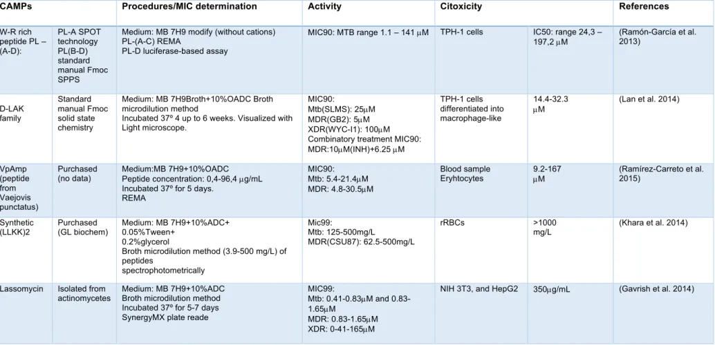
Table 2 - Examples of AMPs showing in vitro or in vivo activity against M. tuberculosis and respective mechanisms of action.
CAMPs Procedures/MIC determination Activity Citoxicity References
W-R rich peptide PL – (A-D): PL-A SPOT technology PL(B-D) standard manual Fmoc SPPS
Medium: MB 7H9 modify (without cations) PL-(A-C) REMA
PL-D luciferase-based assay
MIC90: MTB range 1.1 – 141 µM TPH-1 cells IC50: range 24,3 –
197,2 µM (Ramón-García et al. 2013) D-LAK family Standard manual Fmoc solid state chemistry
Medium: MB 7H9Broth+10%OADC Broth microdilution method
Incubated 37º 4 up to 6 weeks. Visualized with Light microscope.
MIC90:
Mtb(SLMS): 25µM MDR(GB2): 5µM XDR(WYC-I1): 100µM Combinatory treatment MIC90: MDR:10µM(INH)+6.25 µM TPH-1 cells differentiated into macrophage-like 14.4-32.3 µM (Lan et al. 2014) VpAmp (peptide from Vaejovis punctatus) Purchased
(no data) Medium:MB 7H9+10%OADC Peptide concentration: 0,4-96,4 µg/mL Incubated 37º for 5 days.
REMA
MIC90: Mtb: 5.4-21.4µM MDR: 4.8-30.5µM
Blood sample
Eryhtocytes 9.2-167 µM (Ramírez-Carreto et al. 2015)
Synthetic (LLKK)2 Purchased (GL biochem) Medium: MB 7H9+10%ADC+ 0.05%Tween+ 0.2%glycerol
Broth microdilution method (3.9-500 mg/L) of peptides spectrophotometrically Mic99: Mtb: 125-500mg/L MDR(CSU87): 62.5-500mg/L rRBCs >1000 mg/L (Khara et al. 2014)
Lassomycin Isolated from actinomycetes
Medium: MB 7H9+10%ADC Broth microdilution method Incubated 37º for 5-7 days SynergyMX plate reade
MIC99:
Mtb: 0.41-0.83µM and 0.83-1.65µM
MDR: 0.83-1.65µM XDR: 0-41-165µM
D-V13K, D- and L-LL37 Standard solid-phase peptide synthesis t-Boc Medium: MB 7H11 Incubated 37º 7 days CFU/mL MIC99: D(1-5) Mtb: 35.2-200µg/mL MDR: 49-100µg/mL D-LL37 Mtb: 200µg/mL erythrocytes D(15): 3.5 -421.5µg/mL L-LL37: 43.4 µg/mL D-LL37: 125 µg/mL (Jiang et al. 2011) Peptoid 1 1-C134mer 1-Nssb,14mer, 1-11mer, 1-Pro9 Standard solid-phase peptide synthesis
MABA by NIH/NIAID-contracted laboratory MIC99:
Mtb: 1-C134mer more active 6.6 µM and
1-Nssb infective >100µM
RAW 264.7 and J774 mouse macrophage cell line
1-C134mer: >100µM 1-11mer, 1-Pro9: 50µM Peptide 1: 20µM 1-Pro9, 14mer: non toxic (Kapoor et al. 2011) 11 Tic-Oic family peptides Purchased Standard solid-phase peptide synthesis Medium: MB 7H12 Broth
Broth microdilution method (0.5-100µg/mL) of peptides MIC: Visualise MIC99: Mtb: 4.92-40.75µM MDR(CSU45): 4.92-49.26µM Mtb(oflo): 4.92-49.26µM --- --- (Hicks 2016) PG-1 from porcine leukocytes, HBD-1 Standard solid-phase peptide synthesis, (automated Milligen 9050 peptide synthesizer) Medium: MB 7H9
Broth microdilution method 2-fold dilution (0-128µg/mL) of peptides CFU/mL PG-1 Mtb: 128µg/mL (96.7%) MDR(RM22): 128µg/mL HBD-1(45.1%) Mtb: 128µg/mL (49.9%) MDR(RM22): 128µg/mL (45.1%) ---- --- (Fattorini et al. 2004) Callyaerins Cyclic, Peptides Extraction and Isolation from Callyspongia aerizusa Medium: MB 7H9+ 0.5%glycerol+ 0.05%Tyloxapol+10% ADS Incubated 6 days REMA MIC90 Mtb: 2-40µM MIC100 Mtb: 6µM-100µM THP-1 and MRC-5 cell lines THP-1: 30-100µM MRC-5: 2->100µM (Daletos et al. 2015) lacticin 3147 nisin A
Purified Medium: MB 7H9GC broth
Broth microdilution method 0.5 dilution (0.11-60mg/mL) of peptides MABA MIC90 lacticin 3147 Mtb: 7.5mg/mL nisin A Mtb: >60mg/mL --- --- (Carroll et al. 2010)
HNP-(1-3) sNP-1 PG-1 All Purified exception of sNP-1 synthetize by Fmoc chemistry Medium: MB 7H9 Incubated 37º for 48 hours Standard colony count assay
HNP-(1-2),sNP-1 Mtb: 50µg/mL (99%) HNP-3 Mtb: 50µg/mL (98%) PG-1 Mtb: 50µg/mL (91,6%) HNP-1, sNP-1, PG-1 Mtb(ci): 50µg/mL (86.3 to 99%) --- --- (Miyakawa et al. 1996) LL-37 CRAMP E2(Bac8c) E6(Sub3) CP26 Standard solid-phase peptide synthesis (tBOC) Medium: MB7H9+ 10%OADC
Two-fold serial dilution (0.4–12.8 _g/mL) Incubated 37º for 5 days
REMA MIC99 CP26 Mtb: 2.1µg/mL E2 Mtb: 2.6µg/mL E6 Mtb: 3.2µg/mL LL37 Mtb: ~5µg/mL CRAMP Mtb: ~4µg/mL In vivo Mtb: ~53% killing at 32μg/mouse (3xper week, 28-day treatment)
In vivo MDR: ~45% killing a 32 μg/mouse (3x per week, 28-day treatment) --- --- (Rivas-Santiago et al. 2013) Magainin I Mastoparan Cecropin B MIAP standard solid-phase peptide synthesis Medium: MB 7H9+ 10%ADC
Two-fold serial dilution (6,4-1200ug/mL) of Peptides
Incubated 37º for 4 days MTT assay MIC99 Cecropin B, Mastoparan Mtb: 600µg/mL Magainin I Mtb: 1200µg/mL Mtb(clinical isolates): MIAP Mtb: 300µg/mL --- --- (Santos et al. 2012) vgf-1 Isolated from Naja atra venon Medium: MB 7H12 TB Bactec TB-460 MIC99 MDR (clinical isolates): 8.5 mg/mL --- --- (Xie et al. 2003)
Ecumicin Isolated from
actinomycete extracts
Medium: 7H12+ glycerol+ Casitone+ OADC MABA MIC99 Mtb: 0,16-0,36 µM Mtb(MR): 0.12-0.31µM MDR(clinical isolates): 0.31-0.62µM
Vero cells (ATCC CRL-1586) J774.1 macrophage cell line Vero cells: >63µM J774cells: >32µM (Pearson et al. 2016)
IK8-all D IK8-2D IK12-all L Purchased (GL Biochem)
Two fold dilutions (3.9-500mg/L) of peptide Mtb(clinica isolates): 15.6-62.5mg/L
RAW264.7 and WI-38 cells HC10: >125 to >2000 mg/L (Ong et al. 2014) PR-39 Isolated from porcine intestine Rest Synthetic Medium: MB 7H12
BACTEC radiometric assay and
Standard colony count assay activity of PR-39 Incubated 37º for 21 days
PG-39
Mtb: 50mg/L 80%
MDR(P1380/94): 100mg/L 50%
1.13.
Cinnamic acids
Cinnamic acids (example for trans-cinnamic acid in Figure 9) belong to the class of phenolic acid and it’s considered one of the most secondary metabolites in plants (Xu et al. 2009). These secondary metabolites are crucial to plant growth, development, reproduction and disease resistance. From spice cinnamon, Cinnamomum zeilanicum derives the denomination of cinnamic, use as food condiment, stimulant and antibacterial and antifungal compound. Not only is present in cinnamon but also is present in a diversity of aliments such as coffee, apples, berries, potato and more. Recently, the research on the potential proprieties of cinnamic acids has increased, and more articles are being published with potential application in cancer, malaria, diabetes and tuberculosis. In addition, molecular hybridization of cinnamic acid with a selective drug has been used in order alter the spectrum of potency and activity.
1.14.
Cinnamic acids agaisnt Mtb
In 1893, Landerer discovered the therapeutic potential of cinnamic acid in TB conducting an experiment by injecting emulsion form of cinnamic acid in TB patients. Intravenous injections were administrated twice a week for 3 months. Landerer work showed no effect on acute TB in young adults. Although, in cases of chronic TB patients a gradual improvement of the symptoms was observed. Landerer reported, from the 50 patients treatment, a 58 percentage of cure and 20 percentage of improvement and 20 percentage of death (Warbasse 1894).
More recent studies tested natural and synthetic cinnamic acid derivates against Mtb (Tanachatchairatana et al. 2008). For example, studies conducted by Andrade-Ochoa and co-workers reported the capacity of cinnamic acid and cinnamaldehyde against Mtb with associated MICs of 8.16 µg/m and 3.12 µg/mL, respectively (Andrade-ochoa et al.
Yoya at 2009, observed the consequences of punctual additions synthetic cinnamic acid derivates against TB. Was noticed that some modifications contributed to the decrease of activity and other had significant improvements such as the introduction of geranyl chain attributing a MIC of 0.6 µg/mL (Yoya et al. 2009).
Molecular hybridization of cinnamic acid with a selective drug is presented as a solution in order to achieve a more efficient compound (Guzman 2014; Slavchev et al. 2014). For example, the molecular hybridization of INH with trans-cinnamic acid against Mtb resulted in MIC of 3.12 µg/mL (Carvalho et al. 2008). Patel et al. at 2014, used a molecular hybridization of piperazine ring into cinnamic acid derivate which leads to increase on bioavailability and the antitubercular activities (Patel et al. 2014).
1.15.
Solid Phase Peptide Synthesis (SPPS)
Merrifield, in 1963 introduced Solid Phase Peptide Synthesis (SPPS), a brand new method of synthesizing peptides that have enormous advantages relative to the previously known. The SPPS methodology has proved to be faster, simpler, cheaper and more efficient.
The peptide is synthesized on an insoluble solid support being one of the most determinant factors to build the desire peptide sequence. The reaction vessel is connected with a vacuum system which allows the removal of side products and excess of reagents and solvents through washing with Dichloromethane (DCM) and Dimethylformamide (DMF) and filtration.
Briefly, SPPS starts with the coupling of an amino acid N alfa protected into the solid support (resin). The remaining α-amino acids (AAn) of the sequence are sequentially linked together through the formation of an amide bond (peptide bond). After each coupling, the reactivity of the alfa-amino group is protected by a temporary protecting group (usually 9 fluorenylmethoxycarbonyl (Fmoc)) and the other reactive functional groups in the amino acid are blocked by ‘’permanent protection (tert-butyl, tBu). The carboxyl group of the amino acid is blocked by the bond linking the solid support or by the peptide bond.
SPPS is characterised by a group of coupling reactions favoured with activation of the carboxyl group through coupling agent and deprotection reactions with deprotection of the reactive alfa-amide group of the already linked amino acid. Otherwise, in the coupling reaction an unwanted acid-base reaction will happen resulting with the formation of a salt, instead of bonding the two amino acids with amide bond.
Another particularly of SPPS, is the sequence order of amino acids, assembled through Ct à Nt which is the opposite of biosynthetic route for peptides synthesis observed in nature (Nt à Ct sense). The resin not only serve as a peptide support but also as protective group of carboxyl amino acid C-terminal.
The elongation of peptide chain occurs through repetitive cycles of coupling reactions of protected amino acid and subsequent deprotection of the temporary protecting group (Figure 10).
When the sequence is completed all permanent groups are removed and the peptide is cleaved from by chemical solution capable of breaking the bond with the solid support. Nowadays, the SPPS methodology was quickly and easily automated and new equipment were developed in order to produce fast and more efficiently. One example of these equipment’s is the Symphony X Multiplex Peptide Synthesizer, developed by Protein Technologies, Inc.©, which is a 24-channel peptide synthesizer that can run different sequences, scales and protocols on multiple reactors all at the same time, or run up to 12 reaction vessels with preactivation (BACHEM 2014; Behrendt, White, and Offer 2016).
2. Materials and Methods
2.1.
Reagents, Solvents and Equipment
In this work the reagents and solvents were acquired from Novabiochem (Fmoc-Rink Amide MBHA resin and Fmoc-amino acids), Sigma-Aldrich (coupling agents, piperidine,
N, N-Diisopropylethylamine (DIAE)), Resazurin (n-1-naphthylethylenediamine
dihydrochloride), Merck (TFA, solvents).
Peptides were characterized by mass spectrometry (LC/MS), on Finnigan Surveyor LCQ-DECA XP MAX. The purification step of peptides achieved by Reverse Phase
Medium-Pressure Liquid Chromatography (RP-MPLC), using a C18 Vydac® 218TP
stationary phase, by Grace Vydac. The purity of the peptides was determined by high-performance liquid chromatography (HPLC) on a Merck-Hitachi LaChrom Elite equipment, with a quaternary pump, automatic and thermostated by Peltier effect injector and a diode detector. A reverse phase Purospher star RP C-18 (octadecylsilane) column (125 x 4.0 mm), with a particle diameter of 5 μm, was used. The elution was performed with a variable gradient of acetonitrile (ACN) in water containing 0.05% trifluoroacetic acid (TFA), at a 1 mL/min flow.
The purified peptides were lyophilized in a BenchTop Pro 9L with omnitronics from SP Scientific (Department of Chemistry and Biochemistry of Faculty of Sciences of University of Porto).
2.2.
Peptide Synthesis by SPPS
2.2.1. Manual Synthesis developed in this Project
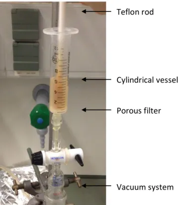
The SPPS reactor consisted in a cylindrical vessel (polypropylene syringe) with a polyethylene porous filter adapted to a vacuum system essential for removal of the excess of reagents from the wash, deprotection and coupling steps. A Teflon rod used to manually stir the resin beads (Figure 11).
2.2.2. Preparation of the resin
The resin chosen as a support for the manual synthesis was the Rink Amide MBHA resin LL (100-200 mesh), a polystyrene-based polymer functionalized with 4-methylbenzhydrylamine (MBHA) groups, further modified with an N Fmoc protected (R,S)-2-{4-[amino(2,4 dimethoxyphenyl)methyl]phenoxy} acetic acid linker (Rink-amide linker). The loading capacity of the resin was 0,38 mmol/g and syntheses was performed at 0.2 mmol scale, so 0.526g of resin was weigh and transferred into the syringe. The swelling step of the resin was made by the addition of N,N-dimethylformamide (DMF) with continuous stir. After 20 min, DMF was rejected, and followed by the addition of dichloromethane (DCM) for 15 min. Before the peptide synthesis, initial removal of Fmoc group was performed using 20% piperidine in DMF (3mL, 1x1min + 1x20min). After deprotection, the resin was washed with DMF (3mL,3x1 min) and DCM (3mL, 3x1 min) and a Kaiser test was performed.
A Kaiser test positive (dark blue resin beads and solution) indicate that the construction of the peptide sequence can be initiated.
Teflon rod
Cylindrical vessel
Porous filter
Vacuum system
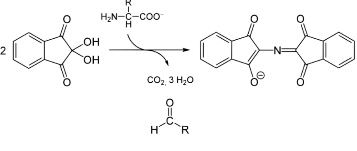
2.2.3. Kaiser Test
The Kaiser Test is used to detect the presence of primary amines, determining if coupling or deprotection reactions are complete. Ninhydrin (yellow) reacts with the deprotected N- terminal amine group leading to the formation of an intense blue chromophore known as Ruhemann’s purple (Figure 12). The tests were performed by transferring few beads to a small glass tube which was added 6 drops of reagent A and 2 drops of B (3:1). The reagent A consisted in a solution of phenol (40 g) in 10 mL of ethanol mix with a solution of aqueous KCN (16.5 mg/25 mL) in 100mL distilled pyridine. The reagent B consisted of a solution of 5 g of ninhydrin in 100 mL of ethanol. After homogenization, the test tube was incubated at 110ºC for 3 min. When N-Fmoc protected amino-acid is present after coupling step ninhydrin, and the result should remain yellow (negative test). When the test indicated a step wasn’t completed, that step was repeated (Kaiser et al. 1980).
2.2.4. Coupling of Amino Acids and Deprotection Cycles
The Elongation step is characterized by the coupling of an amino acid to the peptidyl-resin. Before being transferred to the syringe vessel the amino acids were activated for 5 min by a solution of Fmoc-AA-OH (5 eq.), coupling agent O-(benzotriazol-1-yl)-N,N,N′,N′- tetramethyluronium hexafluorophosphate (HBTU, 5 eq.) and base DIEA (10 eq.) in DMF. The activated amino acid solution was transferred to the syringe to react with the previously deprotected resin or peptidyl-resin for 1 hour with continuous stirring. Once the coupling was completed, the peptidyl-resin was washed with DMF (3 mL, 3 x 1 min) and DCM (3 mL, 3 x 1 min) and Kaiser test was performed. When the test result came negative (yellow) was followed by the deprotection step applying the deprotection solution consisted in 20% of piperidine in DMF (3 mL, 1 x 1 min + 1 x 20 min). After this time, the resin was washed with DMF (3 mL, 3 x 1 min) and DCM (3 mL, 3 x 1 min) and another Kaiser test was made. Confirmed positive (dark blue) the next Fmoc-AA-OH was coupled following the previously described method. The peptide sequence was achieved by repeating the cycles of coupling of N-Fmoc amino acids and deprotection steps.
2.2.5. Structural modification of CAMPs
After the peptide sequence completed, the peptidyl-resin for each peptide was divided into two fractions. The fraction CAMP(n) remained untouched and the fraction Cin+CAMP(n) was modified/coupled with cinnamic acid derivate. Deprotection of the Fmoc-AA-OH of the fraction Cin+CAMP(n) with 20% of piperidine in DMF (3mL, 1 x 1 min + 1 x 20 min). When finished cinnamic acid derivate was coupling to the sequence through activated solution of coupling agent cinnamic derivate, N,N,N′,N′-Tetramethyl-O-(benzotriazol-1-yl)uronium tetrafluoroborate (TBTU,) and base DIEA (10 eq.) in DMF for 3 hours with continuous stirring. The Kaiser test was performed and with a negative result, peptide modification was completed.
2.2.6. Cleavage and deprotection of side chairs of the peptide
When the peptide was completed and with a final deprotection step, the peptidyl-resin was subjected to a cleavage cocktail in order to break the bond linking the peptide to the resin support. In the hood was prepared a cleavage cocktail, containing 95 % TFA, 2.5%
TIS (Triisopropylsilane) and 2,5% H2Odd. Then the dry peptidyl-resin was transferred to
15 mL Falcon tubes in 100 mg portions and 1 mL of cleavage cocktail was added to each Falcon. The tubes were left under orbital stirring for 2h at room temperature. When finished, the contents of tubes were filtered on funnel previously rinsed with TFA, and the resin beads were washed with TFA. The filtrate, containing the soluble peptide, was transferred to new 15 mL Falcon tubes in 1 mL portions and 14 mL of cold tert-butyl methyl ether were added to each tube. The peptide containing tubes were stored at -22 ºC for 30 min. After this time the tubes were centrifuged at 3500 rpm for 5 minutes at -5 ºC then the ether was rejected and other 14 mL of cold ether was added. The addition of ether and centrifugation were repeated 3 more times and finally, the tubes were left in a vacuum desiccator until the crude peptide was dry. Dry peptide pellets were then solubilized in 10% aqueous acetic acid and analysed by HPLC and LC-MS.
2.3.
Purification of the conjugates
CAMP(n) and CAMP(n) conjugates (Cin+CAMP(n) were solubilized in 10% aqueous acetic acid and purified by RP-MPLC, using ACN in water with 0.05% TFA as eluent, in gradient mode (15 – 35%). The collected fractions were analysed by HPLC to determine which contained the conjugate with a purity greater than 92%. These fractions were subsequently pooled, lyophilized and stored at -22ºC until use.
2.3.1. Antimicrobial peptides
Peptide amino acid sequences were based on peptides with 9 amino acid of length previously describe and analysed in Ramon-Garcia (2013) study which included peptides enriched W and R for higher activity against Mtb (Ramón-García et al. 2013). 5 structurally similar CAMP(n) (Table 3) were synthesized using standard manual Fmoc
a modification in the N-terminal side was performed with the coupling of a cinnamic acid derivate (Table 4).
Peptides were weighed and diluted in sterile distilled water to obtain the final stock concentration and then store 4ºC until use.
Table 3 - Sequence of synthesized CAMP(n) in this project.
CAMP(n) Sequence Structure
CAMP1 WKWLKKWIK CAMP2 WRKFWKYLK CAMP3 RLWWWWRRK CAMP5 RIRRWKFRW CAMP7 RQRRVVIWW
Table 4 - Sequence of synthesised Cin+CAMP(n) in this project.
Cin+CAMP(n) Sequence Structure
Cin+CAMP1
trans cinnamic +WKWLKKWIK
Cin+CAMP2 p-methoxycinnamic +WRKFWKYLK