MESTRADO EM ONCOLOGIA
ESPECIALIZAÇÃO EM ONCOLOGIA LABORATORIAL
Demethylation of the epigenetically
silenced androgen receptor gene by a
repurposed drug in castration-resistant
prostate cancer cell lines
Mariana Brütt Pacheco
M
2019Mar
ia
na
Br
ütt
Pa
che
co
D
em
et
hy
lat
io
n o
f th
e e
pig
en
et
ica
lly
sil
en
ce
d a
nd
ro
ge
n r
ec
ep
to
r g
en
e b
y a
re
pu
rp
os
ed
d
ru
g in
ca
str
ati
on
-re
sis
ta
nt
p
ro
sta
te
ca
nc
er
ce
ll li
ne
s
M
.IC
B
A
S
201
9
Mar
ia
na
Br
ütt
Pa
che
co
D
em
et
hy
lat
io
n o
f th
e e
pig
en
et
ica
lly
sil
en
ce
d a
nd
ro
ge
n r
ec
ep
to
r g
en
e b
y a
re
pu
rp
os
ed
d
ru
g in
ca
str
ati
on
-re
sis
ta
nt
p
ro
sta
te
ca
nc
er
ce
ll li
ne
s
IN STI TU T O D E CIÊ N C IA S B IO MÉ DI CA S A BE L S A LA Z A RMariana Carvalho Dias Brütt Pacheco
Demethylation of the epigenetically silenced androgen receptor
gene by a repurposed drug in castration-resistant prostate cancer
cell lines
Dissertação de Candidatura ao grau de Mestre em Oncologia –
Especialização em Oncologia Laboratorial submetida ao Instituto de Ciências
Biomédicas de Abel Salazar da Universidade do Porto
Orientadora: Professora Doutora Carmen de Lurdes Fonseca Jerónimo
Professora Associada Convidada com Agregação
Departamento de Patologia e Imunologia Molecular
Instituto de Ciências Biomédicas Abel Salazar - Universidade do Porto
Investigadora Auxiliar e Coordenadora do Grupo de Epigenética e Biologia do
Cancro
Centro de Investigação
Instituto Português de Oncologia do Porto Francisco Gentil, E.P.E
Coorientador: Doutora Vânia Gomes Camilo
Investigadora Júnior do Grupo de Epigenética e Biologia do Cancro
Centro de Investigação
Instituto Português de Oncologia do Porto Francisco Gentil, E.P.E
Coorientador: Doutora Cristina Joana Marques
Investigadora Auxiliar
Departamento de Genética
Faculdade de Medicina, Universidade do Porto
“A winner is a dreamer who never gives up.”
This study was funded by a grant of FCT
(POCI-01-0145-FEDER-29030-HyTherCaP)
AGRADECIMENTOS
Chegou o fim de mais uma etapa que sem dúvida nunca teria conseguido alcançar sem a ajuda, o apoio e acompanhamento de muita gente. Um sincero agradecimento a todos os membros do Grupo de Epigenética e Biologia do Cancro por me terem acolhido tão bem, por me terem ensinado tanto e por serem um grupo que incrivelmente se apoia e ajuda mutuamente.
Em primeiro lugar, gostaria de agradecer à minha orientadora, Professora Doutora Carmen Jerónimo, por me ter dado a oportunidade de realizar este projeto no Grupo de Epigenética e Biologia do Cancro. Obrigada pela confiança que depositou em mim, pelas críticas e sugestões que sugeriu ao longo deste trabalho e pelas palavras de apoio e de motivação quando precisei.
Ao Professor Doutor Rui Henrique, por todas as críticas construtivas e comentários que surgiram nas reuniões do laboratório e pela constante disponibilidade e contributo para este trabalho.
Ao Professor Doutor Manuel Teixeira, Diretor do Departamento de Genética e do Centro de Investigação do Instituto Português de Oncologia, por ter permitido a realização desta dissertação neste instituto.
Ao Rob Mensink e à Mafalda Rocha do Instituto de Investigação e Inovação da Universidade do Porto, pela colaboração e ajuda na sequenciação das amostras.
À Maria e à Marta do Grupo de Oncogenética, pela ajuda que sempre ofereceram e pela companhia na sala de culturas mesmo aos fins-de-semana.
À minha co-orientadora, Joana Marques, por todos os ensinamentos partilhados ao longo deste ano, sem os quais parte deste trabalho não seria possível.
Um enorme agradecimento à minha co-orientadora Vânia Camilo, que me acompanhou durante este percurso. És das pessoas mais empenhadas e trabalhadoras que conheci com um entusiasmo pelo que faz super contagiante. Obrigada por tudo aquilo que me ensinaste quer profissionalmente quer pessoalmente. Contigo aprendi a ver sempre o lado positivo da situação, desenvolvi o meu espírito crítico e acima de tudo aprendi a confiar mais em mim, deixando as inseguranças de lado. Obrigada por te teres dedicado tanto a este trabalho, pelos fins-de-semana que abdicaste para me ajudar no trabalho prático e por te manteres (e a mim também) sempre calma e estável cada vez que surgia algum problema neste trabalho (que não foram propriamente poucos). Obrigada também pelas palavras de apoio e motivação e por teres acreditado sempre em mim.
À minha querida Ângela, por me ter recebido tão bem neste laboratório. Na verdade, foi contigo que este percurso começou. Ensinaste-me tudo o que sabias com gosto, dedicação e paciência. Agradeço-te muito por isso. Obrigada também por me teres deixado
errar e ao mesmo tempo aprender sozinha. Obrigada por me teres acompanhado sempre, mesmo quando já estavas mais longe e sem nunca teres obrigação de o fazer. Obrigada por todos os conselhos e pela disponibilidade imediata que tiveste, mesmo que isso implicasse pôr de parte os compromissos que tinhas. Obrigada por todas as palavras queridas de força, apoio e motivação. Acima de tudo, obrigada por acreditares em mim e nunca me teres deixado sozinha.
À Filipa, o meu anjinho caído do céu, que foi fundamental para a etapa final desta tese. Obrigada por teres entrado tão facilmente no nosso ritmo e por me teres ajudado sempre no que precisei, mesmo quando eram trabalhos “chatos”. Conseguiste tornar esta reta final mais fácil e mais calma. Acima de tudo, ajudaste-me a conciliar o tempo de escrita com o tempo de trabalho prático que ainda havia para fazer. Obrigada.
Às “mais velhas” do laboratório que me transmitiram muita experiência e conhecimento neste percurso. À Sofia por estar sempre disponível para tirar dúvidas um bocadinho sobre tudo, quer seja protocolos, quer seja dinâmicas do laboratório ou até contratempos que tenham surgido neste trabalho. À Sara pela companhia ao final do dia quando já mais ninguém estava no laboratório, pelos discursos de motivação quando mais precisei de ouvir e pela preocupação que sempre mostrou ter por mim. À Vera por ter me ter trazido à realidade quando mais precisei e se disponibilizar para me ajudar. À Catarina Macedo, por me ter ajudado de livre iniciativa e me ter acalmado nesta reta final. À Lameirinhas, por todas as intervenções que fez neste trabalho e por estar sempre pronta a ajudar. À Dani, pela sua constante boa disposição, disponibilidade para ajudar e resolver problemas de uma forma muito prática. À Carina, um exemplo de força, pela sua amabilidade, piada e alegria que fez com que me risse tanto todos os dias. À Iris, pelos convívios, conselhos e longas conversas depois do trabalho. Ao Zé pela sua ajuda, boa disposição e por me deixar gozar tanto com ele. À Sandra pelos ensinamentos e disponibilidade sempre que precisei. À Helena pelos seus conselhos. À Nair pela sua tranquilidade e simpatia.
Aos mais novinhos no laboratório, Luísa, Moço, Rita, Bela e Diana, desejo as maiores felicidades e sorte para o trabalho que se aproxima. Persistência, calma e dedicação são três conselhos que vos deixo para os próximos tempos.
À Cláudia, a minha companheira das jantaradas e da entrega da tese, obrigada por todo o apoio que me deste. Foste a pessoa que se calhar melhor me compreendeu nesta fase final e, por isso, agradeço-te por poder desabafar contigo, por poder desesperar contigo, mas também por poder disparatar contigo. Obrigada por todos esses momentos divertidos que tanto me fizeram rir. Ao meu companheiro de café, o Gonças, pelo seu sentido de humor que fez com que me risse muitas vezes e pela disponibilidade constante em ajudar. Obrigada também por ires ter comigo aleatoriamente a meio do dia
simplesmente para veres o que estava a fazer e ficares a conversar comigo nem que fosse por uns minutos. À Verita, por estar sempre com os pés assentes na terra e me alertar sempre que achou necessário. Sem te aperceberes, deste-me um grande “boost” de confiança que foi muito importante para mim. À Rita, por se preocupar sempre com tudo e todos e por ser a primeira a oferecer ajuda, independentemente do trabalho dela. À Teixas, por ter trazido sempre ânimo à nossa sala e por me ter feito rir tanto à custa de todas as peripécias e histórias associadas à personalidade dela.
Ao Zé, Taveira e Simão agradeço por terem paciência para falarem comigo mais detalhadamente sobre o meu trabalho e todos os obstáculos associados, já que trabalham na área. Obrigada por todas as jantaradas e momentos divertidos que partilhamos. Sem dúvida que tornaram este percurso muito mais animado.
À Sara Ferreira, que me acompanhou durante o meu percurso académico todo e concluiu mais uma etapa ao meu lado. A minha companheira das aulas, das borgas e agora quase do dia-a-dia. Uma das pessoas que viveu comigo todos os problemas, mas também todas as felicidades que foram surgindo. Obrigada por todo o apoio e carinho e obrigada por nunca desistires de mim.
Aos meus amigos de infância Mafalda Neiva, Inês Pedroso, Carolina Pollmann, Luís Figueiredo e Sara Alves por estarem presentes em mais uma etapa da minha vida. Obrigada por me ouvirem e me aturarem nos maus momentos, mas também por festejarem e ficarem felizes comigo nos bons momentos. Mesmo sem perceberem nada do que fiz, obrigada por me apoiarem e por nunca terem deixado de acreditar em mim. Principalmente, obrigada por compreenderem o meu afastamento nestes últimos tempos.
Ao Bruno, por ter sido sem dúvida o meu maior apoio neste percurso. Pela compreensão em todos os momentos em que descarreguei nele toda a minha irritação, frustração e nervosismo. Por todas as vezes em que deixei de estar com ele por causa de trabalho. Por todas as vezes em que de facto estava com ele, mas na realidade pensava no trabalho, falava do trabalho ou até trabalhava. Pela ajuda e disponibilidade constante que ofereceste durante este percurso. Pelo seu sentido de humor em que as piadas eram tão más que acabava por me rir sem querer. Por ter colocado os seus problemas sempre em segundo plano para se focar nos meus. Por ter tido sempre um discurso de motivação e de força. Obrigada por teres estado sempre presente e por nunca teres desistido de mim.
À Vera, por estar tão longe, mas conseguir manter-me sempre tão perto. Por muitas vezes começar o meu dia já com mensagens tuas a fazerem-me sorrir. Por teres essa personalidade tão forte que me faz tanta falta no meu dia-a-dia. Obrigada por acreditares sempre em mim e por me dares votos de confiança quando precisei. Acima de tudo, obrigada por estares sempre lá quando eu precisei.
Por último, mas mais importante, um profundo e sincero obrigada aos meus pais, que sem eles nada disto teria sido possível. Por acreditarem sempre em mim e estarem sempre ao meu lado. Por me darem força nos momentos menos bons e incentivo nos momentos bons. Obrigada pai por manteres sempre a calma e me alertares sempre para o equilíbrio na vida. Obrigada mãe por viveres tão intensamente (por vezes até mais do que eu) os momentos mais emotivos deste percurso. Obrigada por reconhecerem o meu trabalho, esforço e dedicação. Por me aturarem quando chego a casa de mau humor e de poucas palavras. Pela compreensão quando muitas vezes só ia comer e dormir a casa. Obrigada por perceberem a minha ausência neste último ano, mesmo sabendo que foi difícil e que precisavam mais de mim.
RESUMO
O cancro da próstata (CP) é a segunda neoplasia mais incidente e a quinta causa de morte relacionada com o cancro em homens em todo o mundo, afetando principalmente homens idosos. Embora a maioria dos doentes com CP apresente doença localizada ao diagnóstico, uma proporção significativa progride para doença disseminada resistente à terapia de privação de androgénio (TPA). A alta morbilidade e mortalidade associada a este estádio, bem como a falta de abordagens terapêuticas com intuito curativo, realçam a importância de investigar novos regimes de tratamento.
Atualmente acredita-se que esta progressão pode dever-se à desregulação da via de sinalização do receptor de androgénio (RA) por vários mecanismos moleculares, independentemente dos níveis circulantes de androgénio. Para além disso, alterações ao nível da maquinaria epigenética, como a hipermetilação do DNA, foram associadas a uma perda da expressão do RA em 20 a 30% dos cancros independentes de androgénio. Deste modo, os inibidores da DNA metiltransferase (iDNMT) poderão ser uma abordagem terapêutica promissora neste subconjunto de doentes.
Assim, o objetivo principal desta dissertação de mestrado foi avaliar o efeito da hidralazina, um iDNMT, como drug repositioning em linhas celulares de CP. Neste estudo, este composto induziu inibição da viabilidade celular dependente da dose, bem como um aumento significativo da apoptose na DU145, uma linha celular negativa para RA. Curiosamente, o tratamento sequencial com hidralazina e um inibidor do RA, a enzalutamida, corroborou esses resultados. A hidralazina sensibilizou esta linha celular para a enzalutamida, ao contrário das outras linhas celulares: PC-3 e RWPE. Posteriormente, o padrão de metilação da região promotora do RA foi avaliado nessas linhas celulares através da sequenciação de bissulfito. Em geral, a DU145 exibiu um perfil de metilação mais alto do que as outras linhas celulares de CP especificamente em duas regiões a montante do local inicial da transcrição. Para além disso, estes padrões de metilação diminuíram na DU145 após o tratamento com hidralazina, sugerindo que a hipermetilação nestas CpG do promotor do RA poderá ser um mecanismo que explica a perda da expressão do RA em carcinomas avançados da próstata.
Concluindo, demonstramos que este fármaco tem efeitos desmetilantes em CpG específicos da região reguladora do RA na DU145. De facto, a metilação do RA nessas CpGs pode levar a uma regulação negativa do RA nesse subconjunto de doentes com CP, podendo-se possivelmente associar à resistência à TPA. Assim, a hidralazina constitui um composto promissor para o tratamento destes doentes, pois poderá aumentar a sensibilização dos tumores resistentes à castração para medicamentos já aprovados e direcionados ao RA.
ABSTRACT
Prostate cancer (PCa) is the second most incident malignancy and the fifth cause of cancer-related death in men worldwide, affecting mainly elderly men. Although most PCa patients present with localized disease at diagnosis, an important proportion eventually progresses to a castration-resistant state after androgen-deprivation therapy (ADT). The high morbidity and mortality associated with this disease as well as the lack of curative therapeutic approaches highlight the importance of investigating novel treatment regimens.
It is widely accepted that this progression can be due to androgen receptor (AR) signaling pathway deregulation by several molecular mechanisms, regardless of androgen circulating levels. Additionally, aberrations in epigenetic machinery, such as hypermethylation, have been associated with a loss of AR expression in 20-30% of these androgen-independent cancers. Therefore, DNA methyltransferase inhibitors (DNMTi) might be a promising therapeutic approach in this subset of patients with these reversible modifications.
Thus, the major objective of this master’s dissertation was to evaluate the effect of hydralazine, a DNMTi, as a repositioning drug in PCa cell lines. This compound induced a dose-dependent inhibition of cell viability, as well as a significant increase in apoptosis in DU145, an AR-negative cell line. Interestingly, sequential treatment with hydralazine and an AR inhibitor, enzalutamide, corroborated these results. Hydralazine sensitized this cell line to enzalutamide, contrarily to the other cell lines: PC-3 and RWPE. Afterwards, the methylation pattern of the promoter region of AR was assessed in these PCa cell lines using bisulfite sequencing. Overall, DU145 displayed a higher methylation profile than the other PCa cell lines specifically in two regions upstream the transcription start site. Furthermore, these methylation patterns were decreased in DU145 after hydralazine treatment, suggesting that CpG hypermethylation of AR promoter may be a possible mechanism that explains loss of AR expression in advanced prostate carcinomas.
We demonstrated that this repositioning drug has demethylating effects in specific CpG sites of the AR regulatory region in DU145 cell line. In fact, AR methylation in these CpG dinucleotides may lead to AR downregulation in this subset of PCa patients, possibly being associated with ADT resistance. Thus, it constitutes a promising compound for CRPC treatment, since it could lead to a sensitization of already approved drugs that target AR.
TABLE OF CONTENTS
INTRODUCTION ... 1
Epidemiology of prostate cancer ... 3
Screening and diagnosis ... 4
Grading ... 5
Androgen synthesis in normal prostate ... 6
Prostate cancer ... 7
Localized PCa ... 7
Locally advanced and metastatic PCa ... 8
Castration-resistant PCa ... 9
Androgen receptor...10
Structure ...10
Translocation ...11
Modifications of AR signaling pathway ...12
Loss of AR expression ...13
Local androgen biosynthesis ...13
AR amplification ...13 Genetic alterations ...14 DNA rearrangements ...14 Mutations ...14 Variants ...15 Coregulators ...15 Epigenetics concept ...16 DNA Methylation ...17 DNMTs inhibitors ...17
5-azacytidine and 5-aza-2’-deoxycytidine ...17
Hydralazine ...18
DNMTi in PCa ...20
CRPC Treatment ...21
Antiandrogens ...22
Flutamide and Nilutamide ...22
Bicalutamide...22
Enzalutamide ...23
Abiraterone acetate ...23
xvi
Darolutamide ...24
Chemotherapy ...24
Bone targeting agents ...25
Radium 223 ...25
Zoledronic acid ...26
Denosumab ...26
Immunotherapy ...26
AIMS ...29
MATERIAL AND METHODS ...33
Cell culture ...35
Drug preparation and EC50 value ...36
Cell viability assay ...36
Apoptosis assay ...36
DNA extraction and bisulfite modification ...37
Bisulfite sequencing ...38
Primer design and producing PCR product ...38
Cloning reaction and transformation ...39
PCR product purification and sequencing reaction ...39
Protein extraction and quantification ...39
Western blot ...40
Statistical analysis ...40
RESULTS ...41
Prostate cancer cell lines characterization ...43
Hydralazine treatment validation ...43
EC50 value of hydralazine and enzalutamide in PCa cell lines ...44
Phenotypic Effects of hydralazine on the cell viability of PCa cell lines ...45
Sequential treatment with hydralazine and enzalutamide ...47
Methylation status in wild-type RWPE, DU145 and PC-3 cell lines ...50
...53
Methylation status of DU145 cell line treated with hydralazine ...54
DISCUSSION ...57
CONCLUSIONS & FUTURE PERSPECTIVES ...63
Conclusions...65
Future perspectives ...65
REFERENCES ...67
APPENDIX ...81
FIGURE INDEX
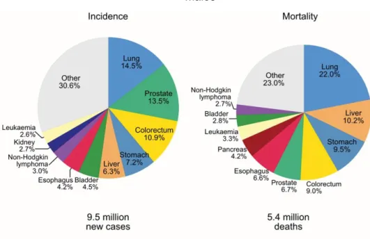
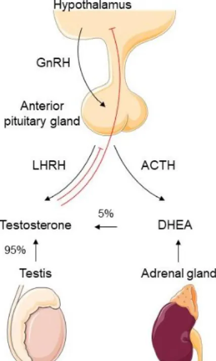
Figure 1: Pie charts present the distribution of cases and deaths for the ten most common cancers in 2018 for males worldwide. Adapted from [1]. ... 3 Figure 2: Regulation of androgen synthesis by the hypothalamic-pituitary-gonadal axis. Steroidogenesis occurs in testis and in adrenal glands, which produce 95% and 5% of testosterone, respectively. Abbreviations: GnRH - gonadotropin releasing hormone; LHRH - luteinizing hormone-releasing hormone; ACTH - adrenocorticotropic hormone; DHEA - dehydroepiandrosterone ... 6 Figure 3: Androgen receptor translocation: the interaction with androgens alters the conformational structure of AR, allowing its transition from the cytoplasm to the nucleus. Abbreviations: A - androgen; Hsp - heat-shock protein ...11 Figure 4: Modifications of AR signaling pathway that contribute to PCa progression independently of androgens’ circulating levels. Abbreviations: T-testosterone; DHEA - dehydroepiandrosterone; DHT - dihydrotestosterone. ...12 Figure 5: The four major epigenetic mechanisms include histone variants, histone post-translation modifications, DNA methylation and non-coding RNAs. ...16
Figure 6: Mechanism of action of nucleoside (5-azacytidine, 5-aza-2’-deoxycytidine) and
non-nucleoside DNMT inhibitors (hydralazine). ...20 Figure 7: Resume of the different treatment options for each PCa stage. Adapted from [181]. ...21 Figure 8. AR promoter gene divided into three regions: region 1 displays 16 CpG sites and a Sp1 binding-site; region 2 is located within the CpG island with 21 CpGs sites; Region 5 includes 14 CpG sites, begins with the ATG codon, continues with the first exon and ends with the CAG repeat...38 Figure 9. PCa cell lines characterization according to AR expression. ...43 Figure 10. AR-expression in DU145 cell line treated with different hydralazine concentrations. ...43 Figure 11. DNMT1 protein levels after hydralazine treatments in DU145 cell line. ...43 Figure 12. Hydralazine and enzalutamide dose-response curves in PCa cell lines on day 3. ...44 Figure 13. Phenotypic effect of hydralazine in PCa cell lines at day 0, 3 assessed by MTT assay. All data are presented as mean of three independent biological replicates with six experimental replicates. ...45 Figure 14. Overall impact in cell number of combined treatment in (A) DU145, (B) PC-3 and RWPE with (C) the separated hydralazine and enzalutamide EC50 in DU145 cell line. ...48
xviii
Figure 15. Impact of hydralazine and enzalutamide different combinations in the cell number of DU145 cell line. Abbreviations: H - hydralazine; E - enzalutamide ...49
Figure 16. Map of the androgen receptor gene 5’ cytidine-guanosine CpG island. The
position of Sp1 is indicated by a vertical line, the +1 and ATG positions by arrows and the exon 1 by a black square. The three selected regions are represented in accordance with sequence’s location. DNA methylation mapping from different replicates is presented for each cell line for each region with the methylation percentage of each region (below the black line at the end of each individual methylation map). ...52 Figure 17. Summarized methylation status of three regions of AR sequence in three independent replicates of each PCa cell line. ...53 Figure 18. Methylation percentage in region 1 in DU145 cell line exposed to different hydralazine concentrations. ...55 Figure 19. Methylation percentage in region 2 in DU145 cell line exposed to different hydralazine concentrations. ...56
TABLE INDEX
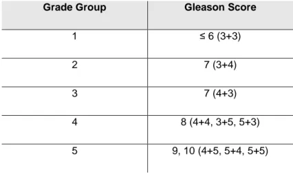
Table 1: Grade group and the corresponding Gleason score. ... 5
Table 2: Clinical trials with hydralazine in monotherapy and in combination. ...19
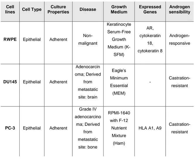
Table 3. Characterization of the different cell lines used in this study. ...35
Table 4: Primers and conditions for each region. ...38
Table 5. All antibodies used in Western Blot and its conditions. ...40
Table 6. EC50 values obtained for each tested drug and for each selected cell line. ...44
Table 7. Percentage of viable cells of three biological replicates at day 3 after hydralazine exposure at different concentrations. ...46
Table 8. Apoptotic levels normalized to vehicle at day 3 after hydralazine exposure at different concentrations in three biological replicates with six experimental replicates of each cell line...46
Table 9. Hydralazine and enzalutamide concentrations used for all cell lines in combination. ...47
Table 10. Methylation percentage in region 1 in DU145 cell lines exposed to different hydralazine concentrations. ...55
Table 11. Methylation percentage in region 2 in DU145 cell lines exposed to different hydralazine concentrations. ...56
LIST OF ABBREVIATIONS
µM – Micromolar 17β-HSD – 17β-hydroxysteroid dehydrogenase 3β-HSD1 – 3β-hydroxysteroid dehydrogenase 1 5-aza-CdR – 5-aza-2’-deoxycytidine 5hmC – 5-hydroxymethylcytosine 5mC – 5-methylcytosine A – AndrogenACHT – Adrenocorticotropic hormone ADT – Androgen deprivation therapy AML – Acute myeloid leukemia
APC – APC regulator of WNT signaling pathway AR – Androgen receptor
AR-V7 – Androgen-receptor splice variant 7 AREs – Androgen-response elements ATCC – American type culture collection CCDS – Consensus coding sequence ChIP – Chromatin immunoprecipitation CpG – Cytosine-phosphate-guanine
CRPC – Castration-resistant prostate cancer CT – Computed tomography
CYP11A1 – Cytochrome P450 family 11 subfamily A member 1 CYP17A1 – Cytochrome P450 family 17 subfamily A member 1 DAC – 5-aza-2’-deoxycytidine
DBD – DNA-binding domain DHEA – Dehydroepiandrosterone DHT – Dehydrotestosterone DMSO – Dimethyl sulfoxide DNMT – DNA methyltransferase DNMTi – DNMT inhibitors DRE – Digital rectal examination
EBRT – External-beam radiation therapy EC50 – Half maximal effective concentration EGFR – Epidermal growth factor receptor ER – Estrogen receptor
xxii ETS – E26 transformation-specific
ETV1 – ETS variant 1 ETV4 – ETS variant 4 ETV5 – ETS variant 5 FBS – Fetal bovine serum
FDA – Food and drug administration
FHIT – Fragile histidine triad diadenosine triphosphatase FSH – Follicle-stimulating hormone
GG – Grade group
GnRH – Gonadotropin releasing hormone GS – Gleason score
GSTP1 – Glutatione S-transferase pi 1 HDAC – Histone deacetylase
HDACi – Histone deacetylase inhibitor HDACi – Histone deacetylase inhibitor Hsp – Heat-shock protein
ISUP – International Society of Urological Pathology LBD – Ligand binding domain
LH – Luteinising hormone
LHRH – Luteinising hormone-releasing hormone
mCRPC – Metastatic castration-resistant prostate cancer MDS – Myelodysplastic syndromes
MGMT – O-6-methylguanine-DNA methyltransferase
MTT – 3-(4,5-dimethylthiazol-2-yl)-2,5-diphenyltetrazolium-bromide N-Cor – Nuclear receptor corepressor
nM - Nanomolar
NSAAs – Nonsteroidal antiandrogens NTD – N-terminal transactivation domain pre-mRNA – precursor mRNA
PCa – Prostate cancer
PET – Positron-emission tomography PIK3 – Phosphoinositide 3-kinase PSA – Prostate-specific antigen
PTEN – Phosphatase and tensin homolog RARβ – Retinoic acid receptor beta RTK – Receptor tyrosine kinase
Sp/KLF – Specificity protein/Krüppel-like factor Sp1 – Specificity protein 1
SRD5A – Steroid 5α-reductase T – Testosterone
TET – Ten-eleven-translocation TSG – Tumor suppressor gene
TMPRSS2 – Transmembrane protease serine 2 TRUS – Transrectal ultrasound
TSS – Transcription start site UTR – Untranslated region
EPIDEMIOLOGY OF PROSTATE CANCER
According to GLOBOCAN data, prostate cancer (PCa) is the second most common malignant neoplasm and the fifth leading cause of cancer-related death in men, worldwide (Figure 1). It is the most common non-cutaneous cancer in men worldwide with an estimated incidence of 1 276 106 cases and 358 989 deaths annually. This number of PCa cases represents 7.1% of the total cancer cases worldwide and 3.8% deaths are due to this malignancy [1]. This cancer type still remains a problem, since its primary benign stages are overtreated, whereas no curative therapies are available for metastatic stages [2]. Despite the high incidence rates in Oceania, Northern and Western Europe and North America, their mortality rates do not correspond to the high incidence ones.
Although PCa is a common disease, its etiology is still unclear [1]. There are some differences in the PCa incidence between different ethnic groups with African men descent in the United States and the Caribbean having the highest incidence and mortality rates which can be explained by genetic factors [1–3]. In fact, having a positive family history and/or a certain ethnic background such as Afro-Caribbean is considered a risk factor for PCa development [3].
Figure 1: Pie charts present the distribution of cases and deaths for the ten most common cancers in 2018 for males worldwide. Adapted from [1].
INTRODUCTION ǀ 4
SCREENING AND DIAGNOSIS
Nowadays, there are two screening tests for prostate cancer: digital rectal examination (DRE) and serum prostate-specific antigen (PSA). Despite its low specificity, PSA is considered the most sensitive biochemical marker for monitoring PCa and its primary regulator is AR [4–7]. This serine protease is produced in prostate and in normal conditions it is secreted into the glandular ducts where it degrades high molecular proteins in order to prevent coagulation of the semen [7,8]. Regarding PCa, this biomarker progressively increases due to aberrant prostate ductal structure formed by neoplastic epithelial cells, thus allowing PSA entering into the serum through leakage into the prostatic extracellular fluid [9,10].
The prostate epithelium is divided into three types: luminal, basal and neuroendocrine [2]. After prostate growth, androgens continue to promote the proliferation of secretory epithelia and stromal cells in the transition zone of the prostate, leading to a physiologic prostate gland enlargement known as hyperplasia [11–13]. Consequently, several men can experience lower urinary tract symptoms [13]. Since luminal cells are constantly multiplying, consequently producing more AR, PSA levels naturally rise, as men grow older. Hence, this hyperplasia is more commonly found in elder man and screening tests in elder men using PSA levels are highly recommended [13]. However, population-based PSA screening for PCa in men with advanced age is still conflicting regarding mortality outcome [4].
Initially, PSA measurement was thought to be able to substitute the digital rectal examination [4]. Nevertheless, PSA test alone is not specific nor sensitive enough to detect prostate cancer, since its levels can be altered with prostatitis, benign prostatic hyperplasia, prostatic biopsies and trauma [14–17]. Therefore, PSA in conjunction with digital rectal examination and transrectal ultrasound (TRUS)-guided biopsy is used as a diagnostic tool for early diagnosis, treatment and monitoring of prostate cancer patients [4,7]. European guidelines recommend a prostatic biopsy in which 10 to 12 cores are sampled in men with abnormal DRE and/or with PSA levels equal or above 2.0 ng/mL [7].
However, PSA alone is not reliable enough for monitoring disease burden in advanced CRPC, since visceral metastases can develop on these patients without an increase in PSA levels. Therefore, it is recommended for these patients to undergo a combination of frequent bone scintigraphy and CT scans along with PSA levels measurements [18]. Furthermore, PET/CT scan can detect a larger number of skeletal events than bone scintigraphy [3].
GRADING
Gleason grading system remains the most frequent approach to histopathological grading, which is one of the most powerful prognostic predictors and treatment selection tools in prostate cancer [19,20]. This system, created by Dr. Donald Gleason, is based on five prostate cancers’ different histologic patterns [21]. However, this system has undergone several modifications by the 2014 International Society of Urological Pathology (ISUP) Consensus Conference on Gleason Grading of Prostatic Carcinoma [7,22]. Since most of the tumors have heterogeneous morphology, having two or more histological patterns, the Gleason Score (GS) was created. It is based on Gleason grading system, but it gathers the two most common grade patterns in a tumor, ranging from 2 to 10. Nevertheless, patients may assume that a diagnosis with GS 6 means having worse prognosis, leading consequently to the possible overtreatment [19]. Hence, it is now recommended to use GG in conjunction with the so-called grade grouping [23]. The grade groups are based on the modified GS and correspond with patient prognosis (Grade Group 1 = Gleason score ≤ 6, Grade Group 2 = Gleason score 3 + 4 = 7, Grade Group 3 = Gleason score 4 + 3 = 7, Grade Group 4 = Gleason score 4 + 4 = 8, Grade Group 5 = Gleason scores 9 and 10) (Table 1) [20,23,24]. This system is simpler and predicts more accurately prostate cancer biology and progression [22].
Table 1: Grade group and the corresponding Gleason score.
Grade Group Gleason Score
1 ≤ 6 (3+3)
2 7 (3+4)
3 7 (4+3)
4 8 (4+4, 3+5, 5+3)
INTRODUCTION ǀ 6
ANDROGEN SYNTHESIS IN NORMAL PROSTATE
Androgen synthesis is regulated by the hypothalamic–pituitary–gonadal axis. Pulsatile release of hypothalamic gonadotropin releasing hormone (GnRH) stimulates luteinizing hormone (LH) secretion from the anterior pituitary gland, which leads to the production of testosterone in the testes (Figure 2). This hormone subsequently regulates not only hypothalamus but also pituitary gland through negative feedback, in order to maintain continued LH secretion. Otherwise, continuous GnRH stimulation would lead to desensitization. This approach is used when administering long-acting GnRH agonists in ADT [25,26].
Although steroidogenesis occurs in both the adrenal cortex and in the testes, the majority of testosterone (95%) is produced in the testes by the Leydig cells. Like all other steroid hormones, its biosynthesis starts with the cleavage of cholesterol by CYP11A1. Testosterone can be later converted to dehydrotestosterone (DHT) within the prostate by the action of enzyme 5α-reductase. At last, testosterone and DHT can exert their biological effects by binding to AR and consequently initiating its transcriptional activity [26,27].
Androgens are the primary regulators of prostate cancer cell growth, proliferation and death. They regulate prostatic epithelial cell number by chronically stimulating cell proliferation and inhibiting cell death at the same time. However, if there is a chronic modification in androgen levels such as castration, these cells die via programmed cell death [28].
Figure 2: Regulation of androgen synthesis by the hypothalamic-pituitary-gonadal axis. Steroidogenesis occurs in testis and in adrenal glands, which produce 95% and 5% of testosterone, respectively.
Abbreviations: GnRH - gonadotropin releasing hormone; LHRH - luteinizing hormone-releasing hormone; ACTH - adrenocorticotropic hormone; DHEA - dehydroepiandrosterone
PROSTATE CANCER
The prostatic adenocarcinoma is a highly heterogeneous disease regarding both pathological and clinical manifestations. Tumors with multiple foci can have different histological characteristics and tumors diagnosed with the same stage and with identical histological characteristics can lead to different clinical outcomes [29–32].
PCa tumors vary depending on their degree, duration of response to primary hormone treatment and clinical manifestations which include an increasing PSA level or doubling time [33,34]. This intrapatient heterogeneity has been associated with the reduced efficacy of the systemic therapies [35,36]. Treatment pressure leads to the development of intra- and intertumor heterogeneity as well as tumor progression either by selection or by divergent differentiation [36]. Therefore, PCa treatment is variable, being chosen according to cancer stage and clinical manifestations such as PSA levels and doubling time [6,7]. Nevertheless, it is important to take in account the balance of benefits and side effects of each therapy modality as well as the patients’ choice, comorbidities and quality of life [7].
Localized PCa
More than 80% of PCa cases are diagnosed while the disease is only confined to the gland representing consequently a low risk, good prognosis [35,37,38]. However, one third of these tumors metastasize to distant organs and can eventually lead to patients’ death. Regarding the median survival, patients presenting localized PCa usually survive more than five years, whereas the advanced type commonly do not exceed three years [37].
Moreover, most early-diagnosed patients are treated with active surveillance or watchful waiting, prostatectomy or radiotherapy which results in optimal survival [37–39].
Patients diagnosed with low grade tumors should be kept under active surveillance, carefully monitor the tumor until the disease clinically progresses [7,40,41]. Patients diagnosed with low grade tumors have serum PSA levels <10 ng/mL, GS≤6, tumor stage of T2a or less and fewer than two-three positive cores with <50% cancer involvement [7]. This treatment modality includes regular PSA measurements, DRE and repeated biopsies although the periodicity remains still unclearly defined [7,40].
If the disease progresses, radical prostatectomy, external beam radiotherapy or brachytherapy is recommended.
Radical prostatectomy is targeted to patients with tumors confined to prostate gland. So, patients with high risk, such as cT2c or cT3 or GS>7 are contraindications. With this procedure, there is a complete removal of the gland preventing consequently future metastasis [7]. However, there are several complications associated with this surgical
INTRODUCTION ǀ 8
procedure, which can compromise the patient’s quality of life. These complications include urinary incontinence, erectile dysfunction as well as bowel and urinary problems [7,42]. Despite high prognosis and long-lasting effect of this treatment option, there may be a recurrence even after the surgery.
External-beam radiation therapy (EBRT) is a non-invasive and less toxic therapy when compared to the three-dimensional conformal radiation therapy. It is recommended for patients with low to high risk only differing in dosage. For low-risk patients, a dose of 74-78Gy is recommended, while for intermediate risk the dose escalation ranges from 76 to 80Gy and brachytherapy or short-term androgen deprivation therapy (ADT) should be added. Concerning high-risk PCa, the modality approach includes EBRT with long-term ADT modality [7]. This type of radiation can also be extended to seminal vesicles or lymph nodes [43].
Low-dose rate brachytherapy uses radioactive seeds implanted within the prostate. This approach is offered to low-risk cases as well as low volume disease. On the other hand, high-dose rate brachytherapy is recommended for intermediate or high-risk PCa and it uses a radioactive source which is temporarily introduced into the prostate [7,43]. Usually, it is combined with EBRT and it can be delivered either in single or in multiple fractions [7].
Locally advanced and metastatic PCa
The disease progression is driven by phenotypical changes caused by genetic and molecular events and is influenced by the tumor microenvironment in which it has spread to [33,35]. Moreover, the progressing tumor can be also influenced by the exposed therapy [44].
Tumors usually invade their adjacent lymph nodes in the first place, followed by the liver, lungs and bones [2,45]. The bone metastasis normally cause severe pain, hypercalcemia and frequent fractures [46]. While the tumors that spread to lymph nodes often regress completely and rarely recur, the ones in bone are rarely eradicated [33,47].
Androgen deprivation therapy becomes the standard treatment strategy for androgen-dependent tumors (80-90% of the initial diagnosed tumors) and for patients with locally advanced or systemically spread disease [32,48]. This treatment occurs either through chemical castration with LHRH agonists or surgical castration resulting in lower levels of circulating androgen [18,35,49,50]. However, the optimal initiation, duration and modality are still not well defined [32]. When testosterone levels reach ≤20 ng/dl, the progression to CRPC is most likely to be delayed [18,32].
When androgens are ablated, more than 70% of normal prostatic secretory epithelial cells undergo apoptosis or survive arresting their cell cycle in G1 phase [51,52]. Thereby,
ADT results in a 90-95% decrease in serum testosterone levels, decreases intraprostatic DHT levels by 50% and inhibits AR [48,52].
Luteinising hormone-releasing hormone (LHRH) agonists have replaced the gold standard surgical castration for ADT. Beyond their potential of reversibility, these agonists avoid the physical and psychological discomfort of resulting from the surgery and have lower cardiotoxicity risks, providing at the same time similar oncologic efficacy. LHRH antagonists decrease rapidly the luteinising hormone, follicle-stimulating hormone and testosterone levels by binding competitively to LHRH receptors [18].
The ADT alone can lead to a positive response in over 80% of the patients and in combination with docetaxel chemotherapy can initially lead to improvements in approximately 80-90% of them [35]. This therapy can inhibit the progression to CRPC for up to 3 years [48]. Despite this initial response to this therapy, it ultimately fails, since the patients develop a resistance to androgens and progress to CRPC in media within 12 to 30 months [37,53]. The cells that were initially resistant to androgen ablation or that adapted to low-androgen environment regrow leading to a clinical progression of the disease [54]. In these cases or in presence of metastases, therapy becomes more challenging [39]. In fact, 1 nM of androgens is sufficient to allow AR signaling and consequent tumor growth [55]. Considering the androgens’ negative feedback, intermittent ADT should be recommended as a therapy in order to delay the development of androgen-resistant tumors [56].
Patients without metastases that are not suitable for curative treatments should report to ADT as a palliative treatment. Contrarily, symptomatic metastatic patients must receive ADT immediately combined with docetaxel, but only if they are fit enough. The toxicities of this therapy combination are mostly hematologic and could be overcome with concomitant use of granulocyte colony-stimulating factor. Moreover, during long-term therapy, bone mineral density and vitamin D should be measured every two years, since ADT increases the risk of fractures and decreases bone mineral density [18].
Castration-resistant PCa
The evolution from localized disease to castration-resistant PCa (CRPC) involves a complex interaction of signaling pathways that collectively promote cell proliferation [33].
Metastatic PCa eventually develops resistance to primary ADT treatments, resulting in CRPC [57]. Although this treatment is effective in 80-90% of the patients, the disease eventually progresses with rising PSA levels despite castrate concentrations levels [18]. This state is defined not only by the serum testosterone levels at <50 ng/dl, but also by biochemical or radiologic progression [18,35,58,59]. In fact, men with nonmetastatic CRPC and rapidly rising PSA level have a high risk developing metastases [34]. Biochemical
INTRODUCTION ǀ 10
progression is characterized by three consecutive rises in PSA one week apart and by a PSA value higher than 2 ng/ml [18]. On the other hand, radiologic progression is when two or more new bone lesions appear on bone scan or a soft tissue lesion [18,35].
Normally, nonmetastatic CRPC remains incurable and patients survive in media from 2-3 years [34,35,58]. The current available therapies for this subset of patients only aim at reducing the symptoms and improve the overall survival (about two months) [37]. Therefore, most of these treatments are ineffective highlighting the interest of investigating new and more effective therapeutic strategies to this aggressive PCa phenotype [37].
The mechanisms responsible for the emergence and progression of CRPC despite low androgen levels are not fully understood. However, it is known that androgen receptor takes an important part in this process [35].
During ADT, several cells undergo apoptosis, while the ones who survive remain in G1 phase of the cell cycle. On one hand, the cells that survive could adapt to the low-androgen environment and regrow after a while. These might acquire new epigenetic and genetic modifications that enable them to survive to this conditions, leading to ADT resistance and consequently tumor progression [51,54]. On the other hand, pre-existing castration-resistant cells that have low androgen dependence and stem-cell properties could be naturally selected, survive and continue to proliferate in the absence of androgens [54,60]. Therefore, ADT might induce expansion of the existing population, allowing a recurrence from only one cell [60].
ANDROGEN RECEPTOR
AR has a significant role in PCa biology in general, in progression to CRPC, in the pathogenesis, as well as in stimulation of PCa cell growth [2,35,61,62]. It is not considered an imperative cause in PCa progression, but it might be oncogenic under circumstances in which AR is inappropriately activated [62,63].
It belongs to the steroid hormone receptor superfamily and in normal conditions is an androgen-activated DNA-binding transcription factor [63,64].
Structure
The AR gene is located on the long arm of X chromosome (Xq11-12), consists of 8 exons and encodes as 110kDa protein composed of three major domains: an N-terminal transactivation domain (NTD), which enables the transactivation of the AR, a central DNA-binding domain (DBD), a hinge region and a C-terminal ligand DNA-binding domain (LBD) [35,64,65]. The AR promoter region displays 27 CpG dinucleotides and an Sp1 protein-binding site instead of a TATA box [66,67].
Translocation
The cytoplasmatic AR is associated with heat-shock proteins/chaperones and co-chaperones that protect the receptor against degradation. The interaction with DHT alters the conformational structure of AR leading to a phosphorylation and a consequent conformational change that allows its transition from the cytoplasm to the nucleus. In there, it dimerizes and regulates the transcription activity of specific target genes involved in growth and survival of the cell by binding to androgen-response elements (AREs) in DNA promoter regions (Figure 3) [48,68–71].
This regulation within the nucleus is influenced by coregulators, which can affect signal transduction pathways without the need of DNA binding in response to growth factors and by post-translational AR modifications: phosphorylation, acetylation, sumoylation, ubiquitinations and methylation [35,72].
Figure 3: Androgen receptor translocation: the interaction with androgens alters the conformational structure of AR, allowing its transition from the cytoplasm to the nucleus. Abbreviations: A - androgen; Hsp - heat-shock protein
INTRODUCTION ǀ 12
MODIFICATIONS OF AR SIGNALING PATHWAY
The inevitable progression to CRPC, despite ADT, cannot be attributed to a single mechanism. Nevertheless, it is known that the AR pathway is generally involved [35,48,73]. Androgens and the functional AR are known to be important mediators for PCa progression [64,74,75]. In addition, it is known that the disease progression is associated with an increase in PSA levels, a bona fide target of AR [33].
There are two main pathways that lead to androgen-refractory PCa development regardless of the androgens circulating levels: those involving AR and the others that bypass this receptor [33,35,39]. However, both pathways are not mutually exclusive, but instead can co-exist [39]. The ones involving AR include loss of AR expression, an increase in local androgens’ biosynthesis, AR overexpression/amplification, activating mutations and enhanced AR activity to other ligands [2,33,35,52,53,76]. The indirect mechanisms include AR variants, increase ligand-independent activity, develop changes in coregulatory molecules and deregulate growth factors or cytokines that lead to AR pathway activation via cross talk of other signaling pathways [33,39,52,53,76]. These modifications are summarized in Figure 4.
Figure 4: Modifications of AR signaling pathway that contribute to PCa progression independently of androgens’ circulating levels.
Loss of AR expression
Although the heterogeneity of AR expression in PCa is not correlated with response to ADT, higher degrees of AR positivity correlate with a greater degree of differentiation as well as a lower Gleason score. This heterogeneity is persistent, suggesting that increased AR expression do not associate with PCa initiation [52,77].
X chromosomes loss, which include loss of AR gene, is an extremely rare event in PCa [78]. On the other hand, one of the major resistance mechanisms is due to the epigenetically silencing of AR by hypermethylation of promoter which occurs in a late state of prostate carcinogenesis (7%) [66,79]. This has been observed in 8% of primary PCa cases [79].
Local androgen biosynthesis
Androgens within the tumor may come either from an adrenal source or from an intratumoral mechanism. It has been shown that prostate tumors do not have a completely androgen-free environment. As described before, the canonical pathway begins with cholesterol, leading through multiple steps to the production of DHT. Alternatively, the “backdoor pathway” uses CYP17A1 to convert pregnanes to androgens that are 5α- and 3-keto-reduced, ending to a terminal conversion to DHT. The other pathway, the 5α-dione pathway, bypasses the need of testosterone as a precursor and uses the 5a-dione instead. These alternative pathways uses additional enzymes: 3β-HSD1, 17β-HSD, and SRD5A [25–27,80,81].
AR amplification
AR gene amplification has been documented in 20%-30% of metastatic castration-resistant prostate cancer (mCRPC) and recurrent primary tumors, but not in hormone-dependent cancers. Contrarily, in untreated PCa AR amplification is very rare (<5%) [64,82,83]. Therefore, treatment possibly induces selective pressure [84]. These amplifications predict resistance to both enzalutamide and abiraterone acetate [85]. In fact, AR amplification allows PCa cells to become sensitive to low levels of androgens after ADT, enhancing AR activity and thus proliferating in a reduced androgen environment [73,86,87]. AR protein is expressed in prostate cancers of all clinical states. This alteration sensitizes the tumor PCa cells to respond to low levels of ligand [88]. Hence, CRPC patients with this amplification survive longer than the ones without it [64,87].
INTRODUCTION ǀ 14
Genetic alterations
There are some genetic alterations that target AR, PI3K, Wnt, DNA repair and cell cycle pathways in nearly all metastatic PCa and several primary PCa [3,84,89]. The most frequent altered genes in mCRPC are AR (62.7%), TP53 (53.3%) and phosphatase and tensin homolog (PTEN) (40%) [84]. Numerous primary PCa tumors have recurrent point somatic mutations, resulting in a single amino acid substitution, copy number alterations and oncogenic structural DNA rearrangements [36,52,90].
DNA rearrangements
The majority of DNA rearrangements (57%) are translocations involving E26 transformation-specific (ETS) family of transcription factors [2,84]. ETS-related gene (ERG), has oncogenic properties, since it activates PI3K signaling pathway leading to PCa progression [91]. Additionally, transmembrane protease serine 2 (TMPRSS2), another AREs, deregulation is also implicated [92,93]. The TMPRSS2:ERG fusion is present in approximately half of localized PCa cases [2,91,92]. TMPRSS2 can also fuse with ETV1, ETV4 and ETV5 [2,84].
Mutations
Most of the point mutations occur in the LBD (49%) which confer hypersensitivity or promiscuity to other ligands, thus activating AR [44,48,94]. Furthermore, most of them are associated with gains of function, thus making the receptor more sensitive to native ligand, to other steroid hormones or to specific antiandrogens used in therapy [52,94]. The proportions of rest of the mutations are 40% in the NTD and 7% in the DBD [48].
In untreated patients, AR mutations normally increase with PCa stage [95]. These mutations are very rare in early-staged prostate tumors [95]. Moreover, the mutations are detected in 10 to 30% of patients previously treated with AR antagonists [96]. Furthermore, since the AR mutations occur before hormonotherapy, it suggests that this therapy does not lead to AR mutagenesis [95]. On the other hand, the most frequent functional consequences of several AR mutations cause AR antagonists to become agonists switching the normal inhibition to inducing proliferation as well as AR transcription by adrenal androgens [52,53]. This antagonist-agonist switch is mostly found in CRPC phenotype [52].
The most common AR mutation is T876A which occurs in approximately 30% of metastatic CRPC after ADT combined with an antiandrogen [52]. AR T878A in another common mutation that cause a gain-of-function in the LBD [48].
Variants
AR genomic structural rearrangements are presented in one-third of mCRPC tumors leading to expression of several AR variants either lacking the LBD, resulting in constant activation of AR signaling [97–99]. They arise due to alternative splicing or AR gene rearrangements [98]. Although more than 20 AR-variants have been identified, androgen-receptor splice variant 7 (AR-V7) is the most common one [84].
AR-V7 comprises the NTD and the DBD and lacks the LBD domain which makes it constitutively active [97,100]. This mutation has been detected in metastatic CRPC patients and in primary PCa tissues associated with poorer outcomes such as biochemical recurrence and shorter survival rates [97,100,101]. Previous studies demonstrated that AR-V7 induces PCa cell growth and progression in the absence of androgens and patients with increased AR-V7 levels do not respond to enzalutamide and abiraterone [84,100,102,103]. However, this variant is sensible to taxane chemotherapies such as docetaxel and cabazitaxel [100,102]. It is suggested that the ADT-induced AR transcription rate and splicing factor recruitment to AR precursor mRNA (pre-mRNA) contribute to the high AR-V7 levels in PCa cells [98]. Moreover, AR-AR-V7 not only activates target genes independently of androgens, but also activates the normally ligand-dependent AR in a ligand-independent manner, facilitating its nuclear localization and transcription of target genes [100,104].
Coregulators
AR transcriptional activity is regulated by coactivators or corepressors that increase or reduce the receptor function respectively [33,105]. They recruit several transcription factors associated with RNA polymerase [105]. Almost 300 nuclear receptor coregulators have been identified [106]. Since the interaction between AR and its coactivators enhance the transcriptional activity of steroid receptors, allowing them to be active despite low androgens concentrations, it has been proposed that overexpression of coactivators may contribute to carcinogenesis in PCa [107].
Coactivator proteins such as ARA54 and ARA70 overexpressed in PCa enhance the activity of AR to alternative ligands, sensitize the receptor to lower concentrations not only of native, but also of nonnative ligands and induce ligand-independent activation by receptor tyrosine kinases (RTK). Consequently, these coactivators may contribute to ADT failure, possibly increasing the onset of CRPC [33]. The coactivator p300 interacts with AR and plays an important role in AR androgen dependent activation. It weakens histone-DNA interactions due to its histone-acetyltransferase activity, facilitating the access of different transcription factors to the DNA molecule [108]. Several tyrosine kinases SRCs overexpressed in PCa can increase the AR transcription by interacting with AR N-terminal portion. SRC-1 is overexpressed in 50% of CRPC cases when compared with normal
INTRODUCTION ǀ 16
prostate, while SRC-3 expression was correlated with increased PCa grade and stage and decreased disease-free survival [48,52].
Corepressors such as nuclear receptor corepressor (N-CoR) coupled with silencing mediator of retinoid and thyroid receptors (SMRT) antagonize the action of bicalutamide and flutamide. These corepressors contribute to the agonist activity of these agents in a ligand-dependent manner, since they inhibit AR function by a direct interaction [33,52].
EPIGENETICS CONCEPT
Epigenetics is defined as heritable and reversible modifications in gene expression patterns that persist during cell division. Unlike genetic abnormalities, epigenetic changes do not alter DNA sequence [109,110]. Moreover, epigenetic plasticity can be driven by genetic, environmental and metabolic stimuli which can lead to cell adaptation and malignant progression [111]. Epigenetic deregulation is present in cancer initiation, thus being considered a hallmark of cancer [112]. Currently, there are four major epigenetic mechanisms: DNA methylation, histone post-translation modifications or chromatin remodeling, histone variants and non-coding RNAs (Figure 5). Since epigenetic abnormalities can be reverted, epigenetic therapies seem to be a promising approach regarding cancer treatment [113].
Figure 5: The four major epigenetic mechanisms include histone variants, histone post-translation modifications, DNA methylation and non-coding RNAs.
DNA Methylation
DNA methylation is the most well studied epigenetic mechanism [113]. In cancer, DNA methylation occurs mostly at cytosines within cytosine-phosphate-guanine (CpG) dinucleotide of gene promoters [92,113]. It consists in a covalent addition by DNA methyltransferases (DNMTs) of a methyl group to the fifth carbon of a cytosine ring, resulting on a new DNA base, 5-methylcytosine (5mC) [114].
The DNMTs enzymes catalyze this process: DNMT1, DNMT3A and DNMT3B. DNMT1 is responsible for maintaining DNA methylation patterns during cell division, while DNMT3a and DNMT3b have both de novo methylation activity [115,116]. Reversal of the DNA methylation process can be achieved by ten-eleven-translocation (TET) proteins which catalyze the oxidation of 5mC to 5-hydroxymethylcytosine (5hmC) [117].
Cancer cells exhibit alterations in DNA methylation profile. In general, they display hypomethylation in normally methylated regions, resulting in genome instability and activation of proto-oncogenes. Additionally, they gain hypermethylation at gene promoters that are normally unmethylated, leading to transcription silencing of tumor suppressor genes involved in several cell functions, such as DNA repair, cell signaling and cell-cycle regulation [118–120].
DNMTs inhibitors
Several compounds are able to restore the normal methylation patterns by irreversibly inhibiting DNTMTs enzymatic activity and stimulating their proteasomal degradation [121,122]. DNMT inhibitors (DNMTi) can activate epigenetically silenced tumor suppressor genes (TGS), resulting in cell death, cell cycle arrest, chromatin extension and induction of cell differentiation [123]. Moreover, these inhibitors may contribute to tumor cell reversion phenotype, bringing significant clinical benefits for patients [124].
There are two types of DNMTi: the nucleoside and non-nucleoside analogues [124]. Nucleoside analogues have a modified cytosine ring that is connected to either a ribose or deoxyribose and may be integrated into RNA or DNA during the S phase of the cell cycle. They covalently bind to DNMTs, inhibiting them and inducing cell death or DNA damage [125]. On the other hand, non-nucleoside analogues bind directly to the catalytic region of DNMT without incorporating into DNA [126].
5-azacytidine and 5-aza-2’-deoxycytidine
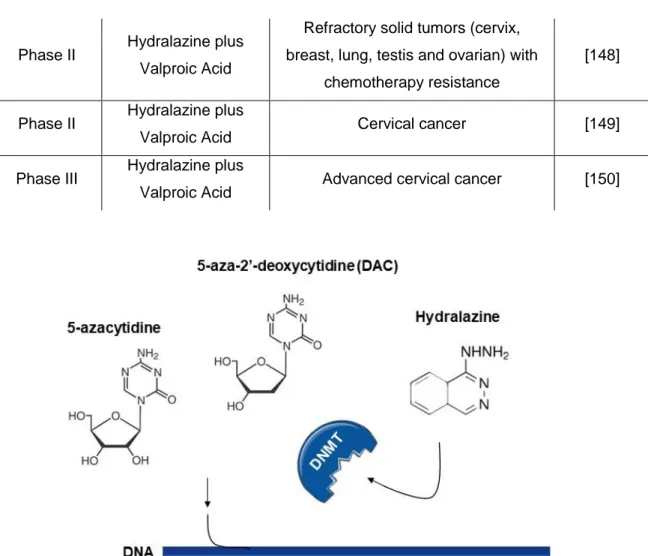
5-azacytidine (Vidaza) and 5-aza-2’-deoxycytidine (Dacogen) are the most well characterized nucleoside analogues (Figure 6). These drugs have been widely used in pre-clinical and pre-clinical trials in several cancer models due to their anti-tumorigenic activity [127,128]. Although these drugs were developed in 1964 as cytostatic agents, their in vitro
INTRODUCTION ǀ 18
cell proliferation induction and their involvement in DNA methylation inhibition was only later discovered [129,130]. Both were FDA-approved for myelodysplastic syndromes (MDS) on 2004 and 2006, respectively, since they demonstrated clinical benefit in clinical trials with hematologic cancer patients [131–134]. Moreover, 5-azacytidine and 5-aza-2’-deoxycytidine are also active against acute myeloid leukemia (AML) and other myeloid malignancies [135]. Nonetheless, these compounds present several limitations in clinical practice due to their cytotoxic effects at higher doses, side effects such as neutropenia and thrombocytopenia and short half-life [136,137].
Comparing to hematolymphoid malignancies, the lack of success of azanucleosides in solid tumors may be explained by the higher proliferative rate of the former ones. Additionally, these agents can potentially cause global hypomethylation resulting in unwanted re-expression of epigenetically silenced genes, which can contribute to tumorigenesis, progression and aggressiveness [127].
Hydralazine
Hydralazine hydrochloride (Apresolin) was approved by the FDA for the treatment of severe hypertension and heart failure. Nowadays, it is commonly used for hypertension in pregnancy [37,138]. Recently, hydralazine has been recognized as a demethylating agent (Figure 6) [139]. It is suggested that this compound interacts directly with the active site of DNMTs through its nitrogen atom, consequently inhibiting DNA methylation [140]. However, its mechanism of action is still not well understood.
Although its half-life in plasma is approximately 1h, the duration of the hypotensive effect lasts up to 12h. The recommended dose varies from 10mg four times a day to 50mg and common side effects include headache, nausea, flushing, low blood pressure, palpitation, tachycardia, dizziness and angina pectoris. Furthermore, it can cause autoimmune reactions, such as drug-induced lupus-like syndrome [138]. In fact, long term use of high doses of hydralazine was associated with a high incidence of lupus erythematosus [141], which is characterized by a decreased global DNA methylation profile. This observation sparked interest in hydralazine as a DNA methylation inhibitor [142].
Pioneer in vitro studies using hydralazine in T cell lines showed that it induced self-reactivity and DNA hypomethylation. Furthermore, later studies demonstrated that hydralazine was able to restore expression of TSG silenced by hypermethylation of their respective promoters in cancer cell lines and primary tumors [139,143], without significant cytotoxic effects [140,144,145]. This hypomethylating effect is related to the decrease of DNMT1, DNMT3a and DNMT3b activity in different cancer models. Several pre-clinical studies confirmed the DNA methylation inhibiting activity of hydralazine upon various genes, including AR [138].
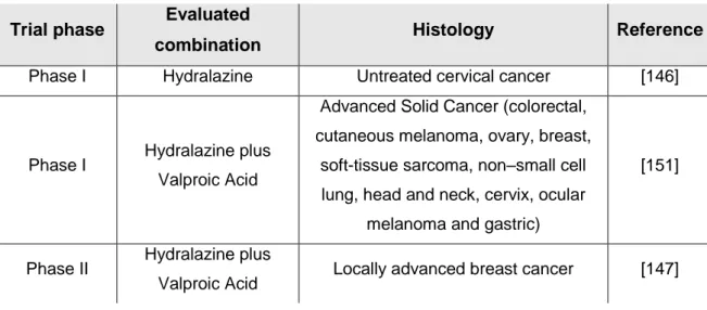
Hydralazine has been investigated as an epigenetic drug in several clinical trials targeting solid tumors (Table 2). One phase I study in cervical carcinoma demonstrated that hydralazine at doses between 50 and 150mg/day is well tolerated and is able to demethylate and reactivate TSG such as APC, MGMT, ER, GSTP1, DAPK, RARβ, FHIT and p16. Importantly, this occurs without affecting global DNA methylation, which is a major caveat of using other FDA-approved, DNMTi drugs, 5-azacytidine and 5-aza-2’-deoxycytidine [146].
A phase II trial in breast cancer also demonstrated the proposed molecular effects of hydralazine and valproic acid as DNMTi and HDACi respectively. In fact, they were able to reactivate TSG in breast cancer tumors. Interestingly, they increased the efficacy of chemotherapy, which was not expected, since epi-drugs have been associated with more myelotoxic side effects [147]. Another phase II trial based on 17 patients with solid tumors (cervix, breast, lung, testis and ovarian) who acquired chemotherapy resistance showed an overall response, disease stabilization and several symptoms’ improvement. They were treated with hydralazine and valproic acid followed by the chemotherapy agents that they were previously submitted to [148]. Almost 1000 downregulated genes of several signaling pathways in untreated cervical cancer patients turned out to be upregulated after hydralazine and valproic acid in one phase II clinical trial, highlighting the importance of investigating the demethylation inhibiting activity of these compounds [149].
Although it was terminated for administrative reasons, preliminary results from a phase III clinical trial in cervical cancer demonstrated that patients treated with hydralazine and valproic acid showed an improved progression-free survival when compared with those subjected to standard combination chemotherapies [150].
Table 2: Clinical trials with hydralazine in monotherapy and in combination.
Trial phase Evaluated
combination Histology Reference
Phase I Hydralazine Untreated cervical cancer [146]
Phase I Hydralazine plus
Valproic Acid
Advanced Solid Cancer (colorectal, cutaneous melanoma, ovary, breast, soft-tissue sarcoma, non–small cell lung, head and neck, cervix, ocular
melanoma and gastric)
[151]
Phase II Hydralazine plus
INTRODUCTION ǀ 20
Phase II Hydralazine plus
Valproic Acid
Refractory solid tumors (cervix, breast, lung, testis and ovarian) with
chemotherapy resistance
[148]
Phase II Hydralazine plus
Valproic Acid Cervical cancer [149]
Phase III Hydralazine plus
Valproic Acid Advanced cervical cancer [150]
DNMTi in PCa
DNA methylation can lead to gene silencing, contributing to drug resistance in antihormone therapies [66]. Therefore, the investigation of DNMTi is important for re-sensitizing malignant cells to antineoplastic agents [66,124,152]. Nevertheless, their clinical effectiveness is not entirely dependent on their DNA methylation inhibitory activity and its efficacy in solid tumors is not fully demonstrated [37].
There are several ongoing phase I and II clinical trials that include an antiandrogen plus an epi-drug as a treatment option for mCRPC. Specifically, there is a phase Ib trial followed by a phase II trial with enzalutamide and decitabine, which will study the side effects and best dose of decitabine and how well it works when given with enzalutamide. Moreover, they intend to determine the 12-month progression-free survival rate. Participants receive decitabine intravenously over 1 hour on days 1-5 and enzalutamide orally once daily on days 1-28. Courses will be repeated every 28 days in the absence of disease progression or unacceptable toxicity (NCT03709550).
Figure 6: Mechanism of action of nucleoside (5-azacytidine, 5-aza-2’-deoxycytidine) and non-nucleoside DNMT inhibitors (hydralazine).


![Figure 7: Resume of the different treatment options for each PCa stage. Adapted from [181]](https://thumb-eu.123doks.com/thumbv2/123dok_br/15598422.1051677/46.892.141.783.800.1090/figure-resume-different-treatment-options-pca-stage-adapted.webp)