The impact of CD6 targeting in T cell
function and immunopathology
Raquel Filipa Reis de Freitas
Orientador: Professor Doutor Luís Ricardo Simões da
Silva Graça
Tese especialmente elaborada para obtenção do grau de Doutor
em Ciências Biomédicas, especialidade em Imunologia
I
Júri: Presidente:
▪ Doutor João Eurico Cortez Cabral da Fonseca, Professor Catedrático e Vice-Presidente do Conselho Científico da Faculdade de Medicina da Universidade de Lisboa.
Vogais:
▪ Doutor Alexandre Valentim Xavier Mourão do Carmo, Investigador Principal do Instituto de Biologia Molecular e Celular do Porto;
▪ Doutora Helena Isabel Martins Soares, Investigadora Principal do Centro de Estudos de Doenças Crónicas da Universidade Nova de Lisboa;
▪ Doutora Íris Maria Ferreira Caramalho, Especialista de reconhecido Mérito e Competência, Investigadora do Instituto Gulbenkian de Ciência;
▪ Doutor Luís Ricardo Simões da Silva Graça, Professor Associado com Agregação da Faculdade de Medicina da Universidade de Lisboa (Orientador);
▪ Doutor Bruno Miguel de Carvalho e Silva Santos, Professor Associado com Agregação da Faculdade de Medicina da Universidade de Lisboa.
The impact of CD6 targeting in T cell function and
immunopathology
Universidade de Lisboa Faculdade de Medicina de Lisboa
Raquel Filipa Reis de Freitas Orientador: Professor Doutor Luís Graça
Tese especialmente elaborada para obtenção do grau de Doutor em Ciências Biomédicas, Imunologia
Instituição Financiadora: Fundação para a Ciência e Tecnologia
(SFRH/BD/52230/2013) 2019
Tese especialmente elaborada para obtenção do grau de Doutor em Ciências Biomédicas, especialidade em Imunologia
II
Todas as afirmações efetuadas no presente documento são da exclusiva responsabilidade do seu autor, não cabendo qualquer responsabilidade à Faculdade de Medicina de Lisboa pelos conteúdos nele apresentados
III
Para a minha avó Maria com um coração demasiado grande para
apenas dois netos. Estarás sempre comigo
.
Lutar&Acreditar
Raquel Freitas
“São meus discípulos, se alguns tenho, os que estão contra mim; porque esses guardam no fundo da alma a força que verdadeiramente me anima e que mais desejaria transmitir-lhes: a de não se conformarem”
IV
Table of Contents
Acknowledgements ... VI Abbreviation list ... VIII
Sumário ... 12
Summary ... 16
INTRODUCTION ... 20
1. The Immune System ... 20
1.1. Innate Immunity ... 21 1.2. Adaptive Immunity ... 25 1.3. B cells ... 27 1.4. T cells ... 29 1.5. Autoimmunity ... 46 1.6. Monoclonal antibodies ... 47
1.7. Aims of the thesis ... 54
• Undertsand the impact of CD6 targeting in the natural course of EAEErro! Marcador não definido. • Identify the cellular and molecular mechanism underlying CD6-targetingErro! Marcador não definido. • Demonstrate the equivalence between mice and human anti-CD6 mAbs ... 54
Autoimmunity ... 57
2. Itolizumab murine equivalente clone 10F12 in a model of EAE ... 57
2.1. Background ... 57
2.2. Material and methods ... 58
2.3. Results: ... 61
2.4. Discussion ... 66
Cancer... 72
3. Itolizumab murine equivalente clone 10F12 in a model of Breast Cancer. 3.1. Background ... 72
3.2. Material and methods ... 73
V
3.4. Discussion ... 80
Itolizumab ... 83
4. Itolizumab a humanised monoclonal antibody against CD6 d1. ... 83
4.1. Background ... 83
4.2. Material and Methods ... 84
4.3. Results ... 85 4.4. Discussion ... 92 General discussion ... 97 References ... 103 5. References ... 103 6. Appendix ... 120
VI
Acknowledgements
Quero agradecer aos meus dois pirilampos Sílvia e Andreia, que quando eu não via mais o caminho, o iluminaram para eu o conseguir acabar de percorrer, gosto muito de vocês e obrigada por tudoooo!!!
Sílvia, obrigada pela tua orientação sempre me defendeste quando eu precisava e trazias-me á terra quando também era necessário, estiveste lá sempre que chamei por ti!!!
Andreia, obrigada por me levares para pensamentos positivos e me distraíres do que não importava , e ás tuas meninas duas luzesinhas.
Obrigada Raquelinha minha homónima que também estiveste sempre lá para mim, ensinavas me truques tinhas paciência para mim e ajudavas me no que podias. Obrigada Filipinha, Catarina, Afonso e Ricardo e claro a nossa cubana favorita Yaqueline.
Obrigada Saumya por teres salvo o Chita e me teres ajudado a projectar o meu CD6zinho lol num modelo 3D lolololol
Obrigada ao meu orientador de tese o professor Luís Graça.
Obrigada ao meu Marquinho por me aturar quando chegava a casa com pouca paciência ou o fazia ir-me buscar ao laboratório de madrugada lol.
Obrigada ao meu Avô por me chatear para despachar de uma vez por todas, esta tese lolololol e à minha Tia , às minhas Primas e á nova aquisição a Matilde, aos meus Pais e ao meu Mano mais o pirilampo que está para chegar, à minha avó e à minha estrelinha do céu que tomou conta de mim lá de cima a minha avó Maria . Obrigada aos Mochos Nocturnos lolololololol, Eduardo, Diana e André, muito importantes na manutenção da minha sanidade mental.
Obrigada à Carina, ao Afonsinho, á Bigui, Maggie e Ricardo a malta das caminhadas
, à Trindade e á Teresa do Yoga que apareceram na minha vida mesmo na hora certa.
Um grande obrigada á AMIRA, a associação de animais que faço parte e me permitiu relembrar o que me motiva na ciência, a vontade de contribuir e ajudar
VII
E aos meus pikachus o meu Kiko a minha rochinha peluda, à nova aquisição a minha Estrelinha e ao Farber, Doroteia e Carolina que me aturaram mais que ninguém durante a escrita da tese.
Quero agradecer ao prof. Luís Graça por me ter aceite na sua equipa como estudante de doutoramento.
Um grande obrigada a todas as pessoas que cruzaram o meu caminho durante este doutoramento e fizeram o que poderam para me ajudar nesta aventura
VIII
Abbreviation list
AD- autoimmune diseases ADCC- antibody-dependent cell-mediated cytotoxicity
AHR- aryl hydrocarbon-receptor AID - activation-induced cytidine deaminase
AIRE- autoimmune regulator genes ALCAM- activated leukocyte cell adhesion molecule
ALL- acute lymphoblastic leukemia AP-1 – activator protein 1
APCs- antigen presenting cells ASCL2 - achaete-scute complex homolog 2
ATP- adenosine thriphosphate BBB- blood brain barrier BCRs- B cell receptors BM- bone marrow
BMDC- bone marrow derived dendritic cells
C57Bl/6- C57 black 6
CDR- complementarity determining regions
CFA- complete Freund’s Adjuvant CIA- collagen-induced arthritis
CK2- casein kinase II
CLL- chronic lymphocytic leukemia CMJ - corticomedullary junction CNS- central nervous system CSR - class switch recombination CTLA4- cytotoxic T-lymphocyte-associated protein 4
CTV- CellTrace violet
CXCL- C-X-C motif ligand CXCR- C-X-C motif receptor DAMPs- damage associated molecular patterns
DBA-1- Dilute Brown Non-Agouti 1 DCs- dendritic cells
DLN- draining lymph node
DN- double negative
DNA- deoxyribonucleic acid DP- double positive
9
EAE- experimental autoimmune encephalomyelitis
EMA- European medicines agency
FCS- Fetal calf serum
FDA- food and drug administration Foxp3- forkhead box p3
Gads- Grb2-related adaptor downstream of Shc
GITR- glucocorticoid-induced TNFR-related protein
GM-CSF - granulocyte-macrophage colony-stimulating factor
GWAS- genome-wide association studies
HSC- hematopoietic stem cells i.p.- intraperitoneal
i.v.- intravenous
IBD- Inflammatory bowel disease ICOS- inducible costimulatory receptor
ICOSL- inducible costimulatory ligand IEL- intraepithelial lymphocyte
IFN-γ – interferon gamma Ig- immunoglobulin
IL- interleukin
ILCs- innate lymphoid cells
iPBMCs- irradiated peripheral blood mononuclear cells
IPEX- immune dysregulation, polyendocrinopathy, enteropathy, x-linked
IS- immunological synapse
LAT- linker for activation of T cells LPS- lipopolysaccharide
LSCL- lymphosarcoma cell leukemia mAb- monoclonal antibody
MAC- membrane attack complex
MAPK- mitogen-activated protein kinases
MFI- median fluorescence intensity
MHC- major histocompatibility complex
MLR- mixed lymphocyte reaction MOG- myelin oligodendrocyte glycoprotein
MS- multiple Sclerosis
mTECs- medullary thymic epithelial cells
10
mTOR- mammalian target of rapamycin
NF-β - nuclear factor-β NK- natural killer cells NKT- natural killer T cells NLR – NOD-like receptor NO- nitric oxide
NOD- nucleotide-binding
oligomerization domain-like receptors
OVA- ovalbumin
PAMPs- pathogen associated molecular patterns
PBMCs- peripheral blood mononuclear cells
PD1- programmed cell death protein1 PI3K-
phosphatidylinositol-4,5-bisphosphate 3-kinase PKC- protein kinase C
PMA- phorbol myristate acetate
PRRs- pattern recognition receptor PTEN- phosphatase and tensin homolog
RA- rheumathoid arthritis
RAG- recombination-activating genes
ROR- RAR-related orphan receptor ROS- reactive oxygen species s.c.- subcutaneous
SAg- superantigen
Satb1- SATB homeobox 1 SCZ- subcapsular zone
SEA- staphylococcal enterotoxin A SEB- staphylococcal enterotoxin B SEE- staphylococcal enterotoxin E
SLP76- Src homology 2 (SH2) domain-containing leukocyte protein of 76 kD SNP- single nucleotide polymorphisms SO- superoxide
SOCS- suppressors of cytokine signaling
SRCR-scavenger receptor cysteine-rich
SRSF1- serine and arginine Rich Splicing Factor 1
STAT- signal transducer and activator of transcription
11
TdT- terminal deoxynucleotidyl transferase
TECs- thymic epithelial cells Teff- T effector cells
Tfh- T follicular helper cells
TGF-β - transforming growth factor beta 1
Th- T helper cells TLRs- Toll-like receptor TNF- tumour necrosis factor
Tregs- T regulatory cells TSAd- T cell-specific adapter TSPs - thymic seeding progenitors
TSST-1 -toxic shock syndrome toxin 1
12
Sumário
Nas últimas décadas, tem se vindo a verificar um aumento crescente no uso de anticorpos monoclonais (mAb) sendo que actualmente são a classe de agentes terapêuticos mais usada, representando uma indústria de milhões com já 30 mAb aprovados para terapêuticas em humanos e mais ainda sob avaliação clínica. As suas principais vantagens em comparação com outras terapêuticas consistem num elevado grau de especificidade e também flexibilidade que se usadas contra alvos na sinapse imunológica irão realçar as suas propriedades imuno-moduladoras.
O CD6 uma glicoproteína transmembranar da sinapse imunológica, importante para estabilidade da apresentação de antigénio, maturação da sinapse imunológica e uma proliferação das células T óptima, foi reavivado enquanto alvo terapêutico aquando da criação de um novo anticorpo monoclonal não-depletante em meados dos anos 90. Apesar de ter sido estudado extensivamente ao longo dos anos, compreender a sua dinâmica tem sido bastante difícil sobretudo devido à existência de resultados paradoxais. Contudo uma coisa é certa, o seu papel nas patologias autoimunes, como é o caso da esclerose múltipla, artrite reumatoide e psoríase. Um exemplo de quão paradoxal o papel do CD6 é, é o caso em que se por um lado em modelos animais de esclerose múltipla ou psoríase a sua ausência (KO) resulta em atenuação ou protecção da doença, por outro lado num modelo de artrite a sua ausência torna a doença ainda pior.
Para compreender melhor a razão de tal comportamento paradoxal, decidimos tirar partido deste novo anticorpo não depletante, anti-CD6 d1, desenvolvido em Cuba pelos nossos colaboradores, e tentar então descobrir como é que o “targeting” do CD6 pode afectar as propriedades funcionais das células T e como é que isso se relaciona com as imuno-patologias aqui estudadas. Aqui mostro como é que ambos os anticorpos, ratinho e humano, contra o CD6 vão fazer exatamente isso.
Primeiro investigámos como é que fazendo “targeting” do CD6 com o nosso anticorpo iria afectar o normal desenvolvimento de um modelo animal de esclerose
13
múltipla, para tal usámos o modelo de EAE, um modelo já bem estabelecido no nosso laboratório.
O tratamento com o nosso anti-CD6 foi intrigante, uma vez que os resultados obtidos estavam fortemente relacionados com as doses administradas. Ou seja, enquanto uma dose baixa era protectora, já uma dose alta ou era equivalente ao controlo ou acentuava a doença ainda mais. Contudo estes resultados em ratinho apesar de inesperados, estão em concordância com relatórios de ensaios clínicos em artrite reumatoide, onde também os efeitos protetores mais persistentes no tempo, advêm da dose testada mais baixa. Para compreender os mecanismos por detrás destas observações, investigámos como é que o “targeting” de CD6 estaria a afetar a normal especialização funcional das células T (polarização). E de acordo com os nossos resultados in-vitro, verificámos uma vez mais um efeito dependente da dose, enquanto doses crescentes de anti-CD6 d1 comprometiam a polarização das células T reguladoras, pelo outro lado eram mais favoráveis à polarização de células Th1.
De forma a tentar excluir um potencial efeito de impedimento estérico, resultante do elevado tamanho de um anticorpo IgG, usámos CD166 solúvel (ligando CD6 d3) como forma de quebrar as interações entre o CD6 nas células T com o CD166 (ALCAM) nas células apresentadoras de antigénio. Contudo o uso do CD166 solúvel não foi capaz de mimetizar o efeito do nosso anti-CD6 d1, sugerindo um efeito independente de impedimento estérico.
Mais ainda, fazer “targeting” do CD6 com o nosso anticorpo, sugere um efeito mais direcionado para a transdução de sinal propriamente dita, porque estas propriedades imuno-moduladoras só são detetáveis se condições de activação fisiológicas forem usadas. Para reforçar o seu impacto a um nível mais específico, temos o facto de que nem a proliferação ou sobrevivência foram afetadas significativamente. Na verdade, o uso de condições supra-fisiológicas, como activação por anti-CD3/anti-CD28 resulta na perda de quaisquer efeitos a nível de polarização.
Seguindo o racional de um efeito dependente de dose aquando do “targeting” de CD6, decidimos explorar o seu potencial terapêutico noutros modelos de doença
14
com uma cinética de acção oposta às doenças autoimunes. Investigámos então, se num modelo de cancro da mama doses altas de anti-CD6, dadas com diferentes estratégias resultariam no total desaparecimento do tumor ou num crescimento mais reduzido. Os nossos resultados confirmaram então que uma dose alta de anti-CD6 d1, ainda que administrada de forma cumulativa, resulta num abrandamento do crescimento tumoral. Contudo a forma como o “targeting” de CD6 especificamente afeta as células T CD4+ permanece inconclusiva, visto não haver significância
estatística. Ainda assim, os dados parecem sugerir um impacto negativo ao nível das células T reguladoras CD25+Foxp3+, especificamente as que infiltram os
tumores. Outra observação a considerar, foi um aumento ao nível da expressão de IL-17 por estas mesmas Tregs infiltrantes, expressão esta descrita como associada com caminhos de activação de MAPKinases previamente também eles associados com activação de CD6.
Sob estas mesmas condições os nossos dados sugerem ainda um impacto negativo por parte do anti-CD6 d1, ao nível da activação propriamente dita como mostrado pelos níveis de MFI de CD25, uma associação previamente estudada em células humanas por outro laboratório.
Como forma de validar o nosso anti-CD6 d1 de ratinho enquanto substituto de estudo adequado para o Itolizumab (human anti-CD6 d1), tivemos então de investigar também como é que o próprio Itolizumab afectava as propriedades funcionais das células T CD4+ in-vitro. E tal como esperado, quando tratadas com
ant-CD6 d1, as células humanas também mostraram um impacto dependente da dose em que doses crescentes impedium a polarização de Tregs enquanto por outro lado favoreciam as Th1s. Contudo, ao contrário do observado em ratinhos, e apesar de não afetar a sobrevivência, o “targeting” do CD6 afectou ligeiramente a proliferação em células humanas. Além disso, os dados das células humanas sugeriram também um efeito independente do impedimento estérico e dependente de condições de activação as mais fisiológicas possíveis de forma a que o impacto do anti-CD6 d1 pudesse ser percetível.
Estratégias de activação como anti-CD3/anti-CD28 ou mistura de SAgs e APCs, precisamente porque não permitem que haja um impacto do anti-CD6 d1 ao nível
15
das céulas T CD4+ ajudam a perceber quais as vias de sinalização em que o CD6
está de facto envolvido.
Em suma, os nossos dados mostram um efeito dependente de dose ao nível da polarização das células T quando “targeted” com anti-CD6 d1, efeito esse que consiste numa menor polarização de Tregs à medida que se aumenta a dose enquanto que ao mesmo tempo a polarização de Th1 é favorecida, observável tanto em células de ratinho como humanas.
Uma explicação potencial para estas observações poderia ser a relação existente entre níveis de ativação e sensibilidade de polarização, isto é, níveis diferentes de ativação causados pelo anti-CD6 podem resultar em fenótipos de polarização específicos.
16
Summary
In the last few decades, monoclonal antibodies have become one of the most widely used classes of therapeutic agents, representing a billion-dollar industry with more than 30 monoclonal antibodies approved for human therapeutics and many others under clinical evaluation.1
Their main advantages regarding other therapeutic agents consist of high specificity and high flexibility, which applied against targets involved in immune synapse will enhance its immunomodulatory therapeutic benefits. CD6 an immune synapse transmembrane glycoprotein, important for the stability of antigen presentation, maturation of immunological synapse and optimal T-cell proliferation, has been revived as a therapeutic target since the creation of a nondepleting anti-CD6 mAb in the early '90s. Despite CD6 has been extensively studied, understanding its biology has been difficult due to paradoxical results. Still, one thing is for sure, which is its role in autoimmune pathologies, as it is the case of Multiple Sclerosis, Rheumatoid Arthritis, and Psoriasis. And an example of CD6 paradoxical impact is, while in experimental autoimmune encephalomyelitis (EAE) and imiquimod-induced psoriasis CD6-deficient mice show disease protection or attenuation, in collagen-induced arthritis (CIA) the absence of CD6 made it even worst.
So, we have decided to take advantage of this new non-depleting mAb against CD6 d1, developed by our collaborators in Cuba and try to understand how targeting CD6 would impact T cell functional properties and how it would interfere in immune pathologies. Here, I show how both murine and human antibodies targeting CD6 domain 1 influenced exactly that.
First, we have investigated how targeting CD6 with our mAb would affect the normal development of a mouse model of MS, to do so we used a well-established EAE model, which had already been used in the lab.
Treatment with anti-CD6 was intriguing, since the outcome was heavily related to the dose being used, meaning while a low dose was protective, high doses showed a level of disease severity equivalent or even worse than the control group. However, our mice results do resemble the reports on RA clinical trials, where lower doses were the ones giving longer-term responses. To uncover the mechanisms behind it, we investigated how CD6 targeting was affecting CD4+ T cell functional specialization. And accordingly, to our in-vitro results, we verified that once more, in a dose-dependent manner, while increasing doses were
17 compromising Tregs polarization, in the case of Th1's it was favoring it. To try to exclude a possible steric hindrance effect due to the mAb size, we used a soluble CD166 (CD6 d3 ligand) as a means to disrupt T cell's CD6 interactions with APC's CD166. However, this did not mimic CD6 targeting with our anti-CD6 d1 mAb, suggesting a steric hindrance independent effect. More, targeting CD6 with our mAb suggests a direct effect over signaling itself, since its modulatory properties are only detectable if under activating physiologic conditions. Under supra-physiologic stimulation like with anti-CD3/anti-CD28, the impact over polarization is lost.
We expected the impact of anti-CD6 d1 to be a fine-tuned one since no major alterations were seen on either T cell survival or proliferation.
Following the rationale of this dose-dependent effect caused by CD6 targeting, we decided to explore its therapeutic potential on other disease models with opposite kinetics to autoimmune diseases. So, we investigated if in a model of breast cancer, high doses of anti-CD6, given under different delivery strategies, would result in total tumor clearance or reduced tumor growth. Our results ended up showing a reduced growth tumor ability if given in in-situ cumulative doses. However, the way CD6 targeting specifically impacted the CD4+ T cell population was not very conclusive due to the lack of statistical significance. But once more the data suggested a negative impact over CD25+Foxp3+ regulatory T cells,
specifically tumor-infiltrating ones. Another observation was a potential increase of IL-17 expression by these very same infiltrating Tregs which has been associated with MAPK activation pathways also associated with CD6 activation. Under these same conditions our data also suggests, a negative impact of CD6 targeting over CD4+ T cell activation as
measured by CD25 MFI levels, a relation previously reported on human cells by literature. To validate the mice anti-CD6 d1 mab as an adequate proxy of itolizumab, we have also investigated how Itolizumab would impact CD4+ T cell's functional properties in-vitro. And as
expected, when treated with anti-CD6 d1, human cells also displayed a dose-dependent negative impact over Treg polarization while on the other side favoring Th1's. But contrary to mice, and despite no impact on survival, targeting CD6 did significantly impact. Besides that, human data also suggested a steric hindrance independent effect and dependence on physiological activation conditions so that an impact on T-cell functional properties could be perceived. Activation strategies like anti-CD3/anti-CD28 or SAg mix and APCs, precisely because did not allow an impact of anti-CD6 d1 on CD4+ T cells, shed some light into which
18 Overall our data show a dose-dependent impact of anti-CD6 d1 over T cell functional specialization, meaning while increasingly high doses reduce T cell's polarization ability towards Tregs also favors Th1 induction, something true for both murine and human cells. A potential explanation for such observations is the relation between activation levels and polarization sensitivity, so different activation levels caused by CD6 targeting might favor specific polarization phenotypes.
Our data highlights the importance of dosage and how the same drug might be beneficial for different disease conditions.
19
20
INTRODUCTION
1. The Immune System
Immunity derived from the Latin word immunis means free or untouched and is one of the most complex systems comparable only to the nervous system. It is essentially a network of molecules, cells, tissues, and organs specialized in keeping the organisms in a state of equilibrium to avoid disease.
This equilibrium requires neutralization of pathogens like bacteria, virus, parasites and fungi, recognition and neutralization of harmful environmental substances and action against damaged or altered cells from the own body. Any alteration in this equilibrium between pro and anti-inflammatory immune responses results in disease.1
We can divide the immune system in innate and adaptive immunity, but we must remember they depend on each other.
To be accurate the first line of defense against infection is anatomical and physiological barriers followed immediately by an innate response, the oldest form of immunity throughout evolution. This readiness happens because of its non-specificity towards antigen, however, adaptive immunity takes longer but is highly specific for each infection and upon re-exposure becomes faster and stronger.
21 Fig.1- Interconnectivities of Human Immune System
As adapted from Turvey et al 2010 The human microbial defense can be divided into three arms: (i) anatomical and physiological barriers; (ii) innate immunity; and (iii) adaptive immunity. With some elements making the connection between the arms. 2
1.1.
Innate Immunity
When anatomical and physiological barriers like intact skin, mucociliary clearance, low stomach pH and bacteriolytic enzymes in secretions fail, the innate immunity gets immediately triggered and an inflammatory response begins. 3
The triggers are damage-associated molecular patterns (DAMPs) and pathogen-associated molecular patterns (PAMPs). The first, DAMPs, are molecules upregulated and released during cell lysis and tissue damage either in sterile or
Intact skin Ciliary clearance Low stomach pH Lysozyme in tears and saliva Macrophages Eosinophils DCs Neutrophils Mast cells NK cells NKT cells T cells B cells Humoral Cellular Cellular Humoral Complement Mannose binding lectin Antimicrobial peptides LPS binding protein C-reactive Protein Antibodies Anatomical and
22
infectious inflammation. The seconds, PAMPs, are highly conserved microbial components essential for survival and virulence of pathogens.
These triggers activate cells when recognized by pattern recognition receptors (PRRs), which are germline-encoded and although this lack of flexibility is a disadvantage it is overcome by its essentiality to pathogen survival and a ready to go response.
But there is also the opposite strategy where innate immune cells must recognize specific molecules expressed only by healthy cells to inhibit their activation. This independence of genetic recombination and developmental phases buys time for the adaptive immunity to get triggered and ready.
PRRs are divided into several classes like the nucleotide oligomerization domain (NOD)-like receptors (NLR), Toll-like receptors (TLRs), C-type lectin receptors (CLRs) scavenger receptors and cytosolic DNA sensors between others. They are found either at the cell surface, cytoplasm or endosomes which allows them to detect both internal and external threats.2
At the cell surface where they detect microbial cell-wall components like LPS (TLR 4), we find TLR 1, 2, 4, 5 and 6. In endosomes where they recognize microbial nucleic-acids like ds-RNAs (TLR3), we find TLRs 3, 7, 8 and 9. But TLRs also recognize DAMPs from the host, like heat shock proteins (TLR2-4) and Chromatin-IgG complexes (TLR9). 4,5
Triggering PRRs activates transcription factors like NF-ƙβ, AP-1, and IRFs, this will start pro-inflammatory cytokines and chemokines production, presentation of co-stimulatory signals (contact-dependent or independent) and finally cell recruitment to the site of injury with activation of the adaptive immunity.
The elements involved in this initial phase are from the humoral and cellular origin. Between the humoral components, we have the Complement, LPS-binding, and C-reactive proteins as well as other pentraxins, collectins and anti-microbial peptides like defensins. Regarding the cellular components they can be of hematopoietic and non-hematopoietic origin. 2
The complement system (C1-C9) it's a liaison between inflammatory triggers and other immune responses like antibodies and phagocytic cells. This is set in
23
motion by a coordinated enzyme cascade resulting in danger clearance, through pathogen recognition, opsonization, and lysis.
Lysis occurs through a structure called membrane attack complex (MAC) that essentially introduces pores on pathogen cell walls and kills them.
This is a highly regulated system, which requires activation of its precursors in a proper sequence to form enzymatic complexes which rapidly dissociate and return to inactivity.6
The hematopoietic group includes both myeloid cells (macrophages, dendritic cells-DCs, neutrophils, eosinophils, mast cells, and basophils) and innate lymphoid cells (ILCs). While the non-hematopoietic, also known as immune stroma includes fibroblasts, endothelial and epithelial cells essentially cells that form the architectural structure necessary for proper cell interactions and proper display of molecular cues needed for position growth and survival.
Macrophages and DCs reside in tissues while scavenging it to find signs of danger, once they get activated and initiate a pro-inflammatory response, blood circulating neutrophils are recruited to the tissue. All the three are highly phagocytic cells, but while macrophages and neutrophils are responsible for removal and disposal of pathogens, infected cells and immune complexes using strategies like nitric oxide (NO), superoxide (SO), reactive oxygen species (ROS), enzymes and pro-inflammatory cytokines with anti-microbial properties, DCs are responsible for the connection with the adaptive immune system.7 They bridge this connection by presenting the antigen to T cells, and although other cell types can also present it (macrophages and B cells) they are the professional antigen-presenting cells APCs.
When the pathogens are large parasites like helminths they cannot be phagocytized, so eosinophils come into action and kill them by releasing cytotoxic granules, cytokines, and lipid mediators. This will increase inflammation as well as tissue destruction, in fact, eosinophils are responsible for allergic diseases like asthma. 8
Allergic diseases are also misdirected immune responses perpetrated by long-lived tissue-resident mast cells and short-long-lived blood circulating basophils which
24
releases inflammatory mediators like histamine and have anti-bacterial properties.9
But lymphoid cells are also involved in innate immunity, they are the ones with the germline-encoded ready to go antigen receptors. For more than 30 years ILCs were all about NK cells but recently, new populations arise, the ILC1, 2 and 3. They look like the innate version of T cells with both ILC1 and Th1 producing IFN-γ, ILC2 and Th2 producing IL-4, 5, 9 and 13, ILC3 and Th17 producing IL-17 and 22 and finally NK and CD8+ T cells both being cytotoxic. However, they are
much better pro-inflammatory and immunoregulatory cytokines producers, and this allows them to direct adaptive immunity in the best way to fight each specific threat. Their localization is also strategic, they are at sites of potential invasion and colonization by pathogens like barrier surfaces: skin, lung, intestine, some adipose tissues, and mucosa lymphoid tissues. 10,11
Innate immunity and adaptive immunity are intrinsically connected. 12
If on the one hand the adaptive immunity activation depends on antigen presentation, where DCs uptake the pathogen, migrate to draining lymph nodes, process it mature and present it to naïve T cells and also on the type of DC stimulus, crucial for T cell polarization, accordingly to the specific group of pathogens to be cleared. On the other hand, the classical activation of the complement system is dependent on antibodies to initiate the enzymatic cascade. Essentially, to function properly they need each other.13
25
1.2.
Adaptive Immunity
Three words: specificity, adaptability, and memory. As its name states it adapts to each new threat by making use of genetic recombination to produce antigen-specific receptors that allow, antigen-specific, faster and stronger responses on subsequent exposures to the same threat.
Like with innate immunity, we can also divide adaptive immunity into humoral and cellular components.
Humoral components include antibodies that depend on B cell activation and cellular components include CD8+ cytotoxic T cells which depend on T cell
activation. However, we must keep in mind that CD4+ T helper cells are also
essential since they provide both B, T and innate cells help.
B cells only require help from T cells if the antigen is a protein, and we call it thymus-dependent immune responses otherwise they independently activate themselves.
Once again both innate and adaptive immunity is triggered by cell receptors in this case, we have the BCRs (B cell receptors) and TCRs (T cell receptors), which account for specificity since they are antigen specific. But contrary to innate immunity here they are a result of random somatic recombination as well as a somatic mutation in B cells' case from germline pools of DNA segments.
These processes called recombination and somatic hypermutation, allow as much as 108 and 1010 possible TCR and BCR combinations to cover all the
pathogens that could ever be encountered in a lifetime as well as increased affinity/avidity
Another advantage of adaptive immunity besides specificity and flexibility is the memory, innate immunity does not have it, no matter how many times they encounter the threat.
26
The immunological memory is the ability to respond specifically, faster and stronger upon antigen re-exposure and both B and T cells can differentiate into memory cells during a first encounter with the antigen, the pillar of vaccination. Again, like some innate cells, B and T cells also derived from hematopoietic stem cells (HSC) which branched into the lymphoid lineage and while B cells mature in the bone marrow, T cell progenitors must migrate into the thymus to do so.
Fig2. Illustration of common lymphoid cell precursors’ production and maturation at
primary lymphoid organs and migration into circulation and secondary lymphoid organs.
27
Bone marrow (BM) and thymus constitute the primary lymphoid organs but is in the secondary lymphoid organs (spleen, lymph nodes, and mucosa-associated lymphoid tissues) where all the action takes place. Here B and T cells meet directly or are presented with the antigen to which they are specific.
While B cells are capable of directly recognize the antigen, T cells require the antigen to be presented within a major histocompatibility complex (MHC). There two types of MHCs the I and II, the MHCI is expressed by all nucleated and healthy cells and the MHCII is expressed only by APCs a group that includes DCs and B cells. Also, MHCI antigen presentation is specific of CD8+ T cells, while MHCII of CD4+ T cells.
When all these conditions are reunited an adaptive immune response begins.
1.3.
B cells
Back in the 1890s when diseases like diphtheria and tetanus were killing thousands of people a year, Behring and Kitasato discovered the importance of a group of circulating antitoxins, in the fight against them.14
Then, later in the twentieth century, Ehrlich came to propose that these anti-toxins (antibodies) were produced and released by certain cells due to antigen stimulation. 15 But it was only in the 1930s and late in the 1940s that both its
physical nature and cellular source (B cells) were finally discovered. 16-18
However, it took until 1965 for B cells to be considered as an independent lymphocyte lineage. 19
Like T cells, B cells also derive from hematopoietic stem cells in the bone marrow, where they will start their development that includes B cell receptor (BCRs) recombination and which is mediated by enzymes like RAG and TdT resulting in higher antigen receptor diversity. 20
BCRs can be divided into two heavy (H-chain) and two light chains (L-chains) connected by disulfide bonds. But it will be their N- and C- terminal regions that will define them.
28
N-terminal regions of both H- and L- chains form the antigen-binding domain, accounting for antibody specificity. This domain is comprised of three hypervariable complementarity determining regions, CDR1-3, the targets of V(D)J recombination guarantying increased diversity of antigen specificity. 21,22
By contrast, the C-terminal regions of both H- and L-chains are constant and define the antibody isotype, responsible for its function.
Naïve immature B cells, only express IgM and IgD isotypes, but when they migrate to secondary lymphoid organs and become activated, class switch recombination (CSR) is triggered. This process is also mediated by an enzyme called activation-induced cytidine deaminase (AID) and the outcome of switching will depend on environmental cues, like cytokines produced by T helper cells. Thus, if supported by Th2s they will produce IgG1 and IgE, if supported by Th1s, they will produce IgG2 but if they are in mucosal tissues, they will produce IgA. These five different isotypes are responsible for activating different types of immune cells specific for each situation while keeping the same specificity. 23-27
To improve specificity and most of all adaptability B cells introduce random mutations into their Igs' CDR domains through a process of somatic hypermutation.
Random mutations will be followed by a process of affinity maturation selecting the B cell clone with the highest antibody affinity, this will ensure a proliferative advantage upon antigen recognition associated with increased survival and growth signals. 28
Maturation of B cells will also result in memory, and these long-lived plasma cells upon antigen re-exposure are capable of rapidly react and secrete higher levels of antibodies for longer periods. This is the basis for vaccination however is also the reason why allergies and autoimmune diseases can perdure. 29
29 Fig.3 Illustration of a germinal center, where activated B cells proliferate,
differentiate and go under several rounds of somatic hypermutation and affinity maturation of their antibody genes.
1.4.
T cells
Like B cells, T cells also derive from bone marrow HSC precursors, but contrary to them they must migrate to the thymus to develop.
These thymic seeding progenitors (TSPs) 30,31, enter the thymus in reduced numbers
and once they get in contact with thymic epithelium the journey begins.
The spatial and temporal organization is the key for proper T cell maturation. Thus, we can divide the thymus into four compartments: subcapsular zone, cortex, medulla and corticomedullary junction (CMJ). 32
30 Fig. 4 Illustration of T cell development and maturation in the thymus.
First TSP enters the corticomedullary junction through postcapillary venules, where these early thymic precursors, also called double negative CD4- CD8 -(DN1) cells, proliferate and begin to differentiate. 32 After leaving the CMJ, DN1 cells migrate
deeply into the cortex towards the subcapsular zone (SCZ). Here, DN1 will receive stimulatory signals from the stroma, mostly thymic epithelial cells (TECs) and fibroblasts.32,33 At this stage (DN2) their fate becomes more restricted and gene
rearrangement at the TCR gene loci begins with upregulation of pre-TCRα chain and Rag1.34 When reaching DN3 stage, T cell lineage is already defined, either αβ or
γδ.35 T cells expressing αβ TCRs, require an additional checkpoint, a β-selection.
This checkpoint demands a fully functional pre-TCR, which is made of a rearranged TCRβ-chain, CD3 chains, and an invariant pre-TCRα chain. Together with Notch1, the cells can now receive signals for survival and metabolism.36,37
If successful, DN4 cells migrate back to the cortex until reach the medulla, along the way pre-TCR signaling allows for CD4 and CD8 expression, at this point Rag1 and 2 are re-expressed so that TCRα rearrangement can begin.38
Functional αβ TCRs will determine if the cell survives and its fate, by establishing the strength and specificity towards peptides in MHC ligands presented by cTECs,
31
DCs, and fibroblasts. This is Positive Selection, where only the TCRs with intermediate avidity for self-peptide-MHC complexes can thrive. 39
After this second main checkpoint, DP thymocytes will commit either to CD4 or CD8 single-positive lineage (SP).
When finally, they reach the medulla as SP thymocytes they are again tested. In this third checkpoint (Negative Selection), the goal is to prevent autoreactivity so, thymocytes with high-affinity TCRs for self-peptides are instructed by mTECs, which can express tissue-specific antigens (encoded by the autoimmune regulator gene- AIRE) to commit apoptosis.39,40
Negative selection in the thymus is the first mechanism of tolerance also known as Central Tolerance, but this is not enough to constrain autoreactive T cells. Thus, the pool of mature T cells leaving the thymus towards the secondary lymphoid organs also include some immature self-reactive naïve cells.
All of us have autoreactive T cells, but not all of us have autoimmune diseases, in fact, a group of self-reactive T cells leaving the thymus and Foxp3+ take a part in maintaining peripheral tolerance.41
At the periphery, activation of naïve T cells requires interaction between the TCR/CD3 complex, and antigen presented at the MHC plus a second signal, co-stimulation.
Co-stimulation is an independent signaling pathway that synergizes with antigen-specific signals to allow lymphocyte activation otherwise they just become anergic. Examples of co-stimulatory partnerships between T and APCs are CD28/CD80 or CD86; OX40: OX40L; ICOS/ICOSL and CD40L/CD40.
However, specific T cell responses are determined by a balance between not only, co-stimulation but also co-inhibition, examples of inhibitory receptors are CTLA4 and PD1.42
Besides the common αβ TCRs (90-95% T cells), also known as conventional T cells, there is also a group of unconventional T cells, γα T cells and NKT cells.43
32
The first to be associated with immunologic memory was the conventional T cells. These cells are characterized by their αβ TCRs plus the co-receptors CD4 or CD8 and their capacity to recognize processed antigenic peptides within MHC groves. While, CD8+ T cells, are a subpopulation of MHC I restricted cytotoxic T cells
responsible for killing cancerous or virally infected cells in a contact-dependent manner through induction of apoptosis. CD4+ T cells, are MHC II-restricted and
mostly responsible for modulating both humoral and cellular immunity.
Unconventional T cells, are kind of a hybrid between adaptive and innate immunity, if on the one hand they also express TCRs, on the other hand, the repertoire is much more limited, and the nature and distribution of the recognized molecules are completely different plus that they bind their antigens directly with no need for classical MHC presentation but instead dependent on conformational shape.43
Intraepithelial lymphocyte (IEL) compartments, like skin, intestine and genitourinary tracts are enriched with unconventional T cells, NKTs but mostly γδ T cells. The latter, by recognizing the so-called, "stress antigens" (e.g. phospholipids and alkyl amines) helps preventing infected or transformed cells dissemination and contributes for tissue homeostasis.43 NKTs on the other hand are mainly specialized
in responding against certain types of bacteria, fungi and parasites, but recently they have also been associated with autoimmunity and immunosurveillance. NKTs, on the other hand, are mainly specialized in responding against certain types of bacteria, fungi, and parasites, but recently they have also been associated with autoimmunity and immunosurveillance. NKTs, as its name implies, share T cell traces like αβ TCR expression and NK traces like the expression of CD56 and NK1.1 marker. Similar to T cells, they must be presented with the antigen. However, instead of peptides, it will be glycolipids and instead of classical MHC presenting it will be a CD1d molecule.44 Another great advantage of this population is, depending on the environment they can be triggered to rapidly release big amounts of several cytokines like IFN-γ, IL-4, IL-10, IL-13, IL-5, GM-CSF, TNF, IL-21, and IL-17. As with conventional T cells, the authors defend this diversity to be a result of different specialized populations and the need for further studies.45-49
33
1.4.1. CD4+ T cells
As previously referred CD4 T cells, also known as T-helpers, provide help to both adaptive and innate immunity. They do it by secreting specific sets of cytokines accordingly to environmental cues, resulting in recruitment and activation of other cells. In fact, they boost, not only primary but also memory immune responses.50
Along with the context of an immune response, mature APCs activate CD4+ T cells, resulting in proliferation and differentiation into a variety of specialized subsets.
Fig.5 Illustration of the different possible CD4+ T cell differentiation pathways and
their associated transcription factors and inducing cytokines. Adapted from Coomes et al 2013.
The concept of differentiation was proposed almost thirty years ago by Mosmann and Coffman to explain how T-helpers could promote completely different immune responses.51 Based on their cytokine profile they were classified into two terminally
differentiated groups, Th1, and Th2 plus a controversial regulatory population, nTregs. Th1 cells are responsible for immune responses like delayed-type hypersensitivity (DTH), cellular immunity, B cell class switching to IgG2a and clearance of intracellular pathogens or transformed cells.26 In these
microenvironments, APC and NK cells are induced to produce cytokines like IL-12 and IFN-γ.52,53 IL-12 is responsible for activating the transcription factor STAT4
which results in more IFN-γ expression, responsible for upregulating STAT1 and resulting in T-BET expression the master regulator of Th1 cells. T-BET activation,
34
will then lead to IL-12Rβ upregulation assuring a positive feedback for Th1 differentiation, but a negative one for Th2 and Th17 (later discussed).54 Once
differentiated they produce mostly IFN-γ and IL-2, essential for phagocytic activity and cytotoxic CD8+ T cells' activation. But when its activity is not properly regulated,
it results in inflammatory and autoimmune diseases, like colitis or multiple sclerosis.55,56
Fig.6 Illustration of CD4+ T-helper 1 polarization.
Th2, on the other hand, are responsible for allergies, humoral immunity, B cell class-switching to IgG1/IgE and clearance of extracellular bacteria or parasites. Th2s are induced when after activation cytokines like IL-4 and IL-2, abundant in these microenvironments bind recently activated T cells.
IL-4 recognition then activates STAT6 resulting in GATA3 upregulation, the master regulator of Th2 cells. Together they activate a set of Th2-related gene loci, responsible for cytokine production, proliferation, and inhibition of Th1 differentiation. Other transcription factors involved are IRF4 and STAT5. While the first activates IL-4 promoter resulting in GATA3 upregulation the second is activated by IL-2 that together with GATA3 promotes IL-4 expression. Again, positive feedback is present. Upon differentiation, they will secrete IL-4, IL-5, and IL-13 which will mostly act upon innate cells, with IL-4 and IL-13 as its key cytokines.57
35
Their main targets are mostly innate cells like macrophages, basophils and mast cells, but the impact also on B cells and non-hematopoietic cells like epithelial cells present in mucosal surfaces.
Seemingly redundant on function, when in physiological conditions IL-4 and 13 have preferential roles. During a helminth infection, IL-4 is essential for IgE production and mast cell activation while IL-13 is essential for goblet cell hyperplasia, mucus production, and parasite expulsion. 58 Even when type 2 response lacks proper regulation and results in allergic diseases like asthma, IL-4, and IL-13 again despite overlapping functions have preferential roles. IL-4 favors IgE and IgG1 production, while IL-13 is essential to goblet cell hyperplasia, smooth muscle contraction, and mucus production.59
Fig.7 Illustration of CD4+ T-helper 2 polarization.
Besides mutual regulation between Th1 and Th2, in the '60s, right after finding out the importance of thymus for immunity60, a population of T-cells capable of immunosuppression was proposed. However, such population was accepted only in the '90s with the induction of autoimmunity by neonatal thymectomy or transfer of T-cell populations depleted of specific T-cell types into lymphopenic mice.61,62
Named regulatory T cells (Tregs) and identified by the markers CD5high, CD25+ and
36
Almost 10 years later, Sakaguchi and his laboratory finally identified Tregs' master regulator, Foxp3, a transcription factor which absence results in scurfy mice and IPEX (immune dysregulation, polyendocrinopathy, enteropathy, x-linked) syndrome, both autoimmune and inflammatory diseases. 64,65,66
Thymic Tregs (tTregs) are the most well studied regulatory T cells, whose specific epigenetic landscape needed for lineage fate commitment, accordingly to Kitagawa et al 2017, which requires a permissive epigenetic remodeling of Tregs' specific super-enhancers. Satb1, a global genome organizer highly expressed during thymocyte development can bind these super-enhancers even within closed chromatin, activating them and assuring lineage commitment. 67 67 Full activation of
specific super-enhancers will also depend on TCR avidity and co-stimulatory signals provided by thymic epithelial and dendritic cells.68,69 Such permissive remodelling
will allow for Foxp3 expression and fixation of Tregs' specific epigenetic signature genes, that also include CD25, CTLA-4 and GITR.70
Immune homeostasis, peripheral tolerance, and regulation of inflammation are mostly dependent on proper Treg function. This function can be either dependent on
cell contact or based at cytokine secretion and metabolic disruption.71
Suppressive mechanisms include direct action upon Teff cells through induction of
apoptosis and cell cycle arrest (granzymes and galectin-1) or indirectly by manipulation of DCs function, either inducing negative signaling for Teff (CTLA-4) or
disruption of proper DCs-T cells interactions (neuropilin).72-75 Also secretion of
cytokines like IL-10, TGF-β, and IL-35 as well as disruption of target cell metabolism, by higher consumption of critical cytokines (IL-2) or even increased expression of proteases (CD39 and CD73) that hydrolyze ATP and compromise DCs' maturation will impact on Teff function.71,76-84
37
Fig8. Illustration of CD4+ thymic Treg polarization.
Still, these populations were not enough to explain the growing complexity of T-helper phenotypes, which despite shared characteristics with the previous subsets presented new ones. But it took extra 5-10 years, until the characterization of these new populations which include Th17, pTregs, Tfh, Th9, and Th22.
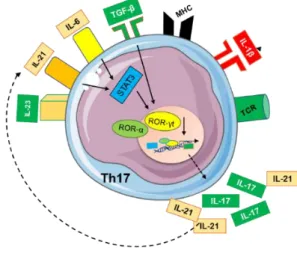
Th17 cells appeared in the early 2000s as a new hypothesis for the regulation of tissue damage present in both microbial infections and autoimmunity, no longer explained by the Th1/Th2 Mosmann and Coffman hypothesis.85-90
When they appeared, researchers considered IL-23 as the main inducer of Th17, but in 2006 three independent labs, showed that IL-23 was most important for survival and expansion while TGF-β and IL-6 were essential for polarization.91-94
Binding of TGF-β to its receptor activates the SMAD signaling pathway which results in both Foxp3 and ROR-γt expression.
At this point depending on microenvironmental cues, these cells can either differentiate into Th17 or a new population discovered around the same time, peripheral induced Tregs (pTregs), which we will discuss later.
So, if the microenvironment is more pro-inflammatory and has higher concentrations of IL-6, STAT3 signaling pathway becomes activated. This results in IL-21 expression initiating an IL-21/STAT3 autocrine loop responsible for a sustained
38
STAT3 activation and expression of Th17 master regulators, γt and ROR-α.93,95
ROR-γt and ROR-α will then induce IL-17A and IL-17F, which will increase inflammatory mediators, like IL-6, IL-1, IL-21, and CXCL8, as well as the recruitment of innate immune cells like neutrophils, essential for clearance and control of either extracellular bacteria or fungal infections at epithelial and mucosal barriers. ROR-γt and ROR-α also upregulates 23R, making Th17 responsive to environmental IL-23, produced mostly by innate cells and essential for their survival and expansion. 91-93,95,96
However, when IL17 production becomes dysregulated it causes chronic inflammation and increased tissue damage culminating in autoimmune diseases like MS, RA, psoriasis and inflammatory bowel disease (IBD).97
MS considered for a long time as a Th1 dependent disease turned out to be also dependent on Th17, in a way still poorly understood. 90,98
Fig 9. Illustration of CD4+ T-helper 17 polarization.
Coming back to pTregs, this is a regulatory population that contrary to tTregs, can be induced in the periphery from conventional naïve CD4+ T cells if under appropriate stimulus.99-101
39
Allergens, food or even non-pathogenic microorganisms (microbiota), in both inflammatory and non-inflammatory conditions, can trigger pTregs differentiation. The main trigger is the host microbiome, with germ-free mice showing reduced numbers of pTregs.102-104
For pTregs differentiation, presentation of lower doses of high-affinity peptides coupled with low co-stimulation (low CD28 signaling) is crucial.105 Also, presentation
by tolerogenic APCs like gut CD103+ DCs capable of synthesizing TGF-β and retinoic acid, both inducers of Foxp3 expression together with a microenvironment rich in IL-2 and other tolerogenic soluble factors, will favor its differentiation. 99,106-108
108Infectous tolerance mediated by tTregs will similarly contribute to pTregs induction. 109
pTregs major advantage versus tTregs is their plasticity of Foxp3 expression, reflected in functional adaptability to evolving immune responses. They permit to balance protective immunity with tissue tolerance to help contain excessive damage without compromising pathogen clearance.110-112
Miyao et al. work supported a notion of time restrained inhibition sensitive to inflammation intensity or antigen availability, where pTregs can revert to Tconv cells. Less inflammation, less damage out of control, thus no need for more inhibition.113
De novo induced pTregs are mostly known for preventing general inflammation and contributing to both fetal and mucosal tolerance (airways and gut), however like tTregs they also contribute to autoimmunity regulation.114-119 119 Its main function is
40 Fig 10. Illustration of CD4+ peripheral induced Tregs.
Since the 60's that scientists have known CD4+ T cells were indispensable for B cell
memory and germinal center formation, a place for B cell somatic hypermutation, affinity maturation and class switch recombination (CSR).122-125
And twenty years later, during the Mosmann and Coffman Th1/Th2 period, it was believed that Th2 cells were the population assuring this help due to IL-4 and IL-10 secretion.126,127 But later, mice data showing preferential CSR towards IgG2a isotype
prompted us to consider Th1 as well. So, this would suggest an unbiased preference of T cell help, only to be contradicted, another twenty years (1999) later, with the discovery of a CXCR5+ population.128 Expression of CXCR5 in CD4+ T cells turned
out to be like in B cells, the "key" for migration into follicles where its ligand CXCL13 is expressed.128,129
In 2000 the term T follicular helper cell (Tfh) arises, when Breitfeld et al. together with Schaerli P. et al show this population superiority in promoting B cells production of immunoglobulins.130,131
But it was only in 2009 with the identification of Bcl-6 as an essential factor for Tfh differentiation that these cells truly become accepted as an independent T helper subset.132-134
Tfh differentiation is a multi-stage, multi-factorial and highly heterogenic process, that comprises an initial priming phase, with DCs at the T-zone of secondary lymphoid organs presenting the peptides and providing co-stimulation in the form of CD80/CD86 and ICOSL in a balanced environment of IL-2 and IL-6 crucial in cell fate determination.135,136
IL-6 will then induce recently activated CD4+ T cells to upregulate Bcl6, Tfh master
regulator that together with ASCL2 results in CXCR5 early expression and migration into the T-B border.132,136-139 Here pre-T
fh cell will finally interact with antigen-specific
41
This recently differentiated Tfh cells can now enter the follicles to establish fully
operational germinal centers, allowing for somatic hypermutation and selection of high-affinity B cells culminating in memory B cells and plasma cells capable of producing antibodies with even greater affinities.125
At later stages, B-cells will then become the major antigen-presenting cells as opposite to DCs essential in priming phases.141,142
So their main function is Germinal Center (GC) development and function and when it goes well we have not only control of pathogens but also commensal microbiota.143
But when it goes wrong we might end up developing allergies or even autoimmunity, due to aberrant generation of autoantibodies or formation and maintenance of ectopic follicles.144-147
Something interesting is their involvement in cancer, which is not so unexpected if we consider all the regulatory checkpoints that may fail and result in disadvantageous mutations. But overall their ability to form and maintain germinal centers as well as promoting antibody affinity selection will be advantageous against cancer (Gu-Trantien et al.), but hazardous in autoimmunity.148,149
Fig 11. Illustration of T cell priming towards a TFH phenotype with consequent
42
For twenty years IL-9 was considered a Th2-derived cytokine, but only until 2008, when Veldhoen et al. and Dardalhon et al discovered a new independent IL-9 producing T cell subset.150-153
Named Th9, this is a population that although independent, can also be re-differentiated from Th2 if TGF-β is present and, that shares a variety of functions with the former.151,154
Both populations are important for resolving parasitic infections and mediating allergic inflammation.155 However, Th9 are reported to be also involved in transplant
tolerance, tumor immunity and autoimmunity.154,156-160
Like any other subset, it needs a proper TCR signal, co-stimulation, and a cytokine cocktail, with a preference for OX40 which selectively enhances 9 over 4 or IL-5 (NF-kB noncanonical pathway). This, in a microenvironment rich in IL-2, TGF-β, IL-4, and IL-1 culminates with the expression of PU.1 and IRF4. Both transcription factors capable of modifying chromatin at the IL9 locus and directly binding to its promotor.161-164
Their close relationship with the Th2 effector program favors a certain degree of plasticity to allow "fine-tuning" of responses accordingly to the need.165 Such lack of
stability or not fully understood flexibility coupled with the absence of a transcription factor are the fuel for the doubts around its identity.
The use of Licona—Limón et al IL-9 fluorescent reporter mice, would ease the doubts around this newbie T helper subset.Even because it would also exclude other sources of IL-9, like mast-cells, eosinophils, ILCs or NKT cells accounting only for T cell production and relevance on the ongoing response.150 , In fact, EAE is one
example of such situation, where other cell types might just be the source of IL-9 which is the case of Th17. Again Licona—Limón et al reporter mice would shed some light on this.
43 Fig 12. Illustration of CD4+ T-helper 9 polarization.
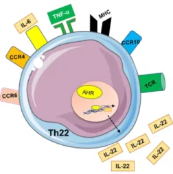
To shake things a little bit more, in 2009 a cytokine first discovered in 2000 which was initially attributed to Th1 cells and latter to Th17 ended up having its own lineage.166-168 We are referring to IL-22, a cytokine with a very important role in skin
homeostasis and inflammation.
Due to its ability to bind epithelial cells, which abundantly express IL-22R, it allows TH22 cell to establish a bridge between adaptive immunity and barrier organs, very important for wound healing and antimicrobial defense. 169-172 Indeed, IL-22 can
trigger anti-microbial peptides (S100 proteins and defensins) and synergise with IL-17 and TNF-α to fight pathogens like Candida albicans.168,169,173
This new lineage, like any other lineage, requires antigen presentation, in this case preferentially by skin DCs', co-stimulation and a microenvironment specifically rich in TNF-β and IL-6.174,175 With a relatively stable phenotype, these cells still lack an
unquestionable master regulator, but like Th9 they also have a candidate, aryl hydrocarbon-receptor (AHR).176
Th22 is a population highly abundant in the skin (upper parts of the epidermis) but scarce in circulation, something explained by skin strong expression of chemokines that bind CCR4, CCR6, and CCR10 present in these cells.168,174,177
And what could be a good thing, left unregulated could result in autoimmunity like psoriasis (hyperkeratosis) or even potentiation of malignancies due to exacerbated anti-apoptotic effect.178-180
44
Still, many questions remain to be answered, like detailed differentiation and regulation mechanisms, dynamics regarding other IL-22 producers and better readouts of Th22 targeting treatments.
Now more than ever, with all these different T cell lineages, differentiation is perceived as a very complex and interdisciplinary process, dependent on factors such as the cytokine pool, antigen concentration, APC phenotype, and co-stimulatory stimulus.181
Fig 13. Illustration of CD4+ T-helper 22 polarization.
But if it was not enough, van Panhuys introduced an extra variable with his temporal signaling model were long term interactions producing high strength TCR signals favor Th1 and short-term interactions (weak TCR signal) with strong co-stimulation favor Th2.182 Also induction of Th17 and Tfh are influenced by TCR strength, but
while strong signals favor Tfh, for Th17 there are divergent data and for Th9 or Th22 is still unknown. 183-186
Another aspect that has been gaining relevance is the plasticity of the CD4+ T cell population contradicting the previously accepted notion of terminal differentiation.
45
Until not so many years ago, each lineage characterized by the expression of a master regulator and a set of signature cytokine(s), was believed to be fully and irreversibly differentiated. Something apparently contradicted by the most recent lineages like Th9 that can be differentiated from Th2 and the well-known dichotomies of Th17/Th1 and Th17/Treg. Lineage tracing systems and phenotypic analyses coupled with TCR sequencing helped to support this concept both in mice and humans, which may suggest an evolutionary advantage to overcome the unstoppable decline of naïve T cells reservoir over time .187-191 And such plasticity,
increases adaptability upon re-challenge, propping the host fitter to face old threats in new scenarios.189,192 The existence of such ability, of reading environmental cues
and act upon that information, is the key behind T-cell plasticity.
Extracellular cues like TCR and co-stimulation strength, responsible for starting polarization, will be decoded in the form of cytosolic signalling cascades, like the PI3K-AKT-mTOR axis and later imprinted in the nucleus establishing new gene expression profiles. Depending on how strong those external cues are and how well established are those gene expression programs, T cells will either resist or remodel their functions to better fit the needs.193,194 With a higher resistance supported for
example by selective expression of cytokine receptors and SOCS proteins and higher plasticity supported by factors like a more lose methylation pattern.195
Metabolic programs also impact on T cell resistance or adoption of new functionalities with glicose favoring inflammatory phenotypes and fatty acids regulatory phenotypes.196-201
For example, PTEN deficient Tregs end up losing Foxp3 and effector cytokines expression as a result of a favored glycolytic metabolism, which normally is inhibited by PTEN.198,199,202
Indeed, deciphering how all these extracellular inputs drive plasticity is challenging, and the answer to clarify this complexity might just be in mathematical modeling. Once we understand this dynamic, we will be able to implement new therapeutic strategies to manipulate immune responses accordingly to our needs. Cancer and autoimmune diseases are authentic niches of reprogramming and we might just be
46
able of turning the tide to our favor.203 Upregulation of IL-10 by self-reactive Th1s
under chronic stimulation (e.g. EAE model) or high antigen dosage is an example of plasticity and a way of manipulating the system.204,205
1.5.
Autoimmunity
Over the last 30 years, western societies have shown a concerning increase of autoimmunity diseases (AD) like inflammatory bowel diseases (IBD), rheumatoid arthritis (RA), psoriasis or even multiple sclerosis (MS). Currently between 5-8% of the worldwide population suffers from at least one autoimmune disease with females being the most affected.206
Autoimmune diseases are a group of disorders where our immune system becomes dysregulated and begins to attack the body own tissues unable to sustain an effective tolerance. Theoretically either environmental or genetic factors could be triggers for such pathologies, however, the geo-epidemiological distribution, socioeconomic status, and impact on migrant populations (developed countries being the most affected) suggest a stronger environmental influence. Factors like better health conditions (old friend hypothesis) and increased consumption of industrial food additives (increased intestinal permeability) seem to be the major causes of this increasing inability to sustain a proper tolerance status.206-209
Accordingly, to the "old friend" hypothesis, the inability to induce tolerance is a consequence of insufficient exposure to probiotics and "friendly helminths", important for Treg polarization and establishment of bystander supression.209-211
Which coupled with a disrupted intestinal barrier by food additives and a consequent leakage of immunogenic antigens will culminate in the activation of the autoimmune cascade.208 Which involves the activity of Th1 and Th17 cells and more recently a
newly identified member of the Th22 cells.
Although this sudden increase is better explained by environmental factors, people with a genetic predisposition to develop autoimmunity, like single nucleotide polymorphisms and specific mutations will be more susceptible.