Faculdade de Ciências
Departamento de Química e Bioquímica
The Influence of epilepsy in synaptic
plasticity in the hippocampus:
Neuroprotective role of VIP and its
receptors
Armando Cruz
Mestrado em Bioquímica
2010
Faculdade de Ciências
Departamento de Química e Bioquímica
The Influence of epilepsy in synaptic
plasticity in the hippocampus:
Neuroprotective role of VIP and its
receptors
Armando Cruz
Mestrado em Bioquímica
2010
Faculdade de Ciências
Departamento de Química e Bioquímica
The Influence of epilepsy in synaptic
plasticity in the hippocampus:
Neuroprotective role of VIP and its
receptors
Armando Cruz
Mestrado em Bioquímica
Faculdade de Ciências
Departamento de Química e Bioquímica
The Influence of epilepsy in synaptic
plasticity in the hippocampus:
Neuroprotective role of VIP and its
receptors
Armando Cruz
Mestrado em Bioquímica
Dissertação orientada pela Doutora Diana Cunha Reis e pelo
Doutor Rodrigo Almeida
2010
Faculdade de Ciências
Departamento de Química e Bioquímica
The Influence of epilepsy in synaptic
plasticity in the hippocampus:
Neuroprotective role of VIP and its
receptors
Armando Cruz
Mestrado em Bioquímica
Dissertação orientada pela Doutora Diana Cunha Reis e pelo
Doutor Rodrigo Almeida
2010
Faculdade de Ciências
Departamento de Química e Bioquímica
The Influence of epilepsy in synaptic
plasticity in the hippocampus:
Neuroprotective role of VIP and its
receptors
Armando Cruz
Mestrado em Bioquímica
Dissertação orientada pela Doutora Diana Cunha Reis e pelo
Doutor Rodrigo Almeida
Esta dissertação é dedicada a minha família, em especial ao meu pai e ao meu avô Vasquito que mesmo não estando fisicamente presentes me estão no coração.
O trabalho experimental descrito nesta tese foi realizado no Instituto de Farmacologia e Neurociências da Faculdade de Medicina de Lisboa e Unidade de Neurociências do Instituto de Medicina Molecular, sob orientação da Doutora Diana Cunha Reis e do Doutor Rodrigo Almeida (Centro de Química e Bioquímica).
Resumo
O VIP (do Inglês ‘vasoactive intestinal peptide’) é um péptido neuromodulador que se encontra largamente distribuído no sistema nervoso central e periférico e tem um papel determinante em muitas acções biológicas em mamíferos. No hipocampo, é expresso exclusivamente em interneurónios, sugerindo um envolvimento na regulação da transmissão GABAérgica (GABA, do inglês ‘γ-Aminobutyric acid’) do hipocampo. O hipocampo é uma estrutura cerebral envolvida fundamentalmente na formação e associação de memórias e orientação espacial. Apresenta um circuito neuronal unidireccional constituído por três áreas principais: giros dentado, com células granulares a constituírem a principal camada (stratum granulosum); área cornu Ammonis 3 (CA3), enervada pelos axónios das células granulares do giros dentado e constituída por uma camada principal de células piramidais (stratum piramidales); área cornu Ammonis 1 (CA1), também constituída por uma camada de células piramidais e enervada pelos axónios projectados da região CA3. Associado às células principais excitatórias está um sistema complexo de interneurónios inibitórios GABAérgicos. Os interneurónios estão envolvidos na regulação do ritmo teta no hipocampo, ritmo relacionado com a aprendizagem. Segundo a classificação de Somogyi e Klausberger existem vários tipos de interneurónios, mas apenas três tipos de interneurónios expressam VIP, sendo estas as únicas células que expressam VIP no hipocampo. Duas das populações de interneurónios que expressam VIP têm como alvo outros interneurónios, que são por sua vez responsáveis pela inibição das dendrites das células piramidais. A outra população (composta por interneurónios denominados células ‘basket’) enerva preferencialmente o corpo celular das células piramidais. Isto sugere que o VIP pode modular a transmissão sináptica para as células piramidais por duas vias alternativas. O VIP actua através da activação de dois receptores selectivos de afinidades semelhantes, o VPAC1 e o VPAC2, acoplados a proteínas G. Estudos
prévios utilizando técnicas de imunohistoquímica, autorradiografia e hibridização in situ, revelaram que estes receptores têm uma distribuição não homogénea pelas diferentes camadas no hipocampo. Os receptores VPAC1 situam-se principalmente no stratum oriens e
radiatum do CA1, enquanto que os receptores VPAC2se encontram principalmente no stratum
piramidale. Portanto, os receptores VPAC1 encontram-se em pontos onde se situam as
sinapses dos interneurónios que expressam VIP que têm como alvo outros interneurónios, sugerindo um papel regulador destes receptores na área CA1. Os receptores VPAC2
localizam-se nas zonas de contacto entre células ‘basket’ e as células piramidais. No Sistema nervoso central é necessário existir um equilíbrio entre a actividade excitatória e inibitória, a transmissão glutamatérgica e a transmissão GABAérgica, caso contrário gera-se uma alteração na actividade eléctrica do cérebro (convulsões), designada por epilepsia. A epilepsia do lobo temporal (TLE, do inglês ‘Temporal Lobe Epilepsy’) é a forma mais comum e tem como consequências dificuldade de aprendizagem e perda de interneurónios no hipocampo. Observou-se, quer em pacientes quer em modelos animais da doença, que há um aumento do VIP endógeno e dos seus receptores no hipocampo. Em estudos prévios no nosso laboratório observou-se que o VIP inibe a potenciação de longa duração (LTP, do inglês ’Long-Term Potentiation’) através da activação dos receptores VPAC1, sendo que a LTP é o modelo in vitro
Sabendo que a cinase A de proteínas (PKA, do Inglês ‘Protein kinase A’) é importante na formação de memórias de longa duração e que a modulação da memória pelo VIP no hipocampo foi associada à selecção de memórias de longa duração, o objectivo deste trabalho foi investigar: 1) o envolvimento da PKA na LTP de fase tardia, 2) o envolvimento dos receptores VPAC1 na LTP de fase tardia, e ainda 3) a influência das convulsões in vitro de
epilepsia na LTP.
Para tal utilizaram-se fatias de hipocampo de ratos jovens (6-7 semanas) e jovens adultos (12-13 semanas) e efectuaram-se registos extracelulares de potenciais excitatórios pós-sinápticos evocados (fEPSPs, do inglês ‘evoked field excitatory postsynaptic potentials’). Foi estimulada a área CA1 no stratum radiatum, ou seja, sobre as fibras colaterais de Schaffer e registaram-se os fEPSPs também no stratum radiatum na zona de contacto entre as fibras colaterais de Schaffer e as dendrites das células piramidais. Foi induzida a late-LTP por estimulação θ –burst utilizando dois protocolos de estimulação diferentes:, strong θ-burst (15x4 pulsos de 100Hz separados por 200ms, durante 3 segundo) e late-LTP θ-burst (3 estímulos strong θ-burst com 6 minutos de intervalo). Usou-se a estimulação weak θ-burst (5x4 pulsos a 100Hz separados por 200ms, durante 1 segundo) como controlo. O efeito na potenciação foi avaliado ao fim de uma hora para todos os protocolos anteriores e ao fim de duas horas para o late-LTP θ-burst. A Late-LTP é uma LTP com uma fase mais tardia, caracterizada no seu todo pela sua persistência de várias horas e dependência de PKA sendo um melhor modelo in vitro para estudo da memória, Para avaliar a influência da PKA e dos receptores VPAC1na Late-LTP induzida por estimulação θ-burst, foram utilizados os inibidores
do PKA, H-89 e PKI 14-22 amida, e o antagonista selectivo dos receptores VPAC1(PG 97-269). A
influência do modelo in vitro de convulsões na plasticidade sináptica, foi avaliada em fatias de hipocampo de rato de 6 semanas de idade com uma pré-incubação de bicuculina (50 µM) durante 30 minutos seguido de uma hora de lavagem. Em seguida foi induzida a LTP utilizando o protocolo de estimulação strong θ-burst, sendo avaliado o efeito na potenciação ao fim de uma hora.
A aplicação do H-89 (1 µM) e do PKI 14-22 amida (1 µM), 20 minutos antes da indução da LTP com o protocolo strong θ-burst em fatias de hipocampo em ratos jovens adultos, não alterou a potenciação quando comparando com a condição controlo (Controlo - H-89: 65,2 ± 10%, n = 4; H-89: 64,4 ± 1,5% n = 4; Controlo - PKI: 69,8 ± 15,5%, n = 3; PKI 14-22 amida: 61,8 ± 4,4%, n = 3). A perfusão do H-89 (1 e 3 µM) e do PKI 14-22 amida (1 µM), 20 minutos antes da indução da LTP com late-LTP θ-burst em ratos jovens adultos, não alterou a potenciação quando comparando com a condição controlo (Controlo: 80,5 ± 8,3%, n = 7, H-89 1μM: 74,2 ± 4,5%, n = 2, H-89 3 μM: n 76,2 ± 9,7%, = 5; PKI 14-22 amida: 75,0 ± 1,5%, n = 2). Estes resultados sugerem que a LTP, induzida tanto pelo protocolo strong θ-burst como pelo protocolo late-LTP θ-burst, é independente do PKA.
O papel dos receptores VPAC1 na LTP foi avaliado com o antagonista do receptor
VPAC1(PG 97-269). A aplicação do PG 97-269 (100 nM) no banho, 20 minutos antes da indução
da LTP com o protocolo strong θ-burst em fatias de hipocampo de ratos de 12 semanas de idade, diminuiu a potenciação em 20% quando comparando à condição controlo (Controlo: 67,1 ± 5,6%, n = 5; PG 97-269: n 53,4 ± 4,8%, = 5, p <0,05) e com uma maior concentração de PG 97-269 (300 nM), o efeito foi similar (Controlo: n 44,9 ± 8,2% = 3; PG 97-269: 37,1 ± 13,4%,
n = 2). Quando se avaliou a influência da intensidade do protocolo utilizado e da idade dos animais, a aplicação do PG 97-269 (100 nM), 20 minutos antes da indução da LTP com o protocolo weak θ-burst em ratos de 12 semanas, aumentou a potenciação quando comparando com a condição controlo (Controlo: 44,0 ± 0,2%, n = 3; PG 97-269: 59,7 ± 11,5%, n = 3) e usando um protocolo strong θ-burst em ratos de 6 semanas, o efeito foi similar (Controlo: 35,2 ± 6,6 %, n = 6; PG 269: 46,4 ± 4,5%, n = 6, p <0,05). A aplicação de PG 97-269 (100 nM), 20 minutos antes da indução da LTP com o protocolo late-LTP θ-burst em ratos de 12 semanas, diminui a potenciação quando comparando com a condição controlo (Controlo: 80,5 ± 8,3%, n = 7, H-89 1μM: 93,9 ± 7,8%, n = 2). Estes resultados sugerem que a activação dos receptores VPAC1 leva a uma diminuição da LTP, excepto quando aplicado um
protocolo de estimulação forte (strong θ-burst e late-LTP θ-burst) em ratos de 12 semanas, em que se observa um aumento da LTP pela activação dos receptores VPAC1.
A potenciação obtida no estudo da influência da actividade epileptiforme in vitro na LTP (46,4 ± 8,9%, n = 2) em fatias tratadas foi semelhante quando comparada com a condição controle (sem tratamento). A temperatura utilizada na condição teste foi superior, sendo por isso impossível tirar qualquer conclusão quantitativa da relação mas os resultados sugerem que a LTP será menor, dado que o aumento de temperatura leva a um aumento da transmissão sináptica.
Em suma, estes resultados sugerem que a LTP induzida por estimulação teta é independente da PKA e o papel do receptor VPAC1na LTP induzida por um protocolo teta não
é clara. Sendo por isso necessários mais estudos sobre a modulação do VIP endógeno na LTP para uma melhor compreensão do papel dos receptores VPAC1na LTP no hipocampo, porque
passados 40 anos após a descoberta do VIP por Mutt e Said, o papel do VIP na transmissão sináptica no hipocampo continua incerto. Ainda assim, os dados obtidos nesta tese sugerem que a modulação pelo VIP poderá funcionar como um filtro na expressão da LTP.
Abstract
Activation of VPAC1 receptors by endogenous VIP, a known modulator of synaptic
transmission to hippocampal CA1 pyramidal cells, was recently reported to inhibit long-term potentiation (LTP) induced by -burst stimulation in young adult rats. The role of endogenous VIP in the modulation of late-LTP was never evaluated either in healthy or epileptic adult rats.
The influence of VPAC1 receptor activation by endogenous VIP on late-LTP was
examined in the CA1 area of young (6-7 weeks) and adult (12-13 weeks) rat hippocampal slices. Late-LTP was induced by strong-burst or late-LTP-burst (a weak-burst was used as control) and potentiation of fEPSP slope evaluated one hour (all protocols) and two hours (late-LTP protocol) later. To select a LTP protocol that would induce a stable late-LTP, lasting for several hours and PKA-dependent, resembling memory formation processes, we studied the involvement of PKA on these late-LTPs. The influence of in vitro epileptiform activity on LTP was studied in 6-week-old rat hippocampal slices pre-treated with bicuculline (50 µM, 30 min). Late-LTP was induced one hour after bicuculline washout using a strong-burst protocol.
PKA inhibitors (H-89 and PKI 14-22 amide) did not change the potentiation obtained for any of the-burst stimulation protocols in 12 week-old rats. The presence of the selective VPAC1antagonist PG 97-269 during the strong-burst protocol: 1) decreased the potentiation
in 12-week-old rats; 2) increased the potentiation in 6 week-old rats. With the weak -burst protocol, the presence of PG 97-269 (100nM) increased the potentiation for both age groups.
When studying LTP induced by strong -burst protocol one hour after in vitro epileptiform activity the potentiation was not changed.
Together, these results suggest that the late-LTP induced by different-burst protocols is PKA independent and that the role of VPAC1 receptors on these late-LTPs varied with the
experimental conditions.
List of abbreviations
AC – Adenylyl cyclaseaCSF – Artificial Cerebrospinal Fluid
AMPA – α-amino-3-hydroxyl-5-methyl-4-isoxazole-propionate ATP – Adenosine triphosphate
CA1-CA4 – Cornu Ammonis, areas 1-4 CaM – Calmodulin
cAMP – Cyclic Adenosine Monophosphate CaMKII – calmodulin kinase II
CREB – cAMP response element-binding CCK – Cholecystokinin
CNS – Central Nervous System
ERK – extracellular-signal-regulated kinase fEPSP – field excitatory postsynaptic potential GABA – γ-Aminobutiric Acid
GABAAReceptor – GABA-gated receptor channel (type A)
HFS – high-frequency stimulation LTP – Long-term Potentiation
E-LTP – early-Long-term Potentiation L-LTP – late-Long-term Potentiation MAPK – Mitogen-activated protein kinases NMDA – N-methyl-D-aspartic acid
PAC1– PACAP-specific receptor
PACAP – pituitary adenylate cyclase activating polypeptide PKA – Protein Kinase A
PKMζ – Protein kinase M, zeta isoform PPF – paired-pulse facilitation
TLE – Temporal lobe epilepsy VIP – Vasoactive intestinal peptide
VPAC1– Vasoactive intestinal peptide receptor 1
Index
Resumo ...ix
Abstract... xii
List of abbreviations ... xiii
Index ...xv List of figures...xvi Background...2 The Hippocampus ...2 Hippocampal Interneurones...4 Hippocampal Rhythms...6
Synaptic Plasticity in Hippocampus ...7
Vasoactive intestinal peptide (VIP)...10
VIP receptors ...11
Temporal lobe epilepsy ...14
Aim ...15
Methods ...16
Animals ...16
Brain dissection and tissue preparation ...16
Evoked field excitatory postsynaptic potentials recordings...16
LTP induction ...17
Results ...20
Influence of PKA on LTP induced by a θ-burst protocol ...20
The role of VIP on LTP induced by a θ-burst protocol ...25
In vitro model of epilepsy in slices...28
Discussion...30
Conclusion ...35
Acknowledgements...36
List of figures
Figure 1. Schematic representation of the limbic system (dark blue) and surrounding
structures...2
Figure 2. Schematic representation of the hippocampus and trisynaptic circuit. ...3
Figure 3. Hippocampal interneurone subtypes in the CA1 area of the hippocampus. ...5
Figure 4. Depth Profile of Theta Oscillation in the rat hippocampus. ...6
Figure 5. Molecular mechanisms involved in the initiation and maintenance of synaptic plasticity...8
Figure 6. Early and late phases of long-term potentiation (LTP)...9
Figure 7. Schematic representation of the molecular pathways activated during the induction and maintenance of NMDA-dependent LTP. ...10
Figure 8. Schematic representation of VIP interneurones in CA1 area. ...11
Figure 9. Intracellular signalling pathways stimulated by VPAC/PAC1R activation. ...12
Figure 10. Distribution of VIP receptors in the Ammon’s horn ...12
Figure 11. VIP modulation of NMDA receptors in CA1 pyramidal cells via cAMP/PKA pathway ...13
Figure 12. Influence of VPAC1 receptor blockade on LTP induced by weak θ-burst stimulation...13
Figure 13.Theta-burst stimulation protocols...17
Figure 14. Schematic representation of electrophysiological experiments designed to study LTP induced by weak-burst or strong--burst protocols. ...17
Figure 15. Schematic representation of electrophysiological experiments designed to study LTP induced by late-LTP--burst protocol. H-89 was present only in test slices...18
Figure 16. Schematic representation of electrophysiology experiments that study how in vitro ictal-like activity may affect LTP. ...18
Figure 17. Influence of PKA inhibition LTP induced by strong θ-burst stimulation...20
Figure 18. Inhibition of PKA with H-89 1 µM did not affect LTP induced by strong θ-burst stimulation...21
Figure 19. Inhibition of PKA with H-89 1 µM did not affect LTP induced by strong θ-burst stimulation (aCSF according to Nguyen and Kandel, 1997)...22
Figure 20. Inhibition of PKA with PKI 14-22 amide 1 µM did not affect LTP induced by strong θ-burst stimulation...22
Figure 21. Inhibition of PKA does not change LTP induced by Late-LTP θ-burst stimulation in the CA1 area of the hippocampus. ...24 Figure 22. Blockade of VIP VPAC1 receptors as opposing effects on LTP induced by strong θ-burst stimulation in the CA1 area of the hippocampus. ...26 Figure 23. The VPAC1 blockade decreased LTP induced by strong and late-LTP θ-burst
stimulation in 12 week-old rats. ...27 Figure 24. Influence of protocol strength and of age on the LTP induced by theta-burst stimulation...28 Figure 25. Ictal-like epileptiform activity observed in the presence of the GABAAreceptor
antagonist, bicuculline 50 µM. ...29 Figure 26. Potentiation of excitatory synaptic transmission in CA1 area after epileptiform activity induced with GABAA receptor antagonist, bicuculline 50 µM, and influence on
Late-LTP induced by strong θ-burst stimulation 1h after Bicuculline perfusion.. ...29 Figure 27. Schematic representation of an electrophysiology experiment designed to study of the effect of VPAC1receptor activation on synaptic transmission, after an
ictal-like epileptiform event in vitro. ...33 Figure 28. Schematic representation of an electrophysiology experiment designed to study how VPAC1 receptor activation may affect LTP after an ictal-like epileptiform event in vitro...34 Figure 29. Schematic representation of an electrophysiology experiment designed to study how VPAC1 receptor activation may affect in vitro seizure-like activity and LTP
Background
The Hippocampus
The hippocampus is a component of the mammalian brain that lies in its central zone, at the base of the temporal lobes of the cerebral cortex. It has the shape of a seahorse, that originated its name, derived from the Greek (Hippo – horse, kampos – sea monster) (Pearce 2001) and is a brain region that is critical to spatial learning, awareness, navigation, episodic and semantic memory and associational recollection (Best et al 2001; Burgess et al 2002; LaBar & Disterhoft 1998).
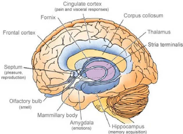
The hippocampus is one of the components of the limbic system that comprises also cortical structures such as the subcallosal, cingulated and parahippocampal gyri and yet sub cortical structures such as the amygdala, the thalamus, the hypothalamus and the septum (See Figure 1) (Lopes da Silva et al 1990). Most of the available information concerning the cellular organization of the hippocampal formation dates from the studies of Ramón y Cajal (Ramón y Cajal (1911) in (Lopes da Silva et al 1990)).
Figure 1. Schematic representation of the limbic system (dark blue) and surrounding structures (Adapted from http://www.daviddarling.info/encyclopedia/L/limbic_system.html).
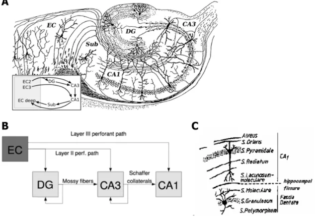
Figure 2. Schematic representation of the hippocampus and trisynaptic circuit. (A)
Neuronal organization of the hippocampus. (Adapted from Ramón y Cajal, 1911) (B) Schematic representation of the hippocampal excitatory tri-synaptic circuit DG – Dentate gyrus, EC - Entorhinal cortex, ff - Fimbrial fibers, mf - Mossy fibers, sc - Schaffer collateral fibers (Adapted from www.wikipedia.com) (C) Representation of the different Cornu
Ammonis and Dentate gyrus cell layers. (Adapted from(Filipe 1991)based on the initial descriptions by(Amaral & Witter 1989)and(Andersen et al 1969).
The hippocampal formation consists of two C-shaped interlocking cell layers: the granular cell layer of the dentate gyrus and the pyramidal cell layer of the Ammon´s horn (Cornu Ammonis or hippocampus proper) and the subiculum. The latter is composed by the molecular and pyramidal cell layers. The dentate gyrus has three layers: the molecular or dendritic layer, the granule cell layer and the hilar region characterized by widely scattered, polymorphic neurons (Lopes da Silva et al 1990). The Ammon´s horn is usually further subdivided into CA1 to CA4 areas (from the latin Cornu Ammonis areas 1-4). The first lies the nearest to the subiculum, that confines with the enthorhinal cortex, and the latter is the one directly contacting the dentate gyrus (Lopes da Silva et al 1990). The Ammon’s horn consists of seven layers (Figure 2.C): the stratum moleculare that lies directly adjacent to the hippocampal fissure and contains predominantly fibers and dendritic terminals; the stratum lacunosum, which consists mainly of bundles of parallel fibers collaterals of pyramidal cells or extrinsic to the hippocampus. Some authors combine these two layers into the stratum lacunosum-moleculare; the stratum radiatum is characterized by rather sparse cell bodies and several fiber systems, the most important of which is formed by the Schaffer collaterals; the stratum pyramidale is formed by densely packed cell bodies of the pyramidal cells; the stratum oriens is a layer that contains the basal dendrites of pyramidal cells, the cell bodies of GABAergic interneurones and (in CA1) some collaterals of CA3 pyramidal cells; the stratum alveus is formed by axons of pyramidal cells and incoming fibers from pyramidal cells and a few cell
bodies that appear to be displaced from the stratum oriens; the epithelial zone forms the lining of ventricular surface of the hippocampus (Lopes da Silva et al 1990).
The circuits of the hippocampus are organized in a looping trisynaptic excitatory circuit going from the dentate gyrus to the CA1 area that comprises (Figure 2.B): 1) the median perforant path projection, consisting in the axons of layer II cells of the enthorhinal cortex that synapse with the granule cells from dentate gyrus; 2) the mossy fibers, that are the axons of granule cells that synapse with CA3 pyramidal cells; 3) the Schaffer collateral/commissural fibers, that consist in the axons of CA3 neurons that innervate the CA1 pyramidal cells. Besides these main circuits there are two other important inputs from the cortex; the lateral perforant path projection from layer II neurons in the enthorhinal cortex to the CA3 area (now known to be very important for activation of CA3 pyramidal cells dendrites in this area; cf. with mossy fibers) and the projections from the layer III of the enthorhinal cortex to the stratum lacunosum-moleculare of area CA1 of the Ammon’s Horn, respectively (Herreras et al 1987).
Hippocampal Interneurones
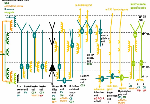
The hippocampus contains interneurones, that make up 10% of hippocampal neurons (Freund & Buzsáki 1996) and that are cells that use GABA (γ-Aminobutyric acid) as a neurotransmitter. Due to the great diversity of interneurones several attempts have been made to classify them based on anatomical, electrophysiological or neurochemical properties (Per Andersen 2006). Since a detailed description of all interneurone subtypes in the hippocampus would be superfluous in the context of this thesis, we will mention only in more detail those subtypes of hippocampal interneurones that are particularly relevant to the work described in this thesis. Using the classification proposed by Somogyi and Klausberger (2005), interneurones are classified taking into account morphological characteristics - layer location and neuronal projections - and neurochemical properties (Somogyi & Klausberger 2005). For a summary description of the main interneurone subtypes present in the CA1 area of the hippocampus please refer to Figure 3.
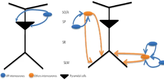
Some interneurones have axonal projections to other interneurones (IS, interneurone-specific). IS-interneurones can be divided in three groups according to the layer to which they project and to their neuropeptide content. Two of these (IS-II and IS-III) express vasoactive intestinal peptide (VIP), whose actions on synaptic plasticity are studied in this work. IS-II interneurons cell bodies are at the border of stratum radiatum and stratum lacunosum-moleculare and receive afferents from the temporoammonic pathway, as inferred from the predominant localization of their dendritic tree at the stratum lacunosum-moleculare (Acsády et al 1996). IS-II interneurons innervate mostly stratum radiatum interneurons that control synaptic transmission to proximal dendrites of pyramidal cells. IS-III interneurons are mainly in stratum radiatum - stratum piramidale border and receive multiple afferents because their dendritic tree spans almost all layers (Acsády et al 1996). IS-III VIP-positive interneurones innervate interneurones of stratum oriens projecting to the stratum lacunosum-moleculare (O-LM cells, (Acsády et al 1996; Gulyás et al 1996). O-(O-LM interneurones receive most (≈70%) of the excitatory inputs to come from retrograde projections of pyramidal cells of CA1 and are
Figure 3. Hippocampal interneurone subtypes in the CA1 area of the hippocampus.
Interneurones 1-12 innervate pyramidal cells whereas interneurones 13-16 innervate selectively interneurones. Somata and dendrites of interneurones are shown in blue or green and axon projections are shown in yellow. The main layers receiving glutamatergic input are shown, as is domain-specific innervation of pyramidal cells/interneurones by interneurone axons. The name and main neurochemical markers of each interneurone subtype is indicated bellow each cell. CB, calbindin; CCK, cholecystokinin; CR, calretinin; LM-PP, lacunosum-moleculare perforant path; LM-R-PP, lacunosum-moleculare radiatum perforant path; m2; muscarinic receptor type 2; mGluR7, 8, metabotropic glutamate receptor 7, 8; NPY, neuropeptide tyrosine; PV, parvalbumin; SM, somatostatin; VGLUT3, vesicular glutamate transporter 3; VIP, vasoactive intestinal peptide (Adapted by Cunha-Reis, 2006 from Somogyi and Klausberger, 2005).
Other type of interneurones relevant for this thesis, are the basket cells. The basket cells play an important role in perisomatic inhibition. These interneurones can be subdivided into three groups using neurochemical criteria. One of basket cells groups express VIP and cholecystokinin (CCK) but do not express parvalbumin, another interneurone marker, as do most basket cells in the hippocampus. VIP/CCK basket cells in the CA1 area receive afferents from CA3 Schaffer collaterals (Acsády et al 1996), suggesting that they can be activated in a feed-forward manner and these basket cells have serotonergic innervations unlike the other groups of basket cells that receive cholinergic innervations (Papp et al 1999) (See Figure 3 and Figure 8).
The electrical properties and firing pattern of interneurones in the hippocampus are different from the ones of pyramidal cells. Interneurones generate fast currents, due to the expression of glutamate receptors of specific subunit composition (Per Andersen 2006). They also generate brief action potentials in response to transient synaptic activation and discharge repetitively at very high frequencies during sustained stimulation. Factors facilitating fast
action potential initiation following synaptic excitation include depolarized interneurone resting potential, sub-threshold conductances and active dendrites. GABA release at interneurone output synapses is rapid and highly synchronized, leading to a faster inhibition in postsynaptic interneurones than in principal cells (Jonas et al 2004). Interneurones also have electrical synapses using gap junctions (Katsumaru et al 1988). These types of synapse allow a synchronization of specific groups of interneurones and work with high speed and temporal precision (Per Andersen 2006).
Hippocampal Rhythms
The theta rhythm is one of the most studied rhythms using electroencephalography and magnetoencephalography. The brain wave can be divided in five rhythms: Delta (4 Hz); Theta (4-8 Hz); Alpha (8 – 12 Hz); Beta (12 – 30 Hz); and Gamma (30 – 140 Hz). The theta rhythm is believed to play a mechanistic role in various aspects of memory (Düzel et al 2010). The loss of hippocampal theta rhythm leads to spatial memory deficits in rats (Winson 1978). In 1997, In 1997, Vertes and Kocsis proposed that ‘‘theta cells’’, cells that fire with the theta rhythm, are responsible for theta rhythms in the hippocampus (Vertes & Kocsis 1997).
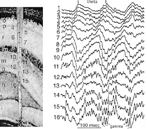
Figure 4. Depth Profile of Theta Oscillation in the rat hippocampus.(Left) o - stratum
oriens, p pyramidal layer, r stratum. Radiatum, lm stratum lacunosummoleculare, g
-granule cell layer, h - hilus. (Right) Theta waves recorded during exploration. Note gradual shift of theta phase from stratum oriens to stratum lacunosum-moleculare. Gamma waves superimposed on theta oscillation are marked by arrows (Adapted by (Buzsáki 2002) from (Bragin et al 1995).
Theta oscillations are most regular in frequency and with larger amplitude in the stratum lacunosum-moleculare of the hippocampal CA1 region. Both the amplitude and phase of theta waves change as a function of depth, whereas in the same layers they are robustly similar along the long axis of the hippocampus (Bullock et al 1990) (Figure 4).
Vertes et al. (2004) argued that the theta rhythm represents a strong depolarizing drive to the hippocampus, while other inputs (mainly cortical) constitute ‘‘information-bearing’’ inputs to the hippocampus that when coupled to theta rhythm produce lasting changes in hippocampal function (Vertes et al 2004). During theta states, theta oscillations would drive large populations of hippocampal neurons to threshold for the activation of N-methyl D-aspartate –sensitive (NMDA) receptor channels, and when combined with the release of glutamate from other afferents to these cells, would result in the opening of the NMDA receptor channels and consequent cellular changes. Accordingly, events occurring together with theta oscillations would have greater access to (and impact on) the hippocampus. In effect, theta oscillations serve as a ‘‘significance signal’’ to the hippocampus, that is, information arriving with theta oscillations would be stored (at least temporarily) in the hippocampus, whereas information arriving in the absence of theta oscillations would not be encoded, or not encoded to the same degree, as that reaching the hippocampus concurrently with theta oscillations (Vertes 2005).
Synaptic Plasticity in Hippocampus
One of the most important and fascinating properties of nervous system is its synaptic plasticity. Synaptic plasticity refers to the activity-dependent modification of the strength or efficacy of synaptic transmission at pre-existing synapses (Citri & Malenka 2008), and since proposed by Cajal and later by Hebb plays a central role in the capacity of the brain to incorporate transient experiences into persistent memory traces (Cajal 1913; Citri & Malenka 2008; Lynch 2004).
Synaptic transmission can be either enhanced or depressed by activity, and these changes span temporal domains ranging from milliseconds to hours, days, and presumably even longer. Furthermore, virtually all excitatory synapses in brain simultaneously express a number of different forms of synaptic plasticity (Zucker & Regehr 2002).
Most forms of short-term synaptic plasticity are triggered by short bursts of activity causing a transient accumulation of calcium in presynaptic nerve terminals. This increase in presynaptic calcium in turn causes changes in the probability of neurotransmitter release by directly modifying the biochemical processes that underlie the exocytosis of synaptic vesicles (Citri & Malenka 2008). Long-term synaptic plasticity can be expressed as Long-term Potentiation (LTP) or long-term depression. As memories are thought to be encoded by modification of synaptic strength, LTP is widely considered one of the major cellular mechanisms that underlies learning and memory (Lynch 2004). LTP has several properties such as input specificity – only occurs in activated synapses , associativity – weak stimulation alone does not potentiate communication but if a neighbouring pathway is strongly stimulated this pathway is reinforced, cooperativity - concurrent weak stimulations of converging afferent
fibers can cooperate to induce LTP in the postsynaptic cell, and durability. Those characteristics are the same that are essential for memory acquiring according Hebb’s postulate (Hebb 1949). Further evidence has been supporting the hypothesis that synaptic plasticity is the cellular mechanism supporting learning and memory. In particular, in the hippocampus, LTP inhibitors also block hippocampal-dependent learning tasks; biochemical changes that occur after LTP induction and memory acquisition are similar; and rhythmic bursts of activity that induce LTP mimic activity naturally occurring during theta rhythm recorded in the hippocampus during spacial memory acquisition (Lynch 2004).
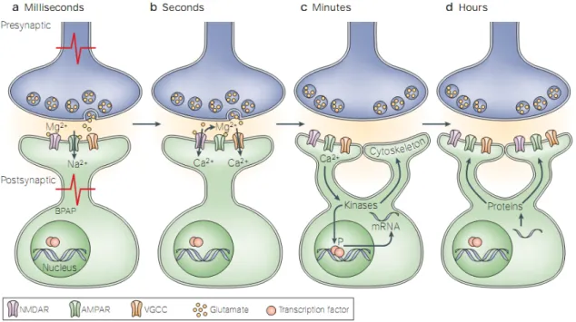
Figure 5. Molecular mechanisms involved in the initiation and maintenance of synaptic plasticity.(A) Activity-dependent release of glutamate from presynaptic neurons leads to the activation of AMPA receptors and to the depolarization of the postsynaptic neuron. Depolarization occurs locally at the synapse and/or by back-propagating action potentials (BPAP). (B) Depolarization of the postsynaptic neuron leads to removal of NMDA receptor inhibition, by Mg2+, and to Ca2+influx through the receptor. Depolarization also activates voltage-gated calcium channels (VGCCs), another source of synaptic calcium. (C) Calcium influx into the synapse activates kinases which, in turn, modulate the activity of their substrates. These substrates contribute to local changes at the synapse, such as morphological alteration through cytoskeletal regulation, or induce the transcription of RNA in the nucleus by regulating transcription factors (TFs). (D) Transcribed mRNA is translated into proteins that are captured by activated synapses and contribute to stabilization of synaptic changes (Adapted from(Lamprecht & LeDoux 2004).
LTP was reported for the first time in 70´s by Bliss and Lomo (Bliss & Gardner-Medwin 1973). They delivered a brief period of high-frequency stimulation (HFS) to the rabbit perforant path causing a sustained increase of synaptic transmission to the granule cells of the dentate gyrus. They termed this phenomenon Long-term potentiation of synaptic transmission. The HFS triggers a depolarization that allows the activation of both α-amino-3-hydroxy-5-methyl-4-isoxazole propionic acid – sensitive (AMPA) and NMDA ionotropic glutamate receptors (See Figure 5). AMPA receptors are channels permeable to monovalent cations (Na+and K+) that
because of the blockade of the channel by extracellular Mg2+and contribute little to the basal
postsynaptic response during low-frequency synaptic transmission. However, when the postsynaptic cell is depolarized during the induction of LTP, Mg2+ dissociates from its binding
site within the NMDA receptor channel, allowing Ca2+as well as Na+influx into the dendrites.
The NMDA receptor activation and Ca2+ influx are crucial for LTP induction. This phase is
usually called early-LTP (E-LTP) and is dependent on the concentration of intracellular Ca2+(See
Figure 6).
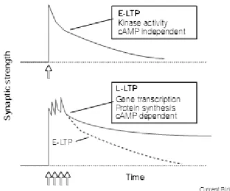
Figure 6. Early and late phases of long-term potentiation (LTP). A train of repetitive stimuli (e.g. HFS, arrows) induces an increase in synaptic strength known as LTP. A single train of stimuli induces E-LTP (red), which decays over the course of a few hours. Multiple trains induce L-LTP (blue), which remains stable for many hours (Adapted from(Huang 1998).
For maintenance of LTP it is necessary to trigger numerous transduction pathways in cell, involving first the activation of various proteins/messengers such as calmodulin (CaM), calmodulin kinase II (CaMKII), and at a latter step adenylyl cyclase (AC), protein kinase A (PKA), CREB, an isoform of PKM (PKMζ) and mitogen-activated protein kinases /extracellular signal-regulated kinases (MAPK-ERK) (See Figure 7). When these latter pathways are activated LTP lasts for hours and this phase is called late-LTP (L-LTP) (Figure 6).
The activation of these transduction pathways ultimately leads to an increased number of AMPA receptors in the synaptic cleft and an increased efficiency of existing receptors, thus providing greater sensitivity to the postsynaptic neuron, thereby suggesting that long-term potentiation is a fundamental molecular process associated with learning and memory formation (Citri & Malenka 2008; Kovács et al 2007; Kullmann & Lamsa 2007; Lynch 2004; Serrano et al 2005).
because of the blockade of the channel by extracellular Mg2+and contribute little to the basal
postsynaptic response during low-frequency synaptic transmission. However, when the postsynaptic cell is depolarized during the induction of LTP, Mg2+ dissociates from its binding
site within the NMDA receptor channel, allowing Ca2+as well as Na+influx into the dendrites.
The NMDA receptor activation and Ca2+ influx are crucial for LTP induction. This phase is
usually called early-LTP (E-LTP) and is dependent on the concentration of intracellular Ca2+(See
Figure 6).
Figure 6. Early and late phases of long-term potentiation (LTP). A train of repetitive stimuli (e.g. HFS, arrows) induces an increase in synaptic strength known as LTP. A single train of stimuli induces E-LTP (red), which decays over the course of a few hours. Multiple trains induce L-LTP (blue), which remains stable for many hours (Adapted from(Huang 1998).
For maintenance of LTP it is necessary to trigger numerous transduction pathways in cell, involving first the activation of various proteins/messengers such as calmodulin (CaM), calmodulin kinase II (CaMKII), and at a latter step adenylyl cyclase (AC), protein kinase A (PKA), CREB, an isoform of PKM (PKMζ) and mitogen-activated protein kinases /extracellular signal-regulated kinases (MAPK-ERK) (See Figure 7). When these latter pathways are activated LTP lasts for hours and this phase is called late-LTP (L-LTP) (Figure 6).
The activation of these transduction pathways ultimately leads to an increased number of AMPA receptors in the synaptic cleft and an increased efficiency of existing receptors, thus providing greater sensitivity to the postsynaptic neuron, thereby suggesting that long-term potentiation is a fundamental molecular process associated with learning and memory formation (Citri & Malenka 2008; Kovács et al 2007; Kullmann & Lamsa 2007; Lynch 2004; Serrano et al 2005).
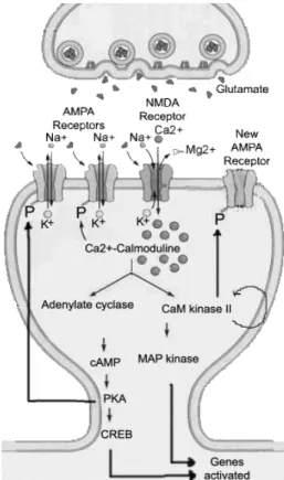
Figure 7. Schematic representation of the molecular pathways activated during the induction and maintenance of NMDA-dependent LTP.The increase [Ca2+]i is essential to
the induction phase. The phosphorylation of MAPK and CREB are essential for the long-term maintenance of LTP (late-LTP) (Adapted from http://thebrain.mcgill.ca).
Vasoactive intestinal peptide (VIP)
Vasoactive intestinal peptide is a neuromodulator peptide that was initially isolated from porcine intestine is a 28-amino acid peptide (Harmar et al 1998; Said & Mutt 1970). VIP is member of the superfamily of structurally related peptide hormones that includes glucagon, glucagon-like peptide, secretin, and growth hormone-releasing factor. This family also includes pituitary adenylate cyclise activating polypeptide (PACAP). PACAP shares 68% sequence similarity to VIP in their N-terminal residues from 1 to 28 (Delgado & Ganea 2001; Laburthe & Couvineau 2002). It was shown to serve potentially as a neuromodulator, a neurotrophic factor and/or a neurotransmitter. Furthermore, it is neuroprotective and enhances cell proliferation in both the peripheral and central nervous systems (Harmar et al 1998). VIP and PACAP also play a neuromodulator role on cardiovascular, circulatory and respiratory systems and on metabolic function, immune system and nervous system (Dickson & Finlayson 2009).
VIP is widely distributed throughout the brain, with considerable expression in the cerebral cortex, hippocampus, amygdala, suprachiasmatic nucleus and hypothalamus (Dickson & Finlayson 2009). In the hippocampus, VIP is expressed exclusively in three types of interneurones (basket cells and Interneurone specific cells II and III), as previously mentioned (See Figure 8). The first study of the role VIP on synaptic transmission on the CA1 area was in 1992 by Haas and Gähwiler and they observed VIP modulates neuronal excitability in
transmission to CA1 pyramidal cells by VIP is also dependent on GABAergic transmission. This action occurs both through presynaptic enhancement of GABA release and postsynaptic facilitation of GABAergic currents in interneurones (Cunha-Reis et al 2004).
Figure 8. Schematic representation of VIP interneurones in CA1 area.SO/A - Stratum
Oriens/Stratum Alveus, SP - Stratum piramidale; SLM - Stratum lacunosum-molecular
(Adapted from Cunha-Reis, 2006).
VIP receptors
At least three receptors for VIP-PACAP have been identified and they all belong to the B family of G-protein-coupled receptors. VIP receptors, Vasoactive intestinal peptide receptor 1 (VPAC1 receptor), and (VPAC2 receptor), have similar affinities for PACAP and VIP whereas
the PACAP-specific receptor (PAC1R) exhibits a much higher (Harmar et al 1998) affinity for
PACAP than VIP .
VIP receptors trigger mainly adenylyl cyclase activation through Gs proteins and cAMP production (See Figure 9). A few other signaling pathways have been described depending on the preparations and species, in particular, activation of phospholipase C through G proteins of the Gq and/or Gi/Go families in various types of cells and species (Dickson & Finlayson 2009). All of VIP receptors couple strongly to the Gαs and stimulate the cyclic AMP (cAMP)/PKA signaling pathway. Shreeve (2002) reported that the VPAC1receptor can couple to Gi/o protein
in the hippocampus.
The VPAC1 receptor is widely distributed in the central nervous system (CNS), most
abundantly in the cerebral cortex and hippocampus, in peripheral tissues including liver, lung, and intestine and in T lymphocytes. In the CNS, the highest VPAC2receptor expression are
found in the thalamus and suprachiasmatic nucleus and lower levels in the hippocampus, brainstem, spinal cord, and dorsal root ganglia. The receptor is also present in several peripheral tissues, including pancreas, skeletal muscle, heart, kidney, adipose tissue, testis, and stomach (Dickson & Finlayson 2009).
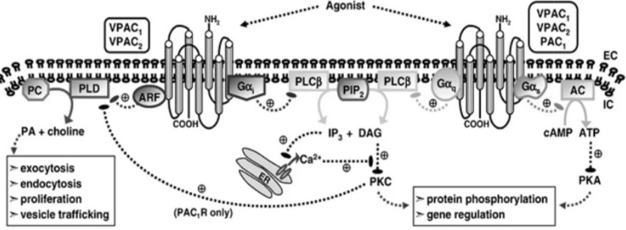
Figure 9. Intracellular signalling pathways stimulated by VPAC/PAC1R activation. The
figure highlights the principal transduction pathways activated by VPAC1, VPAC2and PAC1
receptor coupling to heterotrimeric G-proteins. Upon activation, all three receptors are capable of coupling to Gαs leading to downstream production of cAMP. In addition, the three receptors can also activate PLC leading to an increase in [Ca2+]i, via coupling to Gαq
(all three receptors) and Gαi (VPAC1and VPAC2only). PLD activity can also be stimulated
by the three receptor subtypes via ARF (VPACR) and PKC (PAC1R) sensitive pathways
(Adapted from Dickson & Finlayson, 2009).
Immunohistochemical and autoradiography studies suggest that, in the hippocampus, VPAC1receptor is expressed in stratum oriens and stratum radiatum on Cornu Ammonis and it
is co-localized with glial cells (Joo et al 2004; Vertongen et al 1997). However, functional evidence for the presence of VPAC1receptors in interneurones has been obtained (Cunha-Reis
et al 2006). The same studies show that VPAC2 receptors are majorly expressed in stratum
pyramidale in the CA1 area (Joo et al 2004) (See Figure 10). The distribution of VIP receptors in the hippocampus suggests an important role of VIP in the control of pyramidal cells function.
Figure 10. Distribution of VIP receptors in the Ammon’s horn (Adapted from Cunha-Reis, 2006, based on the data obtained by(Joo et al 2004)and(Vertongen et al 1997).
Recent studies show that VIP enhancement of synaptic transmission to CA1 pyramidal cells is mostly mediated by VPAC receptor activation and is totally dependent on PKC activity
(Cunha-Reis et al 2005). VPAC2 receptor activation also contributes to that enhancement
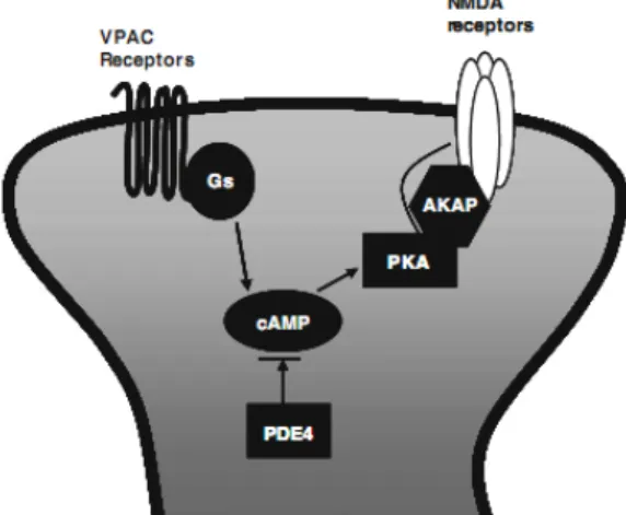
through a PKA-dependent mechanism (Ciranna & Cavallaro 2003; Cunha-Reis et al 2005). VIP also increases NMDA-evoked currents in CA1 pyramidal cells (Yang et al 2009). This increase can be totally mimicked by simultaneous application of VPAC1 and VPAC2 selective
agonists and was blocked by specific PKA inhibitor such as PKI 14–22 amide and Rp-cAMPS (See Figure 11), but not by PKC inhibitor bisindolylamaleimide I (Yang et al 2009). These facts suggest an involvement of both VPAC1 and VPAC2 receptors in the regulation of NMDA
receptors in pyramidal cells (Yang et al 2009).
Figure 11. VIP modulation of NMDA receptors in CA1 pyramidal cells via cAMP/PKA pathway (Adapted from(Yang et al 2010).
It was recently discovered in our lab that endogenous VIP has a restraining effect on LTP elicited in vitro by theta-burst stimulation through a mechanism dependent on VPAC1
receptor activation (See Figure 12) and GABAergic transmission or GABAAreceptors
(Cunha-Reis D. 2008; Rodrigues 2009).
Figure 12. Influence of VPAC1 receptor blockade on LTP induced by weak θ-burst
stimulation, 5 bursts of four pulses at 100 Hz with an interburst interval of 200 msec.
Temporal evolution of the slopes of evoked field excitatory postsynaptic potentials (fEPSP) obtained from a representative experiment in which we evaluated the LTP induced by theta-burst stimulation in the absence and presence of the drug PG 97-269 (red bar). All experiments were performed in two pathways on the same slice (S1 and S2). The independence of the two pathways was tested by studying Paired-pulse Facilitation (PPF) across both pathways, less than 14% facilitation being usually observed independent (Rodrigues 2009).
A deficit in VIP in brain is related with learning impairments in mice (Glowa et al 1992) and intracerebral administration of a VIP receptor antagonist to adult rats resulted in deficits in learning and memory in the Morris water maze task (Glowa et al 1992).
Temporal lobe epilepsy
Epilepsy is the most common severe neurological condition (Duncan et al 2006). It is characterized by spontaneous recurrent seizures, caused by focal or generalized paroxysmal changes in neurological functions triggered by abnormal electrical activity in brain (Dichter 1994). As it involves hyperexcitable neurons, a basic statement links the pathogenesis of epilepsy and the generation of synchronized neuronal activity with an imbalance between inhibitory (GABA-mediated) and excitatory (glutamate-mediated) neurotransmission, in favour of the latter (Dalby & Mody 2001).
Temporal lobe epilepsy (TLE) is the most common form of epilepsy in humans. Individuals affected with TLE typically have comparable clinical description, including an initial precipitating injuring such as status epilepticus, head trauma, encephalitis or childhood febrile seizures (Cendes 2004; Fisher et al 1998; Harvey et al 1997). There is usually a latent period of several years between this injury and the appearance of the chronic TLE characterized by spontaneous recurrent seizures originating from temporal lobe foci that lead to learning and memory impairments (Detour et al 2005; Dütsch et al 2004). In TLE, acute cell death occurs in various limbic regions, including the hippocampal formation. Gliosis and reorganization of axons of the granule cells (mossy fiber sprouting) and reorganization of excitatory and inhibitory circuits in the hippocampal formation following seizure induced neuronal loss are also involved in the development of chronic seizures in TLE (Mello et al 1993).
VIP and its receptors are increased in the human hippocampus of patients with TLE (de Lanerolle et al 1995). It is suggested that the elevated levels of receptor binding in the hippocampal seizure focus may indicate a mechanism for greater excitability of neurons and/or their survival facing the increased excitation and potential for injury in a seizure focus (de Lanerolle et al 1995). In an animal model of TLE, it was also reported an increase of VIP in hippocampus (Marksteiner et al 1989).
Aim
This study aimed to evaluate the influence of VPAC1receptor activation on a Long-term
potentiation in 12-week-old rats, using a LTP induction protocol that result in a late-LTP, characterized by its several hour persistency and PKA-dependence, for this would be a valid in vitro model to study memory.
The study also aimed to elucidate the influence of VPAC1 receptors on synaptic