UNIVERSIDADE DE LISBOA
Faculdade de Ciências
Departamento de Química e Bioquímica
Study of CFTR delivery and stabilization
at the plasma membrane
Bruno José Rother Rocha de Moraes
MESTRADO EM BIOQUÍMICA
(Bioquímica Médica)
UNIVERSIDADE DE LISBOA
Faculdade de Ciências
Departamento de Química e Bioquímica
Study of CFTR delivery and stabilization
at the plasma membrane
Dissertação orientada por:
Doutor Peter Jordan
(Instituto Nacional de Saúde Dr. Ricardo Jorge)
Doutora Margarida Amaral
(Faculdade de Ciências da Universidade de Lisboa)
MESTRADO EM BIOQUÍMICA
(Bioquímica Médica)
Para a minha mãe, como no início, os primeiros passos. --- Para o meu pai, inevitavelmente, o caminho.
“Would you tell me, please, which way I ought to walk from here?”
“That depends a good deal on where you want to get to,” said the Cat.
“I don’t much care where –” said Alice.
“Then it doesn’t matter which way you walk,” said the Cat.
“– so long as I get somewhere,” Alice added as explanation.
“Oh, you’re sure to do that,” said the Cat, “if you only walk long enough.”
Lewis Carroll in “Alice’s Adventures in Wonderland” (1865)
Resumo
A fibrose quística (FQ) é a principal doença genética, autossómica recessiva, a afectar a população Caucasiana, embora se estenda igualmente a outras etnias. Apesar de caracterizada no pâncreas pela primeira vez em 1938, a FQ foi sucessivamente associada a defeitos em tecidos epiteliais exócrinos de outros órgãos, nomeadamente dos pulmões, onde se registam os efeitos mais nefastos, os quais incluem acumulação de muco espesso e sensibilidade aumentada a infecções, que normalmente originam complicações conducentes à morte prematura do doente. Só em 1989 foi possível clonar o gene responsável pela doença, então designado cystic fibrosis transmembrane
conductance regulator (CFTR), o qual codifica um canal selectivo para cloreto
(Cl−), também permeável à passagem de hidrogenocarbonato (HCO3−), expresso na membrana apical dos epitélios que revestem os órgãos afectados. A glicoproteína CFTR é constituída por 1480 resíduos de aminoácidos, de cujo
folding resultam três domínios citoplasmáticos – um regulatório central (RD) e
dois de ligação a nucleótidos (NBD1 e NBD2) – e dois domínios transmembranares (MSD1 e MSD2). A sua actividade é controlada por (i) ligação de ATP aos domínios NBD, que dimerizam e promovem alterações conformacionais que conduzem à abertura e fecho do canal, e (ii) por fosforilação do seu domínio RD pela PKA, que aumenta ou diminui a probabilidade de abertura. Adicionalmente, a CFTR regula ela própria a expressão e actividade de outras proteínas, de que são exemplo os canais de sódio epiteliais (ENaC), sujeitando-os a alterações de estabilidade e probabilidade de abertura. Estas funções conferem à CFTR um estatuto pleiotrópico.
Actualmente sabe-se que diversas mutações, classificadas de I a VI com base no fenótipo celular resultante, estão na origem do desenvolvimento da FQ. Destas, a mais comum, afectando cerca de 90% dos genomas em pelo menos um dos alelos, pertence à classe II e corresponde à delecção de uma fenilalanina na posição 508 da proteína (ΔF508), contida no NBD1. A proteína
mutada, CFTR-ΔF508, apresenta defeitos ao nível do folding no retículo endoplasmático (RE), onde é rapidamente degradada, pelo que o seu tráfego vesicular para a membrana plasmática é muito reduzido. Para além disso, a pequena população de canais mutados que escapam ao controlo no RE, sofre de uma instabilidade acrescida na membrana. De facto, o domínio C-terminal da CFTR é capaz de interagir com diversas proteínas contendo pelo menos um domínio PDZ, como é o caso das proteínas NHE-RF1 e CAL, que asseguram a regulação da sua expressão transmembranar. Notavelmente, o adaptador NHE-RF1 estabelece uma ponte entre o canal e o citoesqueleto de actina filamentosa (F), mediada por proteínas da família Ezrin/Radixin/Moesin (ERM), tendo sido já descrita uma acção estabilizadora do NHE-RF1 sobre a CFTR. Para além destas interacções, também a ubiquitinação do canal na sua região C-terminal e a acção de desubiquitinases como a USP10, que reverte o processo, definem um ponto de controlo da estabilidade da CFTR ao nível da membrana plasmática.
O canal é endocitado por um processo dependente de clatrina. No entanto, quando endocitado, é rapidamente reciclado para a membrana plasmática (mais de 75%), o que justifica a sua elevada expressão transmembranar, apesar da baixa taxa de transcrição do seu gene. De forma a possibilitar um tráfego vesicular eficiente, a célula faz uso de uma complexa rede de F-actina, à qual proteínas motoras, como as miosinas, se podem ligar, promovendo o transporte das vesículas. Para além disso, diversas Rab GTPases medeiam a ligação das miosinas às vesículas e são responsáveis pelo seu correcto endereçamento. Neste contexto, já se observou que a CFTR é internalizada em vesículas contendo Rab5a/miosina Va, e reciclada em vesículas com Rab11a/miosina VI. Contudo, nenhuma destas vias parece estar alterada em células com expressão de CFTR-ΔF508 pelo que, para além do misfolding no RE, deverá ser ao nível da própria membrana plasmática, e não do transporte a partir do Golgi, que é induzida uma destabilização adicional do canal mutado. Tanto as proteínas ERM como o adaptador NHE-RF1, referidos acima, encontram-se normalmente numa conformação inactiva no citoplasma. As
primeiras, quando activadas, ligam directa ou indirectamente (via NHE-RF1) proteínas transmembranares ao citoesqueleto de actina. Esta activação depende de dois factores: a ligação do seu N-terminus ao fosfatidilinositol 4,5-bisfosfato (PIP2) e a fosforilação de resíduos críticos de treonina na região
C-terminal. A actividade de um grupo de pequenas GTPases da subfamília Rho, nomeadamente as GTPases canónicas Rac1, Cdc42 e RhoA, tem sido implicada tanto no metabolismo do PIP2 como na fosforilação das proteínas ERM, pelo que se pensa que a sua acção esteja implicada na activação destas proteínas. Para além disso, estas Rho GTPases também promovem alterações no citoesqueleto que podem condicionar o desempenho do processo de transporte sub-membranar de vesículas, ou seja, a endocitose e a reciclagem de proteínas como a CFTR. Por seu turno, o NHE-RF1 é activado por fosforilação, por exemplo mediada pela PKC, ou por ligação de proteínas ERM ao seu C-terminus, de forma que as Rho GTPases acabam, indirectamente, por também poderem ter um papel na activação do adaptador ao activarem as proteínas ERM.
O presente estudo foi conduzido em duas fases, separadas pela utilização de dois modelos celulares: uma linha estavelmente transfectada com o gene
CFTR normal wt) e uma outra com o gene mutante CFTR-ΔF508
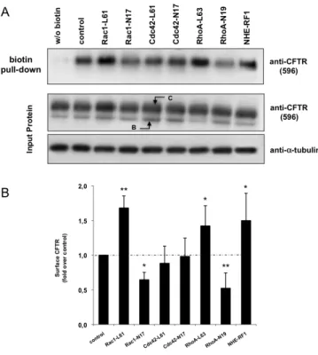
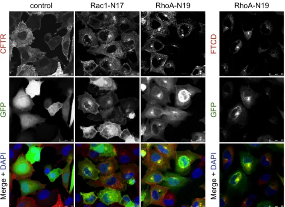
(BHK-ΔF). Começámos por realizar um ensaio de precipitação, com partículas de agarose revestidas com streptavidina, de CFTR marcada com biotina na sua porção extracelular. Para tal trataram-se células BHK-wt com Sulfo-NHS-SS-biotina a 0 °C (para bloquear os processos de endocitose e exocitose), com vista a averiguar se a expressão transiente de mutantes constitutivamente activos (CA) ou dominante negativos (DN) das três Rho GTPases canónicas induziam alterações na expressão da CFTR na membrana plasmática. Descobrimos que, nestas células, tanto o Rac1-CA como o RhoA-CA aumentavam os níveis de CFTR à superfície das células, e que os seus mutantes DN os diminuíam significativamente. A observação, por imunofluorescência (IF), de células transfectadas nas mesmas condições, não só corroborou estes resultados como mostrou que nas células transfectadas
com RhoA-DN havia uma acumulação de CFTR no Golgi, sugerindo uma acção do RhoA no tráfego do canal deste compartimento para a membrana plasmática. Estes resultados indicavam que Rac1 e RhoA poderiam exercer o seu efeito através de mecanismos diferentes pelo que, a fim de averiguar se estes incluíam alterações no processo de endocitose e/ou reciclagem da CFTR, utilizámos um ensaio de biotinilação modificado no qual células transfectadas com mutantes activos de RhoA ou Rac1, depois de biotiniladas a 0 °C, foram brevemente repostas a 37 °C por diferentes períodos de tempo, de modo a reactivar o tráfego vesicular, permitindo-nos assim seguir a cinética de internalização da CFTR. Observámos que enquanto o Rac1-CA provocava um marcado retardamento da endocitose da CFTR (2.0 ± 0.74% CFTR internalizada por minuto, contra 12.0 ± 1.80% para o controlo, transfectado com vector vazio), o RhoA-CA não o fazia (9.0 ± 1.8%, p > 0.05), estimulando, pelo contrário, a reciclagem do canal para a membrana. Estando já descrita a importância do adaptador NHE-RF1 na ancoragem da CFTR na superfície celular, realizámos um ensaio de co-imunoprecipitação (co-IP) dos complexos NHE-RF1/CFTR em células transfectadas com ambas as formas CA e DN das Rho GTPases Rac1 e RhoA e constatámos que, enquanto o Rac1-CA induzia, de facto, um incremento notável na formação dos complexos, o RhoA-CA não apresentava efeitos significativos. Por outro lado, se a redução da actividade do Rac1 produz apenas uma redução parcial na associação NHE-RF1/CFTR, já a diminuição da actividade do RhoA reduz notoriamente esta associação, o que é uma vez mais concordante com um papel desta GTPase no tráfego da CFTR do Golgi para a membrana plasmática, situação na qual ocorre a ligação inicial de NHE-RF1 à CFTR. Analisámos também a actividade global da população de canais na membrana através de um ensaio de efluxo de iodeto (I−). Neste, células BHK-wt transfectadas como na co-IP, previamente incubadas numa solução de NaI, foram estimuladas com Forskolina e IBMX (dois agentes que aumentam os níveis de cAMP na célula, activando a PKA, que fosforila e activa a CFTR) a expelir o I−, detectado com um eléctrodo específico. Observámos que tanto a activação do Rac1 como do RhoA aumentavam a actividade da
CFTR, de acordo com o incremento na expressão membranar do canal observado logo na fase inicial do presente estudo. Pelo contrário, o uso dos mutantes Rac1-DN e RhoA-DN diminuiu significativamente a actividade da CFTR, também concordante com os dados dos ensaios de precipitação e IF. Na segunda fase, usando o modelo celular de FQ, com excepção dos ensaios da cinética de internalização e da co-IP, por limitações técnicas devidas à reduzida quantidade de CFTR na membrana plasmática, os restantes ensaios referidos acima foram conduzidos em células BHK-ΔF transfectadas com os mutantes CA de Rac1 e RhoA. Observámos que apenas a activação do Rac1 conseguia um aumento tanto da expressão de CFTR na membrana plasmática (precipitação com streptavidina) como da sua actividade global (ensaio de efluxo de I−). Estes incrementos foram confirmados por IF, observando-se claramente uma pequena mas notória acumulação de CFTR na membrana plasmática. O mutante RhoA-CA, por outro lado, não induziu quaisquer alterações relativamente ao controlo em nenhum dos ensaios.
O presente trabalho contribuiu para a compreensão dos mecanismos de acção de duas Rho GTPases na expressão e actividade da CFTR ao nível da membrana plasmática de células epiteliais não polarizadas. Com os dados obtidos, articulados com outros da literatura, propomos um modelo de acção para cada GTPase. No que respeita ao RhoA, a sua activação conduzirá à formação de fibras de actomiosina, propícias ao desenrolar dos movimentos de transporte vesicular da CFTR de (endocitose) e para (reciclagem) a membrana plasmática. Estas fibras, constituindo filamentos complexos, em tensão, não formam redes de F-actina que permitam a formação estável de complexos CFTR/NHE-RF1/ERM por ancoragem ao citoesqueleto, não contribuindo, portanto, para a estabilização do canal na membrana mas sim, como vimos, para o aumento da sua taxa de reciclagem. No caso do Rac1, quando activo induz a formação de redes de F-actina na região cortical (mais periférica) das células, permitindo uma maior associação da CFTR com o adaptador NHE-RF1, como observado, formando os complexos referidos acima e justificando, em parte, uma endocitose menos eficiente e, desta forma, retardada. Para além
disso, o Rac1 parece ter um papel preponderante no ajustamento dos níveis de PIP2 na célula, contribuindo directamente para a activação das proteínas ERM e, indirectamente, para a do adaptador NHE-RF1. Infelizmente, o simples aumento da actividade do Rac1 não serve o propósito de terapia para a FQ, uma vez que está descrita a sobre-expressão da GTPase em tumores pulmonares. Devem, portanto, ser realizados no futuro ensaios com efectores a juzante do Rac1 a fim de estabelecer se poderão ser usados como alvos terapêuticos em doentes com FQ.
Summary
Cystic fibrosis (CF), the major autosomal recessive genetic disorder among Caucasians, is caused by mutations in the CFTR gene, which codes for an epithelial chloride-selective channel. Its most common ΔF508 mutation leads to protein misfolding and trafficking impairment, and interferes with CFTR’s stability at the plasma membrane (PM). Both processes associate with increased protein degradation, either through quality control at the endoplasmic reticulum or at the cell surface. Particularly at the latter, interactions between the channel and PDZ-containing proteins, such as CAL or NHE-RF1, and the activity of deubiquitinizing enzymes like USP10, seem to regulate CFTR’s expression at the PM. The Ezrin/Radixin/Moesin (ERM) family of cytoskeleton linkers for transmembrane proteins indirectly anchors CFTR to filamentous (F) actin via interaction with CFTR-bound NHE-RF1 (Na+/H+-Exchanger Regulatory Factor 1). In order to achieve this, ERM proteins first need to be activated by phosphoinositide binding and/or threonine phosphorylation, both proposed to depend on small Rho GTPase activity. Moreover, Rho GTPases play a role in cytoskeleton and F-actin content rearrangements just beneath the PM, whereto CFTR is usually internalized and from where it recycles back to the surface. Using iodide-efflux, biotin pull-down, immunoprecipitation, and immunoflurescence methodologies, we found that although both active Rac1 and RhoA GTPases increased CFTR’s cell surface levels and activity, Rac1 acted by enhancing CFTR/NHE-RF1 complex formation, impairing the channel’s internalization and thus favouring its PM stability, whereas RhoA stimulated CFTR’s recycling but not NHE-RF1-mediated PM tethering. Moreover, using a CF cell line model, we show that only active Rac1 was able to increase CFTR-ΔF508 surface levels and activity, possibly by downregulating the accelarated internalization rate of the mutant channel capable of reaching the PM .
These efforts establish new roles for Rac1 and RhoA GTPases on CFTR PM expression and indicate Rac1 downstream effectors as potential CF therapeutic targets.
Palavras-chave Ancoragem CFTR Estabilidade membranar Fibrose Quística NHE-RF1 Rac1 RhoA Keywords CFTR Cystic Fibrosis Membrane stability NHE-RF1 Rac1 RhoA Tethering
Table of contents
Resumo v
Summary xi
Palavras-chave xii
Keywords xii
Table of contents xiii
Abbreviations xiv
1.
Introduction 1
1.1.
The CFTR gene product 1
1.2.
Epithelia, ERM proteins and the NHE-RF1 adaptor 4
1.3.
Rho family small GTPase function 9
1.4.
Final remarks and Aims of this thesis 14
2.
Materials and Methods 15
2.1.
Plasmid DNA amplification, extraction and sequencing 15
2.2.
Cell lines, culture and transfection 17
2.3.
SDS-PAGE, Western blot and densitometry analysis 18
2.4.
Biotin/streptavidin pull-down assay 18
2.5.
CFTR internalization kinetics assay 20
2.6.
NHE-RF1/CFTR co-immunoprecipitation 23
2.7.
Iodide efflux assay 24
2.8.
Immunofluorescence microscopy 26
3.
Results 28
3.1.
Active Rac1 and RhoA increase wt CFTR cell surface levels 28
3.2.
Rac1 and RhoA affect CFTR internalization kinetics through
different mechanisms 32
3.3.
While Rac1 enhances CFTR/NHE-RF1 complex formation, RhoA
stimulates CFTR’s recycling to the plasma membrane 34
3.4.
Rac1 and RhoA activation increase CFTR-mediated efflux 37
3.5.
Active Rac1, but not RhoA, increases CFTR-ΔF508 levels and
activity at the plasma membrane, in a cystic fibrosis cell line model 39
4.
Discussion and concluding remarks 42
4.1.
Discussion 42
4.2.
Concluding remarks and biomedical implications 48
5.
Bibliography 51
Abbreviations1
AKAP A-kinase (PKA) anchor proteins AP-2 Adapter protein 2
Arp2/3 complex Actin-related protein 2/3 complex BHK-21 Baby Hamster Kidney 21 cell line CA Constitutively activated
CAL CFTR-associated ligand
CAP70 CFTR-associated protein of 70 kDa (NHE-RF3) Cdc2 kinase Cell division control protein 2 homolog kinase Cdc42 Cell division control protein 42 homolog CDK5 Cyclin-dependent kinase 5
CF Cystic fibrosis
CFTR Cystic fibrosis transmembrane conductance regulator CK2 Casein kinase 2
CL Cytoplasmic loop Co-IP Co-immunoprecipitation CTD C-terminal domain
ΔF508 Deletion of exon 508 (Phe) from CFTR’s gene DAPI 4',6-diamidino-2-phenylindole
ddNTP Dideoxynucleotide
DN Dominant negative
DUB Deubiquitinizing protein
EBP50 ERM-binding phosphoprotein 50 (see NHE-RF1) ECL Enhanced chemiluminescence
ECM Extracellular matrix ENaC Epithelial Na+ Channel EPEC Enteropathogenic E. coli
ER Endoplasmic Reticulum ERM Ezrin-Radixin-Moesin ERMAD ERM-association domain F-actin Filamentous actin
FERM 4.1-ERM
FRET Förster resonance energy transfer
FTCD Formimidoyltransferase-cyclodeaminase (also p58k) GAP GTPase-activating protein
GDI Guanine-nucleotide dissociation inhibitor GEF Guanine-nucleotide exchange factor GFP Green Fluorescent Protein
GPCR G protein-coupled receptor
GRK6A G protein-coupled receptor kinase 6A
hnRNP A1 Heterogeneous nuclear ribonucleoprotein A1 IBMX 3-isobutyl-1-methylxanthine
ICAM Intercellular adhesion molecule IF Immunofluorescence
IQGAP IQ motif-containing GAP
ISH In situ hybridization
LB Lysogeny broth
MRCK Myotonic dystrophy kinase-related Cdc42-binding kinase MSD Membrane-spanning domain
NBD Nucleotide-binding domain NEP Neutral endopeptidase
NHE-RF1 Na+/H+ exchange regulatory cofactor 1 (also EBP50) NHE3 Na+/H+ exchanger 3
NOT Non-stimulated
NPF Nucleation promoting factor
N-WASP Neural Wiskott-Aldrich syndrome protein
O/N Overnight
PAGE Polyacrylamide gel eletrophoresis
PD Pull-down
PDZ Postsynaptic-density-95/Disc-large/ZO-1
p58k p58 kinase (see FTCD)
PIP2 Phosphatidylinositol 4,5-bisphosphate [PtdIns(4,5)P2] PIP5KI Phosphatidylinositol-4-phosphate 5-kinase type I
PKA cAMP-dependent protein kinase (former protein kinase A) PKC Protein kinase C
PVDF Polyvinylidene fluoride
Rac1 Ras-related C3 botulinum toxin substrate 1
RD Regulatory domain
RhoA Transforming protein RhoA ROCK Rho-associated protein kinase RTK Receptor tyrosine kinase SDS Sodium dodecyl sulfate SLC26 Solute carrier family 26
SN Supernatant
STK10 Serine/threonine-protein kinase 10
SUR Surface
TC10 Rho-related GTP-binding protein RhoQ USP Ubiquitin-specific-processing protease
1. Introduction
1.1. The CFTR gene product
Mutation of both alleles of the Cystic Fibrosis Transmembrane conductance Regulator (CFTR) gene, positioned at locus 7q31.2 in the human genome, causes cystic fibrosis (CF), the most common autosomal recessive genetic disorder in the Cau-casian population (Ri-ordan, 2008). First characterized by Doro-thy Andersen in 1938 (Andersen, 1938), CF arises from a buildup of dehydrated macro-molecular secretions in most exocrine tis-sues, with its most severe effects in the lungs, where accumu-lation of a thick mucus facilitates microorga-nism colonization, or-chestrating inflamma-tion and lung functio-nal impairment, and in the pancreas, where failure of HCO3− and enzyme secretion dis-arrange intestinal absorption and digestion (Riordan, 2008). The CFTR gene has been cloned and characterized for more than twenty years now (Riordan et Figure 1.1. Structure of the CFTR channel. At both the upper
extended scheme, and the lower theoretical 3D model (reproduced from Serohijos et al., 2008), the protein is shown with its two membrane-spanning domains (MSD1 and MSD2), two nucleotide-binding domains (NBD1 and NBD2) and a middle regulatory domain (RD). It also possesses four cytoplasmic loops (CL), of which only CL4, inside MSD2, is indicated (blue). Phenylalanine 508 (Phe508) at NBD1 is also denoted for its importance in CF disease.
al., 1989) and codes for a chloride-selective channel shown to be expressed at
the apical membrane of epithelial tissues, including the pancreas, salivary glands, intestine, lungs and testis, as assessed by in situ hybridization (ISH) in mice (Trezise and Buchwald, 1991). There its product, the CFTR protein, regulates salt secretion and reabsorption, necessary to maintain cellular and tissue homeostasis (Amaral, 2005). CFTR is composed of two series of 6 transmembrane α-helical loops, each forming a membrane-spanning domain (MSD1 and MSD2), plus two nucleotide-binding domains, NBD1 and NBD2, and a random-coiled middle regulatory domain (RD) between the latter (Amaral, 2005) (Fig. 1.1). Besides, it has a C-terminal domain (CTD) with high affinity for the NHE-RF1 molecular adaptor (see ahead), especially its first Postsynaptic-density-95/Disc-large/ZO-1 (PDZ1) domain, but in some conditions also for its PDZ2 domain. These CTD/PDZ interactions, together with binding of the actin-binding protein filamin to CFTR’s N-terminus, have been shown to be important for CFTR’s stabilization at the cell surface (Riordan, 2008). The chloride transport activity of CFTR is regulated both by multiple PKA-dependent phosphorylation of the R domain and ATP binding to its NBDs. Recent research has shown that, once phosphorylated by PKA, the channel gating cycle is fundamentally irreversible and tightly coupled to the ATPase cycle at the NBDs, which are known to dimerize upon ATP binding to their conserved Walker A and B motifs (Csanády et al., 2010). CFTR then cycles between “open” and “closed” conformations, the probability of each is modulated by the degree of R domain phosphorylation (Riordan, 2008). Furthermore, CFTR has been implied in the direct or indirect regulation of other transmembrane proteins, namely controlling the Epithelial Na+ Channel (ENaC) membrane stability and open probability (Lu
et al., 2007), which gives this chloride channel a pleiotropic status.
The CFTR gene has now more than 1500 mutations described that range through virtually all known types, including missense, frameshift, splicing, nonsense and deletions (Lommatzsch and Aris, 2009). By far the most common, the ΔF508 mutation (class II; see Box 1) does not alter expression of CFTR transcripts (Zabner et al., 2005) but impairs correct protein folding and
processing at the endo-plasmic reticulum (ER), thus driving its rapid degradation, with only a small amount of com-plex glycosylated CFTR reaching the cell surface at physiological tempe-ratures (Luo et al., 2009). Structural and
modelling studies have shown that molecular assembly failure is linked to the absence of the side chain benzyl group in the CFTR-ΔF508 mutant, which normally resides close to the fourth cytoplasmic loop (CL4) in MSD2, at the interface between NBD1/MSD2 CFTR domains (see Fig. 1.1; Serohijos et al., 2008). The ΔF508 deletion is temperature-sensitive, and both low temperatures and chemical correctors promote some CFTR-ΔF508 rescue to the plasma membrane (Cholon et al., 2010). However, when compared to the wild-type (wt) protein, mutant CFTR is much more rapidly endocytosed and degraded, accounting for decreased membrane stability and indicating that the protein is probably recognized as anomalous not only during ER control but also at the cell surface (Gentzsch et al., 2004; Cholon et al., 2010). At present, the altered structure of the ΔF508 region has been only assigned an interference in CFTR’s interaction with protein kinase CK2, which phosphorylates NBD1 at Ser-511 in
vivo, regulating its gating and, thus, its open probability (Treharne et al., 2007).
Nevertheless, several proteins are implicated in CFTR regulated surface expression and function, such as, for example, CFTR-associated ligand (CAL), which increases lysosomal CFTR degradation, and the small Rho GTPase TC10, which binds CAL and prevents CFTR breakdown (Cheng et al., 2005). Besides, it has been demonstrated that surface CFTR misfolding facilitates lysosomal targeting by increasing channel ubiquitination (Sharma et al, 2004).
quality control and membrane recycling. Two deubiquitinizing proteins (DUBs), USP19 at the ER (Hassink et al., 2009) and USP10 at early endosomes (Bomberger et al., 2010), have been recently linked to wt CFTR rescue from degradation. In particular, at the plasma membrane, as most CFTR normally resides in early and recycling endosomes (Cholon et al., 2010), USP10 seems to be responsible for the channel’s permanence at the vesicles, thus accounting for its known rapid endocytic recycling (> 75%) back to the surface (Bomberger
et al., 2010).
Normal CFTR is internalized by clathrin-mediated endocytosis (Lukacs et al., 1997). As for many secretory pathway-transported proteins, CFTR vesicle traffic relies on cytoskeletal roads, namely microfilaments of filamentous actin (F-actin), to which myosin motors can attach. It has been demonstrated that while CFTR endocytosis occurs in myosin VI-powered vesicles (Swiatecka-Urban et
al., 2004), recycling goes through a myosin Vb-powered route
(Swiatecka-Urban et al., 2007). Myosins bind to vesicles through small Rab GTPases, which are molecular switches responsible for the specific targeting of endosomes with their respective protein cargo to their appropriate destinations (Stenmark, 2009). Interestingly, although utterly required for correct CFTR trafficking, neither Rab5a-mediated CFTR endocytosis nor Rab11a-specific CFTR recycling seem to be critically altered in cells expressing CFTR-ΔF508 mutants (Swiatecka-Urban et al., 2005). This, suggests that other processes,
such as cytoskeleton rearrangements or plasma membrane-tethering mechanisms, could have a pivotal role in modifying mutant-CFTR vesicle trafficking and membrane retention.
1.2. Epithelia, ERM proteins and the NHE-RF1 adaptor
Two of the most important epithelium tissues where CFTR is apically expressed are those of the lung airways and the intestine (Ameen et al., 2007). Epithelia are lining sets of polarized cells, meaning their organelle and cell surface domains are arranged in an asymmetrical way (Bretscher et al., 2000),
normally having both apical, lumen-sided, and basolateral, tissue-lining faces. In order to build and maintain this asymmetry, both cytoskeleton and surface domain tethering must be involved in cell polarization. Especially at the cell cortex, the most peripheral region of the actin cytoskeleton, determination of cell shape, cell-cell attachment or interactions with the extracellular matrix (ECM) are essential to maintain cell polarity. Endocytosis, exocytosis, cell migration and division, transmembrane signaling, growth regulation, and differentiation are all important functions involving the cortical cytoskeleton (Bretscher, 1999; Bretscher et al., 2000). Indeed, there has been increasing evidence that, in order to function properly and confer tissue specificity, the expression of some transmembrane proteins, such as CFTR, must be somehow regula-ted at the cell surface, which ei-ther includes en-docytic traffic ma-nagement or cyto-skeleton based anchoring mecha-nisms (Terawaki et
al., 2006; McClat-chey & Fehon, 2009).
A group of pro-teins belonging to the superfamily of 4.1-Ezrin-Radixin-Moesin (FERM) domain-containing proteins, have been successively linked to regulation of membrane receptor distribution (McClatchey & Fehon, 2009). Ezrin-Radixin-Moesin (ERM) proteins belong to this superfamily, sharing a sequence homology of at least 84% at the
Figure 1.2. ERM protein conformations. Shown are two 3D crystal
structures of a representative ERM protein (reproduced from Hennigan
et al., 2010), representing (A) its “open”, active conformation, and (B) its
“closed”, inactive conformation. Different domains are assigned different colours: a 4.1-Ezrin-Radixin-Moesin (FERM) domain (light red) folded into three lobes (F1, F2 and F3), a α-helical middle region containing helices A, B and C (green), and a C-terminal domain (CTD) at the end (violet). Blue arrows indicate phosphorylation sites in Ezrin (T235 and T567), Radixin (T564) and Moesin (T558) (see text for details).
N-terminus, about 70% at their C-terminal halves (Tsukita & Yonemura, 1999), and a preserved α-helical middle region containing three sub-helices (Fig. 1.2A; Hennigan et al., 2010). While their N-terminal ends (containing the FERM domain) bind to cytosolic or transmembrane proteins, the C-terminal half possesses an F-actin-binding domain through which ERM proteins bind to the actin cytoskeleton (Tsukita & Yonemura, 1999). Because of this, their terminal ends have been called N- and C-ERM-association domains (ERMADs). These are the most well-studied membrane protein-to-cytoskeleton connectors, especially Ezrin. This protein’s middle region can bind protein kinase A (PKA) and serve as a bridge for its regulatory actions across membrane proteins (Bagorda et al., 2002), thus also making it a PKA-anchoring protein (AKAP). Several studies show that ERM proteins normally exist in an N-to-C (head-to-tail) conformationally inhibited structure in the cytosol (Gary & Bretscher, 1995; Bretscher et al., 1997; Matsui et al., 1998; Chambers & Bretscher, 2005), but whether this is an intra- or intermolecular association has been a theme for much debate. Gautreau et al., using both T567A non-phosphorylable and T567D phosphomimetic Ezrin mutants, argued that, parallel to some inactivated Ezrin being normally present as oligomers, there is an oligomer-to-monomer transition upon Ezrin activation by phosphorylation (Gautreau et al., 2000). It had been previously noticed that these oligomers were, in fact, essentially dimers (Bretscher et al., 1995), which has been corroborated in recent research (Chambers & Bretscher, 2005); nevertheless, these dimers have been always found in lower numbers when compared to Ezrin monomers, which seem to be predominant in vivo. A very recent and interesting result using FRET conformational analysis of Merlin, another ERM-related protein, not only showed it to fundamentally exist in a stable, closed monomer conformation, but also demonstrated that this inactive state was mainly due to the central α-helical domain, rather than to an N-to-C intramolecular association (see Fig. 1.2B; Hennigan et al., 2010). However, despite renowned homologies between ERM family members, some differences in activation and function exist between Merlin and ERM proteins.
In order to be activated, ERM proteins have to be phosphoinositide-bound at their N-terminal side (Hirao et al., 1996) and/or C-terminal phosphorylated (Matsui et al., 1998). In particular, Ezrin has been shown to need both processes, but in a sequentially specified order: first, Ezrin’s head-to-tail autoinhibited structure is disrupted by phosphatidylinositol 4,5-bisphosphate (PIP2) binding to its N-terminus; second, phosphorylation of threonine 567 (T567) helps maintaining Ezrin’s open conformation (Fievet et al., 2004).
Homologous phosphorylation-prone residues have also been observed in both Radixin (T564) and Moesin (T558) (Matsui et al., 1998), indicating that most probably the same kind of sequential intra-restraining release occurs for these proteins (Fig. 1.2A). Indeed, numerous kinases in vertebrate cells can phosphorylate ERM proteins on this regulatory threonine, including ROCK, protein kinase C alpha (PKC-A) and theta (nPKC-theta) types, mitogen-activated protein kinase kinase kinase 14 (MAP3K14), MST4 and
serine/threonine-protein kinase 10 (STK10) (Fehon et al., 2010). In addition, cyclin-dependent kinase 5 (CDK5) can phosphorylate Ezrin’s T235, which lies on the FERM/C-ERMAD interface directly opposite T567 (Fig. 1.2A), leading to its activation (Ivetic & Ridley, 2004). Besides threonine residues, ERM proteins can be phosphorylated on several tyrosines by several tyrosine kinases, but how these modifications affect the conformation and function of Ezrin is still unclear (Fehon et al., 2010).
Although similar in sequence and sub-cellular localization in most cell lines, it is important to note that ERM family proteins have different, tissue-specific expression patterns in the animal body (Bretscher et al., 1997; Tsukita & Yonemura, 1999), thus probably exerting slightly different functions. Nevertheless, once opened, ERM proteins can in general either directly or indirectly link transmembrane proteins to F-actin (Tsukita & Yonemura, 1999).
Direct interactions have been shown for CD43, CD44, ICAM-1, -2 and -3, NEP, β2-integrin, among several others (McClatchey & Fehon, 2009). In turn, Na+/H+ -exchanger regulatory cofactor (NHE-RF) proteins, and specifically NHE-RF1 (also called EBP50), as early mentioned, serve as molecular adapters on
indirect linkage between ERM proteins and membrane channels (CFTR, NHE3) or receptors (e.g., GPCRs) (Bhattacharya et al., 2010). In particular, concerning CFTR, it is noteworthy that the NHE-RF1-mediated proximity between ERM proteins, such as Ezrin, and the chloride channel facilitates CFTR’s R domain phosphorylation by PKA, since Ezrin has AKAP properties (Bagorda et al., 2002).
Interestingly, though, it has been recently proposed that NHE-RF1, like ERM proteins, also exists in an “off” conformation in the cytosol (Morales et al., 2007; Li et al., 2007). NHE-RF1’s structure comprises a C-terminal domain (CTD), harbouring a 14-residue motif with high affinity for the FERM domain of ERM proteins, and an N-terminal region with two PDZ domains, termed PDZ1 and PDZ2, which bind to transmembrane proteins with different affinities. An interaction between NHE-RF1’s PDZ2 and CTD domains was shown responsible for its observed intramolecular “closed” state (Morales et al, 2007). Subsequent research has shown that the PDZ2/CTD interaction induces a α-helical structure in the last 11 residues of NHE-RF1’s CTD domain, which helps stabilizing the autoinhibited adaptor’s structure (Cheng et al., 2009; Bhattacharya et al., 2010). One of these studies also gave structural support to a mechanism through which Ezrin binding to the CTD domain of NHE-RF1, by
releasing the intramolecular domain-domain interactions in PDZ2/CTD
(Bhattacharya et al., 2010), provides a means to activate NHE-RF1 “off”-molecules. This feature had been previously suggested using small-angle neutron scattering (Li et al., 2009). Most importantly, by increasing the association between “open” Ezrin and NHE-RF1, an increase in adaptor/CFTR interactions was observed, through NHE-RF1’s PDZ2 domain increased affinity for CFTR’s CTD domain (Li et al., 2005). Moreover, besides Ezrin binding, NHE-RF1 phosphorylation of one masked PDZ-localized (S162) and two CTD-localized (S339 and S340) serines by protein kinase C (PKC) has also been shown to mediate release from its autoinhibition and increase its PDZ2 association with CFTR (Li et al., 2007). These mechanisms provide a way for CFTR channels to be stably anchored to the plasma membrane, by a
sequentially ordered process, first involving either NHE-RF1 activation by CTD/PDZ Ezrin binding or PKC-mediated phosphorylation (which can also phosphorylate Ezrin; see above), and then CTD-mediated association with the channel. These findings have contributed to the general acceptance of a long proposed CFTR/NHE-RF1/ERM protein/F-actin tethering model (Short et al., 1998).
Finally, two different studies have reported the existence of NHE-RF1 low molecular products, identified as ERM-binding domain deprived-molecules, indicating that one additional, as yet unappreciated, function of NHE-RF1 might depend on its C-terminus proteolytic cleavage from the full-length protein. In one study, this was reported to happen in cortical brush border membranes of polarized epithelia (Morales et al., 2004), of the same type where CFTR is expressed. Here, although no upstream effector was suggested, cleavage was thought to occur so that ERM proteins could be released from their NHE-RF1-mediated tethering constraint, either permitting their return to an inactive form or their direct binding to other cytosolic or transmembrane protein partners. In the other study, carried in non-polarized epithelial cell lines, infection by enteropathogenic E. coli (EPEC) was shown to induce NHE-RF1 cleavage, which may be involved in the frequently observed EPEC-stimulated child diarrhea in developing countries (Simpson et al., 2006). These efforts give, at least, a possible mechanism by which ERM proteins, once bound to NHE-RF1, can eventually get released from it to play other functions in the cell. Yet, further research on this topic is still needed.
1.3. Rho family small GTPase function
Many proteins inside the cell, like the ERM protein family, exist in an “off” state, until the correct molecular cue triggers their activation. This cue can be a post-translational modification, for example, or an interaction with another protein. The Ras-homologous (Rho) family of GTPases belongs to the latter kind, with its members distributed among six subfamilies: RhoBTB, Rac, Cdc42,
Rho, Rnd and RhoT (Bustelo et al., 2007). The most studied subfamily members, Rac1, Cdc42 and RhoA, are best known for their role in regulating actin cytoskeleton dynamics in cell motility and adhesion (Mackay & Hall, 1998; Zhao & Manser, 2005; Ridley, 2006). Rac1 activation induces the formation of lamellipodia, which are cell surface, sheet-like protrusions, associated with cell motility and arising from intricate cortical F-actin meshworks beneath the plasma membrane of cells. On the other hand, active RhoA drives stress fiber formation: long actomyosin bundles that permit a cell to stretch and contract. Finally, Cdc42 induces the formation of filopodia, actin-based membrane spikes thought to participate in the cellular “sensing” of the extracellular environment. The spatial and temporal coordinated/combinatory action of these three GTPases has already been shown crucial for correct cellular movement (Parsons et al., 2010).
Additional studies have revealed that Rho GTPases influence a much wider range of cellular processes, participating in the regulation of transcription (Mackay & Hall, 1998), endocytic traffic (Qualmann & Mellor, 2003), cell polarity, and cell cycle progression, among others (Zhao & Manser, 2005). Their structure has a strong homology to the α-subunit of heterotrimeric G proteins, and harbors two switch regions, named switch I (S1) and switch II (S2). It is through these regions that GTPases bind to effectors and modulate their activity. However, S1/S2 are not always available for binding: Rho GTPase proteins suffer a conformational change whenever they are bound to GTP, in which case they are in an active state, or GDP, hiding the S1/S2 regions and rendering them inactive. Guanine-nucleotide Exchange Factors (GEFs), GTPase-Activating Proteins (GAPs), and Guanine-nucleotide Dissociation Inhibitors (GDIs) regulate the transition between the GTP- and GDP-bound states (Fig. 1.3). While GEFs promote GDP release and concomitant GTP binding (GTP intracellular levels are normally much higher than those of GDP), thereby inducing S1/S2 region exposure, GAPs increase the GTPase’s intrinsic hydrolytic activity, which is normally low (Beckers et al., 2010), whereby hydrolysis of the last phosphate group from GTP leaves the small G protein in a
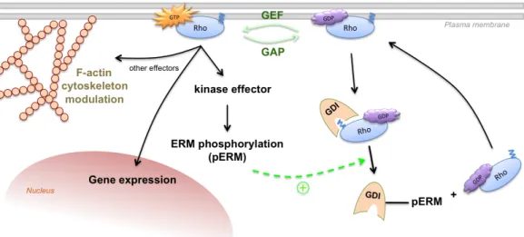
GDP-bound, S1/S2 hidden, inactive conformation (Wennerberg et al., 2005; Bustelo et al., 2007). In vivo, several extracellular signals transduce through GPCRs, RTKs, integrin heterodimers, and other transmembrane proteins to both activate Rho GTPases, usually by stimulating GEF activity (Karnoub et al., 2004), or inactivate them, through GAP stimulation (Beckers et al., 2010). There are now 83 GEFs and more than 70 RhoGAPs identified in eukaryotes, demonstrating their wide regulatory functions in the cell, which occur in a quite specific way. In contrast, only 3 GDIs (α, β and γ) have been characterized until today, showing some redundancy as to their substrates (Beckers et al., 2010). GDI functions include (i) prevention of Rho GTPase interaction with the plasma membrane, where it normally resides, by masking its prenyl moiety (see Fig. 1.3), (ii) inhibition, through sequestration, of the interaction between Rho GTPases and their effectors, and (iii) GEF-mediated GDP-to-GTP exchange impairment, by locking Rho GTPase switch regions in a hidden conformation, thereby maintaining them inactivated (Ridley, 2006). Additionally, it has been Figure 1.3. Rho GTPase function and regulation. Besides GAP, GEF and GDI activities (see text
for details), ERM proteins are shown to be phosphorylated by Rho GTPase downstream effector kinases, rendering them in an “open” state and prone to stimulate Rho/GDI dissociation (green, circled “+” signal and dashed arrow). This permits for Rho release and anchorage to the plasma membrane, where it can be GTP-activated and perform its functions. Abbreviations: ERM – Ezrin/Radixin/Moesin; GAP – GTPase-Activating Protein; GDI – Guanine-nucleotide Dissociation Inhibitor; GEF – Guanine-nucleotide Exchange Factor; pERM – phospho-ERM.
reported that GDIα phosphorylation by PKA enhances its negative effect towards RhoA (Qiao et al., 2008), probably by increasing its affinity. On the contrary, GDIs release Rho GTPases from their control when bound by conformationally active ERM protein’s C-terminal domains (Takahashi et al., 1997). Interestingly, ERM proteins are precisely one class of proteins that might need Rho GTPase activity in order to function. As already established, ERM proteins need conformational activation in order to link transmembrane proteins to the actin cytoskeleton. This is first achieved through PIP2 binding to the N-ERMAD of ERM proteins, followed by C-N-ERMAD phosphorylation, needed to preserve the “open”, active conformation. All three classical Rho GTPases, namely RhoA through ROCK, Cdc42 through MRCK, and Rac1 through still unknown effectors, have been reported to induce ERM protein phosphorylation (Matsui et al., 1998; Nakamura et al., 2000; Jeon et al., 2010; respectively). Moreover, all three GTPases have been, to some extent, implicated in the regulation of phosphoinositol metabolism, although Rac1 has been the most linked to the modulation of ERM activity (Takenawa & Itoh, 2001; Auvinen et al., 2007; Kwiatkowska, 2010). This occurs via activation of type I
phosphatidylinositol-4-phosphate 5-kinase (PIP5KI), which leads to PIP2 production and ERM protein activation (Auvinen et al., 2007). Another phosphoinositide-related Rac1 effector function is negative regulation of clathrin-mediated endocytosis (Lamaze et al., 1996; Jou et al., 2000; Malecz et al., 2000). Several proteins involved in the endocytotic process, including AP-2, AP180, amphiphysin, epsin, endophilin, and dynamin, have PIP2-binding domains, and interactions of these proteins with PIP2 are fundamental to vesicle coating and uncoating events (Ieguchi et al., 2007). The ubiquitously expressed phosphatidylinositol 5-phosphatase Synaptojanin 2 protein has been implied in vesicle inositol lipid composition adjustment, and has a Rac1-binding domain at its C-terminal region. Indeed, both excessive and meager Synaptojanin 2 activities have been linked to endocytic disturbances, namely its inhibition (Ieguchi et al., 2007).
In contrast to Rac1 and RhoA, Cdc42 in part localizes to the Golgi complex
(Matas et al., 2005). Together with its targets N-WASP, IQGAP and the Golgi vesicle coat protein, it appears to have a relevant role in regulating ER/Golgi interface transport (Matas et al., 2005; Ridley, 2006). Cdc42 and N-WASP have also been implicated in the regulation of some endocytic and exocytic pathways (Ridley, 2006) and a Cdc42 close relative, the GTPase TC10, was implicated in targeting CFTR-containing vesicles to the plasma membrane (Cheng et al., 2005).
Although it has been reported that RhoA activation also induced inhibition of receptor endocytosis (Lamaze et al., 1996), there has been some controversy as to exactly how, and if this is actually achieved. Nevertheless, RhoA has been implicated in ENaC’s facilitated trafficking to the plasma membrane (Pochynyuk
et al., 2007), which accounts for some function on vesicle transport. Interestingly, myosin VI vesicles, the type in which CFTR in endocytosed (Swiatecka-Urban et al., 2004), have been shown to walk over stress fiber bundles (Brawley & Rock, 2009), as opposed to myosins V and X. Finally, RhoA has been recently suggested to redistribute phospho-Ezrin to the plasma membrane, necessary to induce a scaffold for CFTR increased NHE-RF1-mediated anchorage and stability. Indeed, NHE-RF1 has been pointed as an upstream activator for RhoA (Favia et al., 2010), probably by inducing its release from GDI, which binds activated phospho-Ezrin, in turn a RhoA/ROCK downstream product, thus completing an auto-stimulatory cycle.
In conclusion, since CFTR is internalized by clathrin-mediated endocytosis (Lukacs et al., 1997) and links to the cytoskeleton through NHE-RF1/ERM protein bridges (Bhattacharya et al., 2010), the data presented above suggest that by modulating the activity of Rho GTPases one could either induce or repress several of the cellular processes that control CFTR’s membrane expression and stability.
1.4. Final remarks and Aims of this thesis
As we have seen, CFTR is essentially expressed at the apical face of polarized epithelial cells. The CFTR surface expression is a regulated process involving PDZ-containing proteins such as NHE-RF1 (stabilizing) and CAL (mediates CFTR degradation), which bind the channel at its C-terminal region (Amaral, 2005). Moreover, CFTR is also ubiquitinated at its C-terminus, normally inducing its degradation unless deubiquitinated by USP10 (Bomberger
et al., 2010). This contributes to make the carboxyl-end of CFTR a key place for protein interactions that, as already suggested by several authors (Bagorda et
al., 2002; Favia et al., 2006; Lee et al., 2007; Cushing et al., 2008), gives to affinity-competition between protein regulators a key role in CFTR stability. As NHE-RF1 over-expression enhances CFTR tethering at the surface (Guerra et
al., 2005), if somehow endogenous NHE-RF1/ERM/F-actin complex formation
could be improved, it would be possible to stabilize both wt and, perhaps, mutant CFTR at the plasma membrane, despite negative pressure exerted by quality control proteins. Small GTPases from the Rho family have already been suggested to facilitate this process, by activating ERM proteins. Also, their role as cytoskeleton modifiers should be considered in transmembrane protein distribution.
The work presented in this thesis attempted to explore the stabilization of both wt and mutant CFTR at the plasma membrane, namely through activity modulation of the three classical small Rho family GTPase molecular switches: Rac1, Cdc42 and RhoA.
2. Materials and Methods
2.1. Plasmid DNA amplification, extraction and sequencing
All the clones used in the present work were already available at the host lab. DH5α bacteria, previously stored at −80 °C, were transformed with Rac1 (L61Q; N17T), RhoA (L63Q; N19T) and Cdc42 (L61Q; N19T) cDNAs [cloned in a pEGFP-C1 mammalian expression vector (BD Biosciences Clontech) in which the coding sequences were attached to the C-terminus of the green fluorescent protein (GFP), so that expression products were functionally stable fusion proteins (Matos & Jordan, 2005; Matos & Jordan, 2006)], along with the empty vector itself. Myc-tagged cDNAs of wild-type, human NHE-RF1 and hnRNP A1, cloned into the pcDNA3.1(+) mammalian expression vector (Invitrogen), were also transformed into DH5α cells. Briefly, 10-50 ng of plasmid DNA were added to 10 µL competent DH5α cells in 1.5 mL Eppendorf tubes and left on ice for at least 30 min to promote plasmid DNA adherence to the cell wall. The mix was incubated for 45 s in a 42 °C pre-heated thermomixer (Eppendorf) and put on ice for another 2 min for thermal shock-mediated transformation to occur. Cells were grown on LB medium (about five times the volume of DH5α cells) for 1 h in the thermomixer, at 37 °C and 900 rpm. Afterwards, about 15 µL cell suspension were plated on either kanamycin- (Sigma) or ampicillin- (Sigma) LB/Agarose (Merck) Petri dishes and left in an incubator at 37 °C overnight (O/N; approximately 16-20 h).
For miniprep mini-cultures, single transformant colonies were picked and inoculated into 15 mL sterile polypropylene centrifuge tubes containing 3 mL LB medium plus selection antibiotic, and subsequently grown at 37 °C and 220 rpm O/N. For maxipreps, a first 8 h mini-culture was carried out as described, followed by a second O/N maxi-culture (100 mL LB medium plus antibiotic) on 500 mL glass flasks. Plasmid extractions were done with solutions from, and according to the peqGOLD HP Plamid Mini or Maxi kits (QIAGEN), yielding
around 150-400 ng/µL DNA (minipreps) or 2-5 µg/µL DNA (maxipreps), respectively.
In order to confirm plasmid sequence identity, about 300 ng of plasmid DNA from each prep were used on sequencing procedures, using the BigDye-terminator cycle sequencing kit (Applied Biosystems). DNA was sequenced through a variation of Sanger’s chain-terminator method. Briefly, cyclic, linear ampli-fication of the template sequence using Taq polymerase was carried out in the presence of a
plasmid-specific primer and fluorescent dye-labelled terminator ddNTPs. Sequencing reactions were performed in 0.5 mL Eppendorf tubes in a TPersonal thermocycler (Biometra), using the following program: an initial denaturation for 10 min at 98 °C, followed by 28 cycles of 10 s at 96 °C, 5 s at 56 °C and 4 min at 60 °C. The sense primers used were: pCMV-1F (5’-GGGACTTTCCAAA ATGTCGTA) that anneals to the 3’ region of the CMV promoter in pcDNA3.1(+), and pEGFP-1F (5’-ACTTCAAGATCCGCCACAACAT) designed in the 3’ region of the EGFP sequence in pEGFP-C1. When required, reverse chain sequencing was carried out using either, respectively, phGH-R1 (5’-TTTATTAGGAAA GGACAGTGGG) that anneals to the human growth hormone polyA signal in the pcDNA3.1(+) plasmid, and pEGFP-1R (5’-AACCTCTACAAATGTGGTATG) that anneals downstream of the multiple cloning site of pEGFP-C1 vector. Reaction products were then analysed on a 3130XL Genetic Analyzer (Applied Biosystems). Results were compared to wild-type reference sequences using the nucleotide BLAST tool (http://blast.ncbi.nlm.nih.gov). DNA integrity was Figure 2.1. Plasmid quality assessment. All
plasmids used in transient transfection protocols were routinely assessed for preserved integrity by electrophoresis in ethidium bromide-stained 1% agarose gels. Shown is a representative gel. High quality was considered for supercoiled (lower, brighter band) plasmids. On the contrary, a preparation such as that of Cdc42 was replaced when it showed only relaxed, probably nicked, and/or linear forms, an indication of poor integrity. MM, molecular weight markers.
routinely assessed by electrophoresis in 1% agarose (Merck) gels stained with ethidium bromide (Sigma) (see Fig. 2.1).
2.2. Cell lines, culture and transfection
Baby Hamster Kidney 21 cells (BHK-21) stably expressing wild-type (wt) human CFTR or the CFTR-ΔF508 mutant, already available at the host lab, were used as normal (BHK-wt) and cystic fibrosis (BHK-ΔF) cellular models (Mendes et al., 2003). Both cell lines were stored in plastic vials on liquid nitrogen (−198 °C). Cells were frozen in 1 mL of foetal bovine serum (FBS; Invitrogen) supplemented with 10% dimethyl sulfoxide (DMSO; Sigma), to avoid cell death during freezing. Low passage cultures were freshly started by rapidly thawing the vials inside an incubator (at 37 °C) to avoid crystal formation and cell lysis, and cells quickly re-suspended in 5 mL of culture medium [DMEM/F-12 (Dulbecco’s modified Eagle’s medium) (Invitrogen) supplemented with L-glutamine and 15 mM HEPES, plus 1% penicillin/streptomycin (Invitrogen), 5% FBS and 0.2% Methotrexate (Mayne) (used as a selection marker for stable CFTR expression)], to swiftly dissolve the toxic DMSO contained in the vial. Cells were then re-pelleted by centrifugation (5 min at 220 × g ) and the DMSO-containing medium removed. Cells were again re-suspended in culture medium and grown in T25 or T75 plastic, tissue-culture flasks (NUNC), as needed, at 37 °C and 4% CO2, never beyond passage 25, to avoid CFTR expression changes and cellular instability. Cell passages were carried out at about 80% confluence, by dissolving the extracellular matrix with 0.05% Trypsin-EDTA 1x (Invitrogen) and re-suspending a fraction of cells into a suitable volume of medium.
For transfection, either 35-mm plastic culture dishes containing five 10×10 mm cover-slips (immunofluorescence) or 60-mm plates (biochemical assays) were seeded with 5 × 10⁵ or 1.5 × 10⁶ cells, respectively, a day before the
procedure. Transfection was done using either 2 (35-mm) or 4 µg (60-mm dishes) of plasmid DNA and 4 (p35) or 8 µL (p60) of Lipofectamine™ 2000 (Invitrogen). Both DNA and Lipofectamine were prior diluted in Opti-MEM® I
Reduced Serum Media (Invitrogen), according to the manufacturer’s transfection protocol. Afterwards, cells were left expressing the transgene for 16-20 h, without medium change.
2.3. SDS-PAGE, Western blot and densitometry analysis
Samples destined to assess CFTR expression were resolved in 10% (w/v) Acrylamide/Bis-acrylamide SDS-PAGE mini-gels containing 10% glycerol, at 4 °C. Input lysates used to assess GTPase expression were resolved in equivalent mini-gels without glycerol addition, at room temperature (RT). Proteins were transferred onto PVDF membranes (BioRad) using a Mini Trans-Blot cell (BioRad). Both the transfer buffer and the transfer process itself were carried at 4 °C whenever CFTR samples were at stake. Otherwise, protein transfer was accomplished at RT. Membranes were blocked with 5% non-fat milk for 1h at RT and washed with a TBS-T 1x (TBS 10%, Triton X-100 0.05%) buffer solution before antibody incubation. Membranes were probed with the following primary antibodies, always for 1 h at RT: mouse anti-CFTR [1:500 (pull-down and immunoprecipitation protocols, see ahead) or 1:5000 (Input), clone 596, produced by the group], rabbit anti-myc (1:250, Santa Cruz) and mouse anti-α-tubulin (1:10000, Sigma). After 10/5/5 min washings with TBS-T 1x, membranes were incubated with goat anti-mouse or anti-rabbit peroxidase-conjugated secondary antibodies (Biorad) for 30 min at RT. Three more 10 min washings were carried out, and double-labelled proteins were finally detected by enhanced chemiluminescence (ECL).
Densitometry analysis was done using the ImageJ software (Abramoff et al., 2004; http://rsbweb.nih.gov/ij).
2.4. Biotin/streptavidin pull-down assay
In order to quantify cell surface CFTR levels, a biotinylation and streptavidin pull-down assay was performed, as depicted in Figure 2.2. The procedure was
carried on BHK-wt and BHK-ΔF cells, which were transfected, as mentioned above, with either NHE-RF1 or the indicated Rho GTPase mutants.
Before starting, a suitable quantity of Streptavidin-Agarose (S-A) bead-slurry (Sigma) was washed with a pull-down buffer (PD-Buffer) solution (Tris-HCl 50 mM (pH 7.5), NaCl 100 mM, Glycerol 10%, NP-40 1%, SDS 0.1%) and left blocking in 2% non-fat milk for at least 30 min. This was found to significantly decrease unspecific protein binding to the agarose beads. All procedures before cell lysis were carried on ice, inside a cold room (4 °C). Cells grown on 60-mm dishes and transfected as previously described were washed with ice-cold PBS-CM (PBS 1x (pH 8.0), CaCl2 0.1 mM, MgCl2 1 mM) to remove all medium contaminants, and incubated for 30 min with 0.5 mg/mL Sulfo-NHS-SS-Biotin (AppliChem), previously dissolved in PBS-CM. This membrane impermeable, cleavable biotin reagent has emerged as an important tool for studying the expression and regulation of plasma membrane proteins. Because this labelling reagent dissolves readily in aqueous solutions and becomes charged in its sodium sulfoxide group on the succinimidyl ring, it cannot permeate the cell membrane. Thus, as long as the cell remains intact, only primary amines exposed on the cell surface will react with the sulfo-NHS ester, becoming attached to the biotin label via a stable amide bond. Moreover, the reducing agent-cleavable disulfide (-S-S-) bond within the molecule makes the biotinylation process reversible, thus allowing the study of surface protein Figure 2.2. Schematic overview of the biotinylation and pull-down assay. Cell surface proteins
were biotinylated at 0 °C (to stop internalization) and cells were subsequently lysed. A sample aliquot was used to measure total (input) CFTR by Western blot analysis. Biotinylated proteins from the remaining sample were pulled-down by streptavidin-agarose beads and also immunoblotted for CFTR quantification. Adapted by Paulo Matos from: Ganeshan et al., 2007.
internalization and recycling kinetics. Next, the biotin reagent was thoroughly removed and the reaction quenched by incubating cells for 15 min with an ice-cold amine-rich solution (TRIS-Q: Tris-HCl 100 mM (pH 8.0), NaCl 150 mM, CaCl2 0.1 mM, MgCl2 1 mM, Glycine 10 mM, BSA 1%). Cells were then washed three times with PBS-CM and lysed in ice-cold PD-Buffer supplemented with a protease inhibitor 1x cocktail mix (100x solutions: aprotinin 1.5 µM, leupeptin 23 µM, E64 10 µM, EGTA µM [cocktail 1, dissolved in water]; pepstatin 15 µM, PMSF 1 mM, 1,10-phenanthroline 1 mM [cocktail 2, dissolved in methanol]; all reagents obtained from Sigma). Cells were scraped from the dishes and collected in 1.5 mL vials. After centrifugation for 5 min at 10000 × g and 4 °C, an aliquot of each supernatant (SN) was saved and mixed with a 2x CFTR Sample Buffer (Tris-HCl 62.5 mM (pH 6.8), SDS 3%, Glycerol 10%, Bromophenol Blue 0.02%) for input protein assessment. The remaining SN samples were passed to new vials and 1/5 of the sample volume in pre-blocked S-A bead-slurry (dry beads 1:1 in PD-Buffer) was added to each tube. The tubes were left rotating for 1 h at 4 °C. Afterwards, samples were centrifuged for 1 min at 6000 rpm, the supernatant discarded and samples carefully washed four times with a Wash-Buffer solution (Tris-HCl 100 mM (pH 7.5), NaCl 300 mM, TX-100 1%) using a 1-mL syringe with a small-diameter hypodermic needle, to avoid S-A bead aspiration. Captured biotinylated protein was finally eluted with 2x CFTR Sample Buffer in the same volume of dried beads used. Pull-down and whole-cell CFTR levels were then compared by Western blot and analysed by densitometry.
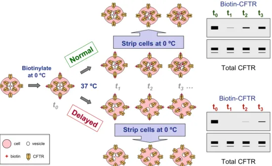
2.5. CFTR internalization kinetics assay
For internalization assays, BHK-wt cells were transiently transfected with the indicated constructs and subjected to a procedure (schematized in Figure 2.3) very similar to the biotin/streptavidin pull-down assay (see subsection 2.4). For this assay, a Lipofectamine/DNA master mix was prepared for each construct to allow the simultaneous transfection of a set of six 60-mm culture dishes under equivalent conditions. Each set of six dishes was then used to analyse the
extent of CFTR internalization at time points 0, 1, 2.5, 5 and 7.5 min. Surface protein biotinylation was then performed on all dishes, as described above, except that after the final wash step with PBS-CM all dishes except one [used for total surface (SUR) CFTR-biotin assessment] were rinsed with warm culture medium and placed inside an incubator (37 °C) for the indicated time points, to restart the endocytic process. Afterwards, cells were quickly rinsed in ice-cold PBS-CM and again placed on ice for 10 min to stop internalization. All dishes, except the SUR control, were biotin-stripped three times for 15 min with a reducing glutathione solution (glutathione 60 mM, NaCl 90 mM, MgCl2 1 mM, CaCl2 0.1 mM, NaOH 90 mM, FBS 10%), leaving only internalized CFTR-biotin molecules untouched. The stripping reaction was then quenched for 15 min with TRIS-Q (see 2.4) and cells were washed three times with ice-cold PBS-CM, lysed and processed as described for the conventional biotinylation assay. Figure 2.3. Schematic overview of the CFTR internalization kinetics assay. Cell surface proteins
were biotinylated at 0 °C (t0, with internalization stopped), and cells subsequently incubated at 37 °C for several time periods (t1, t2, t3, …) to restore CFTR trafficking. Surface biotinylated proteins were biotin-stripped at 0 °C with glutathione, leaving only internalized CFTR biotinlylated. Cells were lysed and a sample aliquot was always taken to measure total (input) CFTR by Western blot analysis. The remaining sample was used to pull-down internalized CFTR-biotin using streptavidin-agarose beads, and immunoblotted for CFTR content assessment. An example is given of both normal (green) and delayed (red) internalization processes, the latter expected if CFTR stabilization occurred at the plasma membrane. Adapted by Paulo Matos from: Ganeshan
Internalized CFTR was assessed by Western blot analysis and densitometry, and the kinetic results expressed as percent internalized CFTR, relative to total surface CFTR (SUR), over time.
To distinguish internalized CFTR recycling from lysosomal degradation a similar methodology to the one just described was applied, but using a non-hydrolysable Sulfo-NHS-LC-Biotin (Pierce). This ensured that any observable decrease in biotin-CFTR levels would be due to lysosomal degradation and not to glutathione-mediated stripping of internalized biotin-CFTR that recycled back to the membrane at later time points.
The theoretical background behind the assay, including kin measurements,
follows from the Wiley-Cunningham steady-state model (Wiley et al., 1982). Briefly, total membrane CFTR (SUR) biotinylated on ice forms the initial “surface” amount (CFTRSUR). Biotinylated CFTR found after 37 ºC exposure for
several time periods, surface biotin stripping and cell lysis, forms the “internalized” protein amount (CFTRIN). Accordingly, the internalization process
is expressed in eqn. 1.
CFTRSUR kin
⎯ →⎯ CFTRIN (1)
As the assay permits CFTRIN measurement overtime, eqn. 1 yields eqn. 2.
d CFTR⎡⎣ IN⎤⎦
dt = kin⎡⎣CFTRSUR⎤⎦dt (2)
Integrating the latter differential equation, ensuring that d[CFTRSUR]/dt ≈ 0
(steady state) and letting [CFTRIN]0 = t0 = 0, gives eqn. 3.
CFTRIN
⎡⎣ ⎤⎦
CFTRSUR