SpAHA1 and SpSOS1 Coordinate in
Transgenic Yeast to Improve Salt Tolerance
Yang Zhou1,2,3☯, Xiaochang Yin3☯, Ruijun Duan1, Gangping Hao4, Jianchun Guo1,2
*, Xingyu Jiang3*
1Institute of Tropical Bioscience and Biotechnology, Chinese Academy of Tropical Agricultural Sciences, Haikou, China,2College of Plant Science & Technology, Huazhong Agricultural University, Wuhan, China,
3Hainan Key Laboratory for Sustainable Utilization of Tropical Bioresources/College of Agriculture, Hainan University, Haikou, China,4Department of Biochemistry, Taishan Medical University, Tai’an, China
☯These authors contributed equally to this work.
*[email protected](XJ);[email protected](JG)
Abstract
In plant cells, the plasma membrane Na+/H+antiporter SOS1 (salt overly sensitive 1) mediates Na+extrusion using the proton gradient generated by plasma membrane H+ -ATPases, and these two proteins are key plant halotolerance factors. In the present study, two genes fromSesuvium portulacastrum, encoding plasma membrane Na+/H+antiporter (SpSOS1) and H+-ATPase (SpAHA1), were cloned. Localization of each protein was stud-ied in tobacco cells, and their functions were analyzed in yeast cells. Both SpSOS1 and SpAHA1 are plasma membrane-bound proteins. Real-time polymerase chain reaction (PCR) analyses showed thatSpSOS1andSpAHA1were induced by salinity, and their expression patterns in roots under salinity were similar. Compared with untransformed yeast cells, SpSOS1 increased the salt tolerance of transgenic yeast by decreasing the Na+content. The Na+/H+exchange activity at plasma membrane vesicles was higher in SpSOS1-transgenic yeast than in the untransformed strain. No change was observed in the salt tolerance of yeast cells expressingSpAHA1alone; however, in yeast transformed with bothSpSOS1andSpAHA1, SpAHA1 generated an increased proton gradient that stimu-lated the Na+/H+exchange activity of SpSOS1. In this scenario, more Na+ions were trans-ported out of cells, and the yeast cells co-expressingSpSOS1andSpAHA1grew better than the cells transformed with onlySpSOS1orSpAHA1. These findings demonstrate that the plasma membrane Na+/H+antiporter SpSOS1 and H+-ATPase SpAHA1 can function in coordination. These results provide a reference for developing more salt-tolerant crops via co-transformation with the plasma membrane Na+/H+antiporter and H+-ATPase.
Introduction
Sodium is a trace element in plants. However, excess sodium in the cytoplasm is toxic to some important physiological and biochemical processes, and efflux of Na+out of the cytoplasm is an important mechanism of salt tolerance. Excess Na+in the cytoplasm can be transported
OPEN ACCESS
Citation:Zhou Y, Yin X, Duan R, Hao G, Guo J, Jiang X (2015) SpAHA1 and SpSOS1 Coordinate in Transgenic Yeast to Improve Salt Tolerance. PLoS ONE 10(9): e0137447. doi:10.1371/journal. pone.0137447
Editor:Keqiang Wu, National Taiwan University, TAIWAN
Received:March 26, 2015
Accepted:August 17, 2015
Published:September 4, 2015
Copyright:© 2015 Zhou et al. This is an open access article distributed under the terms of the
Creative Commons Attribution License, which permits unrestricted use, distribution, and reproduction in any medium, provided the original author and source are credited.
Data Availability Statement:All relevant data are within the paper and its Supporting Information files.
into the external medium across the plasma membrane or into the vacuole across the tonoplast, such that the Na+in the cytoplasm is maintained at a non-toxic concentration. The tonoplast Na+/H+antiporter NHX compartmentalizes Na+into vacuoles using the proton gradient gen-erated by vacuolar H+-ATPase and H+-pyrophosphatase (H+-PPase), whereas the plasma membrane Na+/H+antiporter SOS1, which is driven by the proton gradient generated by plasma membrane H+-ATPases, transports Na+out of cells [1].
NHX proteins from more than 60 plant species have been reported to date [1].Arabidopsis
AtNHX1 was the first NHX protein to be described, and its overexpression can significantly improve the salt tolerance of transgenic yeast or plants [2,3]. Many lines of evidence have indi-cated that tonoplast-bound NHXs function as Na+/H+antiporters and mediate plant salt toler-ance by sequestering Na+into vacuoles [4]. As Na+influx into vacuoles via NHXs is energized by the H+gradient created by vacuolar H+-ATPase and H+-PPase, these two H+-pumps should play important roles in plant salt tolerance. Indeed, the salt tolerance of transgenicArabidopsis
harboringAVP1(vacuolar H+-PPase 1) was stronger than that of wild type [5]. Moreover, the co-expression of NHX and AVP conferred stronger salt tolerance to transgenic plants than NHX or AVP alone, a finding that further demonstrates the relationship between NHX and the H+-pump, with plant NHX proteins functioning as Na+/H+antiporters using the force from the proton gradient produced by the vacuolar H+-PPase [6–8]. SOS1 is the only Na+ efflux protein at the plant plasma membrane reported to date and functions as a Na+/H+ antiport system that is driven by the plasma membrane H+-pump H+-ATPase [9–11], and the salt tolerance ofArabidopsistransformed with theAtSOS1gene is significantly higher than that of wild type [12]. In addition, plasma membrane H+-ATPase activity is involved in plant adaptation to salinity [13]. These findings suggest that the salt tolerance efficiency of the simultaneous expression of SOS1 and plasma membrane H+-ATPase genes should be further enhanced, just as the co-expression of the vacuolar H+-PPase and Na+/H+antiporter results in even higher salt tolerance. However, this hypothesis has yet to be tested. Therefore, the aim of the present investigation was to determine whether the combination of the plasma membrane Na+/H+antiporter with H+-ATPase effectively increases salt tolerance in transgenic organisms. Herein, a plasma membrane Na+/H+antiporter gene,SpSOS1, and a plasma membrane H+ -ATPase gene,SpAHA1, were cloned fromSesuvium portulacastrum, a halophyte with optimal growth at 200–300 mM NaCl [14,15]. Functional analyses indicated that SpSOS1 could trans-port Na+out of cells using the force of the proton gradient generated by SpAHA1. In particular, the co-expression ofSpSOS1andSpAHA1conferred stronger salt tolerance to transgenic yeast cells than the expression of either gene alone. These results may be helpful in developing more salt-tolerant crops through the co-expression of SOS1 and H+-ATPase.
Materials and Methods
Plant material and cultivation
Sesuvium portulacastrum(L.) L. plants, dicotyledonous halophytes belonging to the Aizoaceae family, were collected from the seashore of Haikou, China, and propagated through cuttings (S1 Text). The collection site in the seashore is a wasteland, so no one is responsible for this field. Furthermore,Sesuvium portulacastrumgrows worldwide and is not specifically exploited or an endangered or protected species. Therefore, no specific permission was required for the collection of the plants. Three stem internodes were cut from the mother plants, incubated in Hoagland solution and allowed to grow at room temperature. After 3 weeks, uniform seedlings were treated with 0, 400, 600 or 800 mM NaCl; three replications for each treatment were per-formed. The roots and leaves were harvested at various times (0, 3, 6, 9, 12, 24 and 48 h) after NaCl treatment and immediately stored in liquid nitrogen.
Cloning of
SpSOS1
and
SpAHA1
Total RNA isolation and cDNA synthesis were performed using a reverse transcription poly-merase chain reaction (RT-PCR) kit (Invitrogen, Carlsbad, CA, USA) according to the manu-facturer’s protocol. Degenerate primers were designed forSpSOS1according to the conserved sequences of plantSOS1genes: 5’-ATGAATGATGGGAC(G/T)GC(A/T)AT-3’, 5’-TATAG(A/ T/C/G)CCCAA(A/T)GT(A/G)CTTCCATG-3’. To clone theSpSOS1gene, the cDNA was used as the template in PCR with the degenerate primers. The PCR product, 1,850 bp in length, is homologous with other plantSOS1genes. To obtain the full-lengthSpSOS1sequence, 3’-RACE (rapid amplification of cDNA ends) and 5’-RACE cDNA syntheses were performed with kits according to the manufacturer’s instructions (Invitrogen, Carlsbad, CA, USA). The clones obtained were sequenced, and the overlapping region with the first clone was confirmed. After deducing the open reading frame, gene primers were designed according to the open reading frame (ORF) and including EcoR I and SalI restriction sites (underlined): Forward: 5’-gGAA TTCATGGCGGCGTTGACTGAT-3’; Reverse: 5’-ataGTCGACTCATGGTGCAGGGCGG-3’. A fragment containing the ORF was synthesized via PCR from cDNA and sequenced for fur-ther confirmation. TheSpSOS1ORF was cloned into the yeast expression vector p416 GPD to produce the plasmid p416-SpSOS1. Regarding the cloning ofSpAHA1, anSpAHA1EST frag-ment (GenBank Accession No. DQ925817) was first re-obtained via PCR from cDNA and sequenced for further confirmation. Elongation of the 5’- and 3’- ends ofSpAHA1was per-formed using 5’-RACE and 3’-RACE techniques with two pairs of nested primers and cDNA as the template. One pair of primers deduced from the polylinker sequence was from the RACE kit (Invitrogen, Carlsbad, CA, USA), and gene-specific primers were designed according to EST sequences. The 3’-RACE nested PCR primers were 5’-ATTGTCGGTATGACTGGAG ATGGTGTG-3’and 5’-CGTCTCTATTACCATTCGTA TTGTGCTC-3’, and the 5’-RACE nested PCR primers were 5’- TTGGAAAATAGCCCTACTGGTAAGCAC-3’and 5’- TGAG CATAAATCCGAGCACAATAC-3’. The fragments were cloned into pMD-19T and sequenced. Full-lengthSpAHA1was deduced by overlap of the above two fragments and was obtained via PCR with cDNA as the template and two gene-specific primers (5’-CGGGATC CATGGCCAAGGCCATCAATC-3’; 5’ -CGGAATTCCCTTAGACGGTGTAGTTCTGTTGG-3’) and cloned into the yeast expression vector p414 to produce the plasmid p414-SpAHA1. The complete cDNA sequences ofSpSOS1(accession No. JX674067) andSpAHA1(accession No. JX628604) were deposited in GenBank.
Quantitative real-time PCR
Subcellular localization of SpSOS1 and SpAHA1 proteins
The full-length genesSpSOS1,SpAHA1orArabidopsis SOS1(AtSOS1) without the stop codon at the 3’end were obtained via PCR and fused in-frame to GFP in the modified binary vector pCAMBIA1300. Primers were designed forSpSOS1,SpAHA1andAtSOS1, as follows: SpSOS1–
1300F: 5’-ataGTCGACATGGCGGCGTTGACTGATT-3’, SpSOS1–1300R: 5’-cggGGTA CCTGGTGCAGGGCGGGGGAAAG-3’; SpAHA1–1300F: 5’-gACTAGTATGGCCAAGGC CATCAATC-3’, SpAHA1–1300R: 5’-cgGGATCCGACGGTGTAGTTCTGTTGG-3’; AtSOS1–1300F: 5’- gcTCTAGAATGACGACTGTAATCGACGCG-3’, AtSOS1–1300R: 5’ -cggGGTACCTAGATCGTTCCTGAAAACG-3’. The resulting constructs were transferred intoAgrobacterium tumefaciensstrain LBA4404. For transient expression in plants, different combinations of strains containing these constructs were co-infiltrated intoNicotiana tabacum
leaves [16]. After 5 days, protoplasts were isolated from the infiltrated leaves with a buffer con-taining 0.4 M mannitol, 20 mM KCl, 20 mM MES (pH 5.7), 10 mM CaCl2, 3.5 mMβ
-mercap-toethanol, 0.1% bovine serum albumin, 1.5% cellulase R10, and 0.4% macerozyme R10 [17]. GFP fluorescence in the leaf epidermal cells and mesophyll protoplasts was detected using a confocal laser scanning microscope (FV3000; Olympus Corporation, Tokyo, Japan).
Yeast strains and incubation
Saccharomyces cerevisiaestrain AXT3K (4ena1::HIS3::4ena4,4nha1::LEU2, and4nhx1:: KanMX4), which is deficient in the main endogenous Na+transporters, is a derivative of W303 (MAT ura3–1 leu2–3,112 his3–11,15 trp1–1 ade2–1 can1–100) [10]. To assess the functionality of the SpSOS1 and SpAHA1 proteins, p416-SpSOS1, p414-SpAHA1 and p416-SpSOS1 and p414-SpAHA1 were transformed into AXT3K. The wild-type yeast strain W303 was used as a positive control [18]. W303, AXT3K, and AXT3K transformed with p416-SpSOS1, AXT3K transformed with p414-SpAHA1, and AXT3K co-transformed with p416-SpSOS1 and p414-SpAHA1 were grown in AP medium (10 mM arginine, 8 mM phosphoric acid, 2% glucose, 2 mM MgSO4, 1 mM KCl, 0.2 mM CaCl2, and trace minerals and vitamins) [19].
When these precultures grew to saturation, they were diluted 10-fold, 100-fold, 1,000-fold, and 10,000-fold, and then 10μl of each serial dilution was spotted onto AP plates with the
indi-cated concentration of NaCl. After incubation for 3–5 days at 30°C, growth was imaged and analyzed.
Measurement of Na
+content in yeast cells
Yeast cells were grown in AP medium in the presence or absence of 30 mM NaCl at 30°C. The Na+content in the cells was measured using an atomic absorption spectrophotometer (AA-670, Shimadzu Corporation, Kyoto, Japan), as previously described [19].
Plasma membrane isolation and measurements of Na
+/H
+exchange
and H
+-ATPase activity
7.5), 25 mM BTP-HEPES (pH 7.5), 50 mM KNO3, 250 mM mannitol, 4 mM MgSO4and 50μg
of plasma membrane protein. The assay was initiated by the addition of 3 mM ATP (pH 7.5); when theΔpH reached a steady state, 50 mM Na2SO4was added to the reaction mixture.
Finally, the reaction was stopped with 25 mM (NH4)2SO4. H+-ATPase activity was assayed by
measuring theΔpH across the membrane, which is expressed as the change in fluorescence quenching during the first 30 s after the addition of ATP (ΔF/min/mg protein). Na+/H+ exchange activity was calculated as the change in the initial rate of fluorescence recovery during the first 30 s after the addition of Na2SO4, and this value is expressed as a percentage of the
maximal fluorescence (ΔF (%)/min/mg protein). The reaction was controlled at excitation and emission wavelengths of 430 nm and 500 nm, respectively.
Statistical analysis
A two-tailed Student’s t-test was used to analyze the data, which are presented as the means ± SE.
Results
Cloning and molecular description of
SpSOS1
and
SpAHA1
To clone the genes encoding the plasma membrane Na+/H+antiporter and H+-ATPase fromS.
portulacastrum, we utilized homologs of other plant plasma membrane SOS1s or H+-ATPases and designed degenerate primers. We obtained a fragment of cDNA encoding a putative homolog of the Na+/H+antiporterSOS1gene, and the complete cDNA was then obtained using the rapid amplification of cDNA end (RACE) method and sequenced (GenBank acces-sion number: JX674067). The homology-based computer search identified a 396-bp EST inS.
portulacastrum(GenBank accession number: DQ925817) encoding a putative plant plasma membrane H+-ATPase. The EST was isolated and confirmed via sequencing, and the cDNA was cloned using RACE. TheSpSOS1gene encodes a protein of 1,155 amino acids. The SpSOS1 protein shares 61.53%, 60.58%, 67.67%, 58.94% and 60.39% amino acid identity with similar proteins fromArabidopsis thaliana(AtSOS1), rice (OsSOS1), poplar (PtSOS1), wheat (TaSOS1) andThellungiella halophila(ThSOS1), respectively [11,19,23–25] (Fig 1A). TMHMM software analysis revealed that SpSOS1 contains 12 putative transmembrane domains at the N-terminus and a long hydrophilic C-terminus (http://www.cbs.dtu.dk/ services/TMHMM/), and a conserved phosphorylation site (1143DSPS1146) is also present at the C-terminus. These findings suggest that SpSOS1 may function as a plasma membrane Na+/ H+antiporter that is involved in salt resistance inS.portulacastrum. The 3,394-bpSpAHA1
cDNA fragment for a putative H+-ATPase fromS.portulacastrumwas cloned using the RACE method and then sequenced (GenBank accession number: JX628604). This fragment contains a 2,862-bp ORF encoding a protein of 953 amino acids. An alignment of the amino acid sequences ofArabidopsisH+-ATPases showed that SpAHA1 shares significant similarity with
ArabidopsisAtAHAs and is closest to AtAHA1 (85.83%) (Fig 1B), indicating that SpAHA1 may function as an H+-ATPase at the plasma membrane and assist in energy generation inS.
portulacastrumby catalyzing ATP.
Subcellular localization of SpSOS1 and SpAHA1 proteins
To analyze the localizations of SpSOS1 and SpAHA1 in plant cells, their ORFs fused with the
GFP was expressed alone, a fluorescent signal was clearly observed in the cytosol and nucleus (Fig 2), which agrees with the previous observation indicating that GFP localizes to both the cytosol and nucleus [26,27]. The GFP-fused AtSOS1 protein localized to the cell membrane (Fig 2). The fluorescence images of the SpSOS1-GFP or SpAHA1-GFP fusion protein precisely coincided with the pattern observed for the plasma membrane-localized protein AtSOS1 in tobacco leaf epidermal cells and mesophyll protoplasts, in contrast to the typical cytosolic and nuclear fluorescence distribution of GFP (Fig 2). These results indicate that both SpSOS1 and SpAHA1 are plasma membrane-bound proteins.
Expression of SpSOS1 and SpAHA1 in
S
.
portulacastrum
Sesuvium portulacastrumis a halophyte with optimal growth at 200–300 mM NaCl [14]; thus, 400 mM, 600 mM and 800 mM NaCl were chosen for treatments to assess the expression of
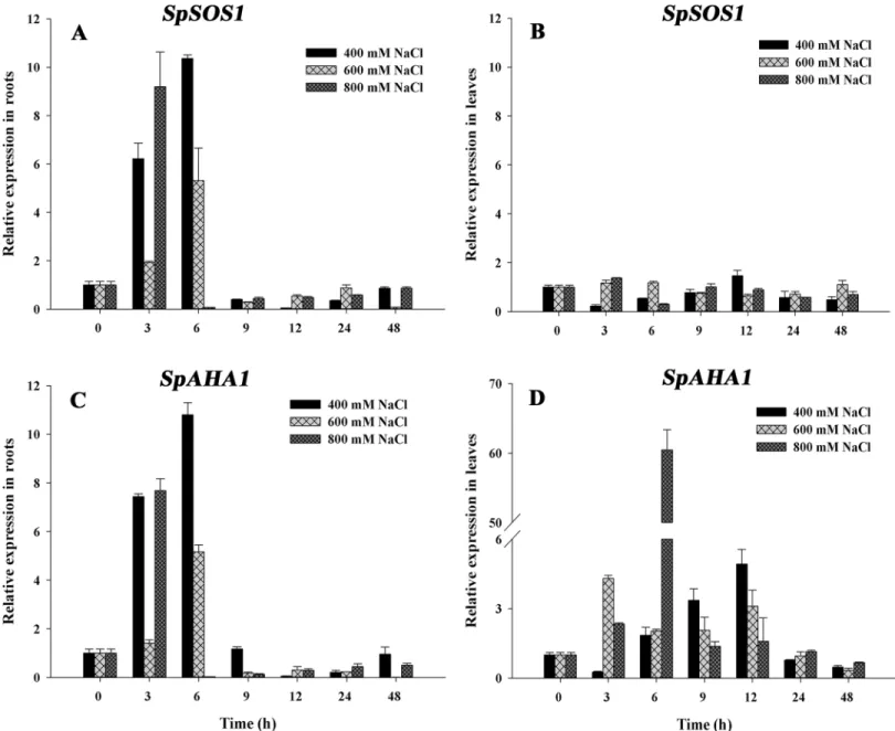
SpSOS1andSpAHA1inS.portulacastrumseedlings under salt stress.SpSOS1expression in roots exhibited a consistent pattern under salt stress: all of the treatment groups exhibited a transient increase inSpSOS1expression in the roots that peaked at approximately 3–6 hours,
Fig 1. Structural analysis of SpSOS1 and SpAHA1 proteins.(A) Phylogenetic tree of plasma membrane Na+/H+antiporters from plants. SpSOS1 (JX674067), AtSOS1 (NM_126259), OsSOS1 (AY785147), PtSOS1 (XM_002315801), TaSOS1 (FN356232) and ThSOS1 (EF207775) indicate Na+/H+antiporters from
Sesuvium portulacastrum,Arabidopsis thaliana,Oryza sativa,Populus trichocarpa,Triticum aestivumand
Thellungiella halophila, respectively. (B) Phylogenetic tree of plasma membrane H+-ATPases from plants. SpAHA1 (JX628604) indicatesSesuvium portulacastrumH+-ATPase. AtAHA1 (NM_127453), AtAHA2 (NM_001203941), AtAHA3 (NM_001203630), AtAHA4 (NM_114664), AtAHA5 (NM_128013), AtAHA6 (NM_126721), AtAHA7 (NM_115897), AtAHA8 (NM_114131), AtAHA9 (NM_001198521), AtAHA10 (NM_101587), and AtAHA11 (NM_125662) indicateArabidopsis thalianaH+-ATPases. The phylogenetic trees were constructed using the MEGA5.0 software based on multiple alignments of full-length protein sequences. Evolutionary distances were computed using the neighbor joining method. Bootstrap values are indicated at the nodes of the tree and are expressed as percentages.
after whichSpSOS1expression gradually decreased to basal levels at 48 h (Fig 3A). In contrast, the change inSpSOS1expression in leaves was not significant under salinity conditions (Fig 3B). The expression pattern ofSpAHA1in roots exposed to salt stress was similar to that of
SpSOS1(Fig 3C). However, compared toSpSOS1,SpAHA1had a more complex leaf expression pattern under salt stress, whereby expression was initially up-regulated and then decreased (Fig 3D).
Effect of salt stress on the growth of yeast cells
To investigate the roles of SpSOS1 and SpAHA1 in halotolerance, the two genes were inserted into the yeast expression vectors p416 GPD and p414 and then transformed individually or co-transformed into AXT3K. The growth of the untransformed and transgenic strains was sim-ilar under normal conditions. Untransformed yeast cells grew very poorly in the presence of NaCl, and these cells could not grow on AP medium containing 75 mM NaCl. The growth of
SpAHA1-transgenic AXT3K was similar to that of the control strain under salt stress, whereas
SpSOS1expression increased the salt tolerance of AXT3K. Interestingly, although the expres-sion ofSpAHA1alone did not influence the response of transgenic yeast cells to salt stress, AXT3K cells co-transformed withSpSOS1andSpAHA1grew better than withSpSOS1alone under salinity conditions. Indeed, the growth of yeast cells expressingSpSOS1andSpAHA1
together was the best among the control and transgenic yeast cells and nearly reached the salt tolerance of the wild-type yeast strain W303 (Fig 4).
Fig 2. SpSOS1 and SpAHA1 proteins are localized at the plasma membrane.SpSOS1-GFP, SpAHA1-GFP and AtSOS1-GFP fusion proteins were produced as described in the Methods and Materials section. Tobacco leaves were transformed with the vector pCAMBIA1300 containing GFP alone (GFP), pCAMBIA1300-SpSOS1-GFP (SpSOS1-GFP), pCAMBIA1300-SpAHA1-GFP (SpAHA1-GFP) or pCAMBIA1300-AtSOS1-GFP (AtSOS1-GFP). GFP signals from epidermal cells (left panel; scale bars = 50μm) and mesophyll protoplasts (right panel; scale bars = 10μm) of tobacco leaves transiently expressing either GFP alone, SpSOS1-GFP, SpAHA1-GFP or AtSOS1-GFP were recorded using confocal microscopy.
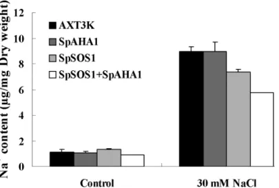
Change in Na
+content
The Na+content in untransformed and transgenic cells was analyzed using atomic absorption spectrophotometry (Fig 5). Although the Na+content did not differ between the untrans-formed strain and various transgenic yeast cells grown under normal conditions, NaCl treat-ment significantly increased the Na+content in all of the yeast strains. A comparative analysis showed that the Na+content increased to a higher level in the untransformed cells than in the
SpSOS1-transgenic cells, whereasSpAHA1expression did not induce a significant change in the Na+concentration in transgenic cells compared with untransformed cells. Interestingly, the
Fig 3. Changes inSpSOS1andSpAHA1mRNAs inS.portulacastrumexposed to salinity.Seedlings were treated with 400, 600 or 800 mM NaCl. The expression ofSpSOS1(A, B) andSpAHA1(C, D) was analyzed in roots (A, C) and leaves (B, D) at different time intervals (0, 3, 6, 9, 12, 24 and 48 h) under salt treatment using real-time PCR. The expression value at the initial time (0 h) was set to 1, and the expression levels at the other time points under salinity treatment were calculated as the fold of the value at 0 h. Values are expressed as the means±SE (n = 3).
Na+content in yeast cells co-expressingSpSOS1andSpAHA1 was the lowest among all yeast strains.
Na
+/H
+exchange and H
+-ATPase activity
The yeast strain AXT3K, which is nearly devoid of background plasma membrane Na+/H+ exchange activity, has been used to investigate the Na+/H+exchange of some plant SOS1 pro-teins [19,23,25]. SpSOS1 suppressed the salt sensitivity of AXT3K (Fig 4). Subcellular localiza-tion showed that SpSOS1 is a plasma membrane-bound protein (Fig 2); thus, AXT3K should be a good tool for studying the Na+/H+exchange activity of SpSOS1 at the plasma membrane. To determine whether SpSOS1 functions as a Na+/H+exchanger in a manner similar to its counterparts AtSOS1, TaSOS1 and ThSOS1 in transgenic yeast cells [11,19,25], we isolated plasma membrane vesicles from AXT3K expressing theSpSOS1gene and from untransformed yeast cells. Little background Na+/H+exchange activity was detected for the plasma membrane vesicles from AXT3K, which contains a mutation in the plasma membrane Na+/H+antiporter
Fig 4. Effect of salt stress on the growth of yeast cells.The wild-type strain (W303), mutant cells (AXT3K) and recombinant strains (SpSOS1, SpAHA1 and SpSOS1+SpAHA1) were grown to saturation, and then 10-μL serial decimal dilutions were spotted onto AP plates supplemented with 0, 50, 75, or 150 mM NaCl. SpSOS1: the AXT3K strain transformed with plasmid p416-SpSOS1; SpAHA1: the AXT3K strain transformed with plasmid p414-SpAHA1; SpSOS1+SpAHA1: mutant strain AXT3K co-transformed with plasmids p416-SpSOS1 and p414-AHA1; AXT3K: untransformed mutant strain AXT3K; W303: wild-type yeast strain W303.
doi:10.1371/journal.pone.0137447.g004
Fig 5. Na+content in yeast cells.The Na+content in unstressed yeast cells and yeast cells treated with 30 mM NaCl was measured. Values are expressed as the means±SE (n = 3). AXT3K: the non-transformed yeast strain; SpAHA1: the AXT3K strain transformed with plasmid p414-SpAHA1; SpSOS1: the AXT3K strain transformed with plasmid p416-SpSOS1; SpSOS1+SpAHA1: the mutant strain AXT3K co-transformed with plasmids p416-SpSOS1 and p414-AHA1.
(Fig 6), whereas the plasma membrane Na+/H+exchange activity of yeast expressingSpSOS1
was higher than that in untransformed yeast cells, indicating that SpSOS1 functions as a Na+/ H+antiporter. As shown inFig 6, the generation of a transmembrane pH gradient is a prereq-uisite for the Na+-dependent movement of H+out of vesicles, suggesting that if the transmem-brane proton gradient increases, the Na+transport capacity of SOS1 should be stronger. To test this hypothesis, the H+-ATPase geneSpAHA1was transformed into yeast cells expressing
SpSOS1, and comparative studies were performed for Na+/H+exchange and H+-ATPase activi-ties at the plasma membrane. H+-pump activity was the highest for plasma membrane vesicles from the yeast cells co-expressingSpSOS1andSpAHA1, followed by that of the yeast cells indi-vidually transgenic forSpAHA1orSpSOS1; the untransformed strain displayed the lowest activity (Fig 6A and 6B). The co-transformed strain showed the highest Na+/H+exchange activity among these tested yeast strains, and the strain transgenic forSpSOS1had a higher Na+/H+exchange activity than the untransformed strain. Although plasma membrane H+ -ATPase activity in theSpAHA1-transformed strain was higher than that in the untransformed cells, inconclusive results were obtained for differences in the Na+/H+exchange activity between theSpAHA1-transformed strain and untransformed cells (Fig 6A and 6C).
Discussion
One key mechanism of salinity tolerance is the ability to remove Na+ions from the cytosol via plasma membrane Na+transporters. SOS1 is the only plant plasma membrane Na+efflux pro-tein reported to date; thus, the propro-tein is likely a key halotolerance factor in plants. SpSOS1 shows similarities with certain reported plasma membrane Na+/H+antiporters, and subcellular localization analysis showed that SpSOS1 is distributed at the plasma membrane in plant cells, suggesting that SpSOS1 can regulate Na+efflux and is involved in salt resistance inS. portula-castrum. SpAHA1 is a plasma membrane-bound protein and has high similarity to the Arabi-dopsisAHA family, indicating that SpAHA1 functions as a H+-ATPase at the plasma
membrane and provides energy for metabolic processes in plants.
Real-time PCR indicated that theSpSOS1expression in roots increased sharply within the first 3–6 h and then decreased to basal levels under salt stress. In leaves, no obvious change in
SOS1transcriptional abundance was found during NaCl treatment (up to 48 h). The salt-induced expression pattern ofSpSOS1is similar to that of riceSOS1, which shows mRNA accu-mulation in roots but not in shoots under salinity [23]. In wheat, theSOS1expression level in leaves was not up-regulated under salinity treatment, but mRNA abundance in leaves was also substantially lower than that in roots [19]. These findings suggest that more active SOS1s can transport excess Na+out of plants through the roots; correspondingly, Na+transport into shoots from roots was decreased, supporting an important salt tolerance mechanism (i.e., retarding Na+transport from roots to shoots). Salt stress induced the up-regulation ofSpAHA1
in both roots and leaves, indicating that the H+-ATPase can provide more energy for the plant to resist salinity. Interestingly, the expression patterns ofSpAHA1andSpSOS1in roots are similar, suggesting that they can work in coordination. Additionally, SpSOS1 functions as a Na+/H+antiporter using the proton gradient produced by SpAHA1.
very important for energizing sodium efflux, and the plasma membrane H+-ATPase is involved in salt tolerance in tomato [13]. In the present study, yeast cells expressingSpAHA1exhibited significantly increased H+-pump activity compared with non-transformed cells, but there were no obvious differences in either salt tolerance or Na+content betweenSpAHA1-transgenic cells and non-transformed cells under salt stress. As a hypothesis, although enhanced plasma mem-brane H+pumping in transgenic yeast cells provides extra energy to support Na+export medi-ated by the Na+/H+antiporter at the plasma membrane, excess Na+in the cytosol cannot be transported out of cells because the Na+transporter utilizing the driving force is deficient at the plasma membrane of AXT3K. This hypothesis is supported by the measurement of the Na+/H+exchange activity of plasma membrane vesicles, showing that Na+/H+antiport activity did not markedly increase at the plasma membrane ofSpAHA1-transgenic cells compared with the non-transformed strain.
Although excess salt is toxic to physiological and biochemical processes in plants, some spe-cies can grow in environments containing high levels of salts because they have evolved the unique ability to resist salinity [28]. One key mechanism in salinity tolerance is the ability to remove excessive Na+from the cytosol via tonoplast Na+/H+antiporters (NHXs) and the plasma membrane Na+/H+antiporter (SOS1). The proton motive force utilized by vacuolar Na+/H+antiporters is generated by H+-translocating enzymes in the tonoplast, such as V-ATPase and H+-inorganic PPase. Recent studies have demonstrated that the vacuolar Na+/ H+antiporter can utilize the electrochemical H+gradient generated by tonoplast H+-pumps to sequester Na+into vacuoles via the simultaneous overexpression of the two tonoplast proteins in plants. The salt tolerance of rice co-expressingSsNHX1andAtAVP1was higher than that achieved by single-gene transformation [6], and rice plants engineered to overexpress native
NHXand H+-PPase genes showed higher salt tolerance than plants expressingOsNHX1or
OsVP1alone [7]. In addition, Gouiaa et al. reported that tobacco plants transformed with both
TNHXS1andTVP1grew better than the single gene-transformed lines under salinity [8]. The plasma membrane Na+/H+antiporter can also transport excess Na+in the cytoplasm out of cells using the driving force of the H+gradient produced by plasma membrane H+-pumps.
Arabidopsisplants transformed with theAtSOS1gene showed higher salt tolerance than wild-type plants [12]. Although plasma membrane H+-ATPase activity correlates with the adapta-tion of plants to salt stress [13], the overexpression of plasma membrane H+-ATPase has not been reported to confer strong salt tolerance to transgenic plants. It is difficult to engineer organisms with vacuolar H+-ATPase because it comprises multiple protein subunits located in the tonoplast, and there is no experimental evidence for enhanced salt tolerance in transgenic organisms with a vacuolar H+-ATPase. In the present investigation, SpAHA1 produced an increased electrochemical gradient of protons to promote the Na+/H+exchange activity of SpSOS1, with more Na+ion being transported out of cells. Correspondingly, yeast cells co-expressing SpSOS1 and SpAHA1 grew better than cells transformed with SpSOS1 or SpAHA1
Fig 6. H+-ATPase and Na+/H+antiport activities at plasma membrane vesicles.Plasma membranes were isolated from mutant cells (AXT3K) and related transgenic strains (SpSOS1, SpAHA1 and SpSOS1 +SpAHA1) using the aqueous two-phase system. Fluorescent quenching of quinacrine was used to monitor the acidification of plasma membrane vesicles from these yeast cells. Formation of a pH gradient, indicating that H+was pumped into the vesicles, was initiated with ATP. Once fluorescence was stabilized, 50 mM Na2SO4was added to the cuvette, and fluorescence recovery, indicating Na+/H+exchange, was monitored. The reaction was terminated by adding 25 mM (NH4)2SO4, which dissipated the pH gradient. The change in fluorescence is expressed as arbitrary units (A). H+-ATPase activity was analyzed by measuringΔpH across the membrane; it is expressed as the change in fluorescence quenching per minute per milligrams of membrane protein (ΔF/min/mg protein) (B). Na+/H+exchange activity is given as the proportion of dissipation of the preformed pH gradient per minute per milligram of membrane protein (ΔF%/ min/mg protein) (C). Values are expressed as the means±SE of three replicates.
alone, demonstrating that the plasma membrane Na+/H+antiporter SpSOS1 and plasma mem-brane H+-ATPase SpAHA1 could work in coordination in transgenic yeast cells. These results provide a reference for the development of more salt-tolerant crops via co-transformation of the plasma membrane Na+/H+antiporter and H+-ATPase.
Supporting Information
S1 Text. The collection site ofSesuvium portulacastrumplants.
(DOC)
Acknowledgments
We thank American Journal Experts (AJE) for English language editing.
Author Contributions
Conceived and designed the experiments: GH JG XJ. Performed the experiments: YZ XY RD GH. Analyzed the data: YZ GH XJ. Contributed reagents/materials/analysis tools: YZ XY RD GH XJ. Wrote the paper: JG XJ.
References
1. Pardo JM, Cubero B, Leidi EO, Quintero FJ. Alkali cation exchangers: roles in cellular homeostasis and stress tolerance. J Exp Bot. 2006; 57(5): 1181–1199. PMID:16513813
2. Apse MP, Aharon GS, Snedden WA, Blumwald E. Salt tolerance conferred by overexpression of a vac-uolar Na+/H+antiport inArabidopsis. Science. 1999; 285(5431):1256–1258. PMID:10455050
3. Gaxiola RA, Rao R, Sherman A, Grisafi P, Alper SL, Fink GR. TheArabidopsis thalianaproton trans-porters, AtNhx1 and Avp1, can function in cation detoxification in yeast. Proc Natl Acad Sci USA. 1999; 96(4): 1480–1485. PMID:9990049
4. Rodriguez-Rosales MP, Galvez FJ, Huertas R, Aranda MN, Baghour M, Cagnac O, et al. Plant NHX cation/proton antiporters. Plant Signal Behav. 2009; 4(4): 265–276. PMID:19794841
5. Gaxiola RA, Li J, Undurraga S, Dang LM, Allen GJ, Alper SL, et al. Drought- and salt-tolerant plants result from overexpression of the AVP1 H+-pump. Proc Natl Acad Sci USA. 2001; 98: 11444
–11449. PMID:11572991
6. Zhao FY, Zhang XJ, Li PH, Zhao YX, Zhang H. Co-expression of theSuaeda salsa SsNHX1and Arabi-dopsis AVP1confer greater salt tolerance to transgenic rice than the single SsNHX1. Mol Breeding. 2006; 17(4): 341–353.
7. Liu SP, Zheng LQ, Xue YH, Zhang Q, Wang L, Shou HX. Overexpression ofOsVP1andOsNHX1
increases tolerance to drought and salinity in rice. J Plant Biol. 2010; 53: 444–452.
8. Gouiaa S, Khoudi H, Leidi EO, Pardo JM, Masmoudi K. Expression of wheat Na+/H+antiporter
TNHXS1and H+-pyrophosphataseTVP1genes in tobacco from a bicistronic transcriptional unit improves salt tolerance. Plant Mol Biol. 2012; 79: 137–155. doi:10.1007/s11103-012-9901-6PMID:
22415161
9. Qiu QS, Guo Y, Dietrich MA, Schumaker KS, Zhu JK. Regulation of SOS1, a plasma membrane Na+/ H+exchanger inArabidopsis thaliana, by SOS2 and SOS3. Proc Nat Acad Sci USA. 2002; 99: 8436
–
8441. PMID:12034882
10. Quintero FJ, Ohta M, Shi H, Zhu JK, Pardo JM. Reconstitution in yeast of theArabidopsisSOS signal-ling pathway for Na+homeostasis. Proc Nat Acad Sci USA. 2002; 99: 9061
–9066. PMID:12070350 11. Quintero FJ, Martı´nez-Atienza J, Caros I, Jiang XY, Kim WY, Ali Z, et al. Activation of the plasma
mem-brane Na/H antiporter SOS1 by phosphorylation of an auto-inhibitory C-terminal domain. Proc Nat Acad Sci USA. 2011; 108: 2611–2616. doi:10.1073/pnas.1018921108PMID:21262798
12. Yang Q, Chen ZZ, Zhou XF, Yin HB, Li X, Xin XF, et al. Overexpression ofSOS(Salt Overly Sensitive) genes increases salt tolerance in transgenicArabidopsis. Mol Plant. 2009; 2: 22–31. doi:10.1093/mp/ ssn058PMID:19529826
14. Yi XP, Sun Y, Yang Q, Guo AP, Chang LL, Wang D, et al. Quantitative proteomics ofSesuvium portula-castrumleaves revealed that ion transportation by V-ATPase and sugar accumulation in chloroplast played crucial roles in halophyte salt tolerance. J Proteomics. 2014; 99: 84–100. doi:10.1016/j.jprot. 2014.01.017PMID:24487036
15. Yang CL, Duan RJ, Li RM, Hu XW, Fu SP, Guo JC. The physiological characteristics of salt-tolerance inSesuvium portulacastrumL. Acta Ecologica Sinica. 2010; 30(17): 4617–4627.
16. Waadt R, Schmidt LK, Lohse M, Hashimoto K, Bock R, Kudla J. Multicolor bimolecular fluorescence complementation reveals simultaneous formation of alternative CBL/CIPK complexesin planta. Plant J. 2008; 56: 505–516. doi:10.1111/j.1365-313X.2008.03612.xPMID:18643980
17. Yoo SD, Cho YH, Sheen J.Arabidopsismesophyll protoplasts: a versatile cell system for transient gene expression analysis. Nat Protocols. 2007; 2(7): 1565–1572. PMID:17585298
18. Xu YY, Zhou Y, Hong S, Xia ZH, Cui DQ, Guo JC, et al. Functional characterization of a wheat NHX antiporter geneTaNHX2that encodes a K+/H+exchanger. Plos One. 2013; 8(11): e78098. doi:10. 1371/journal.pone.0078098PMID:24223765
19. Xu HX, Jiang XY, Zhan KH, Cheng XY, Chen XJ, Pardo JM, et al. Functional characterization of a wheat plasma membrane Na+/H+antiporter in yeast. Arch Biochem Biophys. 2008; 473: 8–15. doi:10. 1016/j.abb.2008.02.018PMID:18316035
20. Menendez A, Larsson C, Ugalde U. Purification of functionally sealed cytoplasmic side-out plasma membrane vesicles fromSaccharomyces cerevisias. Anal Biochem. 1995; 230(2): 308–314. PMID:
7503423
21. Hong S, Cong XL, Jing HY, Xia ZH, Huang X, Hu XW, et al. Characterization of Ca2+/H+exchange in the plasma membrane of Saccharomyces cerevisiae. Arch Biochem Biophys. 2013; 537: 125–132. doi:10.1016/j.abb.2013.07.005PMID:23871844
22. Brandford MM. A rapid and sensitive method for the quantitation of microgram quantities of protein uti-lizing the principle of protein-dye binding. Anal Biochem. 1976; 72: 248–254. PMID:942051
23. Martinez-Atienza J, Jiang XY, Garciadeblas B, Mendoza I, Zhu JK, Pardo JM, et al. Conservation of the salt overly sensitive pathway in rice. Plant Physiol. 2007; 143: 1001–1012. PMID:17142477
24. Tang RJ, Liu H, Bao Y, Lv QD, Yang L, Zhang HX. The woody plant poplar has a functionally conserved salt overly sensitive pathway in response to salinity stress. Plant Mol boil. 2010; 74: 367–380.
25. Oh DH, Leidi E, Zhang Q, Hwang SM, Li YZ, Quintero FJ, et al. Loss of halophytism by interference with SOS1 expression. Plant Physiol. 2009; 151: 210–222. doi:10.1104/pp.109.137802PMID:
19571313
26. Kim BG, Waadt R, Cheong YH, Pandey GK, Dominguez-Solis JR, Schultke S, et al. The calcium sen-sor CBL10 mediates salt tolerance by regulating ion homeostasis inArabidopsis. Plant J. 2007; 52: 473–484. PMID:17825054
27. D’Angelo C, Weinl S, Batistic O, Pandey GK, Cheong YH, Schultke S, et al. Alternative complex forma-tion of the Ca-regulated protein kinase CIPK1 controls abscisic acid-dependent and independent stress responses inArabidopsis. Plant J. 2006; 48: 857–872. PMID:17092313