UNIVERSIDADE DE LISBOA
Faculdade de Medicina Veterinária
THE ECONOMIC IMPACT AND CONTROL MEASURES OF BOVINE RESPIRATORY
DISEASE – A QUALITATIVE APPROACH
JOANA FILIPA DIAS MARTINS DA SILVA
2017 LISBOA
ORIENTADOR
Doutora Magda Alexandra Nobre
Martins Aguiar de Andrade Fontes
CO-ORIENTADOR
Professor Jonathan Rushton
CONSTITUIÇÃO DO JÚRI
Doutor Fernando Jorge Silvano
Boinas
Doutora Magda Alexandra Nobre
Martins Aguiar de Andrade Fontes
Doutor George Thomas Stilwell
UNIVERSIDADE DE LISBOA
Faculdade de Medicina Veterinária
THE ECONOMIC IMPACT AND CONTROL MEASURES OF BOVINE RESPIRATORY
DISEASE – A QUALITATIVE APPROACH
JOANA FILIPA DIAS MARTINS DA SILVA
DISSERTAÇÃO DE MESTRADO INTEGRADO EM MEDICINA VETERINÁRIA
2017 LISBOA
ORIENTADOR
Doutora Magda Alexandra Nobre
Martins Aguiar de Andrade Fontes
CO-ORIENTADOR
Professor Jonathan Rushton
CONSTITUIÇÃO DO JÚRI
Doutor Fernando Jorge Silvano
Boinas
Doutora Magda Alexandra Nobre
Martins Aguiar de Andrade Fontes
Doutor George Thomas Stilwell
i
Agradecimentos / Acknowledgements
À Professora Magda. Por ter assumido a árdua tarefa de ser minha Orientadora. Por todo o seu apoio nesta jornada, pela sua fé no meu potencial e confiança naquilo que sabia podermos alcançar. Obrigada.
Ao Professor Stilwell. Por ter sempre um bocadinho disponível para me ajudar, pelo papel também ele orientador que assumiu durante esta jornada, por tudo aquilo que me ensinou, quer enquanto docente quer enquanto futuro colega. Obrigada.
À minha família. À pedra basilar que esteve sempre lá, nos altos e baixos (e que baixos!) por que passámos. Aos meus pais, pelo apoio incondicional e intemporal, por me fazerem sempre almejar ser melhor. Não melhor que os outros. Melhor que mim mesma, melhor do que ontem. Ainda estou a trabalhar nisso. À titi Madalena, pelo seu exemplo de força. Aos meus avós, por estarem sempre presentes. Obrigada.
À minha irmã. Pelo seu apoio incondicional e não ter medo de me dar um puxão de orelhas (metafórico ou não). Por me trazer de volta à vida quando me sinto a desanimar. Obrigada, Ka. A todos os amigos cujo apoio esteve e está sempre lá, naquele lugar especial. Às meninas da segunda fila: Ana, Anabela, Beatriz, Andreia, Sviatlana, Carolina e tantos outros. Veterinária Cilindra!
To Professor Jonathan. For the warm welcome in the fantastic ride that was my internship at the RVC. For his support and guidance throughout this journey. Thank you.
To Alexis Delabouglise, for his support since day one, for taking the time to ask and consider my opinions and humble expertise. For making me feel part of the group. For his share of knowledge. Merci.
Àqueles que ficaram por mencionar, mas que não estão esquecidos. Obrigada. To those that were not mentioned, but that are not forgotten. Thank you.
“Um navio aportado está seguro, mas não é para isso que os navios são feitos.” “A ship in harbor is safe, but that is not what ships are built for.” Grace Hopper
“Cai sete vezes, levanta-te oito.” Provérbio Japonês “Fall seven times, stand up eight.” Japanese Proverb
ii
The Economic Impact and Control Measures of Bovine Respiratory Disease – A Qualitative Approach
Abstract
Bovine Respiratory Syncytial Virus (BRSV) is a well-known cattle virus, and a key intervenient in the genesis of Bovine Respiratory Disease (BRD). Given its importance, the development of a DIVA vaccine has been established as one of the objectives of a European research project named SAPHIR. BRD is one of the most widespread and costly cattle diseases worldwide but, despite the recognition of its relevance and the substantial investments made in control expenditures, there is still a considerable lack of knowledge concerning its actual economic impact in the dairy and meat cattle value chain. With the objective of collecting primary data concerning epidemiology, presence of risk factors, production losses and expenditures in BRD prevention and treatment on Portuguese farms, two questionnaires were developed and implemented using a convenience sample of five dairy and five meat farms. This case study led to the conclusion that, despite being present in the majority of the farms surveyed, there seems to be an over-all lack of data concerning the quantification of BRD’s economic impacts in primary production, regardless of their recognition and considerable expenditures on medical and prophylactical tools. As proposed, the case study allowed for the identification of gaps concerning BRD and its management, with future work needing to be focused on obtaining a deeper knowledge regarding the meat cattle value chain, evaluating the existence of detailed treatment and vaccination records at farm level, as well as accurate disease prevalence and incidence, and quantification of existing production losses. Considerable control expenditures were also seen in a case study conducted by researchers from the Royal Veterinary College under the SAPHIR project. Despite their presence at farm level, it seems rather difficult to establish a direct correlation between risk factors and disease presence and magnitude. This finding reflects the complex multifactorial nature of BRD, and was transversal to both studies.
Key words: Bovine Respiratory Syncytial Virus; Bovine Respiratory Disease; cattle; economic impact.
iii
O Impacto Económico e Medidas de Controlo da Doença Respiratória Bovina – Uma Abordagem Qualitativa
Resumo
O Vírus Respiratório Sincicial Bovino (BRSV) é um dos vírus bovinos de maior relevo, e um interveniente chave na génese da Doença Respiratória Bovina (DRB). Dada a sua importância, o desenvolvimento de uma vacina DIVA foi estabelecido como um dos objetivos de um projeto de investigação Europeu denominado SAPHIR. A DRB é uma das mais difundidas e dispendiosas doenças dos bovinos mas, apesar do reconhecimento da sua relevância e de substanciais investimentos visando o seu controlo, existe ainda uma considerável falta de conhecimento no que toca ao seu impacto económico concreto na cadeia de valor de bovinos de leite e carne. Com o objetivo de recolher dados acerca da epidemiologia, presença de fatores de risco, perdas produtivas e despesas em termos de prevenção e tratamento da DRB em explorações Portuguesas, dois questionários foram desenvolvidos e implementados numa amostra de conveniência constituída por cinco explorações de bovinos de leite e cinco explorações de bovinos de carne. Este estudo de caso permitiu concluir que, embora a DRB esteja presente na maioria das explorações inquiridas há, de forma generalizada, uma falta de informação no que toca à quantificação dos seus impactos económicos na produção primária, apesar do seu reconhecimento e de despesas consideráveis em ferramentas médicas e profiláticas. Como fora proposto, este estudo de caso permitiu identificar lacunas no que toca à DRB e seu maneio, com trabalho futuro a dever focar-se no aprofundamento do conhecimento relativamente à cadeia de valor da carne bovina, averiguação de existência de registos detalhados de tratamento e vacinações nas explorações, valores precisos de prevalência e incidência da doença e quantificação das perdas produtivas existentes. Despesas consideráveis no controlo da DRB foram igualmente observadas num estudo de caso conduzido por investigadores do Royal Veterinary College ao abrigo do projeto SAPHIR. Apesar da sua presença ao nível das explorações, revela-se bastante difícil estabelecer uma correlação direta entre fatores de risco e a presença e magnitude da doença. Este resultado reflete a natureza complexa e multifatorial da DRB, tendo sido transversal a ambos os estudos.
Palavras-Chave: Vírus Respiratório Sincicial Bovino; Doença Respiratória Bovina; gado bovino; impacto económico.
iv General Index
Agradecimentos / Acknowledgements ... i
Abstract ... ii
Resumo ... iii
Table Index ... vii
Figure Index ... vii
Graph Index ... vii
Abbreviations and Symbols List ... viii
INTRODUCTION ... 1
CHAPTER I: Bovine Respiratory Syncytial Virus Infection ... 3
1.1. The Virus ... 3
1.2. Epidemiology ... 4
1.2.1. BRSV Prevalences in Portugal and in the UK ... 6
1.3. Signs and Pathology ... 8
1.4. BRSV Pathogenesis ... 9
1.5. Diagnosis ... 10
1.5.1. Direct Methods for BRSV Diagnosis ... 11
1.5.2. Indirect Methods for BRSV Diagnosis ... 11
1.6. Treatment ... 12
1.7. Prevention and Control ... 13
1.7.1. Biosecurity and Management Practices ... 13
1.7.2. Vaccination and Immunology ... 14
1.8. Contextualization of BRSV Infection in Bovine Respiratory Disease ... 16
CHAPTER II: Bovine Respiratory Disease – an Overview ... 17
2.1. Enzootic Calf Pneumonia ... 19
2.1.2. Aetiology ... 19
2.1.3. Clinical Signs and Diagnosis ... 20
2.1.4. Treatment ... 21
2.1.5. Prevention and Control ... 21
2.2. Chronic Suppurative Pneumonia ... 24
2.2.1. Aetiology ... 24
2.2.2. Clinical Signs and Diagnosis ... 24
2.2.3. Treatment and Control ... 25
2.2.4. Prevention ... 25
v
2.3.1. Aetiology ... 26
2.3.2. Clinical Signs and Diagnosis ... 27
2.3.3. Treatment ... 28
2.3.4. Prevention and Control ... 28
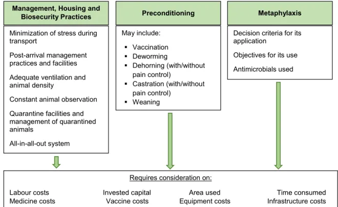
2.3.4.1. Preconditioning ... 30
2.3.4.2. Metaphylaxis ... 30
2.3.4.3. Vaccination ... 31
CHAPTER III: Dairy and Beef Production Sectors: an Overview ... 32
3.1. Dairy Cattle Population and Production Data: Europe, Portugal and the UK ... 32
3.2. Meat Cattle Population and Production Data: Europe, Portugal and the UK ... 33
CHAPTER IV: Economic Assessment of Livestock Disease Impact and Control Strategies... 37
CHAPTER V: The Economic Impacts of BRD on the Bovine Sector ... 47
5.1. Economic Impacts of BRD on the Dairy Sector – Losses ... 47
5.1.1. Daily Gain and Slaughter Weights ... 47
5.1.2. Failure to Reach Growth Targets ... 47
5.1.3. Mortality ... 48
5.1.4. Culling ... 49
5.1.5. Fertility ... 49
5.1.6. Somatic Cell Count ... 49
5.1.7 Milk Yield ... 50
5.1.8. Age at First Calving ... 50
5.2. The Economic Impacts of BRD on the Meat Sector – Losses ... 51
5.2.1. Average Daily Gain, Slaughter Weights and Growth Targets ... 51
5.2.2. Carcase Characteristics and Meat Quality ... 54
5.2.3. Mortality and Culling ... 56
5.2.4. Fertility ... 57
5.3. The Economic Impacts of BRD on Bovine Production – Expenditures ... 58
5.3.1. Expenditures on BRD Prevention ... 58
5.3.1.1. Dairy Calf Disease Control Programmes ... 61
5.3.1.2 Feedlot Disease Control Programmes ... 62
5.3.2. Expenditures on BRD Treatment ... 64
5.4. Summarization of BRD’s Impacts on Bovine Production ... 67
CHAPTER VI: Methodology, Results and Discussion ... 69
6.1. Objectives ... 69
6.2. Questionnaire Design and Sampling Method ... 69
vi
6.4. Results and Discussion ... 71
6.4.1. Sample Characterization ... 71
6.4.2. BRD Occurrence in Dairy and Meat Farms ... 72
6.4.3. Dairy Farm Management and Biosecurity Practices ... 85
6.4.4. Meat Farm Management and Biosecurity Practices ... 87
6.5. Preliminary Results in the UK Study ... 89
CHAPTER VII: Conclusions and Future Work ... 91
Bibliography ... 93
ANNEX I – The SAPHIR Project ... 103
ANNEX II: Medicines Used in BRD Treatment in Portugal and in the UK ... 106
ANNEX III: BRSV vaccines currently available in Portugal and in the UK ... 109
ANNEX IV – Steps of constructing and implementing a questionnaire, adapted from Brancato et al. (2006) and Malhotra (2007) ... 111
ANNEX V – Bovine Respiratory Disease in Dairy Farms: a Questionnaire ... 112
vii Table Index
Table 1: Reported BRSV prevalences in mainland Portugal between 2003 and 2007... 6
Table 2: Reported BRSV prevalences in the UK between 1980 and 2014 ... 7
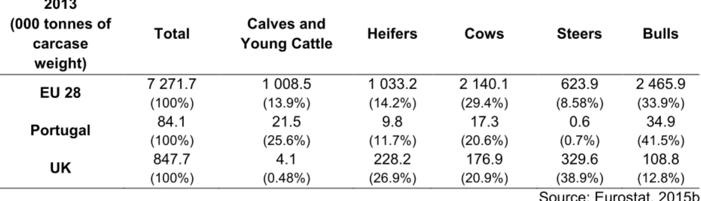
Table 3: Production of beef and veal by class of bovine animals: EU 28, Portugal and the UK, 2013 ... 33
Table 4: Economic methods for the evaluation of animal disease and its control ... 45
Table 5: General characterization of dairy and meat farms (average values) ... 72
Table 6: Current BRD prevalences at farm level ... 74
Table 7: Complementary diagnosis methods used in dairy and meat farms ... 75
Table 8: Expenditures on vaccination in dairy and meat farms ... 78
Table 9: Vaccine effectiveness in dairy and meat farms ... 79
Table 10: Expenditures on treatment in dairy and meat farms ... 81
Table 11: BRD's production impacts in dairy and meat farms ... 82
Table 12: Dairy farm production loss in milk yield ... 83
Table 13: Meat farm production loss with dead unweaned calves ... 83
Table 14: Treatment and vaccination costs for the Portuguese and the Welsh case studies ... 90
Figure Index Figure 1: Most common manifestations of BRD in the dairy and meat sectors ... 18
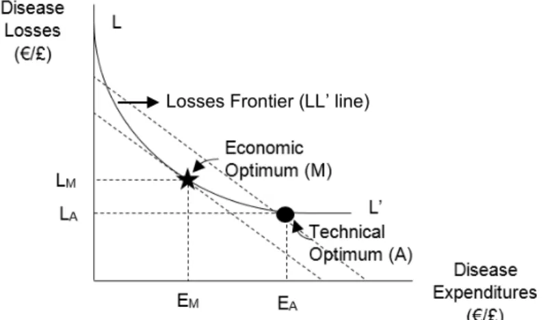
Figure 2: The Loss-Expenditure Frontier (adapted from McInerney et al. 1992) ... 39
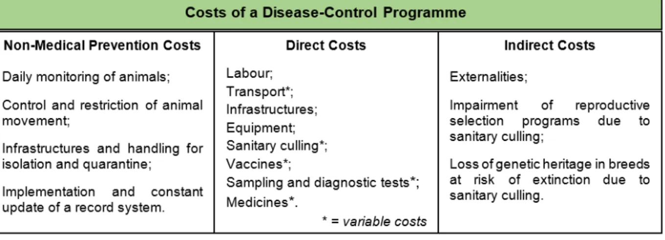
Figure 3: Costs of a disease-control programme (adapted from Henriques et al., 2004) ... 42
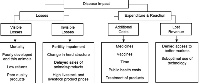
Figure 4: Compartmentalized disease impact on a livestock system ... 43
Figure 5: Considerations on the costs of enzootic calf pneumonia prevention ... 62
Figure 6: Considerations on the costs of shipping fever prevention ... 63
Figure 7: Proportion of the different contributors to the total cost of dairy calf pneumonia ... 65
Figure 8: Proportion of the different contributors to the total cost of suckler calf pneumonia ... 66
Figure 9: The impacts of Bovine Respiratory Disease on bovine production – a model ... 68
Graph Index Graph 1: Production phases present in meat farms ... 71
Graph 2: Number of sick animals in dairy farms by category ... 73
Graph 3: Number of sick animals in meat farms by category ... 73
Graph 4: Clinical signs used for BRD diagnosis in dairy and meat farms ... 75
Graph 5: Diagnosis performance on dairy farms ... 76
Graph 6: Diagnosis performance on meat farms ... 76
Graph 7: Infectious BRD agents identified in dairy and meat farms ... 77
Graph 8: Vaccines used in dairy and meat farms ... 78
Graph 9: Antimicrobials used in dairy and meat farms ... 80
viii Abbreviations and Symbols List
AI Artificial Insemination ADG Average Daily Gain BCoV Bovine Coronavirus
BHV-1 Bovine Herpesvirus Type 1 PI3V Bovine Parainfluenza-3 Virus BRD Bovine Respiratory Disease BRSV Bovine Respiratory Syncytial Virus BVDV Bovine Viral Diarrhoea Virus BAL Bronchoalveolar Lavage CFR Case Fatality Rate
ºC Degree Celsius
DIVA Differentiating Infected from Vaccinated Animals
$ Dollar
ELISA(s) Enzyme-Linked Immunosorbent Assay(s)
€ Euro
EU European Union
FMV-ULisboa Faculdade de Medicina Veterinária - Universidade de Lisboa FCR Feed Conversion Ratio
Ha Hectares
HRSV Human Respiratory Syncytial Virus
IgA Immunoglobulin A
IgE Immunoglobulin E
IgG Immunoglobulin G
IgM Immunoglobulin M
IBR Infectious Bovine Rhinotracheitis
ISO International Organization of Standardization
Kg Kilogram
NSAID(s) Nonsteroidal Anti-Inflammatory Drug(s) NUTS Nomenclature of Territorial Units for Statistics PCR Polymerase Chain Reaction
£ Pound
Lb Pound-mass
® Registered Trademark
RT-PCR Reverse Transcription Polymerase Chain Reaction RNA Ribonucleic Acid
RVC Royal Veterinary College SID semel in die
SCC Somatic Cell Count
m2 Square meters
SAPHIR Strengthening Animal Production and Health through the Immune Response SARA Subacute Ruminal Acidosis
1 INTRODUCTION
Bovine Respiratory Syncytial Virus (BRSV) is one of the most well-known viruses of cattle, being endemic in most countries, and a causal agent of disease in both dairy and meat herds. Despite its ability to cause disease per se, its most important role is as the main viral agent of the scourge that is Bovine Respiratory Disease (BRD). BRD is one of the most studied cattle health problems worldwide, with considerable investments being made towards the minimization of its negative impacts on production. However, and despite the abundant documented knowledge of BRD, this disease continues to be one of the biggest challenges faced by the sector. The multifactorial nature of BRD, with several pathogens, environmental and management risk factors may help explain this fact, in addition to the increased intensification of bovine production, which makes it even more difficult to achieve an effective control. Much emphasis has been put on pharmacological control of this disease, with new molecules being released both in terms of antimicrobials and vaccines. Concerning vaccines, their beneficial effect on BRD prevention, if included in a holistic disease approach, is unquestionable. However, and especially considering BRSV, their field efficacy is far from consensual, and there is still margin for further development. Due to this fact, and as a reflection of this virus’s importance in the establishment of respiratory disease in cattle, the development of a marked BRSV vaccine has been included as one of the goals of a European research project named SAPHIR, which stands for “Strengthening Animal Production and Health through the Immune Response”. The project is still at an early phase, and one of its multidisciplinary foundations is the economic evaluation of the impacts BRSV has on bovine production, as well as the assessment of already existent BRSV-control methods. The Royal Veterinary College (RVC) in London is an active participant in this task and, during the author`s internship at the RVC from January to March of 2016, it was possible to collaborate on a literature review concerning the subject, which was afterwards approved by a panel of experts and whose preliminary results are presented on Annex I. The initial part of this internship, undertaken at the Faculdade de Medicina Veterinária - Universidade de Lisboa (FMV-ULisboa) in Lisbon also encompassed a literature review about BRSV and its impacts, with special focus on its reality in Portugal. The information available proved to be very scarce, despite some data pointing towards a significant BRSV prevalence in Portuguese dairy and meat bovine herds, as well as to the use of vaccines against the virus1.
1Given this paucity of information concerning not just the economic impact of BRSV but of BRD as a whole, a review article of the
2
Given the complex interaction BRSV has with all the other infectious and non-infectious factors involved in the aetiology of BRD, it proves very difficult to attribute specific economic impacts to BRSV alone, and therefore this work proposes to present its economic impact in the context of BRD. It should be noticed that estimating the economic impact of a certain disease is the pillar to evaluate the benefits that can potentially arise from its control, and serves to justify investments such as vaccines, like what is being done under the SAPHIR project. Therefore, the structure of this thesis will be as follows:
A review of the current knowledge concerning BRSV, followed by its contextualization in BRD and a review of the different forms BRD can assume on both the dairy and the meat sectors; A brief description of cattle populations, production data, number and dimension of holdings,
as well as dairy and veal/beef production systems in Portugal and the UK was considered pertinent, especially since some risk factors concerning BRD are practically indissociable from intrinsic aspects of certain production systems;
The importance and implications of economics in the study of animal disease and its control is discussed afterwards, followed by the compilation of production losses and expenditures due to the presence of BRD in the dairy and meat sectors, culminating in a model that presents the impact of BRD on the dairy and meat bovine production chains, as well as data required for the quantification of this impact;
With the objective of collecting primary data concerning the presence and economic impacts BRD has on Portuguese dairy and meat herds, two questionnaires were developed and implemented in a small sample of farms. The methodology for their development, as well as main results, are presented in Chapter VI;
In the final chapter, besides drawing out some conclusions about the present work, we also look at what can be done in the future towards BRD’s control, with focus on some aspects concerning BRSV.
3
CHAPTER I: Bovine Respiratory Syncytial Virus Infection
1.1. The Virus
BRSV is a virus belonging to the Paramyxoviridae family and Pneumovirus genus, and has been recognized as an important agent of respiratory disease in both beef and dairy cattle since its discovery in Switzerland in 1970 (Schreiber et al., 2000; Alm, Koskinen, Vahtiala & Andersson, 2009; Sarmiento-Silva, Nakamura-Lopez & Vaughan, 2012). It is acknowledged as an important causal agent of respiratory disease in dairy calves, nursing beef calves and feedlot calves, but its relevance extends to adult cattle as well (Baker, Ellis & Clark 1997; Hägglund, 2005). As with other syncytial viruses, its name derives from the characteristic cytopathic effect observed both in vivo and in vitro, the formation of syncytium, multinucleated cells derived from the fusion of various cells (Baker et al., 1997).
BRSV is a negative sense, single-stranded RNA enveloped virus, with the viral genome encoding for 11 proteins: the large attachment glycoprotein (G), the fusion protein (F), the small hydrophobic protein (SH), the matrix protein (M), the nucleoprotein (N), the phosphoprotein (P), the RNA polymerase (L), the M2-1 and M2-2 proteins, and two non-structural proteins, NS1 and NS2 (Taylor, 2008; Guzman & Taylor, 2015). The F and G proteins seem to be the most relevant in the development of protective immunity, since neutralizing antibodies are mainly directed at these two proteins (Nettleton et al., 2003; Blodörn, 2015). BRSV exhibits limited genetic variation, especially concerning the F protein, which stands in favour of cattle vaccination. Furthermore, the lipid envelope makes the virus frail outside the host, and sensible to common detergents (Hägglund & Valarcher, 2015). The virus is very closely related to Human Respiratory Syncytial Virus (HRSV), with similarities in terms of epidemiology and pathogenesis allowing the use of calf models infected by BRSV in studies concerning HRSV. Reversely, the study of the human virus has provided valuable information concerning the disease in bovine populations. There are also similar syncytial viruses that affect other animal populations, like ovines and caprines (Baker, 1991; Baker et al., 1997; Valarcher & Taylor, 2007; Gershwin, 2012; Stilwell, 2013).
Apart from being a primary disease agent, BRSV can predispose to secondary bacterial infections by Mannheimia haemolytica, Pasteurella multocida, Histophilus somni and Mycoplasma bovis, culminating in the development of BRD (Valarcher & Taylor, 2007; Klem, Tollersrud, Osterås & Stokstad, 2014; Sacco, McGill, Pillatzki, Palmer & Ackermann, 2014;). Reported values point to up to 40% of viral infections being complicated by secondary bacterial infections (Klem, Kjæstad, Kummen, Holen & Stokstad, 2016). In fact, BRSV has been acknowledged as the main viral component of BRD, mainly due to its high seroprevalence but also to the strong association
4
between viral infection and the development of respiratory disease (Larsen, Tegtmeier & Pedersen, 2001; Blodörn et al., 2015).
1.2. Epidemiology
BRSV is a ubiquitous virus, having been isolated in bovine herds in Europe, America and Asia with seroprevalences that range between 30% and 100% both in dairy farms and in beef herds, and it is considered an endemic agent in many countries. The described levels of morbidity range from 60% to 80% having been reported, in some outbreaks, mortality rates that reach 20-30%, especially when there are other concomitant viral and bacterial infections (Valarcher & Taylor, 2007; Sacco et al., 2014; Blodörn, 2015; Hägglund & Valarcher, 2015). The reported high morbidity levels seem to be due to the rapid spread of the virus within infected herds, which leads to high viral herd prevalence (Klem et al., 2013). Truthfully, it is believed that the majority of cattle populations will be affected by this virus at some point (Woolums, 2010).
The variation in the seroprevalences registered is usually due to the type and age of animals sampled (Hägglund & Valarcher, 2015). It is more difficult to evaluate the frequency of BRSV infections in adult cattle, given the usually high seroprevalence registered in these animals when performing a point prevalence study (Valarcher & Taylor, 2007), and the fact that acquired antibodies are detectable for several years, even in the absence of reinfection (Ohlson, Emanuelson, Tråvén & Alenius, 2010).
The clinical manifestation of disease due to BRSV may vary between herds, depending on the level of viral circulation within the herd. In populations in which the virus is generally present, or in which vaccination programmes are implemented, it is expected that only younger animals develop clinical signs, with calves between one and six months of age being the most affected, and with infection being known to occur even in the presence of maternal antibodies. On the contrary, clinical signs can be transversal to all of the herd when the virus is introduced in previously naïve populations (Valarcher & Taylor, 2007; Woolums, 2010; Stilwell, 2013; Sacco et al., 2014; Hägglund & Valarcher, 2015). In these cases, morbidity levels may reach 100% and studies suggest that adult cattle, especially high production first and second parity cows, pregnant and newly calved cows are in fact more severely affected than other categories of animals within the farm (Raaperi, Bougeard, Aleksejev, Orro & Viltrop, 2012; Hägglund & Valarcher, 2015). Other factors involved in disease expression are concomitant infections with other viruses, environmental factors and stressors such as transportation, gestation, lactation and nutrition (Elvander, 1996).
Regardless of the fact that infection can occur despite the presence of maternal antibodies, and that even seropositive calves may suffer reinfection, antibodies seem to provide at least partial
5
protection, given that both incidence and severity of disease seem to be inversely related to maternal antibody titres (Larsen et al., 2001).
There are several risk factors identified as predisposing to BRSV infection, such as environmental causes like temperature fluctuations, dusty environments, inadequate building ventilation and high humidity levels, as well as stressors related to general management, like weaning, transportation, handling and mixing of animals from different sources. Larger herds, with higher population densities, are also more prone to infection, given the increased contact between animals and increased circulation of farm personnel. Farms located in areas where animal exchanges are common and dual purpose farms also present a higher risk (Raaperi et al., 2012; Sarmiento-Silva et al., 2012; Klem et al., 2013; Sacco et al., 2014; Hägglund & Valarcher, 2015;). Bidokhti, Tråvén, Fall, Emanuelson and Alenius (2009), upon studying the antibody prevalence to BRSV in organic versus conventional farms concluded that organic herds had lower seroprevalences, which could be due to the stricter management practices adopted in these farms, such as closely regulated trading of animals between farms, as well as the implemented quarantine period. Production type seems to also be relevant in the epidemiology of BRSV infections, which have been shown to have increased in parallel with the concomitant intensification of cattle production.
In temperate regions, BRSV outbreaks occur mainly during autumn and winter, but may also occur in the summer (Valarcher & Taylor, 2007; Blodörn, 2015). Infections in winter seem to lead to higher rates of seropositive animals within the herd, though. This may be due to the fact that infectious pressure is higher during the cold months, in which animals are more frequently housed, with high densities and inadequate levels of ventilation and humidity predisposing to infection (Klem et al., 2013).
There is yet no consensus concerning the introduction and maintenance mechanisms of BRSV in cattle populations, with theories of asymptomatic carriers, re-infections and viral mutations, as well as both direct and indirect transmission routes having been proposed (Stilwell, 2013; Blodörn et al., 2015). Despite the fact that cattle are the natural viral host, it is not discarded that other species may play a role in its epidemiology. These may include ovines and caprines, but also species like camelids or bison (Valarcher & Taylor, 2007). It is often theorized that BRSV may lead to persistent infected cattle, which may aid the virus in surviving during the summer, being reactivated and leading to new outbreaks of disease even in herds not subjected to reinfection, but this theory remains yet to be fully clarified (Van der Poel, Kramps, Middel, Van Oirschot & Brand, 1993; Hägglund & Valarcher, 2015).
Viral transmission occurs by direct contact between infected animals and by aerosols, (Valarcher & Taylor, 2007). Airborne transmission, however, doesn’t seem to be very effective, as concluded by several authors. Ohlson et al. (2010) found that in Sweden there were seronegative farms in
6
the midst of areas were BRSV prevalences were very high. Sarmiento-Silva et al. (2012) also consider airborne transmission between herds to be of less importance, strengthening the importance of introduction of infected animals into the herd instead. Indirect transmission, either through humans or fomites is considered of major importance in the epidemiology of BRSV. This may be supported by the occurrence of outbreaks in closed herds or in herds in which outbreaks occur shortly after a visit by animal professionals. In fact, not providing boots for visitors was concluded in one study to augment the risk of BRSV infection (Ohlson et al. 2010). The probability of indirect transmission is directly related to viral load and level of fomite contamination, and also to the existence of contact with vulnerable animals (Hägglund & Valarcher, 2015).
There is some data supporting a possible difference in predisposition to BRSV infection concerning different breeds, indicating that American Red breeds and the Blanc Bleu Belge may be less resistant to the virus. Another study points to a more severe disease manifestation in Holstein-Angus crossbred calves compared to pure Holstein calves (Baker et al., 1997).
1.2.1. BRSV Prevalences in Portugal and in the UK
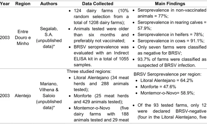
There isn’t an abundant amount of information published concerning the prevalence of BRSV in Portuguese cattle, especially in recent years. The results found in some studies aiming to evaluate the virus seroprevalence in Portuguese herds are presented on Table 1, and demonstrate the expressiveness of the virus in both dairy and meat herds.
Table 1: Reported BRSV prevalences in mainland Portugal between 2003 and 2007
Year Region Authors Data Collected Main Findings
2003 Entre Douro e Minho Segalab, S.A. (unpublished data)* 124 dairy farms (10% random selection from a total of 1208 dairy farms); Animals tested were older
than six months and preferably not vaccinated; BRSV seroprevalence was
evaluated with an Indirect ELISA kit in a total of 1055 samples.
Seroprevalence in non-vaccinated animals = 77%;
Seroprevalence in rearing calves = 57.9%;
Seroprevalence in heifers = 78%; Seroprevalence in cows = 91.1%; Only seven farms were classified
as negative for BRSV;
93.7% of farms were classified as suspected of BRSV infection. 2003 Alentejo Mariano, Vilhena & Saloio (unpublished data)*
Three studied regions:
Litoral Alentejano (34 meat herds and 288 animals tested);
Monforte (25 meat herds and 429 animals tested); Montemor-o-Novo (five
dairy farms with 188 animals tested and 29 meat
BRSV Seroprevalence per region: Litoral Alentejano = 64.2% Monforte = 47.6%
Montemor-o-Novo= 58.9%; Of the 93 tested farms, only 12
were declared BRSV-negative (four in the Litoral Alentejano, five
7
herds with 440 animals tested);
BRSV seroprevalence was evaluated with the indirect ELISA method.
in Monforte and three in Montemor-o-Novo).
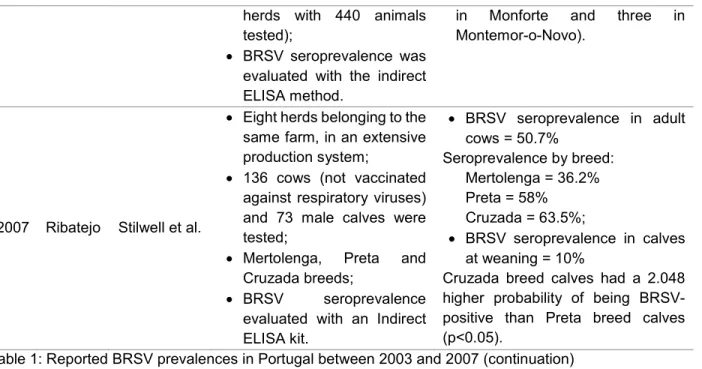
2007 Ribatejo Stilwell et al.
Eight herds belonging to the same farm, in an extensive production system;
136 cows (not vaccinated against respiratory viruses) and 73 male calves were tested;
Mertolenga, Preta and Cruzada breeds;
BRSV seroprevalence evaluated with an Indirect ELISA kit. BRSV seroprevalence in adult cows = 50.7% Seroprevalence by breed: Mertolenga = 36.2% Preta = 58% Cruzada = 63.5%; BRSV seroprevalence in calves at weaning = 10%
Cruzada breed calves had a 2.048 higher probability of being BRSV-positive than Preta breed calves (p˂0.05).
Table 1: Reported BRSV prevalences in Portugal between 2003 and 2007 (continuation) *Source: Stilwell, G. (2016), personal communication.
The prevalence of BRSV in UK dairy and meat herds has been studied by several authors throughout the last decades, and some major findings can be reported (Table 2).
Table 2: Reported BRSV prevalences in the UK between 1980 and 2014
Year Country Authors Data Collected Main Findings
1980 England Stott et al.
Virological survey from 1972 to 1975;
1540 beef-rearing calves; 1143 nasopharyngeal swabs
performed for viral culture; 1069 sera analyzed, with
antibodies titrated in microneutralization tests.
BRSV was detected in 78 samples from a total of 540 viral detections; 58.1% of BRSV infections were diagnosed during outbreaks of disease;
73% of BRSV infections were detected during the winter months; By the age of 9 months, BRSV had been diagnosed in 70% of calves.
1998 England and Wales Paton et al. 341 dairy herds;
Samples collected from July to December 1996;
ELISA testing for antibody detection in bulk tank milk.
100% of the herds tested were positive for BRSV antibodies (however, the vaccination status against BRSV was unknown).
2010 Scotland Hotchkiss et al.
Cross-sectional study;
68 farms (33 beef and 35 dairy); 637 calves;
Deep nasal swabs with real time RT-PCR for RNA viral detection.
Four calves from two farms were positive for BRSV (two calves were from dairy farms and the other two were from beef herds).
2012 Scotland Thonur et al.
541 clinical samples from respiratory or abortion material;
BRSV was detected in 28 samples (5.18%).
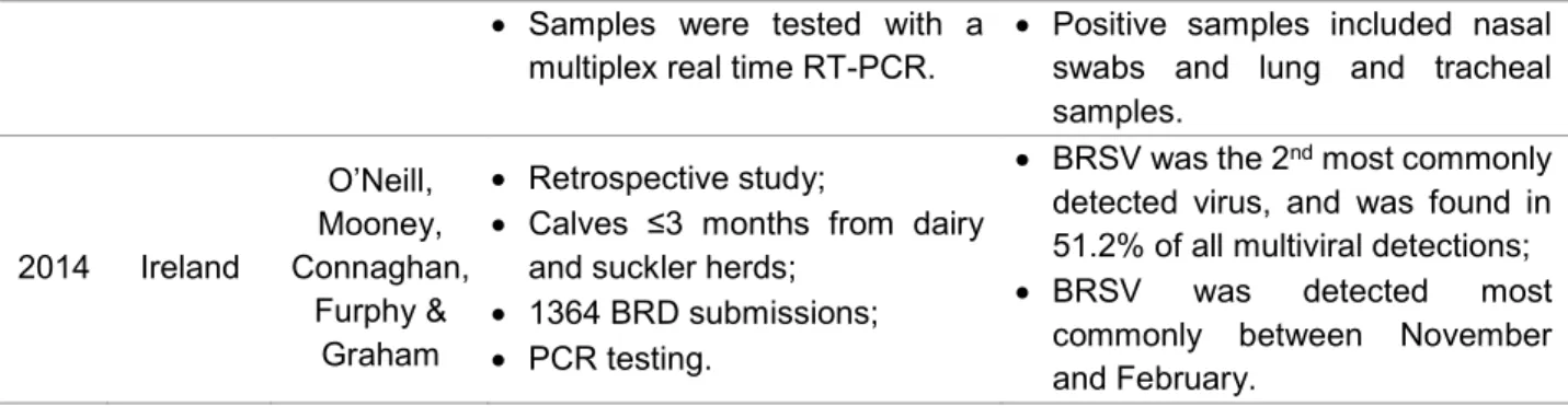
8
Samples were tested with a multiplex real time RT-PCR.
Positive samples included nasal swabs and lung and tracheal samples. 2014 Ireland O’Neill, Mooney, Connaghan, Furphy & Graham Retrospective study;
Calves ≤3 months from dairy and suckler herds;
1364 BRD submissions; PCR testing.
BRSV was the 2nd most commonly
detected virus, and was found in 51.2% of all multiviral detections; BRSV was detected most
commonly between November and February.
Table 2: Reported BRSV prevalences in the UK between 1980 and 2014 (continuation) 1.3. Signs and Pathology
The viral incubation period ranges between two and five days. Clinical signs are usually seen seven to ten days after a stressful event such as transport, but may be seen up to 30 days or more after arrival at destination. The disease developed may be asymptomatic, restricted to the upper areas of the respiratory tree or also involve the lower respiratory tract (Valarcher & Taylor, 2007; Sacco et al., 2014). There are several reasons that justify differences in the severity of disease manifestation, which include: virulence of viral isolates, levels of maternal antibodies, concomitant infections with other pathogens, management practices and environmental conditions (Baker et al., 1997).
The impairment of the upper respiratory tract manifests itself by the presence of cough accompanied by nasal and ocular seromucous discharge, which becomes mucopurulent in the presence of concomitant bacterial infection. Affected animals may exhibit depression, anorexia, milk production decrease, hyperthermia, tachypnea and abdominal breathing. Thoracic auscultation may reveal areas with an increased vesicular murmur, crackles and wheezes, caused by phenomena of bronchopneumonia or bronchiolitis. However, the absence of abnormal sounds is a common finding in this pneumonia, even in the presence of intense dyspnea (Valarcher & Taylor, 2007; Stilwell, 2013). In previously naïve herds, the infection leads to an increase in rectal temperature in two days after exposure and, in three to four days, there is usually a peak in rectal temperature, which reaches values above 40º Celsius (Stilwell, 2013).
Animals in great respiratory distress are usually found exhibiting an orthopneic posture manifested by open mouth breathing, lowered head and stretched neck, as well as sialorrhea, and may sometimes develop pneumothorax, pneumomediastinum or pneumopericardium. In some cases, it is possible to observe the presence of subcutaneous emphysema in the cervical, scapular or perineal areas (Valarcher & Taylor, 2007; Sacco et al., 2014), which is caused by the rupture of alveoli and consequent migration of free air through the mediastinum (Baker et al., 1997).
At necropsy, the most consistent pathological finding of BRSV infection is a cranioventral bronchointerstitial pneumonia, associated with severe bronchiolitis (Baker et al., 1997). The
9
cranioventral lung lobes usually show areas of atelectasis and consolidation, sometimes paired with visible mucopurulent discharge in the bronchus and small bronchi. On the other hand, the caudodorsal lobes frequently show signs of emphysema and edema. In case of secondary bacterial infections, usually with a cranioventral distribution, the lung parenchyma is usually more distended and consolidated (Baker et al., 1997; Valarcher & Taylor, 2007; Sacco et al., 2014). Microscopic lesions in the cranioventral lobes show evidence of proliferative and exudative bronchiolitis with concomitant alveolar collapse, degeneration and necrosis of both ciliated and non-ciliated epithelium, syncytia formation, type II pneumocyte hyperplasia and exudative or proliferative alveolitis. The airway lumen is usually obstructed by neutrophils, macrophages and desquamated epithelial cells, with eosinophils sometimes found both in the lumen and lamina propria of the respiratory tract (Valarcher & Taylor, 2007; Sacco et al., 2014). It should be noted that even though BRSV may be identified as the aetiological agent, necropsies performed at later stages of disease might only reveal the presence of secondary bacteria, and therefore it is important to aim for an early infection diagnosis (Hägglund, 2005).
Reinfections with BRSV usually result in mild disease, with slight pyrexia, dyspnea and, albeit less frequently, cough, or even in subclinical disease, a probable consequence of the development of active immunity following primary infection. Vaccinated herds may also experience sporadic cases of subclinical disease (Baker et al., 1997; Stilwell, 2013).
1.4. BRSV Pathogenesis
Before addressing the pathogenesis of BRSV infection, it is perhaps worth mentioning the peculiarities of the bovine respiratory tract, which can act both as predisposing and aggravating factors for the development of respiratory disease in these animals.
The particular susceptibility of the bovine respiratory tree to disease is due to the following features: bovines have a small respiratory capacity in comparison to their metabolic needs, the bronchial tree also being very narrow; the interalveolar septa are very thick and almost inelastic, which impairs recovery after inflammatory processes; the air speed through the mucociliary apparatus is about 50% slower than in other species of similar size; their rather high respiratory rate aids in aerosol transportation; they usually have a low count of alveolar macrophages and also demonstrate a high susceptibility to infections by M. haemolytica (Stilwell, 2013).
Upon infection, the virus replicates predominantly in the superficial layer of the respiratory ciliated epithelium, but can also replicate in type II pneumocytes (Valarcher & Taylor, 2007; Taylor, 2008). After initial colonization of the nasal cavity epithelium, BRSV extends to the lower respiratory tree, affecting the trachea, bronchi, bronchioles and ultimately reaches the alveoli (Blodörn, 2015).
10
The virus causes a disruption of the ciliated respiratory epithelium by direct lesion of the mucociliary escalator, which affects the clearance of bacterial agents from the lungs, apart from being responsible for the destruction of alveolar macrophages, further undermining respiratory tract defenses (Baker, 1991; Larsen et al., 2001; Stilwell, 2013). This is usually accompanied by the induction of several pro-inflammatory chemokines and cytokines, which recruit cells like neutrophils, macrophages and lymphocytes to the respiratory tract (Taylor, 2008). The direct tissue damage instigated by the virus, added by ventilation and clearance impairment, clear the way for secondary bacterial infections (Blodörn, 2015; Hägglund & Valarcher, 2015).
Resulting disease severity is not always directly connected to the viral load, however, being suggested that the host’s inflammatory response is accountable for a significant portion of the clinical manifestation and pathogenesis of the infection (Gershwin, 2012; Sacco et al., 2014; Blodörn, 2015), leading to the production of Immunoglobulin E (IgE), which is accountable for the mediation of allergic phenomena and anaphylactic reactions (Woolums, 2010). It has been demonstrated that, when in the presence of anti-BRSV IgE, developed disease is more severe (Gershwin, 2012). Even though the virus exhibits cytopathic effects in tissue culture, cytopathic effects following in vitro infection of bovine epithelial cells are much reduced, or even nonexistent. This also adds to the theory that the host response to BRSV infection plays a key role in its pathogenesis (Valarcher & Taylor, 2007).
Due to this apparently hypersensitive reaction, BRSV-induced disease is sometimes described as biphasic, with the first stage of disease being characterized by a short period of uncomplicated respiratory disease, which is then followed by a second stage of extreme respiratory distress, corresponding to the hypersensitive reaction. The time interval between these two stages may vary between days and weeks and this form of the disease, even though not being a regular outcome, is usually fatal (Baker, 1991; Stilwell, 2013). Despite the fact that there is currently no consensual justification for the development of this response, it is admitted that factors like the simultaneous presence of other disease agents or allergen particles, as well as genetic predispositions, may play a role (Woolums, 2010).
1.5. Diagnosis
The diagnosis of BRSV infections can be performed using both direct and indirect methods. The first allow the detection of the virus itself, its antigens or its RNA, while the second aim at detecting specific antibodies against the virus (Blodörn, 2015). The differential diagnosis with bacterial pneumonias is mostly based on clinical presentation: in viral pneumonias, there are no toxemia phenomena and the animals are usually in an alert and active state, contrary to what happens in bacterial pneumonias. The differential diagnosis with other respiratory viruses may also be useful.
11
In Infectious Bovine Rhinotracheitis (IBR), caused by Bovine Herpesvirus Type 1 (BHV-1), for instance, there are usually signs of conjunctivitis and lesions on the nasal mucosa (Stilwell, 2013). 1.5.1. Direct Methods for BRSV Diagnosis
Viral isolation of BRSV is a problematic technique, due both to its extreme lability as well as the fact that inoculation in cellular cultures is not always feasible (Stilwell, 2013; Blodörn, 2015). Therefore, techniques for antigen or viral RNA detection may be preferable. These include immunofluorescent staining in histological sections and antigen Enzyme-Linked Immunosorbent Assays (ELISAs), which can be used to detect BRSV antigens in body fluids. The Polymerase Chain Reaction (PCR) method allows the detection of viral RNA in bronchoalveolar lavage (BAL) fluid up to 13 days following experimental infection in calves (Blodörn, 2015). Given the frequent co-infection of the respiratory tract by different viruses, multiplex PCR is a useful diagnostic tool, since it allows for the simultaneous diagnosis of these viruses, contributing to a more cost-effective diagnosis (Thonur et al., 2012).
Viral replication is detectable from two to three days post infection, continuing up to seven to ten days post infection. In the early stages of clinical disease, tissue samples from typical BRSV lesions in the cranioventral lung lobes are often the best to use in viral detection (Sacco et al., 2014).
Cytology, performed either from samples collected during BAL or during necropsy, has great weight as a diagnostic tool, allowing the identification of inclusion bodies and the characteristic syncytial cells. These cells can be found free in the bronchial lumen, in the bronchial epithelium or in the alveolar walls and lumen (Valarcher & Taylor, 2007; Stilwell, 2013).
1.5.2. Indirect Methods for BRSV Diagnosis
The execution of paired serum analysis with the purpose of detecting seroconversion phenomena or significant raise in anti-BRSV antibody titres is widely used (Blodörn, 2015). A four-time increase in antibody titers 15 days after the establishment of clinical disease is quite consistent with BRSV infection. However, when interpreting the serology, one must consider the virus ubiquity and inclusion in many of the commercialized vaccines, as well as the presence of maternal antibodies in young animals (Stilwell, 2013). Antibody titres may be determined using virus neutralization assays or the ELISA technique (Blodörn, 2015). Of the range of ELISA tests available, the indirect ELISA is perhaps the most frequently used. It should be noted that this test is merely qualitative, serving to differentiate between positive and negative herds (Klem et al., 2014).
12
Antibody levels in bulk tank milk have been used to assess the BRSV status of dairy herds. However, bulk tank milk serology may have a limited use as a diagnostic tool concerning BRSV infections, given that antibody levels can remain high for several years even in the absence of reinfection. It has been found that antibodies against BRSV can be detected in the serum of adult cattle for at least two years post infection. Given this fact, assessment of herd status based on serology from young animals or milk samples from primiparous cows may be preferable than performing bulk tank milk tests (Klem et al., 2014). However, there appears to be a good correlation between serum and milk antibodies levels, and the use of milk samples may be a more convenient screening method for potential health control programmes in dairy herds (Ohlson, Blanco-Penedo & Fall, 2014). Bulk tank milk testing is considered to be a financially attractive and effective method for disease monitoring, having been used in disease eradication programmes as well as epidemiological studies. The fact that it can be used in all lactation stages and that it doesn’t seem to be affected by the presence of subclinical mastitis are some of its advantages (Williams & Winden, 2014).
Upon primary infection of seronegative calves, IgM and IgA may be present from day eight to ten post infection and can be detected ranging from two to four weeks, while IgG1 can persist up to at least four months and IgG2 probably persisting after that period (Larsen et al., 2001; Blodörn, 2015). On the contrary, infection of calves with circulating maternal antibodies doesn’t promote relevant changes in serum antibody titres, with the exception of a feeble IgM and IgA responses. As for adult cattle, IgG1 antibodies are known to persist for at least three years post infection. Selective serological tests (such as ELISAs) aimed specifically at the detection of IgA, IgM and IgG antibodies may help in the establishment of the occurrence of an outbreak of disease, taking into account the persistence of each of those classes of antibodies (Blodörn, 2015).
1.6. Treatment
There is no specific treatment against viral infection. Treatment is merely symptomatic, or aimed at controlling the secondary bacterial infections through the use of antimicrobials.
Glucocorticoids like dexamethasone or nonsteroidal anti-inflammatory drugs (NSAIDs) can be used to control the inflammatory phenomena associated with the infection (Stilwell, 2013). Glucocorticoid treatment can be a useful option in cattle suffering from severe dyspnea. In weaned beef calves, a standardized treatment with dexamethasone (10 mg, SID) for two days has been recommended. NSAIDs will have the advantage of not being immunosuppressive when compared to glucocorticoids. In a study, flunixin meglumine was shown to reduce body temperature of affected calves (Baker et al., 1997). Bronchodilators like atropine and diuretics for pulmonary edema can also be useful, while the use of antimicrobials should be reserved only to cases in
13
which there is a suspicion of bacterial infection (Stilwell, 2013). In addition to the use of medical tools, affected animals should be put under sheltered conditions, with availability of food and water. Dehydration and electrolyte imbalances should be corrected recurring to oral or intravenous fluid therapy (Baker et al., 1997).
1.7. Prevention and Control
BRSV control measures revolve mainly around management practices that aim at reducing viral circulation, as well as vaccination programmes. Despite the weight put on vaccination, there is still little consensus about its efficacy, and field studies concerning this subject are scarce (Glass, Baxter, Leach & Jann, 2012). In order to obtain maximum efficacy, vaccination programmes need to be combined with correct management and biosecurity measures, in a more holistic approach towards BRSV control (Hägglund & Valarcher, 2015).
1.7.1. Biosecurity and Management Practices
Even though the importance of biosecurity practices is widely recognized, the fact that there is still much to understand about BRSV’s epidemiology makes it difficult to define specific measures aiming at controlling the virus. The reliance on biosecurity practices is highly dependable on the type of farm. It is more likely to be successful in farms that implement correct quarantine procedures when introducing new animals and that purchase animals with a known negative BRSV-status, or that do not buy animals at all, than in farms in which comingling animals from a variety of different sources is a common practice (Hägglund & Valarcher, 2015).
Despite its usual association with vaccination programmes, biosecurity practices can be used as a single preventive measure, which comes with both advantages and disadvantages. The non-use of vaccines avoids the introduction of pathogens into the herd, also minimizing immune-induced pathology and saving the costs of the vaccines themselves. It also allows for a viable serological monitoring of virus spreading. On the other hand, having a completely susceptible population comes with the risk of, in case of virus introduction, gargantuan levels of morbidity and mortality transversal to the entire herd, since the virus is likely to cause more severe and rapid establishment of disease in naïve herds (Hägglund & Valarcher, 2015). The risk of severe disease in naïve herds is an argument often used in favour of not aiming for BRSV eradication (Blodörn, 2015).
Management practices such as reducing animal density, grouping animals of similar age, prompt isolation of sick animals, good building ventilation and correct hygiene of materials such as buckets and nipples as well as facilities like maternity pens and calf-rearing installations, associated with timing in the administration of good quality colostrum, dry bedding, correct
14
analgesia during procedures like dehorning or castration and reduced transportation times, may also aid in disease prevention (Hägglund & Valarcher, 2015). Other practices include avoiding the introduction of new cattle into the herd or establishing a good quarantine period. For BRSV, two weeks seems to be a viable choice, considering the viral incubation period (Baker et al., 1997; Woolums, 2010).
1.7.2. Vaccination and Immunology
Vaccination against BRSV aims to protect naïve animals from clinical disease, as well as contribute to minimize viral transmission among and between herds (Blodörn, 2015). It can be used either as a continuous and seen as indispensable method or be reserved for situations when the risk of disease is higher. The categories of animals intended to be vaccinated are very important in the design of a vaccination scheme. In BRSV seronegative animals, or in calves in which maternal antibodies against the virus are no longer present (for example animals intended to be transported to fattening units at six-eight months of age), vaccines of parenteral administration seem to be effective during a temporary period of usually months, even though they require a booster dose (Hägglund & Valarcher, 2015).
In their study, Stilwell, Matos and Carolino, (2007) advise that, upon weaning at five to six months of age, suckler calves should be vaccinated against BRSV, as well as other key respiratory viruses like BHV-1, Bovine Viral Diarrhea Virus (BVDV) and Parainfluenza-3 Virus (PI3V). An alternative to this could be to vaccinate the mothers around the time of birth, aiming for passive antibody transfer through the colostrum. However, it is known that maternal immunity against BRSV is of short duration, the authors stating a maximum of two months.
Calves that are going to be integrated in veal production or heifer rearing units usually need to be vaccinated at a very young age, given the high probability they will come in contact with the virus early in their lives. Concerning the fact that passive immunity against BRSV is at most occasions not ideal, active immunity of calves has a major role in disease prevention. Given the presence of maternal antibodies in these young calves, vaccination usually needs to be performed recurring to several boosts, which will of course come with additional costs (Stilwell et al., 2007; Hägglund & Valarcher, 2015). Despite providing some level of protection against BRSV infection at an early stage of life, maternal antibodies have a negative effect on the establishment and duration of the humoral immune response induced by vaccination, especially when inactivated vaccines are used. The duration of maternal antibodies against BRSV appears to be different between dairy and beef herds, with reported average values of 3.2 months in dairy herds and values around 6.1 months for suckler calves (Klem et al., 2013). The persistence of maternal antibodies may vary accordingly to different factors, such as: nutritional status of both the mother and the young,
15
serological antibody titres in the pre-partum cow, quantity and quality of colostrum ingested by the calf during the first 24 hours after birth and the infectious agent considered (Stilwell et al., 2007; Blodörn et al., 2015).
Maternal antibodies, however, do not seem to impair the cellular immune response. It is known that protection against BRSV is dependent on both the humoral and the cellular immune systems, with antibodies having a role in combating the launch of infection and cells like cytotoxic T cells being indispensable in the clearance of previously established infections in the respiratory tract. Calves without serum antibodies but with circulating BRSV-specific T cells seem to develop stronger humoral and cellular responses when challenged than calves lacking these cells (Blodörn, 2015). Cellular immunity may be strengthened by vaccination (Stilwell et al., 2007). Despite the general use of vaccines against BRSV, its efficacy is still controversial, with different levels of protection reported, as well as some disease enhancement phenomena in calves, and there is therefore a need for improvement in that field. There are some difficulties in the development and appliance of vaccines in young calves, namely: the necessity to vaccinate animals whose immune system is still immature, the interference with maternal antibodies and the successful establishment of an effective and long lasting immune response (Larsen et al., 2001; Sacco et al., 2014). The fact that experimental infection does not usually lead to clinical disease with the same magnitude as natural infection is also an obstacle in the evaluation of BRSV vaccines (Baker et al., 1997; Patel & Didlick, 2004).
There are currently several vaccines available against the virus, both attenuated and killed, but very little has been published concerning their efficacy in calves with maternally derived antibodies (Patel & Didlick, 2004). These vaccines are mainly polyvalent, BRSV being associated with other respiratory viruses (Stilwell, 2013), but there are monovalent vaccines against the virus as well. Under field conditions, and especially in feedlot systems, the identification of the specific viral agents involved in an outbreak of respiratory disease is sometimes not feasible, and therefore the use of polyvalent vaccines is usually favored (Stilwell, Matos, Carolino & Lima, 2008).
The mucosal route of administration, recurring to live vaccines, is known to be more resistant to the effects of the presence of maternal antibodies compared to the parenteral route, and can therefore be more effective in inducing protection in young calves (Larsen et al., 2001; Valarcher & Taylor, 2007). The intranasal administration of live BRSV vaccines has proven to be more efficient in reducing viral shed than parenterally administered vaccines (Vangeel et al., 2007), with the quickly triggered immunity development making them appropriate for use during disease outbreaks (Stilwell, 2013).
All vaccines currently available against BRSV don’t allow the serological distinction between infected and vaccinated animals. Therefore, the production of marked vaccines, for example by
16
deletion of non-essential viral genes, is one of the main goals of vaccine development against BRSV. By enabling the differentiation of infected from vaccinated animals, these so called DIVA vaccines facilitate the monitoring of viral transmission in areas where vaccination is practiced and also allow the monitoring of changes in vaccine efficacy and safety. DIVA vaccines come with the advantage that costs can be reduced by no longer needing isolation and trials of animals aiming for the study of vaccine induced immune responses, since these antibodies will be distinguishable from those induced by natural infection even under field conditions. They also allow for the serologic diagnosis of BRSV infections in previously vaccinated animals (Valarcher & Taylor, 2007; Blodörn, 2015; Hägglund & Valarcher, 2015).
1.8. Contextualization of BRSV Infection in Bovine Respiratory Disease
As mentioned above, BRSV may stand as the main viral aetiological BRD agent. Despite the knowledge of the virus’s pathogenesis and nefarious effects, it is extremely difficult, if not nearly impossible, to assess the economic impact of the virus per se, given its inclusion in the multifactorial disease that is BRD. BRSV’s primary action is often concealed, and it is known that the virus has a synergic association with other respiratory viruses and bacteria (Stilwell, 2013). Given this syndromic nature of BRD, it is often challenging to identify the specific pathogens responsible for disease development (Grissett, White & Larson, 2015). Therefore, it may be difficult to assess the individual weight of each infectious agent in the development of disease and concurrent production losses under field conditions. In fact, most large scale epidemiological studies researching production losses and economic impacts of BRD are commonly based on clinical diagnosis without specific aetiological agent diagnosis (Klem et al., 2016).
Taking that into account, emphasis will be put on the economic impact of BRD as a single entity in the cattle industry, with some particular aspects concerning specific impacts of the virus being brought to attention. Given the description of the impacts of BRD in the dairy and meat sectors presented ahead, a literature review of the subject is needed.
17
CHAPTER II: Bovine Respiratory Disease – an Overview
BRD is a multifactorial cattle disease, involving intricate interactions between infectious agents and environmental, management and host factors (Edwards, 2010; Grissett et al., 2015). It is one of the most extensively studied diseases in cattle, its research going back to the late 1800s (Taylor, Fulton, Lehenbauer, Step & Confer, 2010). However, in spite of all the investment done in BRD, it continues to have a negative impact on bovine production, mainly due to its complex aetiology (Edwards, 2010).
The most common viruses implicated in BRD are BRSV, BHV-1, PI3V, BVDV and Bovine Coronavirus (BCoV), with M. haemolytica, P. multocida, H. somni and M. bovis standing as the main bacterial agents.Besides the capacity of viruses to cause primary disease, they usually act in synergy with bacteria, either in precursor or coexisting infections. By colonizing the upper respiratory tract, viruses compromise the host’s immune system and allow the proliferation and colonization of the lower respiratory tract by bacterial agents, usually commensal of the bovine upper respiratory tract (Edwards, 2010; Taylor et al., 2010).
The establishment of disease is greatly aided by environmental factors such as poor ventilation, dusty environments, extreme temperature oscillations and humidity levels. Several management factors, such as high animal density, transport, commingling, pain caused by mutilations and weaning can also act as ‘triggers’ or ‘stressors’, compromising the immune system and predisposing to disease (Taylor et al., 2010; Stilwell, 2013). The importance of environmental and management factors in the development of BRD is greatly supported by the fact that investigators usually fail to replicate the common manifestations of disease in animals solely exposed to infectious agents (Taylor et al., 2010). It should be noted that the effects of these stressors may vary between animals, given that each animal will react to them differently depending on its physiological and psychological state when challenged, and also on the intensity and duration of the challenge. Therefore, it is expected that, in a group of animals affected, different patterns of disease will arise even in the presence of the same stressors (Hartigan, 2004).
The concept of stress has intensively been used in the discussion of BRD. In general, and despite a lack of clarity concerning practical conclusions on its management, it is assumed to be the major challenge to animal welfare, general health and desired productivity, especially in more intensified production systems. It should be noted that stress is an indispensable phenomenon in all animals, allowing them to deal with challenges to their homeostasis by releasing suitable levels of glucocorticoids, catecholamines and noradrenalin. Glucocorticoids and catecholamines inhibit some leucocyte, macrophage and lymphocyte functions while promoting a decrease in cytokines and inflammation mediators. However, the acute stress response also leads to an increased release of growth hormone and prolactin, enhancing the immune response. The problem arises
18
with chronic stress, in which the combination of the immunosuppressive effects of glucocorticoids and catecholamines, combined with the decrease of growth hormone and prolactin renders the animal more susceptible to infectious diseases, particularly those that affect the respiratory and digestive systems (Hartigan, 2004).
Even though much emphasis has been put on its nefarious effects in feedlot cattle (Snowder, Van Vleck, Cundiff & Bennett, 2006; Schneider, Tait Jr, Busby & Reecy, 2009; Brooks et al., 2011, Stilwell, 2013), BRD also plays a major role in dairy systems, affecting young calves, replacement heifers and adult cows with equally heavy consequences (Gorden & Plummer, 2010; Stilwell, 2013). Given the different categories of animals affected, as well as different risk and management factors involved, BRD may be compartmentalized into distinct clinical entities, addressed by different names, which are presented in Figure 1, and then discussed2.
2Image Sources:
‘Dairy Sector’: http://agrinutrition.com/wp-content/uploads/2014/03/DSC_0809.jpg
‘Meat Sector’: http://img2.allposters.com/images/RHPOD/190-2897.jpg
‘Enzootic Calf Pneumonia’: http://www.farminguk.com/images/News/24828_1.jpg
‘Chronic Suppurative Pneumonia’: Scott, 2013
‘Shipping Fever’: http://www.agweb.com/assets/import/images/Jack-Harrison-160.jpg
19 2.1. Enzootic Calf Pneumonia
Pneumonia in dairy calves can occur both as an endemic disease and in outbreaks. The chronic endemic disease is the most common manifestation, which has led to the term ‘enzootic calf pneumonia’ (Ames, 1997). It predominately affects calves before six months of age, with a peak incidence between two and ten weeks of life. However, it can also affect older animals, up to one year of age (Campbell, 2015). It is mainly a problem of dairy bred calves, either reared for veal or beef or as dairy replacements (Andrews, 2004). In fact, BRD is a major concern in heifer rearing, giving its high incidence and short and long term negative effects on these animals (Stanton et al., 2010). In affected cattle, morbidity levels can be expected to reach 100% while mortality, though variable, may reach a 20% rate (Campbell, 2015). There are apparently some breed differences concerning calf susceptibility to BRD, with Friesian and Jersey calves being pointed as particularly susceptible (Andrews, 2004).
2.1.2. Aetiology
The aetiology of enzootic calf pneumonia is in all similar to the one described for the BRD complex, with interactions between infectious, management and environmental stressors, and usually being initiated by a primary viral infection (Campbell, 2015). All the bacteria involved in the BRD complex have been associated with cases of disease, especially P. multocida and M. bovis, as well as the viruses, with mostly BRSV but also BCoV having been identified as primary agents in outbreaks (Ames, 1997; Gorden & Plummer, 2010; Stilwell, 2013).
The main route of infection is by direct transmission via nasal secretion or droplets (Sivula, Ames, Marsh & Werdin, 1996). Housed animals are therefore at a higher risk for developing disease (Campbell, 2015). Enzootic calf pneumonia is commonly associated with low temperatures and/or sudden drops in environmental temperatures, as well as high humidity levels. The cold seems to be a risk factor for infection in the manner that it somehow damages the respiratory tree defense mechanisms, affecting macrophages, ciliated and mucus-secreting cells as well as impairing lung clearance. Low temperatures also encourage the animals to huddle, which facilitates pathogen spread (Andrews, 2004). The level of noxious gases, like ammonia, methane or carbon dioxide can rise due to poor ventilation and inadequate facility cleaning, contributing to the mucosal lining lesion and impairment of cellular defenses (Ames, 1997).
Other identified risk factors associated with the occurrence of calf enzootic pneumonia are birth from a first-calf heifer, presence of concurrent diseases like diarrhea and inadequate colostrum feeding. Studies show that newborn calves with failure of passive antibody transfer are at a higher risk for developing BRD, with failure of passive transfer also being reported to increase the severity of clinical signs (Ames, 1997; Van der Fels-Klerx, Martin, Nielen & Huirne, 2002b).