Universidade de Lisboa
Faculdade de Medicina de Lisboa
PHENOTYPES IN INFLAMMATORY BOWEL DISEASE
Joana Maria Tinoco da Silva Torres
Orientador(es):
Prof. Doutora Marília Lopes Cravo
Prof. Doutor Jean-Frédéric Colombel
Tese especialmente elaborada para obtenção do grau de Doutor no Ramo de
Medicina, especialidade de Gastrenterologia.
Universidade de Lisboa
Faculdade de Medicina de Lisboa
PHENOTYPES IN INFLAMMATORY BOWEL DISEASE
Joana Maria Tinoco da Silva Torres
Orientador(es):
Prof. Doutora Marília Lopes Cravo Prof. Doutor Jean-Frédéric Colombel
Tese especialmente elaborada para obtenção do grau de Doutor no Ramo de Medicina, especialidade de Gastrenterologia
Júri:
Presidente: Doutor José Luís Bliebernicht Ducla Soares, Professor Catedátrico em regime de tenure e Vice-Presidente do Conselho Científico da Faculdade de Medicina da Universidade de Lisboa
Vogais:
- Doutor Paulo André Vinagreiro Freire, Especialista de reconhecido mérito e Assistente Convidado da Faculdade de Medicina da Universidade de Coimbra;
- Doutor Manuel Guilherme Macedo, Professor Catedrático Convidado da Faculdade de Medicina da Universidade de Porto;
- Doutor Rui Manuel Fraga Martins Maio, Professor Catedrático da Faculdade de Ciências Medicas da Universidade Nova de Lisboa;
- Doutora Cecília Maria Pereira Rodrigues, Professora Catedrática da Faculdade de Farmácia da Universidade de Lisboa;
- Doutora Paula Maria Ferreira Brinca Borralho Nunes, Professora Auxiliar Convidada com Agregação da Faculdade de Medicina da Universidade de Lisboa;
- Doutora Marília Lopes Cravo, Professora Associada Convidada com Agregação da Faculdade de Medicina da Universidade de Lisboa (Orientadora).
Todas as informações efetuadas no presente documento são da exclusiva
responsabilidade do seu autor, não cabendo qualquer responsabilidade à
Faculdade de Medicina de Lisboa pelos conteúdos nela apresentados.
I PROLOGUE AND ACKOWLEDGMENTS
As I began my gastroenterologist training I held on to my long-time desire to dedicate my efforts to both clinical and translational research. In 2009, I had the opportunity to spend 3 months in the Gastroenterology Department in Lille, France, where I met Professor Jean-Fréderic Colombel. At the time of my stay, the Gastroenterology Department in Lille was immersed with a multitude of research projects, from clinical trials, to investigator-initiated studies. Having the opportunity to see how the research was conducted and all the interaction between investigators and clinicians was stimulating and mind-opening. Therefore, despite being a short stay, this period was decisive to foster and blossom my interest in research. It was then in 2011, that the opportunity to start performing research appeared, when Professor Jean-Frédéric Colombel invited me to join him in his sabbatical year at Mount Sinai Hospital (MSH) in New York, working on translational research projects. By coincidence I had just finished my Gastroenterology training so the timing was perfect, and I spent one year in New York. I still remember when I arrived at Mount Sinai, and Professor Colombel told me we needed to start developing a protocol in the field of Primary Sclerosing Cholangitis (PSC) associated with Inflammatory Bowel Disease (IBD), a very rare but distinctive form of IBD. I never had written a protocol before and I must confess I was a little scared, but Professor Colombel, in a seamless way, helped and guided me, always keeping a working environment of dialogue and collaboration, and always challenging me to think out of the box. During this year, I also met Professor Steven H. Itzkowitz and Professor Jianzhong Hu, and alongside with Professor Colombel, we started to work on several projects in the field of PSC-IBD. This year went by fast, but it laid the ground and foundation for the work that followed. After 2011, I went back to Portugal, and spent three years dedicating most of my time to working in the IBD clinic, but we kept collaborating and developing research projects in the field of PSC-IBD. One of the most crucial events in my return to Portugal was the fact and luck that I started to work with Professor Marília Cravo, the Director of the Gastroenterology Department at Hospital Beatriz Ângelo, Portugal. She always trusted in my research and always motivated me to continue and pursue further avenues. She strongly supported me to go beyond clinical work and she provided me the time and means to pursue clinical research, and strongly supported my return to the United States, that happened following the invitation from Professor Jean-Frédéric Colombel, who had in the meanwhile become the Director of The IBD Center at MSH, New York. So, in January 2015, I returned to MSH, to pursue and develop further in IBD research. My goal in going back to the United
II
States was to become fully immersed in translational and clinical research related to Inflammatory Bowel Disease (IBD), with a special focus in the microbiome in specific settings, namely in PSC-IBD but also in the area of preclinical and early IBD. During the time spent at MSH, I worked on large cohort projects devoted to better understanding the preclinical changes that take place before an established IBD diagnosis, and more specifically studying the role of microbiota as causative for disease, with the overall aim of disease prevention. I was involved in several large projects, recruiting a vast number of patients and dealing with large volume of samples. I also resumed the ongoing projects in PSC-IBD, I brought in the projects I had started in Portugal and started new collaborations in the PSC-IBD topic. To compile all these projects and work, and to apply to a PhD was the next natural step, and therefore in 2015, motivated by Professor Marília Cravo, I signed up in the PhD program at the Centro Académico de Medicina de Lisboa.
The work I developed during these years is presented in this thesis, and it was performed mostly in Lisbon (both at Hospital Beatriz Ângelo and Professor Cecília Rodigues´lab) and New York (at Professor Steven H. Itzkowitz and Professor Jianzhong Hu´s labs), also counting with some collaborations from Professor Dominique Rainteau in Paris, and investigators in the Netherlands. I am truly thankful to all them for their generosity and for always welcoming my ideas and helping me to pursue my projects.
This thesis is divided in 5 chapters and a unifying conclusion. The first chapter is an introduction about inflammatory bowel disease and primary sclerosing cholangitis. This chapter results from the adaptation of several review articles and book chapters I had the opportunity to write over this time. The 2nd to the 4th chapters present the results of the work developed and the papers that were generated and published in different journals. Finally, I discuss how the work developed contributed to a better knowledge about the phenotype of PSC-IBD, as well as future avenues for research.
Finally, I would like to acknowledge and thanks all the people I got to work with and that facilitated and made possible the work here presented. I would like to thank my division co-workers that took on my clinical duties while I was away and that provide daily support for all the practical aspects of performing research. I would also like to thank the clinical director of my Hospital, Professor Rui Maio, for allowing me to go away for two years and providing
III me the formal support from my Institution, as well my Hospital´s board of administration (Dr. Artur Vaz, Dr. Francisco Mota) and Engenheira Isabel Vaz, for having supported and keeping on supporting me, and my research and providing me the time to pursue it. Many thanks to Carolina Palmela for helping to recruit patients and samples; Xiuliang Bao, Ruiqui Huang, Shailja Sha, Joren tenHove, Daniel Castaneda, Anli Cheng, Alina Iuga, Noam Harpaz, Ana Paula Krieger and Professor David Sachar, for helping me in handling and processing of samples, collection of clinical data and/or analysis and interpretation of data. A special thanks to Professor Cecília Rodrigues for her availability and help in conducting the work presented in chapter 4 and to Professor Dominique Rainteau for helping to finalize the project. I would also like to thank Professor Steven H. Itzkowitz for his generosity and companionship during this time and for facilitating many of the projects that are presented in this Doctoral thesis. I would like to acknowledge and thanks Professor Jianzhong Hu who was crucial throughout these years and helped me to develop and work in these projects, and contributed immensely to my knowledge and skills in the field of microbiome, both in the lab as well in the analytical side. I also would like to thank all the people I worked with, that despite not being directly involved in the work presented in this thesis contributed immensely to my growth as researcher, by trusting my work and giving me autonomy and liberty to pursue my ideas, namely Judy Cho, Inga Peter and Paula Borralho.
I need to specially thank and acknowledge my two mentors, Professor Jean-Frédéric Colombel and Professor Marília Cravo. Everything I may say to thank them will be an under-statement. Having worked with so many world-wide reputed clinicians and investigators in the field of IBD during my time at Sinai, I can say with confidence that Professor Jean-Fréderic Colombel is the most passionate researcher I have ever met. His motivation and joy conducting research is contagious and everyone working with and around him feels extremely motivated to accomplish and pursue the common projects and the goals he sets for us. He has always worked in a constructive way, teaching me and guiding me through my mistakes and acknowledging my achievements. I am still everyday amazed with the fact that he appreciates my work and forever thankful for all the opportunities he gave me, for involving me in his projects and for fostering my development and career as a researcher. I have the deepest respect, admiration and friendship for him and I will never forget the years I spent at Mount Sinai working with him; I just can hope that all the projects we are still collaborating will keep on growing and that I am up to the task of deserving working with
IV
him. Finally, I need to thank Professor Marília Cravo. If it wasn´t for her I would not be here today writing this doctoral thesis. I need to thank her for always motivating, defending and helping me in every aspect to pursue my goals, and for sharing the interest and passion for research. If it wasn´t for her, I would have probably followed the easiest path and just dedicated myself to clinical work. Her example, being a clinician and performing research, despite all day-by-day obstacles, was inspirational, and she helped me to understand what my goals were, and always helped me to accomplish them. I consider her a friend and a driving force and I admire her immensely.
Last but not the least, I want to thank my friends, parents, sister and family for always supporting me and helping me throughout these years. Being away and facing the uncertainty and unknown was not always easy and they always were there for me. Specially, I want to thank my husband, Gonçalo Leonardo, that always, but always, supported me and always joined, teamed up with me and kept me going. Nothing of this would have been possible without his love, friendship and support. This thesis is dedicated to him and to our beautiful son, João.
V INDEX
ABSTRACT / SUMÁRIO ... IX ABREVIATIONS ... XIX CHAPTER 1
INFLAMMATORY BOWEL DISEASE ASSOCIATED WITH PRIMARY SCLEROSING
CHOLANGITIS: A SPECIAL PHENOTYPE ... 1
INFLAMMATORY BOWEL DISEASE ... 3
Clinical symptoms and diagnosis ... 3
Epidemiology ... 6
Etiology and pathophysiology ... 6
Genetics and family history ... 6
Environmental factors ... 7
Microbiota ... 8
Intestinal immune system in IBD ... 8
The management of IBD ... 11
PRIMARY SCLEROSING CHOLANGITIS ... 12
The pathogenesis of PSC-IBD – what is known ... 14
Genetics ... 15
Gut microbiome ... 16
Gut lymphocyte homing ... 17
Environment ... 17
The special phenotype of PSC-IBD ... 18
The impact of the PSC on the IBD ... 19
The impact of IBD on the PSC ... 20
Increased colorectal dysplasia and cancer in PSC-IBD patients ... 20
Increased hepatobiliary malignancy ... 21
The management of PSC ... 21
RATIONAL TO CONDUCT RESEARCH PRESENTED IN THIS THESIS ... 24
CHAPTER 2 THE ROLE OF BILE ACID RECEPTORS IN THE SPECIAL PSC-IBD PHENOTYPE AND ITS RELATION WITH COLORECTAL NEOPLASIA ... 27
INTRODUCTION ... 29
RATIONAL AND AIMS ... 31
MATERIAL AND METHODS ... 33
Case Selection ... 33
Histologic grading of inflammation and neoplasia ... 34
Immunohistochemistry ... 35
Evaluation of immunohistochemistry ... 35
FXR gene de-methylation experiments ... 36
Quantitative real –time PCR analysis of FXR mRNA expression: ... 36
VI
RESULTS ... 37
FXR expression in non-neoplastic mucosa ... 37
FXR expression in neoplastic mucosa ... 40
In Vitro Studies with Colon Carcinoma Cell Lines ... 41
DISCUSSION ... 42
CHAPTER 3 THE FEATURES OF MUCOSA-ASSOCIATED MICROBIOTA IN PRIMARY SCLEROSING CHOLANGITIS ASSOCIATED WITH INFLAMMATORY BOWEL DISEASE ... 47
INTRODUCTION ... 49
RATIONAL AND AIMS ... 52
MATERIAL AND METHODS ... 53
Subjects and sampling ... 53
Tissue DNA extraction and 16S ribosomal RNA (rRNA) amplification ... 53
Metagenomic 16S rRNA data analysis ... 54
Blautia-specific long 16S rRNA sequencing ... 55
RESULTS ... 56
Study population ... 56
Samples ... 58
The mucosa-associated microbiota is stable across different locations within each individual ... 58
PSC associated left colon mucosa microbiota features ... 60
Differential OTUs by PSC status ... 64
Differential OTUs at Blautia genus between PSC and non-PSC by long-read 16S rRNA sequencing ... 64
DISCUSSION ... 65
CHAPTER 4 THE GUT MICROBIOTA, BILE ACIDS AND THEIR CORRELATION IN PRIMARY SCLEROSING CHOLANGITIS ASSOCIATED WITH INFLAMMATORY BOWEL DISEASE ... 69
INTRODUCTION ... 71
RATIONAL AND AIMS ... 72
METHODS ... 73
Subjects and samples ... 73
Serum bile acid profiles ... 74
Stool Bile acid profiles ... 75
Bile acid analysis ... 75
Stool DNA extraction ... 75
16S ribosomal RNA (rRNA) sequencing ... 76
Data analysis ... 76
Correlation networks ... 77
VII
Study population ... 77
Nutritional assessment and nutrient intake ... 79
Serum Bile acid ... 81
Stool bile acids profiles ... 82
Survey of gut microbiota ... 84
Differential OTUs by PSC status ... 85
Correlation between microbiota genera and nutrients ... 87
Correlation between microbiota genera and stool bile acids ... 87
DISCUSSION ... 89
CHAPTER 5 HIGH RISK OF ADVANCED COLORECTAL NEOPLASIA AND ACCELERATED DYSPLASIA-CARCINOMA SEQUENCE IN PATIENTS WITH PRIMARY SCLEROSING CHOLANGITIS ASSOCIATED WITH INFLAMMATORY BOWEL DISEASE ... 93
INTRODUCTION ... 95
RATIONAL AND AIMS FOR THIS STUDY ... 97
MATERIAL AND METHODS ... 99
Study population and case identification ... 99
Patient selection (inclusion and exclusion criteria) ... 99
Data collection ... 99
Histology ... 100
Primary and secondary outcomes ... 100
Statistical analysis ... 101
RESULTS ... 101
Baseline demographic and clinical characteristics ... 101
Inflammatory activity ... 104
Occurrence of aCRN and associated risk factors ... 106
Risk of aCRN following a diagnosis of IND and/or LGD ... 110
DISCUSSION ... 113 CONCLUSIONS ... 117 SUPPLEMENTARY MATERIAL ... 125 Supplementary tables ... 127 Supplementary Table 1 ... 127 Supplementary Table 2 ... 128 Supplementary figures ... 133 Supplementary Figure 1 ... 133 Supplementary Figure 2 ... 133 Supplementary Figure 3 ... 133 Supplementary Figure 4 ... 134 REFERENCES ... 139 ADDENDUM ... 179
IX
ABSTRACT / SUMÁRIO
XI ABSTRACT
Inflammatory bowel diseases represent a group of chronic conditions characterized by relapsing inflammation within the gastrointestinal tract. Crohn's disease and Ulcerative colitis are the 2 major forms of idiopathic inflammatory bowel disease.
IBD is an extremely heterogeneous disease in what regards disease presentation, course and prognosis. The coexistence of other diseases may influence IBD´s clinical course. For example, patients with concomitant IBD and primary sclerosing cholangitis (PSC) have a distinctive phenotype, with milder colonic disease activity, more right-sided inflammation, more extensive disease and higher incidence of colorectal neoplasia. Furthermore, the co-existence of IBD bears a poor prognosis in PSC, increasing the risk of hepatobiliary malignancy, need for liver transplant or death. The reason for this distinctive phenotype is unknown, but a deeper understanding of the mechanisms underlying it could provide further insights into disease pathogenesis.
PSC is an obstructive cholestatic disease associated with changes in the bile acid pool. Therefore, bile acids (BA) and BA receptors could play a hypothetical role. BA homeostasis is tightly regulated by the activation of BA receptors expressed in the intestine. The interaction between BA and their intestinal receptors has been shown to play a key role not only in enterohepatic circulation, but also in the regulation of inflammatory liver and intestinal responses, intestinal barrier function and antibacterial defense. Furthermore, the reciprocal interaction between BA and gut flora in the intestine may be of relevance in the context of IBD, where an imbalance between the protective and harmful bacteria (dysbiosis) has been demonstrated. Altogether, these data suggest that in PSC-IBD patients, the gut-liver axis could be involved and contribute to disease phenotype. The goal of this project is to provide new insights into this special phenotype, focusing on the interactions between BA, BA receptors, and microbiome, and studying specific aspects of the natural history of patients with PSC-IBD.
We started by studying the expression of the main bile acid receptor (FXR) in PSC-IBD versus IBD alone, and its relationship with inflammation, dysplasia and location in the colon. We observed that FXR expression is inversely correlated with neoplastic progression and
XII
severity of inflammation in UC. Furthermore, patients with PSC-UC had diminished FXR expression in the proximal colon compared to UC patients. This finding could contribute to the higher risk of proximal neoplasia in PSC patients.
Recognizing the involvement of FXR in the control of inflammation and bacterial responses in the gut, and the important role of the gut microbiome in the pathogenesis of IBD, we next explored the differences in the mucosa-associated and stool-associated microbiota in PSC-IBD versus PSC-IBD alone. Our first work in this area compared the mucosa-associated microbiome between PSC-IBD and IBD alone patients, in different locations in the colon. Biopsies taken during surveillance colonoscopy from the terminal ileum, right and left colon from patients with PSC-IBD, IBD alone and healthy volunteers were collected and sequenced using 16s rRNA for the study of the microbiome. We observed that the overall microbiome profile was similar across multiple locations in the gut from the same individual regardless of disease status, and therefore the phenotypic differences observed between PSC-IBD and PSC-IBD alone in the right versus the left colon are not explained by variations in the gut microbiome alone. We also showed that the mucosa associated-microbiome of PSC patients was characterized by an enrichment of bacteria known to be involved in bile acid handling, and gut homeostasis, therefore suggesting indeed a role of gut flora in PSC-IBD’s special phenotype.
Motivated by these findings we then sought to explore the correlations between stool microbiome and bile acid profile. For this study, we recruited Portuguese patients with PSC-IBD and PSC-IBD alone patients. All patients had extensive colitis. From each patient demographic and clinical information as well as disease clinical and endoscopic activity scores were recorded. Each patient conducted a food-frequency questionnaire and nutritional analysis as well. In this study patients collected a paired fasting stool and serum sample for bile acid analysis. The stool sample was also studied and analyzed with 16S sequencing for the characterization of the gut microbiota. In this study, we observed that patients with PSC-IBD had distinct microbiota and microbiota-stool BA correlations as compared to PSC-IBD alone. Interestingly, one of the taxa enriched in PSC-IBD in this study was the Fusobacterium genus, a taxon known to be involved in colorectal cancer.
Finally, we wanted to study two very clinical and important questions in the management of patients with PSC-IBD: the rates of colorectal neoplasia in PSC-IBD in the current ear of improved endoscopic surveillance, and the fate of low-grade dysplasia. These are very important questions from the clinical standpoint since there is an increasing tendency to
XIII manage low-grade dysplasia in IBD in a conservative way with increased surveillance, since the progression to high-grade dysplasia and colorectal cancer seems to be infrequent. However, whether this premise was true for PSC-IBD as well remained to be defined. Therefore, we conducted a multicentre study on almost 300 patients with PSC-IBD and 1600 patients with IBD alone and considered their longitudinal endoscopic and pathological data. We confirmed that PSC remains a strong independent risk factor for aCRN in IBD. Furthermore, we observed that there is a faster progression to advanced colorectal neoplasia, highlight the peculiarities of PSC-IBD´s special phenotype. Therefore, our findings add further credence to current recommendations for careful annual colonoscopic surveillance in this high-risk population and consideration of colectomy once LGD is detected.
It is evident that the cross talk between the liver and the colon in PSC-IBD patients, is worth exploring as it can provide important pieces of information that could lead to the development of new strategies in the management of this disease.
Keywords: Inflammatory bowel disease, Primary Sclerosing Cholangitis, Bile Acids, Farnesoid X Receptor, Gut Microbiota
XV SUMÁRIO
As doenças inflamatórias intestinais (DII) representam um grupo de doenças crónicas caracterizadas por inflamação recidivante do trato gastro-intestinal. A doença de Crohn e a Colite Ulcerosa são as duas principais formas de doença inflamatória intestinal idiopática.
A DII é uma doença extremamente heterogénea em termos de apresentação da doença, curso e prognóstico. Além disso, é uma doença multissistémica, podendo fazer-se acompanhar de várias manifestações extra-intestinais (MEI). A coexistência de algumas destas outras doenças pode influenciar o curso clínico da DII. Por exemplo, doentes com DII e colangite esclerosante primária (CEP-DII) concomitante, têm um fenótipo e um comportamento clínico distintos. Estes doentes apresentam frequentemente doença cólica mais extensa (pancolites), contudo com atividade endoscópica e histológica mais ligeira e daí com curso clínico da sua doença inflamatória mais favorável. Curiosamente, e ao contrário do que se verifica na colite ulcerosa, em que a atividade inflamatória é mais marcada no cólon esquerdo, vários estudos demonstraram que na CEP-DII a atividade inflamatória é mais marcada no cólon direito. Além disso, estes doentes apresentam um risco de neoplasia coloretal significativamente aumentado, que por motivos não conhecidos, e também em contraste com a colite ulcerosa (onde a neoplasia coloretal é mais frequente no cólon esquerdo), é mais frequente no cólon direito. A razão para este fenótipo distinto permanece desconhecida, mas uma compreensão mais profunda dos mecanismos subjacentes poderia fornecer mais informações sobre a patogénese da doença.
A CEP é uma doença colestática obstrutiva, associada com alterações no pool de sais bilares. Assim, os ácidos biliares (AB) e os recetores dos AB podem ter um papel hipotético. A homeostasia dos AB é fortemente regulada pela ativação de recetores expressos no intestino. A interação entre AB e os seus recetores intestinais mostrou desempenhar um papel fundamental não apenas na circulação entero-hepática, mas também na regulação das respostas inflamatórias no fígado e intestino, função de barreira intestinal e defesa antibacteriana. Além disso, a interação recíproca entre os AB e a flora intestinal podem ser particularmente relevante no contexto da DII, onde um desequilíbrio entre bactérias protetoras e prejudiciais (disbiose) foi demonstrado. Globalmente, estes dados sugerem que em doentes com CEP-DII o eixo fígado-intestino pode estar envolvido e contribuir para o
XVI
fenótipo da doença. O objetivo deste projeto é fornecer novos conhecimentos sobre este fenótipo especial, com um foco nas interações entre AB, recetores dos AB e microbioma, assim como estudar aspetos específicos da história natural da CEP-DII.
Começámos por estudar a expressão do recetor principal dos ácidos biliares, o recetor Farnesóide X, em doentes com CEP-DII e DII, em relação com o grau de inflamação no cólon, a localização no cólon e os vários graus de displasia e cancro colorectal. Para o efeito recuperámos amostras em parafina armazenadas no Hospital Mount Sinai em Nova Iorque e realizámos imuno-histoquímica para este recetor. Verificámos que existe um gradiente proximal-distal da expressão do recetor ao longo do cólon, uma vez que significativamente mais amostras do cólon direito apresentavam marcação forte em relação com amostras do cólon esquerdo. Além disso, verificámos que quer em doentes com DII e doentes com CEP-DII havia uma correlação negativa da expressão do recetor com a gravidade da atividade inflamatória no colon. De forma interessante, verificámos que em doentes com CEP-DII, o recetor se encontrava globalmente diminuído no cólon direito, mesmo na ausência de inflamação ativa. Finalmente, em amostras com displasia observámos uma marcação diminuída do recetor inversamente proporcional ao grau da lesão. Tendo em conta o papel do FXR na carcinogénese coloretal, os nossos dados sugerem que a sub-expressão deste marcador poderia estar envolvida no processo de carcinogénese acelerada na CEP-DII. Além disso, reconhecendo o envolvimento do FXR no controle da inflamação e regulação das respostas bacterianas no intestino, assim como o papel importante do microbioma intestinal na etiopatogenia da DII, decidimos explorar as diferenças do microbioma entre doentes com CEP-DII e DII. O primeiro trabalho realizado nesta área foi realizado em doentes que se apresentavam para realização de colonoscopia de vigilância (CEP-DII e DII) e voluntários saudáveis. Atendendo às várias diferenças entre cólon direito e cólon esquerdo em doentes com CEP-DII e DII, estávamos também interessados em explorar se diferenças na composição do microbioma associado à mucosa poderiam estar envolvidos neste aspeto do fenótipo. Assim, foram colhidas biopsias adicionais para isolamento de DNA bacteriano e sequenciação do gene 16rRNA. Observámos que não havia diferenças significativas entre a composição microbiana entre os diferentes locais do cólon, pelo que as diferenças fenotípicas observadas entre CEP-DII e a DII entre o cólon direito e esquerdo não são explicadas pelas variações do microbioma intestinal. Contudo verificámos que o microbioma da mucosa de doentes com CEP-DII se caracterizava por enriquecimento de
XVII grupos bacterianos que se sabe estarem envolvidos no metabolismo dos sais biliares, sugerindo, portanto, um eventual papel da flora intestinal, no fenótipo especial da CEP-DII. Motivado por esses achados, procurámos explorar as correlações entre o microbioma fecal e o perfil de AB fecais. Para esse efeito recrutámos doentes portugueses com CEP-DII e DII apenas. Todos os pacientes tinham colite extensa. Recolhemos informações demográficas, clínicas, assim como os scores de atividade clínica e endoscópica de todos os doentes. Além disso, cada doente realizou também um questionário de frequência alimentar e análise nutricional. Todos os doentes colheram uma amostra de soro e fezes para análise de ácidos biliares. A amostra de fezes também foi estudada e analisada com o recurso a sequenciação 16S para a caracterização do microbioma intestinal. Neste estudo, verificámos que embora não existissem diferenças estaticamente significativas na proporção de AB fecais individuais, o pool de AB era globalmente distinto entre a CEP-DII e a DII. Além disso, comprovámos que os doentes com CEP-DII apresentavam interações distintas entre o microbioma e os sais biliares fecais em comparação com os doentes com DII apenas. Curiosamente, um dos grupos taxonómicos enriquecidos na CEP-DII neste estudo foi o género Fusobacterium, previamente descrito como estando envolvido no cancro coloretal. Finalmente, pretendemos estudar duas questões clínicas de especial relevância no manejo de pacientes com CEP-DII: as taxas de neoplasia coloretal numa era de melhor vigilância endoscópica e o grau de progressão da displasia coloretal de baixo grau. Estas são questões muito importantes do ponto de vista clínico, uma vez que na DII há uma tendência crescente para o manejo de displasia de baixo grau de forma conservadora, uma vez que a progressão para displasia de alto grau e cancro coloretal parece ser pouco frequente. No entanto, permanece por esclarecer se esta premissa também se pode aplicar em doentes com CEP-DII, que têm um risco basal muito elevado. Assim, realizámos um estudo multicêntrico com quase 300 doentes com CEP-DII e 1600 doentes com DII, e estudámos os seus dados endoscópicos e histopatológicos longitudinais. Confirmámos que, apesar de todos os avanços no tratamento da DII e nas técnicas de vigilância endoscópica, a CEP continua a ser um fator de risco independente para o desenvolvimento de displasia de alto grau e cancro coloretal. Além disso, observámos que, quando existe o diagnóstico displasia de baixo grau, existe uma rápida progressão para a neoplasia coloretal avançada, destacando mais uma vez as peculiaridades do fenótipo especial da CEP-DII. Assim, os nossos achados reforçam as recomendações atuais de vigilância endoscópica anual cuidadosa nesta população de alto risco e consideração de colectomia, uma vez diagnosticada displasia de baixo grau.
XVIII
Deste conjunto de estudos é pois evidente que o cross-talk entre o fígado e o cólon em doentes com CEP-DII, pode fornecer informações importantes que poderiam levar ao desenvolvimento de novas estratégias na abordagem desta doença.
Palavras-chave: Doença Inflamatória Intestinal, Colangite Esclerosante Primária, Sais biliares, Recetor Farnesóide X, Microbiota Intestinal
XIX
ABREVIATIONS
XXI
Abbreviation Description
5-ASA 5-Amynosalicilates
AIH Auto-Immune Hepatitis
ALP Alkaline Phosphatase
AOM Azoxymethan
APC Adenomatous Polyposis Coli
ASBT Apical Sodium Bile Acid Transporter
ATG16L1 Autophagy-Related Protein 16-1 gene
BMI Body Mass Index
BSA Bovine Serum Albumin
CA Cholic Acid
CCA Cholangiocarcinoma
CD Crohn´s Disease
CDCA Chenodeoxycholic Acid
CRC Colorectal Cancer
CRN Colorectal Neoplasia
CRP C-Reactive Protein
CYP7A1 Cholesterol 7-Α-Monooxygenase
DAB Diaminobenzidine
DCA Deoxycholic Acid
DCs Dendritic Cells
DMEM Dulbecco's Modified Eagle's Medium
DSS Dextrane Sodium Sulfate Models
XXII
ERCP Endoscopic Retrograde Cholangiopancreatography
FBS Fetal Bovine Serum
FDR False-Discovery Rates
FGF19 Fibroblast Growth Factor-19
GGT Gammaglutamyl Transpeptidase
GWAS Genome Wide Association Studies
HAI Histological Activity Index
HER Electronic Health Record
HGD High-Grade Dysplasia
HLA Human Leukocyte Antigen
HPLC High-Performance Liquid Chromatography
HR Hazard ratios
IBABP Ileal BA-Binding Protein
IBD Inflammatory Bowel Disease
IBDU Inflammatory Bowel Disease Unclassified
ICD International Classification Of Disease
IFN-γ Interferon gamma
Il Interleukin
IM Immunomodulator
IQR Interquartile range
IRGM Immunity-related GTPase family M protein nege
JAK2 Janus Kinase
LC Left colon
XXIII
LCA Lithocolic acid
LDA Linear discriminant analysis
LefSe Linear discriminant analysis effect size
LGD Low-grade dysplasia
LRKK2 Leucine-rich repeat kinase 2 hene
MAPK Mitogen-activated protein kinase
MRCP Magnetic resonance cholangiopancreatography
mRNA Messenger RNA
MUC2 Mucin 2 gene
NF!B Nuclear factor !B-dependent
NGS Next-generation sequencing
NK Natural killer
nMDS Non-metric multiple dimensional scaling
NOD nucleotide-binding oligomerization domain
NPR National Patient Registry
OLT Orthotopic liver transplantation
OR Odds ratio
OST Basolateral organic solute transporter
OTU Operational taxonomic unit
PANDAseq PAired-eND Assembler for DNA sequences
PBS Phosphate Buffered Saline
PCA Principal component analysis
PCoA Principle coordinates analysis
XXIV
PD Phylogenetic Diversity
PerMANOVA Permutational Multivariate Analysis of Variance
PRDX5 Peroxiredoxin-5 gene
PSC Primary Sclerosing Cholangitis
QIIME Quantitative insights into microbial ecology
RC Right colon
sdPSC Small-duct PSC
SES-CD Simple endoscopic score for CD
SHARE Sinai-Helmsley Alliance for Research Excellence Network STAT3 Signal transducers and activators of transcription 3 pathway
TH T helper
TI Terminal ileum
TLR Toll-like receptor gene
TMAs Tissue microarrays
TNBS Trinitrobenzensulfonic acid
TNF Tumor necrosis factor
TREG T regulatory cells
UC Ulcerative colitis
UDCA Ursodeoxycholic acid
1
CHAPTER 1
INFLAMMATORY BOWEL DISEASE ASSOCIATED WITH PRIMARY SCLEROSING CHOLANGITIS: A SPECIAL PHENOTYPE3 INFLAMMATORY BOWEL DISEASE
Inflammatory bowel disease (IBD) represents a group of chronic conditions characterized by relapsing-remitting inflammation within the gastrointestinal tract. There are two major types of IBD: Crohn´s disease and Ulcerative colitis. All ages can be affected, but IBD typically affect young patients. Being a chronic disease with a young age of onset, IBD has a major impact on the personal and professional life of patients. It can lead to progressive bowel damage and disability, with a significant number of patients needing surgery over
time1, 2. Besides the intestinal manifestations, other organs and several types of
extra-intestinal manifestations can also affect patients and have an impact in quality of life and prognosis.
Clinical symptoms and diagnosis
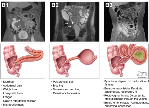
Clinical presentation varies depending on the segment affected and type of involvement. In Crohn’s disease, any segment of the GI tract, from the mouth to the anus, can be affected but most frequently disease is located in the terminal ileum and proximal colon. The inflammation in CD is typically asymmetric, segmental and transmural1. Three patterns of involvement can be seen: inflammatory, stricturing and fistulizing. The typical symptoms in CD are abdominal pain, chronic diarrhea and other features suggestive of IBD (eye, skin, joints complaints, perianal manifestations and family history of IBD). Weight loss, anorexia, fatigue and low-grade fever are frequently present, independent of disease location. In some patients, subclinical inflammation over the years, results in fibrotic strictures, and postprandial abdominal pain, distension and vomiting may be the presenting complaints. Due to the transmural nature of inflammation patients can present with abscesses, inflammatory masses, or fistulae to adjacent organs or skin (Figure 1). Perianal disease occurs in almost 1/3 of patients and may be the chief complaint3.
4
Figure 1 – Crohn´s disease classification based on disease behaviour. The figure depicts the three
types of behaviour of Crohn´s disease as per the Montreal classification represented in magnetic resonance enterography (MRE) and illustrated, with typical symptoms. Upper left MRE (B1) shows mural thickening and enhancement in the distal ileum in a patient with active CD (T1 weighted imaging with fat saturation after injection of gadolinium chelates). The middle MRE panel (B2) shows a narrowed luminal segment with thickened wall and upstream dilation suggesting the presence of a stricture (T2 weighted imaging). The right MRE panel (B3) shows multiple converging enhancing loops of small bowel suggestive of entero-enteric fistulae (arrows) (T1 weighted imaging with fat saturation after injection of gadolinium chelates); in the lower illustration, a deep and transmural fissure/ulcer leads to the formation of an abscess. Reproduced with permission from
Torres J et al. (2017) Crohn´s disease. The Lancet. doi: 10.1016/S0140-6736(16)31711-11.
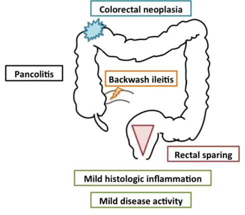
In UC, inflammation is usually restricted to the colon only, involving only the mucosa and sometimes the sub-mucosal layers; usually the inflammatory process starts in the anal canal and extends proximally. Patients may have disease limited to the rectum (proctitis), up to the splenic flexure of the colon (left-sided colitis) or beyond the splenic flexure (extensive colitis) (Figure 2)1. This classification has prognostic implications, since more extensive
5 forms of disease (left-sided colitis, pancolitis) are usually associated with higher need for steroids, hospitalization and surgery4, 5.
Figure 2 – Ulcerative colitis classification based on disease extent. The figure depicts the 3 types
of mucosal involvement usually seen in ulcerative colitis, as per the Montreal classification. This classification has prognostic and therapeutic implications, since proctitis is usually associated with a milder disease course, lower therapeutic requirements, less need for hospitalizations and surgery4, 5.
Reproduced with permission from Ungaro R et al. (2017) Ulcerative colitis. The Lancet. doi:
10.1016/S0140-6736(16)32126-26.
Occasionally mild inflammation can be seen in the terminal ileum, a process known as backwash ileitis. Typical symptoms in UC include rectal bleeding, bloody diarrhea, fecal urgency and incontinence, mucus discharge, and abdominal pain. Clinical presentation might vary based on disease extent. Patients with proctitis might predominantly have urgency and tenesmus (sensation of incomplete evacuation), while in pancolitis, bloody diarrhea and abdominal pain might be more prominent.
The diagnosis of IBD relies on a combination of clinical, laboratorial, endoscopic, histological, and radiological findings after exclusion of alternative diagnosis1. Endoscopy with biopsies is required to establish the diagnosis. Typical endoscopic findings in CD are ulcerations interspersed with areas of normal mucosa, cobblestoning, and rectal sparing. Focal, discontinuous and segmental chronic inflammation is the key pathological finding. Non-caseating granulomas can sometimes be found1, 7 . In UC, typical endoscopic findings
6
ulcerations. The typical histologic findings are distortion of crypt architecture, increased lymphocytes and plasma cells in the lamina propria, basal plasmocytosis, mucin depletion, and Paneth cell metaplasia67.
IBD is a multisystemic disease, and multiple other organs can be affected, including the bones, joints, mouth, skin, eyes, hepatobiliary system, lungs and kidneys8-10. The overall prevalence of these extra-intestinal manifestations (EIM) ranges from 20-50%, and they may present prior, in conjunction or following the diagnosis of the bowel disease8. Some EIM run in parallel with the bowel disease activity while other have an independent course. One of such EIMs is Primary Sclerosing Cholangitis (PSC), described for the first time in 196511. Patients with IBD and concomitant PSC have a distinct phenotype and disease course
(see below).
Epidemiology
IBD is a chronic disease that can affect all ages. There is no sex-specific distribution in adult IBD, and the onset of the disease usually occurs in the 2nd- 4th decade of life, with the highest incidence reported among 20 to 29 year-old individuals; a second and smaller peak has been described inconsistently between 50-60 years12. There has been a steady increase in IBD frequency in most regions of the world12. Usually there is a rise in the incidence of UC, followed by a rise in the number of new cases of CD12. The incidence and prevalence of IBD is highest in westernized nations, and in urban compared to rural areas. The highest reported incidence rates come from Canada (20.2/105), Northern Europe (10.6/105), New Zealand (16.5/105) and Australia (29.3/105)12. Prevalence rates are highest in Europe (322/105)9,
Canada (319/105)4 and United States (214/105)1, 13-16. Portugal is considered to be an
intermediate incidence and prevalence region, with a growing number of new cases16.
Etiology and pathophysiology Genetics and family history
Several lines of evidence link genetics to the risk of IBD. First, around 12% of patients have a family history of IBD, with the risk being higher if first-degree relatives or multiple family members are affected17, 18. Second, concordance rates in monozygotic twins range from 20– 50%, with higher concordance rates for CD than UC19. Third, certain ethnic groups are specifically affected- the risk of IBD is 3-4 times higher in the Ashkenazi Jewish19, while
7 African-American and Asian ancestry is associated with the lowest risk19. Finally, Genome-wide association studies (GWAS) have identified more than 200 risk alleles associated with IBD, of which 37 are specific for CD and 32 for UC20. Interestingly, 70% of the loci are shared with other diseases, such as type 1 diabetes, ankylosing spondylitis and psoriasis20. Furthermore, there is a striking overlap between loci for IBD and primary immunodeficiencies and mycobacterial infection. Among the identified genes, notable examples include mutations in genes associated with bacterial sensing and innate immunity (NOD2, ATG16L1, LRKK2, IRGM, Il23R, HLA, STAT3, JAK2 and Th17-pathway) and altered mucous layer (MUC2)21. However, only 13.1% of disease heritability is explained by genetic variation, highlighting the importance of epigenetic and other non-genetic environmental factors1.
Environmental factors
In the past years, areas of the world where IBD was previously considered to be very rare, have witnessed a sharp increase in their incidence rates, almost in parallel with the fast industrialization rates ongoing in these areas122. Changes in lifestyle, diet, better sanitation and Westernization” of lifestyles has been proposed as a potential explanation to the rise of allergic and immune-mediated disorders, including IBD over the past few decades (‘hygiene hypothesis’). However, breastfeeding, living in rural environments, contacting with animals in childhood, etc, have been only inconsistently identified as being “protective” for IBD1, 23,
24. Cigarette smoking is the best studied environmental factor; it is associated with twofold
increase in risk for developing CD (OR, 1.76; 95% CI 1.40-2.22), while it is protective from developing UC1, 6, 25-27. Appendectomy performed before the age of 20 years confers protection against ulcerative colitis28. Antibiotic exposure in early childhood, and during
pregnancy has been shown to increase the risk of IBD29, 30, again highlighting a putative role
for the microbiota in disease pathogenesis. Other medications potentially associated with higher risk of developing IBD include oral contraceptives, aspirin and nonsteroidal anti-inflammatory drugs, while statins have been linked with a decrease in the incidence of IBD, especially in the elderly31, 32. A reduction in dietary fiber and an increase in saturated fat intake have also been associated with increased risk33-35. Micronutrients such as zinc or iron, and Vitamin D have been proposed to potentially play a role. Causative association remains
8
to be proven for many environmental factors, as only association studies with major methodological limitations have been conducted.
Microbiota
The role and involvement of the gut microbiota in the pathogenesis of IBD has long been supported by many clinical observations, such as disease location in areas of highest bacterial population, the positive role of antibiotics for inducing remission and for preventing post-operative recurrence, and remission of disease upon diversion and recurrence upon re-anastomosis36. The advent of next-generation sequencing, allowed the characterization of an abnormal composition of commensals called “dysbiosis”. Generally, IBD is characterized by a decrease in species diversity, namely in Bacteroides and Firmicutes, and specifically in bacteria from the Clostridium cluster XIVa and IV, and an increase in Gammaproteobacteria37 and Actinobacteria38, 39. Faecalibacterium prausnitzii, a butyrate-producing commensal with anti-inflammatory properties, has been shown to be reduced in mucosal samples from patients with CD and UC38, 39. Approximately 1/3 of CD patients display in ileal biopsies increased numbers of mucosa-associated Escherichia coli, designated adherent-invasive E. coli (AIEC)30,31 . These E.coli strains are able to cross the mucus barrier, adhere and invade intestinal epithelial cells (IEC), and survive and replicate within macrophages, provoking the secretion of high amounts of TNF-α30,31. The potential causative role of the gut microbiota in disease pathogenesis, had led many investigators to seek a microbial-derived therapy. So far the experience with antibiotics and probiotics has been disappointing40, but the recently published results showing a promising role for fecal transplant in the setting of ulcerative colitis has again led to very active investigation in this area41.
Intestinal immune system in IBD
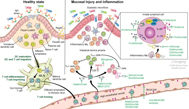
IBD is a complex disease with a complex ethiopathogenesis. It is commonly accepted that IBD results from the interplay between environmental factors, genetic susceptibility, and intestinal microflora resulting in an abnormal mucosal immune response and compromised epithelial barrier function. Multiple and overlapping pathways of the intestinal immune system are dysregulated in IBD (Figure 3).
The mucous barrier and epithelial barrier defects are strongly implicated in the pathogenesis of UC and CD. The epithelium establishes a “buffer zone” about 50 µm thick between the
9 luminal contents and itself42. Defects in the barrier function of the intestinal mucosa can prime increased microbial and antigen presentation and lead to immune activation. After triggering by antigen presentation, both innate and acquired immune responses are activated with subsequent loss of tolerance to enteric commensal bacteria. This results in sustained Th1 and/or Th17 responses and production of pro-inflammatory cytokines (Il1, Il2, Il6, INF-γ, TNF-α, Il17, etc.). TNF-α is one of the most relevant mediators in intestinal inflammation. Together with Il1 and Il6 it also contributes to symptoms such as fever, anorexia and weight loss. Disruption of this buffer zone by emulsifiers, ubiquitous in western diet43 or by mutations in MUC2 gene44 are associated with IBD. Further, epithelial cells are armed with an evolutionarily conserved process called autophagy, in which unwanted cytoplasmic contents are targeted to the lysosome for degradation, preventing the dissemination of invasive bacterial species. Notably, defects in autophagy-related genes such ATG16L1 and IRGM have been identified as further important risk factors for the development of CD45. Finally, defects in intestinal tight junctions, comprising members of the claudin and occludin families, are associated with IBD3. Many studies performed in patients with IBD have shown that intestinal barrier function is disrupted both in active and in quiescent disease states. Indeed, several studies have shown that increased intestinal permeability to inert tracer molecules such as PEG molecules or 51Cr-EDTA occurs before inflammation and can predict disease relapse in patients with CD46. Interestingly, increased permeability has also been documented in first-degree relatives of patients with CD, underlining possible shared genetic susceptibility to disease17.
An array of innate immune mechanisms coordinates to preserve mucosal function and integrity. The NOD-like receptors (NLRs) are one such class of innate immune proteins that mobilize host defense to intracellular fragments of bacterial peptidoglycan, by initiating nuclear factor !B (NF!B)-dependent and mitogen-activated protein kinase (MAPK)-dependent gene transcription, producing multiple protective cytokines. NOD2 was the first gene to be associated with CD47 and it remains a prominent genetic risk factor for CD. Dendritic cells (DCs), key antigen-presenting cells, are tolerogenic at steady state. However, under inflammatory conditions they transform into cells with inflammatory potential. In patients with IBD, intestinal DCs have enhanced expression of TLR2, TLR4, and co-stimulatory molecules, and they secrete increased pro-inflammatory cytokines compared to healthy controls. Intestinal macrophages, play essential housekeeping functions, such as the clearance of apoptotic or senescent cells and tissue remodeling at steady state48. Finally,
10
neutrophils choreograph the early response to microbial stimuli and likely modulate the adaptive responses beyond the acute state by the production of cytokines and reactive oxygen species (ROS). Adaptive immune cells have long been implicated in the pathogenesis of IBD. CD4+ T helper (TH) cells can be functionally compartmentalized into TH1, TH2, TREG,
TH17, TFH and TH9 cells49. Broadly, ulcerative colitis is a modified T-helper-2 (Th2) disease,
while Crohn’s disease is Th1 driven disease. Intestinal inflammatory infiltrate in CD contains both TH1 and TH17 cells. Such effector T cell responses to bacteria or fungi are
implicated in the pathogenesis of CD49. In addition, impaired functional activity of intestinal TREG has been reported in CD patients50. The colonic lamina propria cells from patients with
ulcerative colitis contains Th2-T cells that produce interleukin-5 (IL5) 49. Il4 and IL13 have
also been implicated in the pathogenesis of ulcerative colitis. IL13, produced by non-classical natural killer T cells is a key mediator of epithelial cytotoxicity and barrier dysfunction in ulcerative colitis49.
Figure 3 - Overview of the intestinal immune system in health and IBD. During the healthy state
(left side of the figure), the intestinal epithelium and IgA dimers work in concert to regulate and separate the luminal microflora from the mucosal immune system. During the healthy state, barrier function is maintained by the mucus layer and epithelial cells bound across tight junctions The intestinal epithelium also contains specialized cells such as Paneth cells (that produce antimicrobial peptides) and M cells (that sample luminal antigens). M cells are in close contact with antigen
11
presenting cells such as dendritic cells (DC). Contact with the antigen leads to DC maturation and antigen presentation to T and B cells. DCs default to inducing a “tolerizing” phenotype in the mucosa unless danger signals such as bacterial LPS induce the switch to an inflammatory/immunizing DC phenotype. Intestinal DCs also imprint T and B lymphocytes to express gut homing molecules α4β7 and CCR9. Lymphocytes thus imprinted within the GI tract enter the systemic circulation and upon reaching intestinal high endothelial venules, the gut-imprinted, α4β7-expressing lymphocytes engage locally expressed MAdCAM and egress the circulation to enter the intestinal lamina propria. The intestinal lamina propria has multiple families of T cells. These include TH1, TH17 and TREG subtype. At steady state TREG regulate the activity of TH1 and TH17 cells and prevent unchecked inflammation. During mucosal injury and inflammation such as seen in Crohn’s Disease (right side of the figure), the epithelial barrier is breached as a primary or secondary event and the luminal microflora stimulates a pro-inflammatory immune response by DCs and inflammatory macrophages (Mφ). The regulatory ability of TREG is outstripped by inflammatory activity of TH1 and TH17. Additionally, innate lymphoid cells (inset), homeostatic at steady state contribute to the cytokine production, perpetuating inflammation. Mucosal injury and damage is associated with dysbiosis, which perhaps perpetuates the inflammatory cascade. Increasing understanding of the mucosal immune system has led to an expanding array of therapeutic targets. Of these, TNF-α antagonists and homing inhibitors are currently in clinical practice while the others are in early to advanced stages of clinical development. DC= dendritic cell, MAdCAM=Mucosal addressin Cell Associated Molecule, IL=interleukin, TH= T helper cell, TREG= Regulatory T cell, IFN= interferon, Mφ= macrophage, TGF= transforming growth factor. Illustration by Jill Gregory. Reproduced with
permission from Torres J et al. (2017) Crohn´s disease. The Lancet. doi:
10.1016/S0140-6736(16)31711-11.
The management of IBD
The enormous advances in the knowledge about disease pathogenesis, resulted in the development of new targeted therapies directed at specific cellular processes, that led to major improvements in the care of patients. This resulted in a treatment paradigm change that evolved in from mere control of symptoms and improved quality of life towards attaining deep remission (absence of symptoms and endoscopic healing), with the goal of blocking the natural progression of disease and avoiding complications, bowel damage and disability5, 51. However, there is no cure for disease, and medical-free remission is not possible in most patients52.
12
The choice of medication depends on disease location, behavior, and severity and response to previous therapies, and considers prognostic factors that are known to be associated with higher probability of complicated disease41. The treatment of IBD involves an induction and maintenance regimen. The most widely used drugs in IBD currently are corticosteroids, 5-aminosalicilates, immunosuppressants (IM) [thiopurines (azathioprine, mercaptopurine), and methotrexate], biologics [(TNF (infliximab, adalimumab and golimumab), anti-adhesion molecules (vedolizumab) and anti-IL23 (ustekinumab)]1, 3, 6. Corticosteroids are potent anti-inflammatory drugs that are used to rapidly induce clinical remission; they should never be used as maintenance therapy, since they have multiple side-effects. Patients who are dependent on steroids to maintain clinical remission have indication to start steroid-sparing agents such as immunomodulators or biologic drugs1, 3, 6. The 5-aminosalicylates
drugs are not effective in the treatment of Crohn´s disease but are usually the first-line therapy for the treatment of mild UC (administered as suppositories, enemas, or oral formulations). Antibiotics use should be restricted to CD complicated by fistulas and/or abscesses. Biological drugs are usually reserved for patients with moderate-severe UC. There is an increasing tendency to consider early aggressive therapy (IM and/or anti-TNF) in the subgroup of patients with poor prognostic factors (perianal disease, extensive small bowel disease, young age at diagnosis, deep ulcers in endoscopy, penetrating disease) complicated and/or severe disease (top-down therapy)1, 3, 5, 6. Patients with refractory medical disease, who develop complications (abscesses or malignancy), and/or do not tolerate medical therapy are candidates for surgery1, 3, 6. The decision to advance for surgery should always be discussed in the context of a multidisciplinary team involving dedicated surgeons and gastroenterologists, and should include appropriate pre-operative imaging, patient counselling, optimization of nutritional status of the patient, and reduction in the thromboembolic risk1, 6.
PRIMARY SCLEROSING CHOLANGITIS
PSC is a chronic and progressive cholestatic disease, characterized by inflammation and fibrosis of the intrahepatic and/or extrahepatic ducts53, that may result in liver cirrhosis and
eventually end-stage liver disease53. Orthotopic liver transplantation (OLT) is the only
13 respectively53. Without OLT, half of symptomatic patients die within 12-15 years. In Western countries, the reported incidence of PSC is 0.07-1.3 per 100.000/year, and the prevalence is 8.5-13.6 per 10.00054,55. There is no epidemiological data available in Portugal for PSC, but it is generally considered a very rare disease.
Having a diagnosis of IBD is the strongest risk factor for PSC development, since 70% of patients with PSC have underlying IBD, most frequently ulcerative colitis (UC) in over 56-72%% of cases56, 57. Conversely, in patients with known IBD, PSC is found much less commonly, occurring in about 2-8% of UC patients and 3% of Crohn’s disease (CD) cases58.
Although there may be a possible common pathogenesis between PSC and IBD, the two disorders can occur at different times. PSC may be diagnosed many years after proctocolectomy for colitis, and conversely IBD can appear many years after the initial diagnosis of PSC or even after OLT altogether59. In most reports, IBD diagnosis precedes the diagnosis of PSC60, 61. In patients with known IBD, the presence of persistent of unexplained cholestasis obliges one to exclude concurrent PSC through magnetic resonance cholangiopancreatography (MRCP) or endoscopic retrograde cholangiopancreatography (ERCP). When PSC is diagnosed first, half of the cases have only abnormal laboratory tests; the typical diagnostic hallmarks of fever, itching, and jaundice are rarely seen nowadays53. Symptomatic patients usually present with fatigue and pruritus and can also exhibit jaundice, hepato-splenomegaly or scratching injuries. Recurrent episodes of bacterial cholangitis with fevers, chills, right upper quadrant pain and jaundice can also be a part of the clinical presentation, and usually develops in about 10–15% of patients during the course of their disease62. The diagnosis of PSC is based on the findings of diffuse multifocal strictures and dilations in the intrahepatic and/or extrahepatic biliary tree53 (Figure 4).
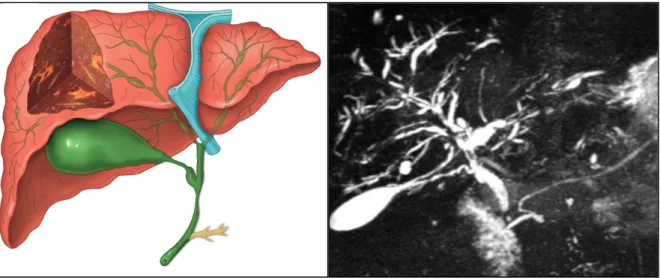
14
Figure 4 – Typical findings of Primary Sclerosing Cholangitis. Diagram (left) and magnetic
resonance cholangiopancreatography imaging (right) showing the typical findings of primary sclerosing cholangitis. Several strictures with intervening saccular dilatations of both the intrahepatic and the extrahepatic bile are seen conferring a beading aspect to the intra-hepatic biliary tree. Diagram in the left reproduced with permission from Hirschfield G et al. (2013) Lancet. Primary
Sclerosing Cholangitis. doi: 10.1016/S0140-6736(13)60096-363. Cholangiogram-MRI image
courtesy of Afonso Gonçalves, MD
Patients with a confirmed diagnosis of PSC should undergo colonoscopy with biopsies to exclude concomitant IBD or any malignancy53, even if they report no gastrointestinal symptoms. As most of PSC-IBD patients have mild colonic disease activity and even possible normal endoscopic appearances, histological sampling is crucial to avoid underdiagnosis58. Although no evidence-based guidelines are available, if the index colonoscopy is negative for IBD, a repeat colonoscopy every 3-5 years should be performed to monitor for possible onset of IBD64.
The pathogenesis of PSC-IBD – what is known
PSC is likely to have an underlying multifactorial etiology, with a predominant immune-mediated process53, 65. PSC and IBD are interrelated conditions that may share an underlying predisposition. Both diseases share common antibodies, such as those directed against cytoplasmic and nuclear antigens of neutrophils with a characteristic perinuclear staining pattern (p-ANCA). The p-ANCA antibodies have been found in 26–85% of PSC patients
15 and in up to 68% of patients with UC53. The available evidence points towards a complex interaction between genetic, immunologic and environmental factors (Figure 5).
Figure 5 - Possible hypothesis linking PSC and IBD pathogenesis, including genetic
predisposition, immune-mediated processes, altered gut microbiota and altered bile acids (BA) metabolism. Reproduced with permission from Palmela C. et al (2017). Gut Liver. Inflammatory
Bowel Disease and Primary Sclerosing Cholangitis: A Review of the Phenotype and Associated
Specific Features. doi: 10.5009/gnl16510 65.
Genetics
From a genetic standpoint, there is increasing evidence that PSC is distinct from UC and CD. Large-scale genome-wide association studies (GWAS) have identified close to 200 independent loci associated with IBD20, 65, 66. Most of these loci are shared between UC and
CD20. Genome-wide association studies (GWAS) have allowed for the identification of 23
genetic risk loci associated with PSC.67 Most of these genetic risk loci play an important role
in the immune system, such as the HLA complex, IL2, or PRDX5, suggesting that PSC may be an immune-mediated disorder. Furthermore, there is some overlap between some genetic risk loci of PSC and IBD. However, the known genetic defects only explain less than 10%
PSC$IBD
Genetic.predisposition. ! GWAS%studies%have%identified%more%than%200%IBD% and%23%PSC%susceptibility%loci% ! However,%there%is%limited%genetic%overlap% between%PSC%and%IBD Gut.lymphocyte.homing hypothesis ! Presence%of%shared%chemokines%and%adhesion% molecules%by%the%liver%and%gut ! Activated%lymphocytes%from%the%inflamed%gut% enter%the%enterohepatic%circulation%and%cause% hepatic%inflammation The.“leaky.gut”.hypothesis ! Increased%intestinal%permeability%and% translocation%of%bacterial%metabolites%from% the%inflamed%gut%to%the%liver ! Microbiome%dysbiosis%may%contribute%to% biliary%injury Bile.acids$microbiome.interaction ! Possible%altered%BA%excretion%in%the%colon% due%to%cholestasis ! Impaired%microbiota%enzymatic%activity may% be%associated%with%BA%dysmetabolism16
of PSC disease predisposition68, pinpointing the possible importance of the environment in the pathogenesis of the disease. Genetic predisposition to autoimmune bile duct injury triggered by toxic or infectious agents that may gain access through the diseased colon is potentially a major mechanism leading to PSC in IBD patients65.
Gut microbiome
Several lines of evidence support the involvement of gut microflora in PSC´s pathogenesis. Bacteria and fungi are more frequently found in the bile ducts of patients with PSC, as compared to patients with other cholestatic liver diseases69, 70. Several reports and case series of pediatric patients with PSC treated with vancomycin with positive results further suggest a role for the gut microbiome, and have paved the way for the trial of antibiotics in PSC71.
Indeed, in a pilot small randomized controlled trial where patients were allocated to vancomycin high or low doses, or metronidazole, high or low doses, it was shown that patients receiving vancomycin reached the primary endpoint of decrease in alkaline phosphatase at 12 weeks72. Moreover, addition of metronidazole to ursodeoxycholic acid (UDCA) showed some beneficial effects in biochemical test results and liver histology73. The “leaky gut” hypothesis suggests that increased intestinal permeability may lead to translocation of bacterial metabolites from the gut74 (Figure 5). The liver receives approximately 75% of its blood supply from the splanchnic circulation and is constantly exposed to both beneficial and noxious molecules from the intestinal microbiome75. This so-called ‘gut-liver axis' is essential for the maintenance of health but may also play an important role in pathogenesis of liver and intestinal diseases75,76. This dysbiosis may be associated with mucosal immunity dysregulation by modulating intestinal permeability and altering homing of gut-specific lymphocytes77. Recently, evidence for an etiologic role of the intestinal microbiome in PSC has been provided by animal model studies. Multidrug resistance gene 2 knockout (Mdr2-/-) mice, a murine model for PSC, exhibited a more severe phenotype when maintained under germ-free conditions.78 However, NOD.c3c4 mice, a murine model for biliary inflammation, exhibits a less severe phenotype when maintained in germ-free conditions.79 These contradictory findings probably result from the different murine models used and the different intestinal microbiota of these mice, highlighting the complex interaction between the intestinal microbiota and the liver.
The interaction between microbiota and bile acid (BA) metabolism may also play an important role in the PSC-IBD phenotype. It is well known that there is a reciprocal relation
17 between the microbiome and BA pool. Reduced BA in the gut (such as in situations of obstructive cholestasis like PSC) leads to bacterial overgrowth and inflammation. In the other way around, BA metabolism is a property of the gut bacteria80.
Recent evidence supports the existence of BA (dys)metabolism in IBD patients due to impaired microbiota enzymatic activity81. One of the contributing factors for the difference in phenotype between PSC-IBD patients and IBD controls could potentially be altered concentration and/or composition of colonic bile acids impacting on gut microbiota and stool BA metabolism.
Gut lymphocyte homing
Activated T lymphocytes from the inflamed and permeable gut may enter the enterohepatic circulation and persist as memory cells that cause hepatic inflammation82, 83. Some molecular
features, such as chemokines and adhesion molecules, are shared by the liver and intestine and could contribute to lymphocyte binding at both sites82. T cells activated in the gut during active inflammatory bowel disease could differentiate into effector cells with the ability to bind to both hepatic and mucosal endothelium. The activation and expansion of these memory cells in the liver could eventually lead to the induction of MAdCAM-1 and CCL25 in the liver, promoting the recruitment of CCR9+ α4β7+ mucosal T cells and the development of inflammation84. The enterohepatic circulation of lymphocytes may explain the interaction between the colonic immune system triggered by dysbiotic intestinal microbiota and biliary inflammation. This theory is further supported by the finding of memory T-cells with common clonal origin in both the gut and the liver of patients with PSC-IBD.85
Findings such as PSC development after colectomy for IBD, or the development of IBD after OLT for PSC, have led some investigators to suggest that aberrant homing of lymphocytes between the intestine and liver could be involved in the pathogenesis of the PSC-IBD phenotype82.
Environment
Very little is known about the impact of environment in the pathogenesis of PSC. Smoking has been repeatedly associated with a lower risk for developing PSC, independently of the protective effect of smoking in UC.86-88 Coffee consumption also seems to be associated with


![Figure 8 - Illustration of the role of FXR in entero-hepatic circulation. Primary Bile acids [chenodeoxycholic acid (CDCA) and cholic acid (CA)] are produced in the liver by the conversion of cholesterol into primary bile acids, a re](https://thumb-eu.123doks.com/thumbv2/123dok_br/15247487.1023798/59.892.155.762.347.875/figure-illustration-circulation-primary-chenodeoxycholic-produced-conversion-cholesterol.webp)