FABIANNE
DE ARAÚJO RIBEIRO
EFEITOS COMBINADOS DE QUÍMICOS E RADIAÇÃO
ULTRAVIOLETA EM Daphnia magna
COMBINED EFFECTS OF CHEMICALS AND
ULTRAVIOLET RADIATION ON Daphnia magna
Dissertação apresentada à Universidade de Aveiro para cumprimento dos requisitos necessários à obtenção do grau de Mestre em Ecologia,
Biodiversidade e Gestão de Ecossistemas realizada sob a orientação científica da doutora Susana Patrícia Mendes Loureiro, investigadora auxiliar CESAM, Universidade de Aveiro e co-orientação do Prof. Doutor Amadeu Soares, Professor catedrático do Departamento de Biologia da Universidade de Aveiro
Prof. Dra. Lúcia Maria Candeias Guilhermino
Professora Catedrática da Universidade do Porto, Instituto de Ciências Biomédicas Abel Salazar
Prof. Dr. Amadeu Mortágua Velho da Maia Soares
Professor Catedrático do Departamento de Biologia da Universidade de Aveiro
Dra. Susana Patrícia Mendes Loureiro
Investigadora auxiliar, Centro de Estudos do Ambiente e do Mar, Departamento de Biologia, Universidade de Aveiro
todos os processos que me trouxeram até aqui,
Aos meus avós, pelo amor e carinho incondicionais que me dedicaram em vida, por me proporcionarem uma formação com base em respeito e dignidade, e pelos exemplos que me foram transmitidos!
Aos meus tios Luciene e Eduardo, pela adoção aqui da sua quarta filha, pelo respeito e principalmente apoio às minhas escolhas. Sem a presença e apoio constante de vocês dois em tudo, eu não teria chegado até aqui.
Ao professor Amadeu Soares, pelas oportunidades que me tem concedido, pelo apoio, e principalmente pelo seu voto de confiança.
A Susana Loureiro, pela orientação, que ao longo deste ano foi marcada por uma paciência admirável, atenção e disponibilidade sempre constantes, e pelos ensinamentos.
À minha família brasileira em Portugal: Nessa e Valdir, Rhaulzinho, Dani, Carol, Jamilica, Darlan, Talita, Ísis e Negão, obrigada pela companhia nos vários finos e empalhadas da vida, pelo ombro, pelas broncas de mãe (é, pra Nessa e Ísis).
À Salomé, Carla, Marta, Maria, Joanne e Sandrina, pelo agradável convívio diário, pela recepção, pelas milhões de dicas e ajudas desde programas de estatística a preenchimento de envelope de correio… e à mé em especial pela formatação da tese.
Às minhas queridas irmãs, Luna, Carla, Rosa, Laura, Carol e Lara, pelos momentos inesquecíveis que foram e são partilhados
À Fátima, ao Abel e ao Pedro Serra pelas produtivas discussões em laboratório sobre o trabalho, e eventuais ajudas.
Ao Gonçalo pela ajuda com as reservas energéticas
Aos professores do curso de Biologia da UFT, pela contribuição na minha formação profissional.
E ao marquinhos, por ter gentilmente cedido esta frase: A todos os que sabem que merecem o meu agradecimento!
actividade humana e dos processos de alteração climática. Os pesticidas são geralmente usados em práticas agrícolas para controlar doenças em vegetais e o aparecimento de pragas, e podem ser levados do solo para os sistemas aquáticos adjacentes aos locais de aplicação, onde representam um factor de stress para os organismos não-alvo. Além das exposições a químicos, o ambiente está sofrendo as consequências dos processos de alterações climáticas. Uma destas consequências é o aumento da radiação ultravioleta que chega à superfície terrestre devido à diminuição da concentração de ozono na estratosfera. O presente trabalho teve como objectivo principal elucidar alguns padrões e comportamentos biológicos relativamente a mudanças no ambiente. Para isto, com o intuito de prever as interacções entre stressores naturais e químicos, a radiação ultravioleta (RUV) e o fungicida carbendazim foram escolhidos como fontes de stressores natural e químico, respectivamente, e foram aplicados em combinação, como um exemplo das possíveis condições adversas que podem ser encontradas no ambiente. Os efeitos isolados da radiação ultravioleta em Daphnia magna foram avaliados através da utilização de uma lâmpada artificial de RUV, à qual os organismos foram expostos por um período máximo de 5 horas. Os experimentos de combinação entre RUV e carbendazim foram conduzidos com exposição constante ao químico, e uma única dose de radiação ultravioleta. Os parâmetros analisados foram sobrevivência, actividade alimentar, reservas energéticas e produção de juvenis de Daphnia magna. Para prever os efeitos das combinações, um dos modelos utilizados na análise de misturas de químicos e combinação de químicos com stressores naturais foi o utilizado. O modelo da Acção independente (AI) assume que ambos os componentes da combinação têm diferentes modos de acção, e actuam de forma independente sobre o organismo. Os efeitos são avaliados de acordo com as probabilidades de não-resposta do organismo a ambos os componentes da combinação. Há ainda outras formas de interacção entre os componentes da combinação que podem produzir um efeito mais severo (sinergismo) ou menos severo (antagonismo); os efeitos podem ser também dependentes do nível da dose aplicada ou do rácio entre os dois componentes da combinação Os resultados da exposição de Daphnia magna à radiação ultravioleta somente demonstraram um decréscimo na sobrevivência, na actividade alimentar e na produção de juvenis, com valores de dose-efeito muito próximos para todos os parâmetros, o que pode ser explicado pela diferença da sensibilidade deste organismo à radiação, de acordo com a idade em que são expostos. Os resultados das combinações entre carbendazim e RUV para o parâmetro sobrevivência foram bem ajustados ao modelo da acção independente, e não demonstraram nenhum desvio. Para a reprodução e a actividade alimentar, houve um desvio dependente do rácio entre os componentes, que demonstrou maior toxicidade para o carbendazim quando a radiação ultravioleta era o item dominante na combinação. Este estudo mostra a importância da avaliação de combinações entre químicos e stressores naturais. Neste caso, espera-se que
abstract The natural environment and wildlife are often exposed to several chemicals, physical and biological stressors originated from human activities and climate changes. Pesticides are often used to control plant disease and pest in agricultural practices, and can runoff from the soil to adjacent aquatic systems, where it represents a stress factor for non-target organisms. In addition to chemical exposures, the natural environment is suffering from climate change processes. One of the consequences of that is the increasing amount of ultraviolet radiation reaching the earth’s surface due to depletion on stratospheric ozone. The present work aimed to elucidate some biological behaviors and patterns regarding changes in the environment. For that, to predict interactions between natural stressors and toxicants to Daphnia magna, the ultraviolet radiation (UVR) and the pesticide carbendazim were chosen as the source of natural and chemical stressors, respectively and were employed in combination with each other as an example of possible stress conditions that can be found worldwide in the environment. Single effects of ultraviolet radiation on Daphnia magna were assessed using an artificial UV source, by exposing the organisms to UV and visible light simultaneously, to a maximum period of 5 hours. Combined experiments of carbendazim and ultraviolet radiation were conducted with a constant chemical exposure and a single UVR dose. The parameters analyzed were survival, feeding activity, energy budget and offspring production of Daphnia magna. To predict effects of combined exposures, one of the reference models used for analysis of mixture toxicity and combination of chemical and natural stressor was applied. The Independent Action (IA) model assumes that both components of the combination have different modes of action, and act independently from each other; the effects of the combinations are based on the probabilities of non-response of the organism to both stressors. There are some deviations from the independent action model which can cause a more severe effect (synergism), or a less severe effect (antagonism); they might be also dose-level dependent or dose-ratio dependent. Results from single exposure of Daphnia magna to ultraviolet radiation showed a decrease in survival, feeding activity and offspring production, with similar dose-effect values, due to differences in the sensibility of the organism to UVR according to their age. Combined exposures of carbendazim and UVR for survival endpoint fitted to the IA model, showing no deviation patterns, while the response of reproduction and feeding activity were dose-ratio dependent, indicating a higher toxicity of carbendazim when ultraviolet radiation was the dominant item in the combination. This study shows the importance of evaluate the combined effects of chemicals and
Chapter I General Introduction and objectives
General Introduction
3Ultraviolet Radiation
4UV effects on aquatic organisms and counteracting strategies
5Phytoplankton and macrophytes
5Zooplankton
6Daphnia magna as a model organism for ecotoxicology tests
8Chemical compounds
9Carbendazim
9Predictions of joint effects of natural stressor and chemical
compounds 10
Objectives
12References
13Chapter II Effects of Ultraviolet Radiation single exposures to life
parameters of Daphnia magna
Abstract
19Introduction
20Material and methods
21UV-R experiments
22Immobilisation tests
22Reproduction tests
23Feeding inhibition tests
23Energy Budget
24Statistical Approach
25Results
25Spectral composition of exposures
25Acute and chronic bioassays
26Energy reserves
29Discussion
30References
34Chapter III Toxicity predictions for combined exposures of Carbendazim
and ultraviolet radiation to Daphnia magna
Abstract
39Introduction
40Material and methods
42Test-organisms
42Single toxicity end points
42Reproduction
42Combination of Carbendazim and UVR
43Immobilisation test
43Feeding inhibition test
43Reproduction test
45Data analysis
46Results
47Chemical analysis
47Chronic Single toxicity tests
48Binary combinations of carbendazim and UV-Radiation
49Discussion
52Single exposures
52Combined exposures
52Acknowledgments
55References
55Chapter IV General discussion and conclusions
Final remarks and conclusions
63at a distance of 30cm.
II. Fig.2 - Effects of a pre-exposure to UV-radiation on the feeding rates of Daphnia magna. The feeding rates are related to the 24h post-exposure period. Data is expressed as
mean values± standard errors. Asterisks indicate significant difference from the control (p<0.05)
II. Fig.3 - Number of neonates produced by Daphnia magna after UV radiation exposure.
Data is shown as mean values± standard errors. Asterisks indicate significant differences from the control (p<0.05)
II. Fig.4 – Body length of 21d old Daphnia magna pre-exposed to UV radiation. Data is
shown as mean values± standard errors. Asterisks indicate significant differences from the control. (p<0.05)
II. Fig.5 - Number of neonates produced per Daphnia magna in each brood released after
exposure to ultraviolet radiation. Data is presented as the mean value ±standard error.
II. Fig.6 - Total sugar and protein contents of Daphnia magna measured after the release
of first-brood upon a pre UV-R exposure. Data is shown as mean values ± standard errors, where black bars stand for sugar content and gray bars for protein contents.*represents significant differences from control. (p<0.05)
II. Fig. 7 - Total lipids in Daphnia magna after release of first brood upon a pre UV-R
exposure. Data is shown as mean values ± standard errors. *indicates significant differences from the control (p<0.05)
III. Fig.1 - Number of neonates cumulative produced by Daphnia magna exposed to
carbendazim for 21 days. Data is shown as mean values ±standard errors. Asterisks indicates significant differences from control (p<0.05)
III. Fig.2 - Number of aborted eggs produced by Daphnia magna during exposure to
carbendazim after 21 days. Data is shown as mean values ±standard errors. Asterisks indicates significant differences from control (p<0.05)
III. Fig. 3 – Body length of 21 d old Daphnia magna exposed to carbendazim. Data is
shown as mean values± standard errors. Asterisks indicate significant differences from the control (p<0.05).
III. Fig.5 - Dose-response pattern for combination of carbendazim and ultraviolet radiation
to the feeding activity of Daphnia magna, showing a dose-ratio deviation from the IA model. a represents the 3-D mesh, and b the isobols surfaces.
Fig.6 - Dose-response pattern for combination of carbendazim and ultraviolet radiation to
the reproduction output of Daphnia magna, showing a dose-ratio deviation from the IA model. a represents the 3-D mesh, and b the isobols surfaces.
List of tables
II.
Table 1. Values for UV radiation intensities, presented as kJ.m-2, applied inDaphniamagna exposure experiments. kJ.m-2
= mean value (mW.m-2. m-2.nm-1) from 280 to 320nm x 40J.m-2was obtained multiplying the intensity (mW.m-2.nm-1) for the time of exposure in seconds.
III. Table 1. Values obtained from single stressor exposures of ultraviolet radiation and
carbendazim at different endpoints, and respectively bibliography.
III. Table 2. Values for UV radiation intensities, presented as kJ.m-2, applied in Daphnia
magna exposure experiments. kJ.m-2
= mean value (mW.m-2. m-2.nm-1) from 280 to 320nm x 40. Values in J.m-2 were obtained multiplying the intensity (mW.m-2.nm-1) for the time of exposure in seconds.
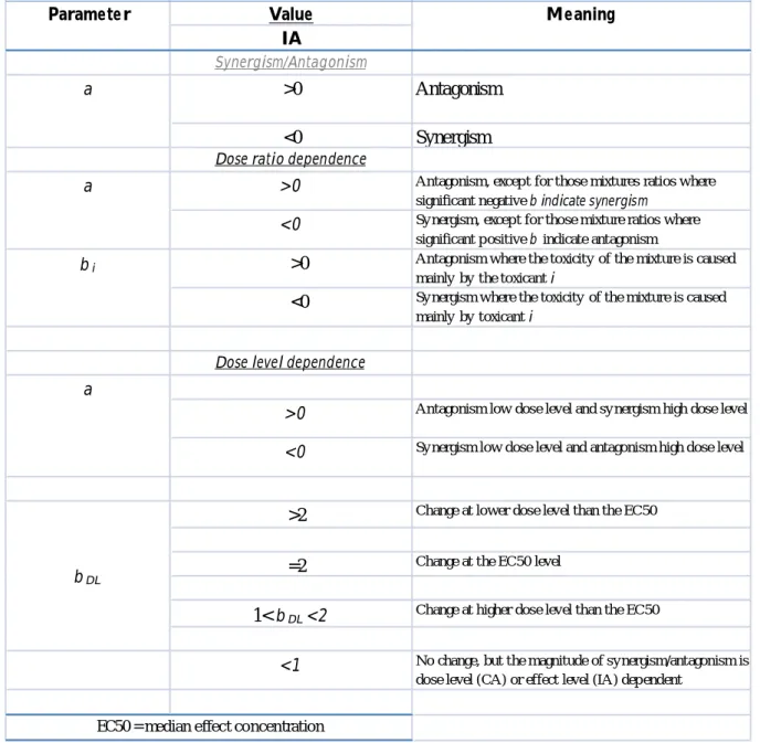
III. Table 3. Interpretation of additional parameters substituted into the independent action
model (IA) reference model that define the functional form of the deviation patter
III. Table 4. 24-h and 4-h post-exposure feeding inhibition EC50obtained for carbendazim
at different UVR intensities
III. Table 5. 48-h LC50obtained from nominal concentrations of carbendazim at different
UVR intensities
III. Table 6. 21-days EC50 Values obtained from nominal concentrations of carbendazim
_________________________________
General Introduction and Objectives
I. General introduction
The natural environment and wildlife are often exposed to several chemicals, physical and biological stressors originated from human activities and climate changes. One of the anthropogenic activities that have a high impact on ecosystems is agricultural practices, because of the large use of pesticides in order to control plant diseases and pest. These chemicals are designed according to their target organism, and exist in a variety of types and classes. Pesticides, once applied in the soil, can runoff to adjacent aquatic systems, potentially representing a risk for non-target organisms (e.g. algae, aquatic invertebrates and fish). The fate of pesticides in the environment is linked with some natural properties, like precipitation levels (high rate of precipitation can lead to more runoff of chemicals from the soil), temperature, dissolved oxygen levels and ultraviolet radiation income, which are closely related to chemical degradation, bioavailability and volatility. (Noyes et al. 2009). Climate change phenomenon is leading to alterations of precipitation levels, increases of temperature and decrease of stratospheric ozone concentration, which acts as a filter to the shortest ultraviolet rays from the sunlight. As a consequence, the environment and wildlife is now experiencing a combination of several natural stressors, in addition to the already mentioned chemical exposure.
Environment risk assessment (ERA) usually evaluates effects of single stress exposures, and environment management regulations have been established based on that. Nowadays, there is an increase and urgent need to evaluate the combined exposures of stressors to obtain a more realistic prediction of environment risk.
In this work, a situation where the combination between a pesticide and increasing levels of ultraviolet radiation input to the water system was carried out in laboratory conditions, with the purpose to analyse the biological response of organisms to both stress sources.
Ultraviolet Radiation
Ultraviolet Radiation (UVR) is composed by wavelengths below 400nm, between X-rays and visible radiation, and divided into UV-A, or long wave (from 320nm to 400nm), UV-B, or medium wave (from 320 nm to 280) and UV-C, short wave or germicidal (from 280nm to 100nm). From these wavelengths, the UV-A and UV-B ranges can reach the earth’s surface, and the UV-C is completely filtered by ozone layer present in the atmosphere (Madronich et al. 1998). The germicidal denomination for UV-C wavelengths is usually employed due to its action on microorganisms; this wave-range has the shortest and thus more energetic rays capable to damage and/or destroy microorganism’s cells and DNA, and stop their reproductive ability.
The amount of UVR that reaches the earth’s surface is determined by a series of factors interacting with the radiation as it passes through the atmosphere. These include the state of the atmosphere, position on the earth (latitude and altitude) and season (relative position of the sun to location on Earth) (Blumthaler and Webb 2003). Besides that, when reaching aquatic and/or terrestrial ecosystems, the UVR light is also modified by natural factors. For instance, in aquatic environments there are several natural features to which the solar radiation input can interact with. Dissolved organic carbon (DOC) and various humic substances present in ocean and lakes contribute tremendously for the penetration of UV radiation in the water column by absorbing the shortest UV wavelengths and blocking the amount of harmful radiation coming to the organisms. DOC is degraded by a slowly process in water, but it can be broken down by solar UVR and became more available for the bacterioplankton’s metabolism (Moran et al. 2000) which leads to an increase in lake’s UVR transparency (Madronich, McKenzie et al. 1998).
Dissolved organic matter (DOM) is the designation for the carbon-containing compounds derived from the decomposition of dead organisms that are present in aquatic systems. The chromophoric DOM (cDOM) absorbs the radiation that incomes aquatic ecosystems in the UV and blue ranges of the solar spectrum. The presence of cDOM gives to natural waters a yellow-brown colour. The fate of UV-R in lake system as been demonstrated to be dependent on the amount of DOM present in natural waters due to the attenuation of light penetration, or to effects on phytoplankton photosynthesis, which ultimately change the
amount of DOM (Häder et al. 2007). Williamson et al. (2009) has argued that the quantity and quality of DOC in inland waters is much more likely to control UV transparency than the ozone itself, meaning that although environmental policies for healing the ozone layer are showing beneficial results, other climate change factors can interfere with harmfulness of UV radiation income, mainly changes in DOC concentration that leads to more UV transparent waters.
UV-R can cause indirect effects on freshwater nutrient cycles by breaking down the DOC into dissolved inorganic carbon (DIC). When exposing the macrophyte Vallisneria
gigantean to UV-B levels, Anusha (2008) observed that at the end of ten weeks, the
amounts of DOC and DIC had a significant decrease and increase, respectively. The transformation of DOC into DIC by UV-R increases the microbial population due to the availability of inorganic carbon substrates and increasing bacterial population are likely to decompose organic carbon under environmental sunlight (McCallister et al. 2005). In addition to decrease of stratospheric ozone concentration that leads to increase of UVR input, climate change events can also cause severe situations of drought or flood, depending on precipitation levels alterations (Whetton et al. 1993; Le Houérou 1996) Drought reduces the export of terrestrially-derived CDOM to aquatic ecosystems and may also influence water export from wetlands that are important determinant of CDOM inputs to other downstream aquatic ecosystems (Williamson et al. 2003). Flood can increase the rate at which anthropogenic-introduced compounds in agricultural areas are leachate or run off to adjacent water bodies.
UV effects on aquatic organisms and counteracting strategies
Phytoplankton and macrophytes
Phytoplankton represents the major primary producers in oceans, being the basis of food webs. The sensibility of phytoplankton to ultraviolet radiation is reported to ecosystems from tropical to polar regions (Helbling et al. 1994). Phytoplankton are potentially subject to harmful UV-B radiation which can cause DNA damage, inhibited photosynthesis and growth, and finally, cell death (Klisch et al. 2001). UVR is likely to alter photosynthesis through photo-inhibition, at relatively high doses; some studies have
shown that UV-A causes more photosynthetic inhibition rather than UV-B because their natural levels are higher. UVR potentially impair the performance of the three main photosynthesis component processes: photophosphorylation reactions, on the thylakoid membrane, the CO2 fixation reactions of the Calvin cycle and stomatal control of CO2 supply (Allen et al. 1998).
This inhibition is highly variable, depending on the irradiance/dose received by the cells, the sensibility of the cell and other environmental factors that can mask the observed effects (Villafaiie et al, 2003). The mechanism in which UVR impairs photosynthesis is linked with the bleaching of photosynthetic pigments. Freshwater phytoplankton are likely to be more inhibited by solar UV than marine phytoplankton (Häder, Kumar et al. 2007). Cyanobacteria, as well as phytoplankton and macroalgae can synthesize ultraviolet absorbing/screening compounds that can absorb the radiation before it can reach intracellular targets. One of the most common compounds is mycosporine-like amino acids (MAAs), which has a maximum absorption range from 310 to 360nm. (Klisch, Sinha et al. 2001).
Artificial UV-B was shown to induce the activity of nitrate reductase in cyanobacteria; for these organisms, the primary photosynthetic reactions and CO2uptake is affected by UV-B (Häder, Kumar et al. 2007).
Macrophyte species play an important role on nutrient cycling in freshwater systems. One of the consequences related to the inhibition of photosynthesis caused by UVR is the decrease of dissolved oxygen concentrations in shallow freshwater type systems (Anusha and Asaeda 2008).
Zooplankton
Zooplankton species have a major role in aquatic food chains, and alterations in any level (individual, population and community) can lead to changes in aquatic systems functionality. For instance, the decrease in filter-feeding species can generate accumulation algae cells in lakes during the late spring and early summer seasons; moreover, changes on zooplankton community represents depletion on food source for planktivores fishes (Confer et al. 1978). For that reason, zooplankton are widely used in ecotoxicology testing with the objective of predict environmental risk. As climate changes introduce alterations
in environmental components and compartments, zooplankton species as been used for the assessment of natural stressors effects in freshwater environments, mainly the role of increasing ultraviolet penetration through water column (Rhode et al. 2001; Alonso et al. 2004; Marinone et al. 2006). In earlier researches, the vertical migration of zooplankton was believed to happen only due to visual predation avoidance (Zaret and Suffern 1976). Nowadays, the UVR exposure is assumed to be an important factor that guides the vertical migration of zooplankton into deeper waters (Boeing et al. 2004; Fischer et al. 2006b). In daphnia species case, UV-tolerance is linked with pigmentation; melanin pigments as well as carotenoids increase their tolerance to UVR (Herbert and Emery 1990). Rhode and
co-workers (2001) observed a deeper distribution of Daphnia that presented low
pigmentation, under both laboratory and natural sunlight exposures. In order to minimize and balance metabolic costs of deeper cold waters, a less pronounced downward migration is predict for zooplankton with higher UV-R tolerance (Rhode, Pawlowski et al. 2001). Moreover, for the high-pigmentedDaphnia living in low DOC lakes, there is a conflicting factor of visual predation by fish (Confer, Howick et al. 1978).
The main target of UVR in organisms is the DNA molecule (Teoule 1987; Huot et al. 2000). UV-B can cause dimerization of DNA bases, leading to the formation of cyclobutane pyrimidine dimmers (CPDs) and 6-4 pyrimidine photoproducts (PPs). These photoproducts block DNA transcription and replication as only a single distortion of DNA may be sufficient to stop DNA replication (Buma et al., 2003). The molecular repair of these damages is strictly dependent on UV-A and visible light. In general, there are two types of DNA repair described for organisms: nucleotide-excision repair (NER) and photoenzymatic repair (PER). NER is a complex and multi-enzimatic process, which depends on energy provided by ATP. Therefore, it is a metabolic cost process to the cell (de Laat et al. 1999). PER is performed by one single enzyme, the photolyase and requires energy from visible light and UV-A range (Carell et al. 2001). PER is specific for repair damage caused by ultraviolet light, and it is found in many diverse species, such as zooplankton, fishes and bacterioplankton (Huot, Jeffrey et al. 2000; Gonçalves et al. 2002; Dong et al. 2008). Considering the enzymatic nature of DNA photo-repair, temperature has a crucial role in this process as in all other enzymatic mechanisms. MacFadyen (2004) observed that ectotherms that depend on temperature-dependent enzyme processes might
be less able to repair DNA damages at low temperatures, and argued that low temperatures are likely to favor photoprotection rather than photorepair.
Zooplankton also shows impairments in reproduction caused by UVR exposure as a consequence of metabolic alterations. Karanas et al (1981) observed that when the copepod
Acartia clausii survived to UV radiation exposure, its ability to reproduce was impaired.
Another study showed differences in survival and reproductive output ofDaphnia magna aging from 1 to 4 days during the post-exposure period to UVR (Huebner et al. 2006). Lacuna and Uye (2000) also described a marked reduction on gut content and egg production of Sinocalanus tenellus females that were exposed to high sub-lethal doses of
UV-B. Changes in respiration rates of Daphnia catawba pre exposed to ultraviolet
radiation were related to metabolic costs of DNA repair as described by Fischer et al (2006a) Another consequences aroused from zooplankton exposure to UVR is the formation of reactive oxygen species (ROS) causing oxidative stress. Vega and Pizarro (2000) observed an increase on catalase activity in Daphnia longispina exposed to UV-B radiation, probably as a strategy to avoid oxidative stress. Oxy-radicals might lead to tissue damages through lipid peroxidation, eventually causing impairments of vital cellular functions and alterations in physicochemical properties of cell membranes. (Barata et al. 2005).
Daphnia magna Straus, 1820 as a model organism for ecotoxicology tests
The genus Daphnia is one of the most widely known groups of freshwater invertebrates. Many member species are used as model-organisms in ecotoxicological tests. The genus is most diverse and abundant in temperate regions, but it has representation through all climate zones and continents, and is one of the dominant members of the world’s freshwater zooplankton. Daphnia reproduces largely by cyclic parthenogenesis; females lay eggs into the brood pouch that do not require fertilization, and which produces only females; after many cycles of this asexual process, and usually in response to adverse environmental conditions (e.g., temperature, population density, pH, etc), the parthenogenetic females produce males or mixed brood of male and female. Some evidences suggests that the induction of sexual females can be due to changes in photoperiod, food levels or crowding while males can be induced by photoperiod or by a chemical sign emitted when there is high density of females. (Ferrari and Hebert 1982).
Sexual females produce haploid eggs which are fertilised by males and develop diapausing embryos, encased in a protective structure called ephippium. These resting eggs can be hatch when the conditions became favorable again, and during the dorment time they might have been dispersed to other location. Sexual reproduction provides a generation of novel genotypes through recombination. Individuals that hatch from epphippia not only have the potential to survive in unfavorable conditions, but also the sexual process increases the probability that some of the new genotypes produced are better adapted to novel environments conditions in which the eggs can hatch.
After released into the brood pouch, parthenogenic eggs and the ones destined to become males, develop juveniles. Once neonates are released to the external environment, they go through five events of moulting to become mature; after which they continue to growth. Moulting process also happens after each occasion of brood release. Daphnids feed on both live and dead suspended matter, including protozoa and bacteria, but mainly phytoplankton. Organisms inhabiting shallow ponds can feed on material settled to the bottom by creating water currents with the beating of their thoracic limbs to suspend the matter, and afterwards by filtering it.
Members of the genus Daphnia posses a large distribution and can be often found in freshwater and continental saline lakes, but is not found in the marine environment. Temperate and higher latitudes are likely to present the most diversity ofDaphnia species. Both abundance and number of species are decreased in tropic regions, and for this areas, higher latitudes and thus lower temperatures are the most often places of records of Daphnia species. Species living on clear waters at high latitudes have developed intense pigmentation as a protection from high UV radiation levels.
The large applicability of Daphnia magna in laboratory assays is due to several reasons. Among them are the small size and short life-cycle of the species, which allows to obtain new organisms in a relatively short time period, and do not demand large spaces for cultures. Another feature is the significance of this species to the ecology of freshwater trophic chains and the overall environment.
Chemical compounds
Carbendazim
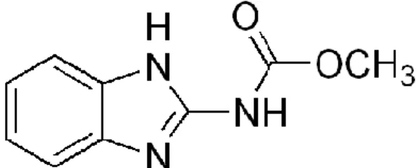
Carbendazim (methyl-2-benzimidazole carbamate) is a fungicide that belong to the benzimidazole carbamate class with a wide applicability in agricultural activity against fungal diseases.
Figure 1. Methyl 1H-benzimidazol-2-ylcarbamate structure.
Carbendazim is a metabolite of thiophanate-methyl, which breaks down rapidly in the environment, generating carbendazim (A.P.V.M.A 2007). The fungicide is used to control a broad range of diseases on arable crops (cereals, oilseed rape), fruits, vegetables and ornamentals. It is also used in post-harvest food storage, and as a seed pre-planting treatment. Carbendazim acts by interrupting the development of fungal germ tubes, the formation of appressoria and the growth of mycelia (A.P.V.M.A 2007). Effects on non-target individuals has been related to impairments on cell division and to the inhibition of the enzyme acetylcholinesterase (Cuppen et al. 2000). Mitosis in plants and mammalian cells is affected by carbendazim by the impairment of formation and functioning of microtubules (Davidse 1977). Ferreira et al. (2008) observed that carbendazim affects feeding activity ofDaphnia magna at concentrations above 70µg/L, with 50% of reduction near 100µg/L. In the same study, a LC50 value of this fungicide to Daphnia magna was established at 156µg/L. The photochemical behaviour of carbendazim in aqueous solution was investigated and a relationship with environmental characteristics was observed; the photodegradation rate is accelerated in alkaline solutions, and under UV radiation levels, while under natural sunlight, carbendazim showed to be a stable compound (Boudina et al. 2003). Carbendazim affects the structure of aquatic ecosystems indirectly, by promoting increase in abundance of phytoplanktonic algae, through impairments on grazing pressure by zooplankton in a microcosm experiment, as observed by Van den Brink et al (2000).
Predictions of joint effects of natural stressor and chemical compounds
In natural systems, wildlife species are often exposed to mixtures of chemical compounds and combinations between chemical and natural stressors. Climate change leads to alterations of environmental natural conditions, such as temperature, precipitation, increase of ultraviolet radiation input, polar ice melting, water acidification, changes on carbon cycling, etc. (Justic et al. 1996; Koinig et al. 1998; Parmesan and Yohe 2003). The effects of single natural stressors have being evaluated for aquatic and terrestrial species; for instance, effects of increasing amount of ultraviolet radiation, temperature and decrease of dissolved oxygen have been reported for some zooplankton species (Häder, Kumar et al. 2007; Ferreira, Loureiro et al. 2008), as well as the effects of several single chemical exposures that occur in aquatic systems. Indeed, the environmental risk assessments is now taking into consideration that possible mixtures of chemicals and combinations of chemicals and natural stressor are crucial when evaluating ecological and human risk.
For the prediction of joint toxicity, an approach based in two foundation concepts has been used for chemical mixtures but also for the combined effects of chemicals and natural stressor (Kienle et al, 2008). First, if chemicals stressors are believed to have the same mode of action, their combined toxicity will be described by the concentration addition model (CA); on the other hand, if they present dissimilar modes of action, the independent action model (IA) is used to predict the joint toxicity. Concentration addition was first formulated by pharmacologists in 1926 (Loewe and Muischnek, 1926) and is based on the idea that all chemicals in the mixture will act by the same mode of action, i.e. on the same target of the organism. The CA model is mathematically expressed by the formula:
n
Ci / ECi = 1
i=1
∑
Where Ci is the concentration of the chemical i in the mixture, and ECxi is the effect concentration of the chemical i that causes the same effect as the mixture does.
The assumption behind independent action is that chemicals in a mixture do not physically, chemically or biologically interact (Bliss 1939) therefore, the effects are based on
probabilities of non-response of the organism to both chemicals in the mixture (Cedergreen et al. 2008). The mathematical expression for IA model is:
1
Y
max
(
)
n iqi Ci
µ
==
∏
Where Y means the biological response, Ci the concentration of chemical i in the mixture; qi(Ci) is the probability of non-response, µmax is the control response for a certain end point, is the multiplication function.
For evaluation of environmental parameters interacting with chemicals compounds, one can preview that both factors could act by independent ways. For that reason, with exceptions of some environmental variables that produces similar effects of those caused by a certain class of chemicals (e.g. oxidative stress caused by dissolved oxygen, ultraviolet radiation and metals) when the CA model can also have a applicability (Ferreira, Loureiro et al. 2008), the IA model is the most applicable in cases of natural stressor and chemical combinations assessments.
There are some mixtures that although the mode of action of its chemical components is known, show some deviations patterns from the models. These deviations are described as Synergism/antagonism when the mixture can cause a more severe (synergism) or less severe (antagonism) effect to a target organism; dose-level dependency, where the toxicity of the mixture is variable depending on whether the mixture is applied in a high or low dose, and dose-ratio deviation, which is reliant on the mixture composition, i.e. which chemical is mainly responsible for the mixture toxicity. These deviation occur when chemicals affect each others bioavailability (related to the environmental conditions), modes of action and behaviour after uptake (Cassee et al, 1998)
II.
Objectives
The present work aimed to elucidate some biological behaviors and patterns regarding changes in the environment. For that, to predict interactions between natural stressors and toxicants toDaphnia magna, the ultraviolet radiation (UVR) and the pesticide carbendazim were chosen as the source of natural and chemical stressors, respectively and were employed in combination with each other as an example of possible stress conditions that
can be found worldwide in the environment. The first section describes the effects of ultraviolet radiation as a single stressor to lethal and sub-lethal end points of Daphnia
magna, as survival, feeding activity, reproduction and energy budget. In the second
section, it is presented the study on the combined effects of carbendazim and ultraviolet radiation on the survival, feeding activity and reproduction response of Daphnia magna. Third section will address the further discussions and final conclusions about results presented along the work.
References
A.P.V.M.A (2007). The Reconsideration of Registrations of Products Containing Carbendazim or Thiophanate-methyl and Their Associated Approved Labels: 27 pages. Allen, D. J., S. Nogués and N. R. Baker (1998). Ozone depletion and increased UV-B radiation: is there a real threat to photosynthesis? Journal of Experimental Botany49(328): 1775-1788.
Alonso, C., V. Rocco, J. P. Barriga, M. A. Battini and H. Zagarese (2004). Surface avoidance by freshwater zooplankton: Field evidence on the role of ultraviolet radiation. Limnology and Oceanography49(1): 225-232.
Anusha, K. and T. Asaeda (2008). Indirect mechanisms accelerated due to ultraviolet-B radiation on nutrient cycling in a freshwater ecosystem. Journal of Photochemistry & Photobiology, B: Biology93: 1-8.
Barata, C., I. Varo, J. C. Navarro, S. Arun and C. Porte (2005). Antioxidant enzyme activities and lipid peroxidation in the freshwater cladoceran Daphnia magna exposed to redox cycling compounds. Comparative Biochemistry and Physiology, Part C140(2): 175-186.
Blumthaler, M. and A. R. Webb (2003). UVR climatology. UV Effects in Aquatic Organisms and Ecosystems. D. P. Hader and G. Jori. Cambridge, Royal Society of Chemistry.1.
Boeing, W. J., D. M. Leech, C. E. Williamson, S. Cooke and L. Torres (2004). Damaging UV radiation and invertebrate predation: conflicting selective pressures for zooplankton vertical distribution in the water column of low DOC lakes. Oecologia138(4): 603-612. Boudina, A., C. Emmelin, A. Baaliouamer, M. F. Grenier-Loustalot and J. M. Chovelon (2003). Photochemical behaviour of carbendazim in aqueous solution. Chemosphere50(5): 649-655.
Carell, T., L. T. Burgdorf, L. M. Kundu and M. Cichon (2001). The mechanism of action of DNA photolyases. Current Opinion in Chemical Biology5(5): 491-498.
Cedergreen, N., A. M. Christensen, A. Kamper, P. Kudsk, S. K. Mathiassen, J. C. Streibig and H. Sorensen (2008). A Review of Independent action compared to concentration addition as reference models for mixtures of compounds with different molecular target sites. Environmental Toxicology and Chemistry27(7): 1621–1632.
Confer, J. L., G. L. Howick, M. H. Corzette, S. L. Kramer, S. Fitzgibbon and R. Landesberg (1978). Visual predation by planktivores. Oikos: 27-37.
Cuppen, J. G. M., P. J. Van den Brink, E. Camps, K. F. Uil and T. C. M. Brock (2000). Impact of the fungicide carbendazim in freshwater microcosms. I. Water quality, breakdown of particulate organic matter and responses of macroinvertebrates. Aquatic Toxicology48(2-3): 233-250.
Davidse, L. C. (1977). Mode of action, selectivity and mutagenicity of benzimidazole compounds. European Journal of Plant Pathology83: 135-144.
de Laat, W. L., N. G. J. Jaspers and J. H. J. Hoeijmakers (1999). Molecular mechanism of nucleotide excision repair. Genes & development13(7): 768-785.
Dong, Q., W. Todd Monroe, T. R. Tiersch and K. R. Svoboda (2008). UVA-induced photo
recovery during early zebrafish embryogenesis. Journal of Photochemistry &
Photobiology, B: Biology93(3): 162-171.
Ferrari, D. C. a. and P. D. N. Hebert (1982). The induction of sexual reproduction in Daphnia magna: genetic differences between arctic and temperate populations. Can J Zool
60: 2143-2148.
Ferreira, A. L. G., S. Loureiro and A. M. V. M. Soares (2008). Toxicity prediction of binary combinations of cadmium, carbendazim and low dissolved oxygen on Daphnia magna. Aquatic Toxicology89(1): 28-39.
Fischer, J. M., P. A. Fields, P. G. Pryzbylkowski, J. L. Nicolai and P. J. Neale (2006a). Sublethal Exposure to UV Radiation Affects Respiration Rates of the Freshwater Cladoceran Daphnia catawba. Photochem. Photobiol. Sci.82: 547-550.
Fischer, J. M., J. L. Nicolai, C. E. Williamson, A. D. Persaud and R. S. Lockwood (2006b). Effects of Ultraviolet radiation on diel vertical migration of crustacean zooplankton: an in situ mesocosm experiment. Hydrobiologia563: 217-224.
Gonçalves, R. J., V. E. Villafañe and E. W. Helbling (2002). Photorepair activity and protective compounds in two freshwater zooplankton species (Daphnia menucoensis and Metacyclops mendocinus) from Patagonia, Argentina. Photochemical & Photobiological Sciences1(12): 996-1000.
Häder, D.-P., H. D. Kumar, R. C. Smith and R. C. Worrest (2007). Aquatic ecosystems: effects of solar ultraviolet radiation and interactions with other climatic change factors. Photochemistry and Photobiology.
Helbling, E. W., V. Villafane and O. Holm-Hansen (1994). Effects of ultraviolet radiation on Antarctic marine phytoplankton photosynthesis with particular attention to the influence of mixing. Antarct. Res. Ser62: 207-227.
Herbert, P. D. N. and C. J. Emery (1990). The adaptive significance of cuticular pigmentation in Daphnia. Functional Ecology: 703-710.
Huebner, J. D., D. L. W. Young, N. L. Loadman, V. J. Lentz and M. D. Wiegand (2006). Age-Dependent Survival, Reproduction and Photorepair Activity in Daphnia magna (Straus, 1820) After Exposure to Artificial Ultraviolet Radiation. Photochemistry and Photobiology82(6): 1656-1661.
Huot, Y., W. H. Jeffrey, R. F. Davis and J. J. Cullen (2000). Damage to DNA in Bacterioplankton: A Model of Damage by Ultraviolet Radiation and its Repair as Influenced by Vertical Mixing¶. Photochemistry and Photobiology72(1): 62-74.
Justic, D., N. N. Rabalais and R. E. Turner (1996). Effects of climate change on hypoxia in coastal waters: A doubled CO2 scenario for the northern Gulf of Mexico. Limnology and Oceanography: 992-1003.
Karanas, J. J., R. C. Worrest and H. Dyke (1981). Impact of UV-B radiation on the fecundity of the copepod Acartia clausii. Marine Biology65(2): 125-133.
Klisch, M., R. P. Sinha, P. R. Richter and D.-P. Häder (2001). Mycosporine-like amino acids (MAAs) protect against UV-B-induced damage in Gyrodinium dorsum Kofoid. Journal of Plant Physiology158: 1449–1454.
Koinig, K. A., R. Schmidt, S. Sommaruga-Wögrath, R. Tessadri and R. Psenner (1998). Climate change as the primary cause for pH shifts in a high alpine lake. Water, Air, & Soil Pollution104(1): 167-180.
Lacuna, D. G. and S. Uye (2000). Effect of UVB radiation on the survival, feeding, and egg production of the brackish-water copepod, Sinocalanus tenellus, with notes on photoreactivation. Hydrobiologia 434(1): 73-79.
Le Houérou, H. N. (1996). Climate change, drought and desertification. Journal of Arid Environments34(2): 133-185.
MacFadyen, E. J., C. E. Williamson, G. Grad, M. Lowery, W. H. Jeffrey and D. L. Mitchell (2004). Molecular response to climate change: temperature dependence of UV-induced DNA damage and repair in the freshwater crustacean Daphnia pulicaria. Global Change Biology: 408–416.
Madronich, S., R. L. McKenzie, L. O. Björn and M. M. Caldwell (1998). Changes in biologically active ultraviolet radiation reaching the Earth's surface. Journal of Photochemistry & Photobiology, B: Biology46(1-3): 5-19.
Marinone, M. C., S. M. Marque, D. A. Suarez, M. C. Dieguez, P. Perez, P. Rios, D. Soto and H. E. Zagarese (2006). UV Radiation as a Potential Driving Force for Zooplankton Community Structure in Patagonian Lakes. Photochemistry and Photobiology82(4): 962-971.
McCallister, S. L., J. E. Bauer, J. Kelly and H. W. Ducklow (2005). Effects of sunlight on decomposition of estuarine dissolved organic C, N and P and bacterial metabolism. Aquatic Microbial Ecology40(1): 25-35.
Moran, M. A., W. M. Sheldon Jr and R. G. Zepp (2000). Carbon loss and optical property changes during long-term photochemical and biological degradation of estuarine dissolved organic matter. Limnology and Oceanography: 1254-1264.
Noyes, P. D., M. K. McElwee, H. D. Miller, B. W. Clark, L. A. V. Tiem, K. C. Walcott, K. N. Erwin and E. D. Levin (2009). The toxicology of climate change: Environmental contaminants in a warming world. Environment International35: 971–986.
Parmesan, C. and G. Yohe (2003). A globally coherent fingerprint of climate change impacts across natural systems. Nature421(6918): 37-42.
Rhode, S. C., M. Pawlowski and R. Tollrian (2001). The impact of ultraviolet radiation on the vertical distribution of zooplankton of the genus Daphnia. Nature412(6842): 69-72. Teoule, R. (1987). Radiation-induced DNA damage and its repair. International Journal of Radiation Biology51(4): 573-589.
Van den Brink, P. J., J. Hattink, F. Bransen, E. Van Donk and T. C. M. Brock (2000). Impact of the fungicide carbendazim in freshwater microcosms. II. Zooplankton, primary producers and final conclusions. Aquatic Toxicology48(2-3): 251-264.
Vega, M. P. and R. A. Pizarro (2000). Oxidative stress and defence mechanisms of the
freshwater cladoceran Daphnialongispina exposed to UV radiation. Journal of
Photochemistry & Photobiology, B: Biology54(2-3): 121-125.
Whetton, P. H., A. M. Fowler, M. R. Haylock and A. B. Pittock (1993). Implications of climate change due to the enhanced greenhouse effect on floods and droughts in Australia. Climatic Change25(3): 289-317.
Williamson, C. E. and K. C. Rose (2009). Ultraviolet Insights: Attempting to Resolve Enigmatic Patterns in Pelagic Freshwaters – The Historical Context and a View to the Future. Internat. Rev. Hydrobiol94: 129-142.
Zaret, T. M. and J. S. Suffern (1976). Vertical migration in zooplankton as a predator avoidance mechanism. Limnology and Oceanography: 804-813.
Chapter II
_______________________________
Effects of Ultraviolet Radiation
single exposures to life parameters
of
Daphnia magna
Effects of Ultra-Violet Radiation on Survival, Feeding activity, Offspring
production and Energy Budget of Daphnia magna
Fabianne Ribeiro1*, Susana Loureiro1and Amadeu M.V.M Soares1 1
Department of Biology & CESAM, University of Aveiro, 3810-193 Aveiro, Portugal. *fabianne@ua.pt
ABSTRACT. Ozone layer is a natural Ultra-violet Radiation (UV-R) filter present in
atmosphere, which has suffered serious deleterious impacts. Nowadays, although the policy for reduction on use of the ozone depleting compounds and recovery of ozone layer is showing good results, this process of healing is slow and depends on several components. The amount of UV-R that reaches earth’s surface is an issue of concern among the scientific community, and has caused the execution of a number of studies to predict effects of R on living organisms both aquatic and terrestrial. By taking the UV-R as a natural stressor, and considering it a threat to aquatic organisms, UV-related effects have been well reported for many zooplankton species. In this study, we used a Daphnia
magna model organism exposed to different intensities of UV radiation (artificial source)
and Photo Reactivating Radiation (PRR) simultaneously. Intensities of irradiation applied in experiments varied from 5.7kJ.m-2 to 31.87 kJ.m-2. Immobilisation, reproduction, and feeding inhibition tests were carried out with adaptations from the already applied and described protocols. Results showed decrease on survival rates of neonates after 48h post exposure to irradiation, with a Lethal-dose LD50 value of 14.7kJ.m-2 and significant differences on feeding rates and offspring production after exposure, with Effect-Dose ED50 values of 14.78 kJ.m-2 and 21.11 kJ.m-2, respectively. The energy budget of adult females after 1st brood showed a significant decrease on sugar and lipids content. The results obtained showed similar ED50 values, for all three different end points used (survival, feeding and reproduction), meaning that ultraviolet radiation exposure can alter the physiological status of the organism in every life-stage, with serious consequences to ecosystem functionality and food-web dynamics.
1. Introduction
Over the past decades, global concern associated to climate changes has grown as a consequence of many anthropogenic events that changed the environmental natural characteristics. One of these changes is the increase of Ultraviolet Radiation (UV-R) that reaches the earth’s surface, due to stratospheric ozone depletion (Madronich et al. 1998) that can be considered a natural stressor to terrestrial and freshwater environments. Freshwater ecosystems usually have a high UV penetration, depending on their
eutrophication’s level, i.e., amount of dissolved organic carbon (DOC). Lakes containing low DOC absorb more UVR energy. The presence of organic particles in water can absorb the damage energy carried by short wave-lengths of UV radiation and thus, decrease the amount of energy that reach organisms, or even prevent them to be in contact with the harmful radiation (Williamson et al. 2001). It has been also observed that climate changes negatively alter the DOC concentration of adjacent lakes by altering the events of inundation and water saturation of soils and watersheds, hence increasing the transparency of lakes, that become more vulnerable to radiation input (Williamson et al. 1996). Moreover, DOC can be broken down into smaller subunits by solar UV-R, that are taken up by bacterioplankton, which can increase the transparency of lakes and thus the penetration of radiation. (Häder et al. 2007). Besides the effects of changing amounts of DOC, MacFadyen et al (2004) found that at lower temperatures, the photoprotection in ectotherms is favored rather than the temperature-dependent repair, and this factor can change the distribution of animals in the water column, with consequences to lake communities. The primary and main target of UV-R to organisms is the DNA molecule. UV-B radiation can cause dimerization of DNA bases, leading to the formation of cyclobutane pyrimidine dimmers (CPDs) and photoproducts, that block the action of DNA polymerase in repairing errors during the replication process, leading to impairments on production of basic cellular components (Buma et al. 2003).
The molecular mechanisms carried out by zooplankton to prevent UV-R damage is closely linked to UV-A radiation and visible light (approximately from 320 to 400nm) because this wavelength range provides the enzymatic repair on daphnids. The repair, defined as the PER- Photo-enzymatic Repair, is carried by the enzyme photolyase that is
activated by light and acts on the DNA repair system(MacFadyen et al. 2004). The sum of the photoprotection process and molecular repair is defined by Williamson et al (2001) as “UV tolerance”.
Zooplankton also shows behavioral response to UV-R by altering their distribution through the water column as a strategy of avoidance, showing deeper distribution at daytime when higher UV-R intensity occurs. The diel vertical migration is a natural process as consequence of fish predation avoidance by zooplankton, but has been described to happen also due to increasing amounts of UV-R on the surface. These patterns can interfere with the food web dynamics of lakes (Alonso et al. 2004; Boeing et al. 2004; Fischer et al. 2006b). The physiological status of zooplankton is also changed as a consequence of combating deleterious effects caused by UV-R. Among these changes we can observe impairment on respiration rates (Fischer et al. 2006a), decrease in the number of neonates produced after irradiation exposure (Huebner et al. 2006), alteration on fecundity (Karanas et al. 1981), and decrease on feeding rates (Lacuna and Uye 2000). The metabolic costs of natural or chemical stressors to a organism is an important key for higher level response predictions, in terms of ecosystem functionality, which is expected to alter under non-favorable conditions.
Approach. In this study we aimed to evaluate the effects of UV-B radiation on life-cyle
parameters of Daphnia magna, with the purpose to predict long-term consequences of climate changes, from organism to ecosystem level, by assessing the immediately immobilization after irradiation exposure, feeding activity, reproduction output and allocation pattern for proteins, sugar and lipids as a complement to the reproduction response.
2. Material and methods
All the experiments were conducted with the cladoceranDaphnia magna Straus, clone k6, originally from Belgium, and maintained in culture in our laboratory for more than 3 years. Cultures were kept in aquariums with 3L of ASTM hard water (ASTM 1980), in controlled light and temperature chambers (16h:8h light-dark), 20°C± 1°C. The green algae Pseudokirchneriella subcapitata was used as food source (3x10⁵ cells/ml) together with a seaweed extract, at a concentration of 6ml/L. The cultures were renewed three
times a week. Neonates or juveniles from third to fifth brood were used in the experiments and the ones from fifth and sixth brood were used for replace old cultures. To assure test’s validation, an acute test with the reference compound potassium dichromate is being performed at least twice a year in our laboratory.
2.2 UV-R experiments. To assess the UV-B radiation effects on Daphnia magna, all tests
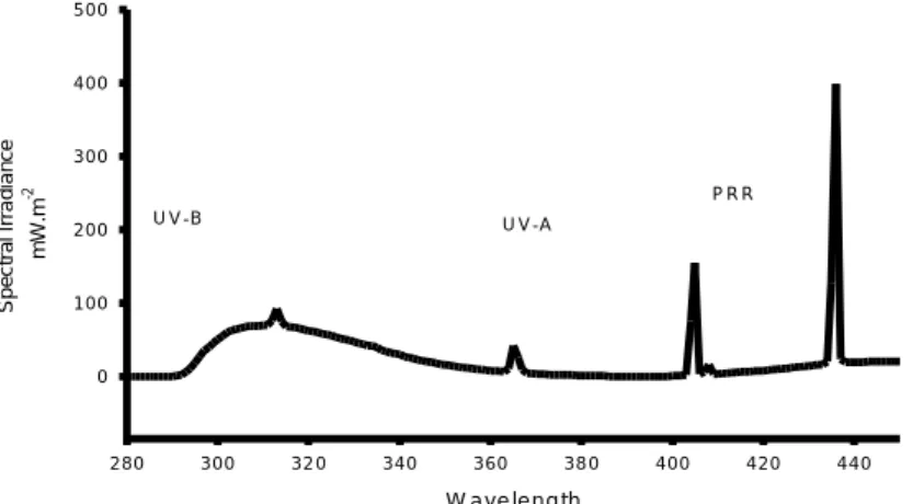
were performed in a controlled temperature room (20°C± 1°C), in which a UV-B apparatus was set. Ultraviolet light source was provided by an UV lamp (Spectroline XX15F/B, Spectronics Corporation, NY, USA, peak emission at 312nm) and two fluorescent tubes (Philips Master TL-D 18W/840) to allow photo-activating repair (PAR) during the experiments. The UV lamp was placed 30cm above the vials and clear cellulose acetate sheets (0,003mm) were used to cut-off UV-C range wavelengths. These cellulose acetate sheets were UV irradiated for 12 (twelve) hours before used in the experiments; the 12h pre-burned period has demonstrated to be suitable to minimize great differences in radiation energy throughout experiments. The exposure was made in transparent glass vials containing 500mL of ASTM hard water, placed on a high- adjustable basis below the UV lamp. Different times of exposure were used to achieve different radiation intensities (Table 1). For acute testing, UV-B intensity reaching the surface of experimental vials ranged from 5.74 kJ.m-2 to 31.87 kJ. m-2 while for chronic tests ranged from 5.74 kJ. m-2 to 19.43 kJ.m-2. Photo Reactivating Radiation (PRR), approximately 320-400nm, was held constant through all experiments. Measurements were made every hour, using an spectroradiometer placed at the surface water level and connected to a monochromator, that provide information on energy per nanometer. Espectral irradiation was obtained by the BenWin+ Software (Bentham Instruments, Reading, UK). Data transformation from mW.m-2to kJ.m-2, considering the time of exposure, is shown in table 1.
2.2.3 Immobilisation tests. The acute test was adapted from the OECD guideline (OECD,
2004) using neonates <24h old exposed to different UV-B treatments. Neonates from the third to fifth brood were used in the experiments and exposed to UVR and PAR light simultaneously during 1, 2, 3, 4, 5, 6 and 7 hours, corresponding to 5.7; 10.1; 15.9; 19.4; 25.6; 30.2 and 31.8 kJ.m-2 respectively. After each irradiation time, organisms were transferred into vials with 50 ml of ASTM, and placed inside a climate chamber with a
photoperiod of 16h:8h (light-dark) and temperature of 20°C±1°C. The mean time for the transfer of the individuals from the lamp to the medium test was less than 5 minutes, and conducted under normal light conditions. For each treatment, five replicates were used, with five neonates each. After 24 and 48h (counting from the beginning of each irradiation time), immobile and dead daphnids were recorded. The end point for immobilization indicates a near future lethality and is defined as their inability to swim within 15 seconds, after gentle agitation of the test vessel (even if they can still move their antennae). Control replicates were kept in the climate chamber while the treatments received irradiation. No food was provided during both exposure and post-exposure periods.
2.2.4 Reproduction tests. Chronic tests were conducted following the OECD 211 guideline
(OECD, 1998), with adaptations. For assessment of UVR effects on reproduction, neonates from the third to fifth brood were separated from the main culture and maintained in the same conditions until they complete their fourth instar, i.e. the release of the third moult, after which the beginning of egg formation take place. Daphnids within this age were exposed to the UV-lamp at the same regime described above, except for the irradiation period. The intensities applied ranged from 5.7 to 19.4 kJ.m-2. After the exposure phase, organisms were then placed individually in 50ml glass vials, containing ASTM hard water,
P.subcapitata as food source (5x10⁵
cell/ml), and the algae extract, as an organic complement, and kept in a climatic chamber until they complete 21-days old. One individual per replicate and ten replicates per treatment were used. The medium test was renewed every other day; organisms were fed daily. The number of neonates was recorded and removed from the vials every day. For test validation, dissolved oxygen, temperature and pH were measured and recorded once in a week.
2.2.5. Feeding inhibition tests. Individuals with less than 24h old were separated from the
laboratory cultures, and maintained at the same conditions until the release of their third moult (4-5 days old) equivalent to the fourth instars, to avoid moulting processes during the test, that are know to unstable feeding activities. The procedure for the feeding test was adapted from McWilliam and Baird (2001). Exposure to UV-R was carried without food to the ASTM medium. 4d-oldDaphnia magna were exposed in glass vials containing 500mL
of ASTM (75 daphnias/500mL). After each time of irradiation (treatment), five individuals were transferred to 200ml vials containing 100ml of ASTM hard water and the green algae
P. subcapitata at a concentration of 5x10⁵
cells/ml, and allowed to feed for 24h. The experimental setup included five replicates per treatment, and five individuals per replicate Controls were exposed to visible light only. For the UV-B light effects recovery, the post exposure (feeding period) was carried in a 16h:8h light-dark regime (Huebner, Young et al. 2006). To determine the initial algae concentration, a blank set of one replicate per treatment (with algae and no daphnids) was carried out. The initial algae concentration was considered as the concentration of algae in the blank vial after 24h, for each treatment. Individual feeding rates (cells/individual/h) were determined according to the method described by Allen et al. (1995)
2.2.6 Energy Budget. Following the procedure described for the reproduction test, 300
neonates were separated from the main cultures, and exposed to UV light when they reached 6-days old. The same UV intensities used for the reproduction test were applied. After each irradiation period, individuals were separated into two glass beakers of 1L contained ASTM hard water (30 individuals/beaker) and placed inside the climate chamber, until the release of their first brood. After this event, daphnids were immediately frozen in liquid nitrogen. This specific period was chosen in order to asses the effort for reproduction of Daphnia magna when the initial phase of recovery process from UV-B damage was being completed. For each treatment, 3 replicates of 10 organisms were used for quantifying protein/sugar, and 3 replicates for lipids quantification (Janssen 1997) Total lipids were extracted following the methodology described by De Coen and Jansen (1997). Daphnids were homogenized in 300µL of water using a sonicator. After homogenization, 500µl chloroform (spectrofotometric grade) and 500µl methanol (spectrofotometric grade) were added and vortexed. After centrifugation, the top phase was removed and the remaining lipid extract was diluted into 500µl of sulfuric acid, and heated for 15min. at 200oC. After cooled down, 1.5 µl of water was added and samples pipetted into the microplate for absorbance measurement at 375nm.
For total protein and sugar content measurements, daphnids were homogenized in 300µL of water with a sonicator. After homogenization, 15% trichloroacetic acid (TCA) was added and samples incubated at –20oC for 10 min. Following centrifugation, a pellet was
formed and then washed with 5% TCA and the supernatant fractions formed were combined and used for the total sugar analysis. The remaining pellet was re-suspended in
NaOH and incubated at 60oC for 30 min, after which was neutralized with HCl.
Absorbance was measured at 590 nm in a microplate reader; standard curves were obtained using bovine serum albumin. Total carbohydrate content of the supernatant fraction was quantified by adding 5% phenol and H2SO4to the extract. After 30 min incubation at 20oC, the absorbance was measured using glucose as a standard at 492nm.
2.3 Statistical Approach
The 48h Lethal Dose of 50% (LD50) for UV-Radiation exposure toDaphnia magna was calculated using a probit analysis with the Minitab software (Minitab, 2003). For reproduction and feeding inhibition tests, data were analyzed by a one-way ANOVA, using the SigmaPlot software (Systat, 2004). Whenever data showed a normal distribution, the differences between control and treatments were obtained by the Dunnett’s test. For data that failed the normality testing, a non-parametric Kruskal-wallis test was used and the multiple comparisons Dunn’s method conducted. All significant differences were established at P<0.05. The effect-dose ED50 for feeding activity and reproduction was calculated using a non-linear regression, a logistic 3-parameter equation (SigmaPlot). Effect-dose ED10and ED20 were calculated by the ToxRat professional software, (ToxRat Solutions) using a linear maximum likelihood regression and ECx- confidence limits based on Fieller’s theorem.
3. Results
Spectral composition of exposures
Spectral composition of exposures
The integrated spectrum of 1 UV lamp and two fluorescent bulbs measured below the cellulose acetate filter is given on figure 1. For data transformation (mW.m-2to kJ.m-2), the main value for each measurement (from 280nm to 320nm) was multiplied by 40, in order to cover the whole range of intensities during the exposure, and the value obtained
was multiplied by the time (in seconds) that lasted each exposure treatment. Data transformation and respective values are given on Table 1.
W a v e le n g th 2 8 0 3 0 0 3 2 0 3 4 0 3 6 0 3 8 0 4 0 0 4 2 0 4 4 0 S p e c tr a l Ir ra d ia n c e m W .m -2 0 1 0 0 2 0 0 3 0 0 4 0 0 5 0 0 U V -B U V -A P R R
Figure. 1. Spectral composition of a UV-lamp and two fluorescent bulbs (PRR) measured at a distance of
30cm.
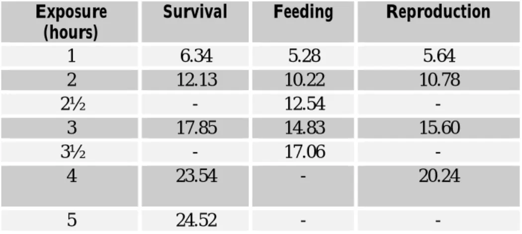
Table 1. Values for UV radiation intensities, presented as kJ.m-2, applied in Daphnia magna exposure experiments. kJ.m-2 = mean value (mW.m-2. m-2.nm-1) from 280 to 320nm x 40 J.m-2 was obtained multiplying the intensity (mW.m-2.nm-1) for the time of exposure in seconds.
Exposure (hours) Survival Feeding Neonates* Energy Budget+
1 5.7 5.28 5.64 6.0 2 10.2 10.22 10.78 11.52 2½ - 12.54 - -3 14.5 14.83 15.60 17.0 3½ - 17.06 - -4 19.4 - 20.24 22.5 5 6 7 25.6 30.2 34.8 - -
-Acute and chronic bioassays
Immobilization records of neonates after 24h and 48h exposed to different intensities of UV-R showed decrease of survival among treatments. After 48h, no survival was observed in any of replicates of the treatments 25.6 kJ.m-2, 30.2 kJ.m-2and 34.8kJ.m-2 equivalents to 5h, 6h and 7h of exposure, respectively. The Lethal-Dose for 50% of animals calculated for this exposure was 14.78 kJm-2. Control treatment showed 100% of survival.
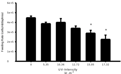
Feeding activity, measured as feeding rate (cells/mL) also showed a decrease in values with increasing UV-intensity exposure to Daphnia magna. (Fig.2) Statistically differences from the control on feeding rates were detected for UV-intensity of 15 kJm-2
and 17.3 kJm-2, corresponding to times of exposures of 3h and 3h30min. (ANOVA, F 5.24=7.570, p<0.05 Dunnett’s test). The ED50value for feeding activity of Daphnids at 24h post-exposure was 17.88 kJ.m-2. (st.error = 1.11; r2=0.971). No mortality was observed during the feeding experiments neither in the controls nor in UV-treatments.
U V -In te n s ity k J .m-2 0 5 ,3 5 1 0 ,3 6 1 2 ,7 2 1 5 ,0 5 1 7 ,3 2 F e e d in g R a te (c e lls /m l/ d a p h n ia ) 0 1 e + 5 2 e + 5 3 e + 5 4 e + 5 5 e + 5 6 e + 5 * *
Figure 2. Effects of a pre-exposure to UV-radiation on the feeding rates of Daphnia magna. The feeding
rates are related to the 24h post-exposure period. Data is expressed as mean values± standard errors. Asterisks indicate significant difference from the control (p<0.05)
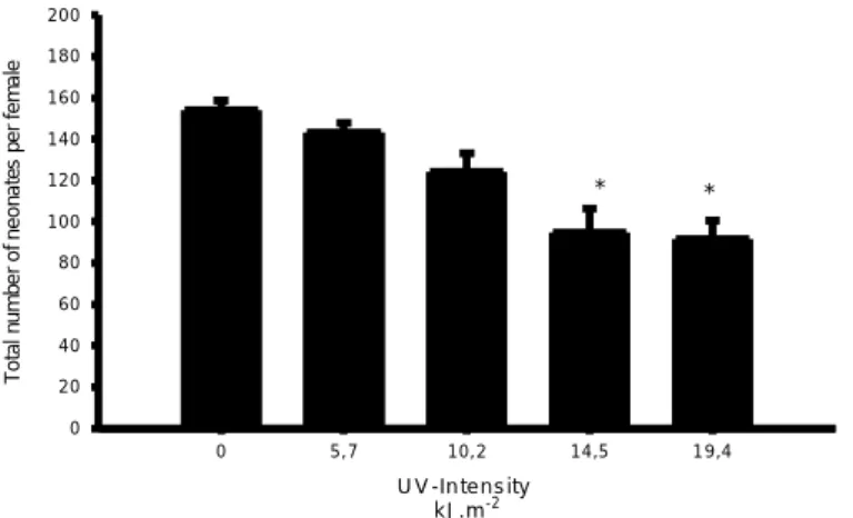
The reproduction effort, measured as the number of neonates produced per live female, during 21days post exposure to UVR was detected to be different among the tested treatments. (ANOVA, F4.34 = 10.26, p<0.001). Significantly differences from the control were observed from exposure of 10.1 kJ.m-2onwards, which corresponds to 2h, 3h and 4h of UV lamp exposure. (Fig.3) (Dunnett’s method, p<0.05). The ED50 calculated for number of neonates was higher than 19.4 kJ.m-2, which was the highest intensity used for this exposure. The length of D.magna at the end of the 21-days post exposure presented different mean values from the control at intensities of 14.5 kJ.m-2 and 19.4 kJ.m-2 (Fig.4) (ANOVA, F4.34=7.76; p<0.05; Dunnett’s method). UV-levels that affected the production of neonates in 10% and 20% for the organisms applied in this experiment were 6.4 kJ.m-2 and 9.9 kJ.m-2respectively (ToxRat Professional)
U V -In te n s ity k J .m- 2 0 5 , 7 1 0 , 2 1 4 ,5 1 9 ,4 T o ta l n u m b e r o f n e o n a te s p e r fe m a le 0 2 0 4 0 6 0 8 0 1 0 0 1 2 0 1 4 0 1 6 0 1 8 0 2 0 0 * *
Figure 3. Number of neonates produced by Daphnia magna after UV radiation exposure. Data is shown as
mean values± standard errors. Asterisks indicate significant differences from the control (p<0.05)
U V - In te n s ity k J .m-2 0 5 ,7 1 0 ,2 1 4 ,5 1 9 ,4 L e n g th (m m ) 0 1 2 3 4 5 * *
Figure 4.Body length of 21d old Daphnia magna pre-exposed to UV radiation. Data is shown as mean
values± standard errors. Asterisks indicate significant differences from the control. (p<0.05)
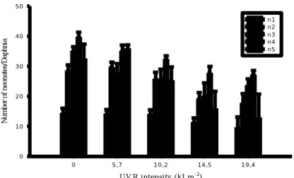
For the analysis of the number of neonates per brood after exposure to ultraviolet radiation, a one way ANOVA was conducted for each treatment in comparison with the control, and significant differences were obtained from the second brood onwards. (Fig. 5)
U V R in te n sity ( k J.m-2) 0 5 ,7 1 0 ,2 1 4 ,5 1 9 ,4 N u m b er o f n eo n at e s/ D a p h n ia 0 1 0 2 0 3 0 4 0 5 0 n 1 n 2 n 3 n 4 n 5
Figure 5. Mean number of neonates produced per Daphnia magna for each brood released after exposure to
ultraviolet radiation. Data is presented as the mean value ±standard error.
Energy reserves. Sugar content, measured after the release of daphnid’s 1st
brood was reduced post UV-R exposure when compared to the control, being statistically significant from 10.2 kJ.m-2 onwards (Fig.6) (Kruskal-Wallis ANOVA, H=23.948, DF=4; p<0,001; Dunn’s method p<0.05). The same pattern was observed for the lipids content (Fig.6) (Kruskal-Wallis ANOVA, H=21.01, DF=4; p<0.001 Dunn’s method p<0.05)
Daphnids did not show any significant differences in their protein content post UV-R exposure after production of the first brood. (ANOVA, F4.25=2.72, p=0.052).
A c u c a re s U V -in te n s ity 0 5 ,7 1 0 ,2 1 4 ,5 1 9 ,4 F ra c ti o n c o n te n t (m J /o rg a n is m ) 0 5 0 0 0 1 0 0 0 0 1 5 0 0 0 2 0 0 0 0 2 5 0 0 0 * * *
Figure6. Total sugar and protein contents of Daphnia magna measured after the release of first-brood upon
a pre UV-R exposure. Data is shown as mean values ± standard errors, where black bars stand for sugar content and gray bars for protein contents.*represents significant differences from control. (p<0.05)