Ciência Rural, v.48, n.10, 2018.
P wave dispersion in obese dogs with and without mitral valve disease
Dispersão da onda P em cães obesos apresentando ou não doença valvar mitral Gustavo Dittrich1 Gabriela Marin Van der Broocke Campos1
Marlos Gonçalves Sousa1* Simone Tostes Oliveira1
ISSNe 1678-4596
Received 01.24.18 Approved 08.21.18 Returned by the author 09.19.18 CR-2018-0047.R2
INTRODUCTION
P wave dispersion (Pd) is an
electrocardiographic index defined as the difference between the minimum and maximum durations of P wave, which are obtained from different leads of the surface electrocardiogram (NOSZCZYK-NOWAK et al., 2008). An increased P wave lenght and dispersion
may be the consequence of an abnormal atrial depolarization secondary to chronic injuries such as elevated atrial pressure, myocardial ischemia and metabolic stress (MAGNANI et al., 2009). In people, Pd was shown to consistently predict the development of atrial fibrillation and to be a prognostic surrogate (DILAVERIS & GIALAFOS, 2001; DOGAN et al., 2003; NIELSEN et al., 2015; LAZZERONI et al.,
1Departamento de Medicina Veterinária, Universidade Federal do Paraná (UFPR), Rua dos Funcionários, 1540, Cabral, Curitiba, PR, Brasil.
E-mail: [email protected]. *Corresponding author.
ABSTRACT: Pwave dispersion (Pd) is an electrocardiographic index defined as the difference between the minimum and maximum Pwave duration in multiple leads. The augmentation of Pd reflects the discontinuous and inhomogeneous atrial depolarization resulting from cardiac and non-cardiac conditions. In humans, an increased Pd is associated with the development of cardiac arrhythmias, particularly atrial fibrillation. To investigate Pd in obese dogs, we enrolled 76 dogs, which were classified in four distinct categories according to body condition and the existance of valve insufficiency: obese dogs (O), dogs with both obesity and cardiac disease (O+CD), lean dogs with cardiac disease (CD) and healthy controls (H). To be included in the study, all dogs underwent an electrocardiographic and echocardiographic assessment. We reported significantly higher Pd in the animals included in categories O, O+CD and CD (18.0±7.6ms, 16.1±4.4ms, 12.1±4.3ms, respectively) as compared to the healthy subjects (7.3±2.2ms). Also, significant correlations between Pd and both the body mass index and body fat percentage were documented for the obese dogs. However, no association between Pd and LA/Ao could be identified in patients belonging to the O, O+CD and H categories. Thus, we have demonstrated that obese dogs, regardless of their valvular competency status, present high Pd values, suggesting an impaired propagation of atrial electrical impulse.
Key words: mitral degeneration, P wave indices, adiposity, obesity, left atrial size.
RESUMO: A dispersão da onda P (Pd) é um índice eletrocardiográfico definido como a diferença entre as durações máxima e mínima da onda
P em múltiplas derivações. O aumento da Pd reflete a despolarização discontínua e não homogênea resultante de condições cardíacas e não cardíacas. Em seres humanos, uma Pdaumentada está associada com o desenvolvimento de arritmias cardíacas, particularmente fibrilação atrial. Com o intuito de investigar a Pd em cães obesos, foram selecionados 76 cães, os quais foram classificados em quatro categorias distintas, de acordo com sua condição corporal e a existência de insuficiência valvar: cães obesos (O), cães com obesidade e doença cardíaca (O+CD), cães magros com doença cardíaca (CD) e cães saudáveis usados como controle (H). Uma vez selecionados para o estudo, todos os cães foram submetidos às avaliações eletrocardiográfica e ecocardiográfica. Os resultados mostraram maior Pd nos animais pertencentes aos grupos O, O+CD e CD (18.0±7.6ms, 16,1±4,4ms, 12,1±4,3ms, respectivamente) quando comparados aos cães saudáveis (7.3±2.2ms). Além disso, foram verificadas correlações significativas entre Pde tanto o índice de massa corporal quanto o percentual de gordura corporal nos cães obesos. Entretanto, não se identificou associação entre Pd e a relação AE/Ao nos pacientes das categorias O, O+CD e H. Dessa forma, foi possível demonstrar que cães obesos, independentemente do estado de competência valvar, apresentam Pd elevada, fato que sugere comprometimento da propagação do impulso elétrico atrial.
Palavras-chave: degeneração mitral, índices da onda P,adiposidade, obesidade, tamanho atrial esquerdo.
2016). Also, an increased Pd was documented in subjects
with arterial hypertension and diabetes, which are both recognized as independent factors for the development of atrial fibrillation (YAZICI et al., 2007; DAGLI et al., 2008). Other studies reported a direct relationship between augmented P wave indices and diseases that cause diastolic dysfunction, coronary syndromes and dilated cardiomyopthy (SENEN et al., 2004; GUNDUZ et al., 2005). Among several other conditions in people, obesity was shown to impair P wave indices, which are related with an increased risk for developing cardiac arrhythmias (MAGNANI et al., 2009). Studies have demonstrated an association between the increase of Pd in obese individuals and atrial arrhythmias (SEYFELI et al., 2006; KOSAR et al., 2008).
Pd values have been previously documented in healthy dogs, and in dogs with either chronic valvular disease or supraventricular conduction disturbances (NOSZCZYK-NOWAK et al., 2011). However, at least to the authors’ knowledge, no information exists in literature concerning the role played by body condition, especially obesity, on both the duration and dispersion of P wave in dogs. Therefore, the aims of this study were two-fold: to investigate P wave indices in obese dogs and to determine whether these indices correlate with adiposity parameters and the left-atrial size.
MATERIALS AND METHODS
Seventy-six dogs of either sex were enrolled in this prospective observational study. Inclusion criteria included the animal’s age (≥4y) and body weight (≥4 and ≤30kg)and meeting the selection characteristics for each of the studied categories (obese, obese+cardiac disease, cardiac disease, healthy). Having any cardiac alteration other than mitral valve disease was considered an exclusion criterion. Several breeds and crossbred dogs were admitted. All dogs underwent a thorough clinical examination and were assessed for body condition. Also, an electrocardiogram and an echocardiogram were performed. Prior to enrollment, the animals had blood aliquots drawn for laboratory analyses. Dogs which exhibited any abnormality in either CBC or bichemistry profile (albumin, alanine aminotransferase, alkaline phosphatase, blood urea nitrogen, creatinine, glucose, triglicerides, cholesterol, and free T4) were not admitted into the study.
The assessment of body condition included the determination of body condition score using the 1-9 scale, which is based on fat deposition over the costal and abdominal areas (LAFLAMME, 1997). Dogs were classified as either having a normal body
condition score (4 or 5) or being obese (8 or 9). Also, two adiposity indices were obtained: the body mass index (BMI) described by MUELLER et al. (2008), and the body fat percentage (BF%) as proposed by BURKHOLDER & TOLL (2000).
Computed-based electrocardiographic tracings were recorded for a minimum of three
minutes (ECGPC®, TEB, São Paulo, Brazil) with the
dogs restrained in right lateral recumbency. Six limb leads (I, II, III, aVR, aVL and aVF) were recorded in all animals, while in 18 dogs the precordial leads (rV2, V2, V4 and V10) were obtained as well. All recordings were obtained with a speed of 50mm/s and a sensitivity of 1cm per millivolt. Each recorded lead was carefully assessed by a single experienced veterinarian to determine the duration of P wave, which was measured from its beginning (the start of either the positive or negative deflection at the isoelectric line) until its end (the return of the deflection to an isoelectric line). The minimum (Pmin) and maximum (Pmax) durations of P wave were documented. P wave dispersion was calculated as the diference between Pmax and Pmin (Pd = Pmax-Pmin). All parameters were considered as the average of three distinct measurements obtained from cardiac beats (cycles) of sinus origin.
All dogs underwent a transthoracic
echocardiogram (Esaote® MyLab™ 30 Vet, Genova,
Italy). That exam was the gold standard to confirm the diagnosis of degenerative mitral valve disease and to rule out any other cardiac anomaly. Also, using transverse views of the heart obtained from the right paraesternal window, the left atrial dimension and the aortic diameter were measured at the very beginning of diastole, which were later used to calculate the left atrium-to-aorta ratio (LA/Ao). In this study, we considered 1.37 as the cut-off for increased left atrium (PRADA et al., 2012).
After the assessment was complete, the dogs were classified in four different categories:
Obese (O): dogs with body condition score indicative of obesity, but free from cardiac disease;
Obese + Cardiac Disease (O+CD): obese dogs which also had mitral valve disease documented in the echocardiogram;
Cardiac Disease (CD): lean dogs with documented mitral valve disease;
Ciência Rural, v.48, n.10, 2018. Statistical analyses were based on
measures of central tendency and dispersion for heart rate, Pmin, Pmax and Pd. Normality of data was assessed with D’Agostino & Pearson test. Either an analysis of
variance (ANOVA) followed by the post hoc Tukey
test or the Kruskal-Wallis test followed by Dunn’s test were used to compare groups in accordance with data normality. Also, Pearson’s correlations were calculated between Pd and BMI, BF% and LA/ Ao. All analyses were performed using the software GraphPad® Prism 5 (La Jolla, California, USA).
RESULTS
This study investigated 76 dogs, which were separated in four distinct categories. The obese group (O) included 18 dogs (15 female/3 male; 4-15 y; 17.5±5.2kg), while 19 animals (13 females/6 males; 9-20 y; 14.2±4.2kg) were enrolled into the obese with cardiac disease group (O+CD). In that group, 47% of the dogs (9/19) had an increased left atrium (LA/Ao>1.37) documented on the echocardiogram, i.e. belonged to either stages B2 (n=3) or C (n=6). For the lean and cardiac disease group (CD), another 19 dogs (6 females/13 males; 10-16 y; 7.4±2.3kg) were included, of which 58% (11/19) had left atrial enlargement, i.e. were classified as either stage B2 (n=3) or C (n=8). Finally, 20 dogs (14 females/6 males; 9-17 y; 7.3 ± 1.9kg) were assessed as healthy controls (H).
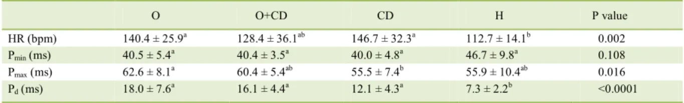
Table 1 brings the results obtained for heart rate, Pmin, Pmax and Pd in all four groups. A significant difference existed for P wave dispersion, which was higher in O, O+CD and CD dogs as compared to the healthy controls (P<0.0001).Also, Pmax was higher in O in comparison with CD dogs (P=0.016). We documented no difference between males and females regardless of the category to which the subjects were
assigned (P=0.453), however our data derives from a small population of dogs. Figure 1 illustrates the individual and mean results of Pd for all groups.
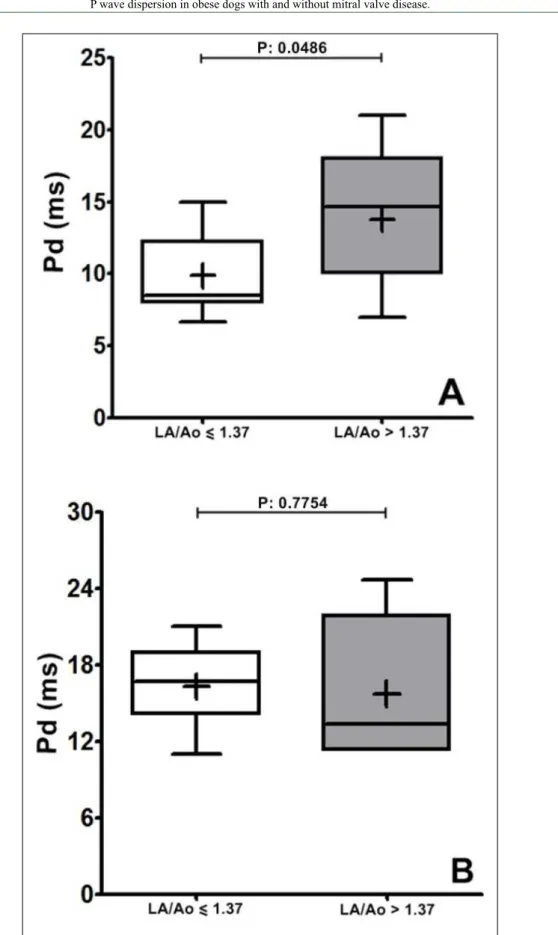
For the dogs with mitral valve disease (groups O+CD and CD), we also compared the results of Pmin, Pmax and Pd between subjects with normal and remodeled left atria (LA/Ao>1.37) regardless of their body condition. While no difference existed for Pmin
and Pd (Table 2), dogs with an increased left atrium had a significantly greater Pmax. However, when the role played by left atrium remodeling was assessed within CD and O + CD individually, a significantly higher Pd was documented for dogs with dilated left atria only in category CD (Figure 2).
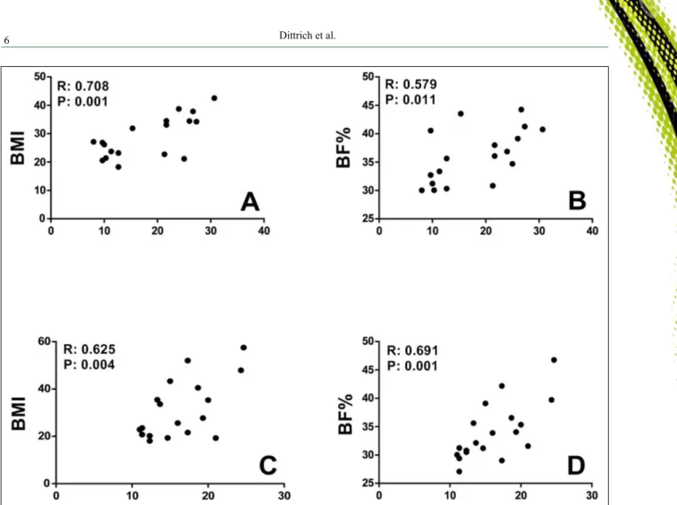
When the correlations between Pd and body weight, BMI, BF% and LA/Ao were investigated, significant results were reported only between Pd and the adiposity indices for obese dogs (O) and obese dogs with cardiac disease (O + CD) (Figure 3). On the contrary, no correlations were shown to exist between Pd and both body weight and LA/Ao ratio for categories O, O + CD and H. However, specifically for CD results, significant correlations were documented between LA/Ao and Pd, Pmin and Pmax (Table 3).
The 18 dogs in which precordial leads were also recorded belonged to the four different categories of this study (O=4; O+CD=4; CD=5; H=5).
No differences existed (P=0.847) when comparing Pd
calculated in consideration of the precordial leads with the results obtained from the standard limb leads. Again, this data is fragile in view of the small number of dogs that have been assessed.
DISCUSSION
Although simple, this study adressed a fairly “novel” electrocardiographic index in a population of obese dogs either with or without mitral
Table 1 – P wave parameters obtained in dogs. Results shown as the mean and standard deviation for heart rate, minimum and maximum P
wave duration, and P wave dispersion.
O O+CD CD H P value
HR (bpm) 140.4 ± 25.9a 128.4 ± 36.1ab 146.7 ± 32.3a 112.7 ± 14.1b 0.002 Pmin (ms) 40.5 ± 5.4a 40.4 ± 3.5a 40.0 ± 4.8a 46.7 ± 9.8a 0.108
Pmax (ms) 62.6 ± 8.1a 60.4 ± 5.4ab 55.5 ± 7.4b 55.9 ± 10.4ab 0.016
Pd (ms) 18.0 ± 7.6a 16.1 ± 4.4a 12.1 ± 4.3a 7.3 ± 2.2b <0.0001
O: obese dogs (n=18); O+CD: obese dogs with mitral valve disease (n=19); CD: lean dogs with mitral valve disease (n=19); H: healthy
control dogs (n=20); HR: heart rate; Pmin: minimum duration of P wave; Pmax: maximum duration of P wave; Pd: P wave dispersion. (a,b)
valve disease. At least in people, P wave dispersion has been demonstrated to be associated with both cardiac and non-cardiac diseases (DOGAN et al., 2003; GOUDIS et al., 2015).
Our results showed no differences in Pd for males and females, which is likely supportive of its independency of gender. However, the effect of age on Pd could not be assessed in this study since only dogs aged at least four years were enrolled.Also, Pd
calculated from either six (I, II, III, aVR, aVL, aVF) or ten (I, II, III, aVR, aVL, aVF, rV2, V2, V4, V10) electrocardiographic leads was considered similar in this investigation. In a study that evaluated Pdin healthy dogs using six-lead electrocardiogram both unipolar and bipolar leads showed to be appropriate to calculate P wave dispersion, making uneccessary the use of precordial electrodes (NOSCZCZYK-NOWAK et al., 2008).
Figure 1 – Individual values and means of P wave dispersion (ms) obtained in dogs. (O: obese dogs; O+CD: obese dogs with mitral valve disease; CD: lean dogs with mitral valve disease; H: healthy control dogs).
Table 2 –Comparison of P wave indices between dogs with mitral valve disease (groups O+CD and CD) presenting either normal or
remodeled left atria. Means and standard deviations are shown.
LA/Ao ≤ 1.37 LA/Ao > 1.37 P value
Pmin (ms) 38.5 ± 3.7 41.5 ± 4.0 0.066
Pmax (ms) 54.1 ± 6.0 60.0 ± 6.1 0.006
Pd (ms) 13.2 ± 4.5 14.8 ± 4.9 0.322
Ciência Rural, v.48, n.10, 2018.
Figure 2 – Comparison between Pd in (A) CD and (B) O+CD dogs according to the size of the
It has been well accepted that obese people have an increased P wave dispersion, which seems to be a risk factor for developing cardiac arrhythmias, especially atrial fibrillation (DOGAN et al., 2003; GOUDIS et al., 2015). In veterinary medicine, no information exists concerning the association of obesity and changes in P wave duration and dispersion. Our results demonstrated, for the first time, that obese
dogs, either with or without valvular disease and atrial remodeling, have an increased Pd in comparison with healthy animals. Although, no significant difference was documented, a trend of increase in Pmax was observed in the obese animals (categories O and O+CD) as well. The effect of obesity in the P wave indices was already demonstrated in human beings. These studies showed a significant increase in Pmax and Pd, which could be fully
Figure 3 – Significant correlations documented between Pd and (A) BMI of obese dogs; (B) BF% of obese dogs; (C) BMI of O+CD dogs;
and (D) BF% of O+CD dogs.
Table 3 –Correlation coefficients obtained between P wave indices and the LA/Ao ratio for each of the studied categories.
O O+CD CD H
Pmin (ms) R: -0.030 P: 0.904 R: 0.090 P: 0.713 R: 0.561 P: 0.012 R: -0.119 P: 0.618
Pmax (ms) R: 0.047 P: 0.852 R: -0.002 P: 0.993 R: 0.778 P:<0.0001 R: -0.040 P: 0.867
Pd (ms) R: 0.110 P: 0.664 R: -0.043 P: 0.860 R: 0.606 P: 0.006 R: 0.305 P: 0.190
O: obese dogs (n=18); O+CD: obese dogs with mitral valve disease (n=19); CD: lean dogs with mitral valve disease (n=19); H: healthy
Ciência Rural, v.48, n.10, 2018.
reversed after weight loss (DURU et al., 2006; SEYFELI et al., 2006; KOSAR et al., 2008; FALCHI et al., 2014). Moreover, in obese subjects a positive correlation between those indices and the BMI was shown to exist. In people, increases in duration and dispersion of P wave is associated with prolonged atrial conduction, left atrial remodeling and the predominant sympathetic tone, which impair the velocity of impulse propagation. Also, there are evidences of structural cardiovascular alterations and impairment of myocardial electrical conduction attributable to obesity (ABEL et al., 2008; ADOLPHE et al., 2014).
In our study, obese dogs and dogs with chronic valvular disease had similar results for Pd. NOSZCZYK-NOWAK et al. (2011) reported an increased Pd in
dogs with valvular disease as compared with healthy subjects, which is similar to our own findings. Chronic valvular disease results in inhomogeneous propagation of electrical impulses within the atria, which together with atrial remodeling cause the increase of such index. Nevertheless, it is not completely clear whether Pd might be an independent index to evaluate both inter-andintra-atrial electrical conduction.
We reported a correlation to exist between Pd and the adiposity indices in obese dogs, which is similar to the findings of Kosar et al. (2008). In their study, a significant correlation between Pd and BMI
was documented in obese people. In our study, Pd increased in parallel with the adiposity indices of the obese dogs. This finding suggested that obesity may indeed play a role in atrial electrical activity.
P wave indices were also investigated in view of atrial remodeling regardless of body condition. In that sense, a significant correlation was found between Pmax and LA/Ao ratio only for dogs with left atrial dilatation. Hence, it is reasonable to speculate that the larger the electrically excited atrial tissue, the longer the duration of P wave as suggested by SURAWICZ (1986). Moreover, once specifically Pd was compared between dogs with remodeled and non remodeled left atria, a significant difference was documented only for the experimental group CD. A reasonable explanation for the contrasting results between CD and O + CD in this regard lies on the conflicting population of stage B1 (larger in O + CD) and stage C (larger in CD) in these two groups. Curiously, the duration of P wave was already shown to be poorly related to left atrial size in dogs, with just a mild correlation with LA/Ao. For instance, SAVARINO et al. (2012) showed that P wave duration is not a reliable surrogate for left atrial remodeling. Concerning Pd, no correlation existed with LA/Ao, which is similar to the findings of NOSZCZYK-NOWAK et al. (2011). We speculated that Pd may be more dependent on inter-
and intra-atrial conduction disturbances as well as an inhomogeneous conduction of electrical impulses than the degree of left atrial dilatation itself.
Among the limitations of this study are the small number of dogs enrolled in each category, as well as the absence of stages B1 and D of MMVD. Although measurements of P wave duration seem quite easy to accomplish, we neither evaluated intra- and inter-observer repeatability for measurements of P wave duration, nor assessed how the level of training would interfere with the results of that parameter, i.e. an experienced veterinarian versus a resident or student. Finally, assessing how P wave dispersion might predict the development of supraventricular arrhythmias in either lean or obese dogs was not the purpose of this investigation.
CONCLUSION
P wave dispersion was higher in obese dogs, either presenting or not mitral valve insufficiency. Our results are indicative of the role played by excess of weight and the accumulation of fat on the canine cardiovascular system. Further studies might add information on the prognostic aspect of P wave dispersion.
BIOETHICS AND BIOSSECURITY COMMITTEE APPROVAL
This study was approved by Universidade Federal do Paraná (UFPR) Committee on Animal Research and Ethics (CEUA) under protocol 29/2014.
ACKNOWLEDGEMENTS
The authors thank Conselho Nacional de
Desenvolvimento Científico e Tecnológico (CNPq) for the scholarship.
DECLARATION OF CONFLICTING INTERESTS
The authors declare no conflict of interest. The founding sponsors had no role in the design of the study; in the collection, analyses, or interpretation of data; in the writing of the manuscript, and in the decision to publish the results.
AUTHORS’ CONTRIBUTIONS
The authors contributed equally to the manuscript.
REFERENCES
ABEL, E. D. et al. Cardiac remodeling in obesity. Physiological Reviews, v.88, p.389–419, 2008. Available from: <http://dx.doi.
ADOLPHE, J. L. et al. Short-term obesity results in detrimental
metabolic and cardiovascular changes that may not be reversed with weight loss in an obese dog model. British Journal of Nutrition, v.112, p.647-656, 2014. Available from: <http://dx.doi.
org/10.1017/S0007114514001214>. Accessed: Sept. 12, 2018.
ATKINS, C. et al., Guidelines for the diagnosis and treatment of
canine chronic valvular heart disease. Journal of Veterinary Internal Medicine, v.23, p.1142-1150, 2009. Available from: <http://dx.doi.org
/10.1111/j.1939-1676.2009.0392.x>. Accessed: Sept. 12, 2018.
BURKHOLDER, W.J.; TOLL, P.W. Obesity. In: HAND, M.S. et
al. Small animal clinical nutrition. 4.ed. Topeka: Mark Morris Institute, 2000, p.401-430.
DAGLI, N. et al. Are maximum P wave duration and P wave dispersion a marker of target organ damage in the hypertensive
population? Clinical Research in Cardiology, v.97, p.98–104,
2008. Available from: <http://dx.doi.org/10.1007/s00392-007-0587-8>. Accessed: Sept. 12, 2018.
DILAVERIS, P.E.; GIALAFOS, J.E.P-wave dispersion: a novel predictor of paroxymal atrial fibrillation. Annals of Noninvasive Electrocardiology, v.6, 159-165, 2001. Available from: <http://
dx.doi.org/10.1111/j.1542-474X.2001.tb00101.x>. Accessed:
Sept. 12, 2018.
DOGAN, A.et al. A comparison of P-wave duration and dispersion in patients with short-term and long-term atrial fibrillation. Journal of Electrocardiology, v.36, p.251-255, 2003. Available from: <http://dx.doi.
org/10.1016/S0022-0736(03)00049-9>. Accessed: Sept. 12, 2018.
DURU, M. et al. Effect of weight loss on P wave dispersion in obese
subjects. Obesity, v.14, p.1378-1382, 2006. Available from: <http://
dx.doi.org/10.1038/oby.2006.156>. Accessed: Sept. 12, 2018.
FALCHI, A.G. et al. Weight loss and P wave dispersion: a
preliminary study. Obesity Research & Clinical Practice, v.8,
p.614-617, 2014. Available from: <http://dx.doi.org/10.1016/j. orcp.2014.08.005>. Accessed: Sept. 12, 2018.
GOUDIS, C.A. et al. Obesity and atrial fibrillation: A comprehensive review of the pathophysiological mechanisms and links. Journal of Cardiology, v.66, p.361-369, 2015. Available from: <http://dx.doi.
org/10.1016/j.jjcc.2015.04.002>. Accessed: Sept. 12, 2018.
GUNDUZ, H. et al. The relationship between P wave dispersion and diastolic dysfunction. Texas Heart Institute Journal, v.32,
p.163-167, 2005. Available from: <https://www.ncbi.nlm.nih.gov/ pmc/articles/PMC1163463/>. Accessed: Sept. 12, 2018.
KOSAR, F. et al. P-wave duration and dispersion in obese
subjects. Annals of Noninvasive Electrocardiology, v.13,
p.3-7, 2008. Available from: <http://dx.doi.org/10.1111/j.1542-474X.2007.00194.x>. Accessed: Sept. 12, 2018.
LAFLAMME, D. P. Development and Validation of a Body Condition Score System for Dogs. Canine Practice, v.22, p.10-15, 1997.
LAZZERONI, D. et al. P-wave dispersion predicts atrial fibrillation following cardiac surgery. International Journal of Cardiology,
v.203, p.131-3, 2016. Available from: <http://dx.doi.org/10.1016/j. ijcard.2015.10.143>. Accessed: Sept. 12, 2018.
MAGNANI, J.W. et al. P wave indices - Current status and future directions in epidemiology, clinical and research
applications. Circulation: Arrhythmia and Electrophysiology,
v.2,p.72-79, 2009. Available from: <http://dx.doi.org/10.1016/j. ijcard.2015.10.143>. Accessed: Sept. 12, 2018.
MUELLER, D.C.M. et al. Adaptation of human body mass index for dogs. Ciência Rural, v.38, p.1038-1043, 2008. Available from:
<http://dx.doi.org/10.1590/S0103-84782008000400020>. Accessed:
Sept. 12, 2018.
NIELSEN, J.B. et al. P-wave duration and the risk of atrial fibrillation: Results from the Copenhagen ECG Study. Heart Rhythm, v.12, p.1887-1895, 2015. Available from: <http://dx.doi.
org/10.1016/j.hrthm.2015.04.026>. Accessed: Sept. 12, 2018.
NOSZCZYK-NOWAK, A. et al. P-wave dispersion in healthy dogs. A preliminary study. Bulletin of the Veterinary Institute in Pulawy, v.52, p.683-688, 2008. Available from: <http://dx.doi.
org/>. Accessed: Sept. 12, 2018.
NOSZCZYK-NOWAK, A. et al. Comparison of P-wave dispersion
in healthy dogs, dogs with chronic valvular disease and dogs with
disturbances of supraventricular conduction. Acta Veterinaria Scandinavica, v.53, p.1-6, 2011. Available from: <http://dx.doi.
org/10.1186/1751-0147-53-18>. Accessed: Sept. 12, 2018.
PRADA, D.G. et al. Echocardiographic evaluation of the left atrium of healthy dogs using the conventional M-mode and
the bidimensional mode. Arquivo Brasileiro de Medicina Veterinária e Zootecnia, v.64, p.585-592, 2012. Available
from: <http://dx.doi.org/10.1590/S0102-09352012000300009>. Accessed: Sept. 12, 2018.
SAVARINO, P. et al. Diagnostic performance of P wave duration in the identification of left atrial enlargement in dogs. Journal of Small Animal Practice, v.53, p.267-272, 2012. Available from:
<http://dx.doi.org/10.1111/j.1748-5827.2012.01200.x>. Accessed:
Sept. 12, 2018.
SENEN, K. et al. P-wave duration and P-wave dispersion in
patients with dilated cardiomyopathy. European Journal of Heart Failure, v.6, p.567-569, 2004. Available from: <http://dx.doi.
org/10.1016/j.ejheart.2003.12.020>. Accessed: Sept. 12, 2018.
SEYFELI, E. et al. Effect of obesity on P-wave dispersion and QT
dispersion in women. International Journal of Obesity, v.30,
p.957–961, 2006. Available from: <http://dx.doi.org/10.1038/ sj.ijo.0803233>. Accessed: Sept. 12, 2018.
SURAWICZ, B. Electrocardiographic diagnosis of chamber
enlargement. Journal of the American College of Cardiology,
v.8, p.711-724, 1986.Available from: <http://dx.doi.org/10.1016/ S0735-1097(86)80207-8>. Accessed: Sept. 12, 2018.
YAZICI, M. et al. The effect of diabetes mellitus on the P-wave
dispersion. Circulation Journal, v.71, p.880–883, 2007. Available from: