http://dx.doi.org/10.1590/s2175-97902017000417141
Article
*Correspondence: M. J. S. Salles. Universidade Estadual de Londrina, De-partamento de Biologia Geral. Rodovia Celso Garcia Cid, Km 380 - Campus Universitário, P.O. box: 10.011, CEP 86057-970 - Londrina, Paraná, Brasil. Phone: 55 43 3714527 / Fax: 55 43 33714207. e-mail: [email protected]
Effect of alcoholic beverages on progeny and reproduction of mice
Fernanda Dias Figueiro
1, Ana Clara Cerato Bispo
1, Karla Lorena Guarido
1, Priscila Marianno
1,
Gabriel de Araújo Costa
1, Helena Kaminami Morimoto
2, Maria José Sparça Salles
1*1Department of General Biology, Londrina State University, Londrina, Paraná, Brazil, 2Department of Pathology, Clinical and Toxicological Analysis, Londrina State University, Londrina, Paraná, Brazil
Alcohol is the most commonly consumed substance in the world. The objective of this study was to evaluate the influence of alcoholic beverages on male reproduction and possible alterations in their offspring. The mice were divided into 4 groups: beer, wine, cachaça (a type of sugarcane rum), with ethanol concentrations of 1.9 g/kg, and control group treated with PBS. The treatment period was 35 days. The animals which received cachaça, demonstrated significant weight loss in the testes and epididymis. The alcoholic beverages promoted significant testosterone level and fertilization index diminution, and morphological alterations in the spermatozoa. The beer group presented decreased implantation sites and a high frequency of dominant lethal. The number of reabsorptions in the wine group was increased. The fermented beverages presented higher potential to induce visceral malformations, while the cachaça caused fetal skeletal malformations. The cachaça treated group presented a negative impact on semen quality and fertilization potential. The treatment with different alcoholic beverages, during spermatogenesis, demonstrated contrasting degrees of induction of toxic effects, interfering in a general aspect in male reproductive performance, fetal viability during intrauterine life, and birth defects. From the data, it is possible to infer that the distillated beverage caused more harmful effects to reproduction in this study.
Keywords: Alcoholic beverages/analysis. Wine/adverse effects. Beer/adverse effects. Cachaça/adverse
effects. Reproduction/drugs effects.
INTRODUCTION
Alcohol is the most commonly consumed substance when compared with other psychoactive elements, with a world prevalence which reaches almost 2 billion consumers worldwide, attaining the “health problem” category, attributed as the cause of 3.8% of deaths and 4.6% of causes of illness worldwide, and has been singled
out as an agent of more than 60 types of illness (Anderson, Chishol, Fuhr, 2009; Rehm, Mathess, Popova, 2009).
The teratogenic effects related to alcohol usage
were initially described in 1968 and 1973, when a specific
pattern of alcoholic mother child malformations were
defined, named as fetal alcohol syndrome (Hoyme, May, Kalberg, 2005).
The majority of alcohol ingestion is metabolized by
the liver and converted to acetaldehyde. The conversion product is extremely toxic, since it can cause DNA fragmentation and therefore interfere in the cellular
metabolism (Halsted, 2004).
The excessive use of alcohol is involved in hormonal, metabolic, and pathophysiological alterations,
which severely influence the development and growth of offspring (Burgos et al., 2002). The decrease in serum
testosterone concentration can stimulate the activity of the
aromatase enzyme, present in peripheral fat cells, which
is responsible for conversion of testosterone to estrogen. This substance can promote a decrease in sperm volume
MATERIAL AND METHODS
All the procedures described here are in accordance with the Animal Experimentation Ethical Principles of the
Brazilian Federal Veterinary Medicine Council (CFMV) and Brazilian law n. 9605 (regulated by Decree n. 3179, 21/12/1999) and were approved by the Ethics Committee
on animal experimentation of the Universidade Estadual
de Londrina (Londrina State University – UEL), number
8198.2013.22.
Experimental design
Male (n=10 per group) and female (n=20 per group) Swiss mice weighing approximately 45 grams
and 60 days old, sexually mature, were chosen. The animals were kept in a controlled light environment, with a 12-hour light/dark cycle, at 22 ± 2 ºC, with free access to food and water. The mice were distributed equally into four experimental groups. Three groups received,
via gavage, different kinds of alcoholic beverages (G1)
= beer, (G2) = wine, and (G3) = cachaça. Cachaça is a
Brazilian beverage obtained from the fermentation of sugarcane juice (Andrade-Sobrinho et al., 2002). The
ethanol concentration administrated in the groups was
1.9 g/kg (Doremus et al., 2003). The control group
(G0) received phosphate buffered saline (PBS). The
treatment of the male mice took place on 45 consecutive days. On the 45th day, the male mice were euthanized
by cervical dislocation, and a laparotomy to remove certain organs and blood collection via cardiac puncture were performed. The collected blood was submitted to testosterone dosage via the radioimmunoassay method.
The study design included fifteen days of mating, to
guarantee at least two estrous cycles of the female mice. The mating occurred on the 35th day of male treatment, a
period corresponding to a spermatogenic cycle in mice
(Adler, 2000). The proportion of mating was one male to
two females. The females used were not submitted to any treatment. During this period, every 12 hours, the female vaginas were examined to verify the occurrence of the
“vaginal plug” which determined day zero of pregnancy.
On the 18th gestational day, the female mice were
euthanized, and laparotomy and hysterectomy carried out
to evaluate the uterine content.
Analyzes of the male mice
Paternal toxicity was analyzed through the
weight loss, presence of piloerection, diarrhea, motor coordination, death in male mice, external analysis, and
weight of the organs: lungs, kidneys, livers, testes and epididymis.
For histological procedures, the testes were fixed
in Bouin solution, dehydrated at increasing alcoholic
concentrations and included in paraffin (Pesce, 1987). Histological sections (5 μm thick), were obtained by
microtome and stained using the hematoxylin-eosin
technique (Michalany, 1980).
The protocol of Johsen (1970) was adopted for analysis of the spermatogenesis, since it allows definition of the average result of each group. It is difficult, without
this protocol, to detect changes in the spermatogenesis, since each seminiferous tubule contains germ cells in
different stages of maturation. The seminiferous tubules were analyzed (n = 40) per animal. The histological
evaluation was classified as a score from 1 to 10. The minimum score represents the majority of the cells of the germinal line absent and few Sertoli cells in the epithelium of the seminiferous tubule. The maximum score is given when the epithelium of the seminiferous tubule exhibits all the cells that compose the spermatogenesis, including Sertoli cells.
To evaluate interstitial tissue the Leydig cells were counted and evaluated by the number of cells in a unit area
(mm2), in 10 images per animal, with a magnification of
400X.
The methodology described by Wyrobek et al.
(1983) was used for the spermatozoa morphology evaluation. The spermatozoa were collected from the tails
of the epididymis and 400 cells per animal were evaluated under a light microscope.
Analyzes of the female mice
Uterine content was evaluated to verify the presence
of reabsorptions (embryo death), number of live and
dead fetuses, and weight of the placentas. The parameters reabsorption index, post-implantation loss index, and fetal viability index were measured. The dominant lethal assay is an important method for testing mutagenic substances
(Shively et al., 1984). In this context, the presence of
implantation sites that develop properly in females mated with males exposed to xenobiots is used as a criterion
for success in insemination and fertility (Sarkar et al.,
2000). The dominant lethal assay was calculated using the formula described by Haseman, Soares (1976). The formula corresponds to 1- (average of live fetuses per
pregnant female of the treated group / average of live
fetuses per pregnant female of the control group x 100). This index aims to analyze the lethal dominant mutations
Analyzes of the fetuses
All live fetuses were weighed and measured.
In addition, the weight of fetuses for pregnancy age
was evaluated, according to Calderon et al. (1992).
A stereoscopic microscope was used for the systemic analysis of the fetuses to detect possible external structural malformations.
For the evaluation of skeletal and visceral anomalies,
half the fetuses were fixed in Bodian mixture for visceral analysis. The other half were fixed in acetone for skeletal analysis (Staples, Schnell, 1964). The visceral analysis was
performed through cuts/ micro dissections as proposed by
Barrow, Taylor (1969) to verify the chest and abdomen,
and to evaluate the head, the strategic cuts proposed
by Wilson (1965) were used. The skeletal analysis
evaluated the skull, sternum, vertebrae, ribs, pelvis, collarbone, phalanges, metacarpus, and metatarsus to
detect anomalies, according to the Taylor (1986) method.
These evaluations were performed using a stereoscopic microscope.
Statistical analysis
The quantitative data, such as male weight gain;
testosterone dosage; male organs; Johsen score; number of Sertoli cells; Leydig cells/mm2, and parameters related
to intrauterine development were analyzed by the ANOVA
test followed by Tukey’s test.
The qualitative data, such as fertility index;
spermatozoa alterations; and skeletal and visceral malformations were analyzed by the Chi-Square test. All analyzes were performed in GraphPad Prism. The significance level adopted was 5%.
RESULTS
Analyzes of the male mice
Table I presents parameters of paternal toxicity. The
body weight gain was decreased in males of the G1 and G3 compared with the G0. In relation to the organ weights
there were no statistically significant data. The presence
of piloerection, diarrhea, motor coordination, and deaths of male mice were not detected.
The histological testes evaluations are presented in
Table II. This Table demonstrates a Johsen score reduction
in the G1, G2, and G3 compared to G0. Concerning the
Sertoli cell and Leydig cell count, there was a significant
reduction in the G1, G2, and G3. In addition, there was a
TABLE I - Parameters related to male mice toxicity
Parameter G0 G1 G2 G3
Number of male mice 10 10 10 10
Weight gain[g] -0.554 ± 0.669 -3.201±0.595* -0.850 ± 0.799 -3.263 0.531*
Lung % 0.50 0.60 0.58 0.53
Kidneys % 1.23 1.32 1.18 1.08
Liver % 4.56 4.99 4.43 4.47
Average represented data ± SEM and relative weight (organ weight/body weight *100). *p < 0.5.[ANOVA/Tukey].G
0 - Control; G1-Beer treated group with 1.9 g/kg of ethanol; G2 – Wine treated group with 1.9 g/ kg, of ethanol and G3 – Cachaça treated group with 1.9 g/kg of ethanol.
TABLE II - Histological testes and testosterone evaluation
Parameter G0 G1 G2 G3
Number of male mice 10 10 10 10
Score Johsen2 10 7* 7* 6**
Sertoli cells2 15 12* 13* 7***
Leydig cell/ mm2 1.400 1.300* 1.200* 1.200*
Testosterone [ng/mL]1 259.5 ± 55.68 198.5 ± 38.85* 183.0 ±49.08* 119.0 ± 20.830**
*p <0.5 , **p < 0.01 and ***p < 0.001 [Chi-square test]. G0 - Control; G1-Beer treated group with 1.9 g/kg of ethanol; G2 – Wine treated group with 1.9 g/ kg of ethanol and G3 – Cachaça treated group with 1.9 g/kg of ethanol.
1Average represented data ± SEM
significant reduction in the testosterone dosage in the G1,
G2, and G3 in relation to G0.
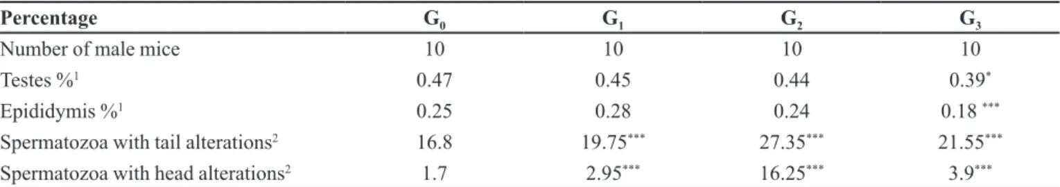
The male mice spermatozoa morphology evaluations are presented in Table III. Anomalous spermatozoa were
found in all groups, presenting tail and head alterations. The G1, G2, and G3 demonstrated statistically significant alterations when compared to G0. Furthermore, the G3
males presented testicle and epididymis weight reduction.
Table IV shows the male mice reproductive
performance. The treated groups presented a reduction in
the number of fertile animals, being significant in the G3.
Analyzes of the female mice
Parameters related to the intrauterine progeny
development are presented in Table V. The G1 demonstrated
a reduced number of implantations and live fetuses, when compared to G0. The G2 presented a higher number of reabsorptions and consequently a rise in the reabsorption index and post-implantation loss, these parameters
being statistically significant. The LPF (Lethal Prevalent Frequency) (%) analysis demonstrates that all groups
treated with the alcoholic beverages demonstrated
prevalent lethal induced mutations, statistically significant
in the G1 group which showed high mutation indices.
Analyzes of the fetuses
Table VI presents analyses of the fetuses. The weight
and length of fetuses were not statistically significant. In total, 397 fetuses were analyzed and no external congenital malformations were found. In the skeletal analysis
the following malformations were found: Sternum, interparietal, and supraoccipital. The malformations in the G3 were statistically significant. Concerning the visceral analysis, the G1 and G2 showed statistically significant
alterations in the heart, such as reduced myocardium and sponge appearance.
DISCUSSION
The ingestion of alcohol may induce impotence, infertility, and alterations in functions of male reproductive
organs (Emanuele, Emanuele, 2000). There are different
alcoholic drinks, based on the production process, through which the beverage is prepared by distillation or
fermentation processes, in addition to differences in the
raw material from which the beverage is produced, such as barley, grapes, and sugarcane.
In the toxicological analyses, negative weight
gain was observed. This weight loss can be justified by the effects of alcohol on the metabolism as, when used chronically, alcohol potentiates the Mitochondrial
System of Oxidation of Ethanol (SMOE), which occurs
on the cytochrome p-450 system. SMOE increases the thermogenic potential of food, thus increasing the basal
metabolism rate in alcoholics (Suter, Hasler, Vetter, 1997).
The Johsen score showed the spermatogenesis
TABLE III - Male mice testes, epididymis, and spermatozoa morphological evaluation
Percentage G0 G1 G2 G3
Number of male mice 10 10 10 10
Testes %1 0.47 0.45 0.44 0.39*
Epididymis %1 0.25 0.28 0.24 0.18 ***
Spermatozoa with tail alterations2 16.8 19.75*** 27.35*** 21.55***
Spermatozoa with head alterations2 1.7 2.95*** 16.25*** 3.9***
*p <0.5 and***p < 0.0011Average represented relative weight (organ weight/body weight *100) [ANOVA/Tukey]. 2[Chi-square test]. G0 - Control; G1-Beer treated group with 1.9 g/kg of ethanol; G2 – Wine treated group with 1.9 g/ kg of ethanol and G3 – Cachaça treated group with 1.9 g/kg of ethanol.
TABLE IV - Parameters related to the male mice reproductive performance
Parameter G0 G1 G2 G3
Fertile males/number of paired males Pregnant females/ number of paired females Fertility index [%]
10/10 20/20 100
8/10 14/20**
80
7/10 12/20**
70
5/10* 9/20**
50* *p < 0.5 and **p < 0.01. [Chi-square test]. G
were immature in all treated groups. Many studies have evidenced that germ cells are the target of ethanol toxicity, and this agent can promote damage during the production
of spermatozoa and differentiation of spermatozoa. The
alcohol can act directly on the testes, which may decrease
dosages of some hormones responsible for germinal maturation and reduce the nutrition that Sertoli cells provide
to germ cells. (Raychoudhury, Flowers, Millette, 2000).
The number of Sertoli cells was reduced in all treated groups. These cells have important functions in the
TABLE V - Parameters related to the intrauterine progeny development
Parameter G0 G1 G2 G3
Number of female mice 20 14 12 9
Implantations number 11.230 ± 2.618 7.916 ± 3.528* 11.250 ± 2.187 11.250 ± 2.187
Live fetuses number 10.307 ± 2.287 7.166 ± 2.979* 9.875 ± 1.457 10.125 ± 2.587
Reabsorption number 0.846 ± 0.800 0.833 ± 1.193 2.125±1.125* 0.875 ± 0.479
Reabsorption index 7.059± 5.407 8.300± 3.244 13.983± 9.739* 8.515 ± 5.323
Post-implantation loss 7.609 ± 7.495 6.912± 3.244 13.983± 9.739* 10.546 ± 5.165
Fetal viability 92.390 ± 5.767 93.087± 3.244 86.016± 9.739 89.450 ± 5.165
Placental individual weight [g] 0.106 ± 0.009 0.111± 0.011 0.101 ± 0.006 0.112 ± 0.006
Placental index 0.078 ± 0.023 0.082 ± 0.007 0.070 ± 0.003 0.079 ± 0.004
Evaluation of the weight of fetuses for pregnancy age
---- WFAPA1 WFAPA1 WFAPA1
Frequency of dominant lethal ---- 20.76%* 4.19% 1.76%
Average represented data ± SEM. *p < 0.5. G
0 - Control; G1-Beer treated group with 1.9 g/kg of ethanol; G2 – Wine treated group with 1.9 g/ kg of ethanol and G3 – Cachaça treated group with 1.9 g/kg of ethanol.1 WFAPA = Weight of fetuses was adequate for pregnancy age.
TABLE VI - Analyses of fetuses
G0 G1 G2 G3
Total number of fetuses 126 98 76 97
Individual fetal weight [g]1 1.372 ± 0.032 1.344 ± 0.055 1.430 ± 0.021 1.419 ± 0.030
Fetal length [cm]1 2.762 ± 0.047 2.711 ± 0.060 2.664 ± 0.054 2.716 ± 0.071
G0 G1 G2 G3
Skeletal Malformations2
Number of analyzed fetuses 66 53 40 51
Number of normal fetuses 57 46 33 33
Total number of malformed fetuses 9 7 7 18
Sternum malformation 5 5 5 9
Interparietal malformation 2 1 1 4
Supraoccipital malformation 2 1 1 5
%MF 13.6 13.2 17.5 35.29**
Visceral
Malformations2
Number of analyzed fetuses 60 45 36 46
Number of normal fetuses 60 38 32 45
Total number of malformed fetuses 0 9 4 1
Reduced Myocardium 0 6 3 1
Sponge Appearance of Myocardium 0 3 1 0
%MF 0 19.14*** 11.1* 2.17
*p < 0.5.**p < 0.01 and o***p < 0.001.G
0 - Control; G1-Beer treated group with 1.9 g/kg of ethanol; G2 – Wine treated group with 1.9 g/ kg of ethanol and G3– Cachaça treated group with 1.9 g/kg of ethanol.
1Average represented data ± SEM [ANOVA/Tukey].
spermatogenesis: mechanical support, germ cell nutrition and movement, paracrine regulation, phagocytosis, steroid hormones synthesis, and spermiation. The Sertoli cells are susceptible to the action of numerous toxic substances, presenting morphological and functional alterations,
induced by xenobiotics (Hess, Nakai, 2000).
The alcohol negatively affected the activity of Leydig cells, compromising the production of testosterone, which is responsible for the development and maturation of germ cells. The testosterone dosage reduced in all treated groups, in addition to which the number of Leydig cells
decreased. Testosterone is synthesized from cholesterol by a sequence of enzymatic chains within the Leydig cells (Rosenblatt et al., 2010). This can infer there is evidence
of a degenerative testicular process considering the Johsen score and decrease in testosterone, suggesting a negative impact on semen quality and fertility potential.
The morphological analysis of spermatozoa
contained in the epididymis presented tail and head
alterations. According to Villata, Ballesca, Nicolás (1997) stated that fertility disturbance is related to ethanol ingestion, besides which it can promote low spermatozoa
concentration with damage to mobility and raise the
teratozoospermia number (Gomathi, Balasubramanian, Vijayabanu, 1993). These alterations can compromise
motility, and consequently fecundation. These changes associated with chromosomal mutation can wreck the
embryo implantation by natural selection (George, Gramath, Johansson, 2006).
There was a reduction in the average weights of the testes and epididymis in the treated groups. According
to Basth, Oko, (1989) testes and epididymis weight
reductions can induce important functional variations,
compromising the spermatozoa morphology, considering
that spermatogenesis starts in the seminiferous tubules, the
formed spermatozoa migrate to the epididymis, where they mature and are stored. Gonçalves et al. (2017) showed that
the number of abnormal seminiferous tubules increased in ethanol drinking rats.
In the female analyses, the high frequency of
dominant lethal were presented in the G1. This suggests
that beer may promote mutations in germ line cells present
in the testicles. According to Leber (1988) these genes do not compromise fertilization but interfere with the normal
development of the embryo resulting in lethality in the
early stages prior to implantation. This statement justifies
the lower number of implanted and live fetuses observed in this group.
The G2 favored the abortion of fetuses in more
advanced development phases, evidenced by the rise in reabsorption numbers, reabsorption index, and
post-implantation loss. The G3 did not show a statistically
significant result related to fetal viability during the intrauterine life. This demonstrates that the fetuses that crossed the barrier of all or nothing were able to overcome the intrauterine development, but still presented a higher index of skeletal malformations. The fetuses which were exposed to fermented beverages presented higher visceral malformations.
It can be concluded that treatment of male mice
with different types of alcoholic beverages induced
different degrees of toxic effects, compromising the male
mice reproductive performance, fetal viability during
the intrauterine life, and congenital malformations (birth defects). Among the malefic effects mentioned, the
distillated beverage caused more damage to reproduction, when compared to the other beverages tested in this study.
REFERENCES
Adler ID. Spermatogenesis and mutagenicity of environmental hazards: extrapolation of genetic risk from mouse to man. Andrologia.2000;32(4-5):233-7.
Anderson P, Chishol D, Fuhr DC. Effectiveness and
cost-effectiveness of policies and programmes to reduce the harm
caused by alcohol. Lancet. 2009;373(9682):2234-46.
Andrade-Sobrinho LG, Boscolo M, Lima-Neto BS, Franco DW.
Carbamato de etila em bebidas alcoólicas (cachaça, tiquira, uísque e grapa). Quím Nova. 2002;25(6):1074-7.
Barrow MV, Taylor WJ. A rapid method for detecting malformations in rat fetuses. J Morphol. 1969;127(3):291-306.
Basth AD, Oko RJ. Abnormal morphology of bovine
spermatozoa. Ames: Iowa State University Press; 1989. 285 p.
Burgos MGPA, Medeiros MC, Bion FM, Pessoa DCNP. Efeitos de bebidas alcóolicas em mães lactantes e suas repercussões na
prole. Rev Bras Saúde Matern Infant. 2002;2(2):129-35.
Calderon IMP, Rudge MVC, Brasil MAM, Henry MACA.
Modelo experimental em ratas para estudo do binômio diabete
e gravidez. Acta Cir Bras. 1992;7(4):136-44.
Doremus TL, Brunell SC, Varlinskaya EI, Spear LP. Anxiogenic effects during withdrawal from acute ethanolin adolescent and adult rats. Pharmacol, Biochem Behav. 2003;75(2):411-8.
Emanuele MA, Emanuele N. Alcohol and the male reproductive
Effect of alcoholic beverages on progeny and reproduction of mice
George L, Granath F, Johansson AL. Risks of repeated
miscarriage. Paediatr Perinat Epidemiol. 2006;20(2):119-26.
Gomathi C, Balasubramanian K, Vijayabanu N. Effects
of chronic alcoholism on some studies on lipid profiles. J
Androl.1993;16(3):175-81.
Gonçalves DG, Vieira AN, Vieira HR; Valério AD, Siervo GEML, Pinheiro PFF, Martinez FE, Guarnier FA, Teixeira
GR, Fernandes GSA. Role of resistance physical exercise in preventing testicular damage caused by chronic ethanol
consumption in UChB rats. Microsc Res Tech.
2017;80(4):378-86.
Halsted CHMD. Nutrition and alcoholism liver disease. Sem
Liver. 2004;3:289-304.
Haseman JK, Soares ER. The distribuition of fetal death in
control mice and its implication on statistical tests for dominant
lethal effects. Mutat Res. 1976;73:133-42.
Hess RA, Nakai M. Histopathology of the male reproductive system induced by the fungicide benomyl. Histol Histopathol.2000;15(1):207-24.
Hoyme HE, May PA, Kalberg WO. A practical approach to diagnosis of fetal alcohol spectrum disorders: clarification of the 1996 Institute of Medicine criteria. Pediatrics.
2005;115(1):39-47.
Johsen SG. Testicular biopsy score countra method for registration of spermatogenesis in human testes: normal values
and results in 335 hypogonadalmals. Hormones.1970;303:2-25.
Leber AP. Chemical induction of dominant lethal mutations in
mammals. In: Allantyne B, editor. Perspectives in basic and
applied toxicology. London: John Wrigth; 1988. p. 199-205.
Michalany J. Técnica histológica em anatomia patológica: com instruções para o cirurgião, enfermeira e citotécnico. São Paulo: EPU; 1980. Operações fundamentais da técnica histológica;
p.22-31.
Pesce CM. The testicular biopsy in the evaluation of male
infertility. Sem Diagn Pathol.1987:4(4):264-74.
Purohit V. Can alcohol promote aromatization of androgens to estrogens? a review. Alcohol. 2000:22(3):123-7.
Raychoudhury SS, Flowers AF, Millette CF. Toxic effects of
polychlorinated biphenyls on cultured rat Sertoli cells. J Androl.
2000;21(6):964-73.
Rehm J, Mathers C, Popova S. Global burden of disease and injury and economic cost attributable to alcohol use and alcohol-use disorders. Lancet. 2009;373(9682):2223-33.
Rosenblatt C, Delgado Filho MA, Delgado DR, Delgado FR.
infertilidade masculina – novos conceitos. Urologia
2010;71:85-92
Sarkar M, Gangopadhyay P, Basak B, Chakrabarty K, Banerji
J, Adhikary P. Chatterjee A. The reversible anti- fertility effect
of Piper betle Linn on Swiss albino male mice. Contraception.
2000;62(5):271-4.
Shively CA, White DM, Blauch JL, Tarka Jr SM. Dominant lethal
testing of theobromine in rats. Toxicol Lett. 1984;20(3):325-9.
Staples RE, Schnell VL. Refinements in rapid clearing technic in the KOH-alizarin red S method for fetal bone. Stain Technol.
1964;39:61-3.
Suter PM, Hasler E, Vetter W. Effects of alcohol on energy
metabolism and body weight regulation: is alcohol a risk factor
for obesity? Nutr Rev. 1997;55(5):157-71.
Taylor P. Skeletal examination. In: Practical teratology. London:
Academic Press; 1986. p.77-100.
Villata J, Ballesca JL, Nicolás JM. Testicular function in
asymptomatic chronicalcoholism: relation to ethanol intake.
Alcohol Clin Exp Res. 1997;21(1):128-33.
Wilson JG. Methods for administering agents and detecting
malformations in experimental animals. In: Teratology:
principles and techniques. Chicago: University of Chicago Press; 1965. p.262-77.
Wyrobek AJ, Gordon LA, Burkhart JG, Francis MW, Kapp RW
Jr, Letz G, et al. An evaluation of the mouse sperm morphology
test and other sperm tests in non-human mammals: a report of the US. Environmental Protection Agency. Amsterdam: Gene-Tox Program; 1983. p.1-72.