UNIVERSIDADE DA BEIRA INTERIOR
Ciências da Saúde
Novas abordagens terapêuticas para regeneração
óssea
Kevin Domingos de Sá
Dissertação para obtenção do Grau de Mestre em
Ciências Biomédicas
(2º ciclo de estudos)
Orientador: Professor Doutor Ilídio Joaquim Sobreira Correia
Co-orientadores: Mestre Sónia Alexandra Pereira Miguel
Mestre Tiago Ruivo Correia
iii
List of publications
Article published in a peer reviewed international journal:
Fradique, R., Correia, T. R., Miguel, S. P., de Sá, K. D., Figueira, D. R., Mendonça, A. G., &
Correia, I. J. (2016). Production of new 3D scaffolds for bone tissue regeneration by rapid prototyping. Journal of Materials Science: Materials in Medicine, 27(4), 1-14. doi: 10.1007/s10856-016-5681-x.
Correia, T. R., Figueira, D. R., de Sá, K. D., Miguel, S. P., Fradique, R., Mendonça, A. G., &
Correia, I. J. (2016). 3D printed scaffolds with bactericidal activity aimed for bone tissue regeneration. International Journal of Biological Macromolecules. In press.
v
"When the aerials are down, and your spirit is covered with snows of cynicism and the ice of pessimism, then you are grown old, even at 20, but as long as your aerials are up, to catch waves of optimism, there is hope you may die young at 80.”
vii
ix
Acknowledgments
Firstly, I would like to thank to my supervisor professor Ilídio Correia for the opportunity to develop the theme of my master's thesis with him. For all the dedication and time spent in carrying out the whole of this project. I also thank him for the confidence deposited on me and for the opportunities he provided me during this year.
To professor Abílio Silva, for all availability to perform the mechanical characterization of the scaffolds.
To my co-supervisors and friends Sónia and Tiago, for the lunches, soccer games, daily help, and for the acquisition of the scanning electron microscopic images.
To my work group, in special to Elisabete and Marco, for all the patience and good vibes. To my best friend and little fellow in life, Daniela. Thank you for always being on my side, for all the love, confidence and support. Thank you for everything, my darling. Now, it’s time that
we began
♪.
Finally, to my extraordinary parents, Rosa and António. A very special word of recognition for them, for all the love, patience and support who gave me during all these years.
xi
Resumo
A capacidade limitada que o osso apresenta para reparar os defeitos críticos na sua totalidade requer o desenvolvimento de novos implantes capazes de melhorar o processo de recuperação. Neste contexto, têm sido desenvolvidas novas abordagens terapêuticas de forma a promover o processo de regeneração óssea, assim como evitar o surgimento de diferentes problemas associadas às terapias convencionais. Uma destas abordagens envolve o desenvolvimento de matrizes tridimensionais também designadas de scaffolds, que atuam como suportes temporários e que promovem a adesão e proliferação celular fornecendo ainda suporte mecânico durante o processo de regeneração óssea. Neste estudo desenvolveram-se scaffolds híbridos compostos por Tricalcio Fosfato/Ácido Alginico com recurso a prototipagem rápida (Fab@Home). Posteriormente, as superfícies destas estruturas foram funcionalizadas com membranas nanofibrosas produzidas pela técnica de electrofiação. Nestes revestimentos, compostos por Policaprolactona e Gelatina, foram ainda incorporados dois agentes antimicrobianos diferentes, nanoparticulas de Prata e Ácido Salicílico. Os resultados obtidos revelaram que os scaffolds produzidos apresentaram propriedades mecânicas, capacidade de absorção de água, porosidade, biodegradabilidade e biomineralização, compatíveis com a sua aplicação na área de engenharia de tecidos. Além disso, a presença da malha nanofibrosa na superfície do scaffolds melhorou a adesão e proliferação de células eucariótcas em contacto com estas estruturas tridimensionais. Por outro lado, a funcionalização da superfície dos
scaffolds permitiu evitar a formação de biofilmes na superfície dos scaffolds, durante pelo
menos 5 dias. Estes dados experimentais evidenciam o grande potencial destas estruturas para aplicação na regeneração do tecido ósseo.
Palavras-Chave
Ácido Salicílico; Atividade Bactericida; Electrofiação; Nanopartículas de Prata; Prototipagem Rápida; Regeneração Óssea;
xiii
Resumo alargado
O osso é um tecido altamente dinâmico e vascularizado responsável por conferir suporte ao corpo humano. Além disso, é responsável por permitir a locomoção do corpo atuando como escudo protetor dos principais órgãos vitais. A sua estrutura é formada por matriz orgânica (maioritariamente colagénio), matriz inorgânica (hidroxiapatita), células (osteoblastos, osteócitos e osteoclastos) e água. Apesar da elevada capacidade de auto-regeneração deste tecido, esta não é suficiente para reparar defeitos críticos que surgem com o envelhecimento, doenças ou traumas. Atualmente, o tratamento destes defeitos ósseos pode ser realizado através de autoenxertos, aloenxertos ou xenoenxertos. No entanto, a disponibilidade limitada, a frequente ocorrência de infeções e rejeições imunológicas por parte do hospedeiro podem comprometer o sucesso destas alternativas.
Neste contexto, a engenharia de tecidos tem vindo a desenvolver novas abordagens de forma a promover o processo de regeneração óssea. A engenharia de tecidos é um ramo interdisciplinar que aplica os princípios da biologia e engenharia, visando o desenvolvimento de substitutos biológicos que restaurem, mantenham ou melhorem as funcionalidades de tecidos humanos que tenham sido lesados ou comprometidos. Desta forma, têm-se desenvolvido matrizes tridimensionais (3D), também designadas por scaffolds, as quais atuam como suportes temporários no processo de formação do novo tecido ósseo. Além disso, a sua superfície deve ser capaz de mimetizar a matriz extracelular do local de implantação de forma a promover a adesão e proliferação celular. Na atualidade os scaffolds são produzidos através de uma grande variedade de técnicas, onde são usados diferentes materiais, nomeadamente polímeros, cerâmicas e metais.
O presente estudo descreve a produção e caracterização das propriedades químicas, mecânicas e biológicas de scaffolds 3D híbridos compostos por Tricalcio Fosfato/Ácido Alginico (TCP/AA). Estas estruturas foram produzidas por prototipagem rápida usando uma impressora 3D (Fab@Home), sendo posteriormente revestidas com dois tipos diferentes de matrizes nanofibrosas obtidas por eletrofiação de forma a melhorar o seu comportamento biológico. Estes revestimentos, tinham na sua composição Policaprolactona (PCL) e Gelatina (GEL). Por outro lado, de forma a conferir propriedades antimicrobianas aos scaffolds, estes foram funcionalizados com dois agentes antimicrobianos diferentes, nanopartículas de Prata (AgNPs) e Ácido Salicílico (SA).
As propriedades físico-químicas dos diferentes scaffolds produzidos (TCP/AA, TCP/AA_AgNPs e TCP/AA_SA) foram caracterizadas por Espectroscopia de Infravermelho por Transformada de Fourier, Espectroscopia de Energia Dispersiva e Microscopia Eletrónica de Varrimento. Os resultados revelaram que nenhum dos componentes sofreu qualquer tipo de alteração química
xiv
durante o processo de manufatura dos scaffolds. Além disso foi ainda possível observar a presença de uma densa malha nanofibrosa na superfície dos scaffolds TCP/AA_AgNPs e TCP/AA_SA. Este revestimento melhorou a aptidão dos scaffolds para suportar adesão e proliferação celular, em comparação com o grupo sem revestimento (TCP/AA). A porosidade e resistência mecânica apresentadas por todas as estruturas produzidas foram semelhantes às do osso trabecular. Os ensaios de citotoxicidade realizados in vitro revelaram que todas as formulações são biocompatíveis durante pelo menos 7 dias. Por outro lado, as imagens de microscopia eletrónica de varrimento revelaram que as formulações previamente revestidas (TCP/AA_AgNPs e TCP/AA_SA) apresentaram um maior número de células aderidas na sua superfície. Tal deve-se sobretudo às propriedades bioadesivas que o revestimento de nanofibras conferiu à superfície dos scaffolds. Por fim, a capacidade antimicrobiana dos materiais foi analisada através do método de Kirby-Bauer. Os resultados obtidos revelaram que os scaffolds TCP/AA_AgNPs e TCP/AA_SA são capazes de inibir o crescimento bacteriano, um facto constatado através da formação de halos inibitórios após incubação destes scaffolds com S.
Aureus. A formação de biofilmes na superfície dos materiais, foi ainda caracterizada por
microscopia eletrónica de varrimento sendo que os resultados confirmaram as propriedades antimicrobianas dos scaffolds.
Em suma, os resultados obtidos no presente estudo revelaram que os scaffolds 3D produzidos apresentam propriedades químicas, biológicas e mecânicas, que são compatíveis com a sua aplicação futura na regeneração de tecido ósseo.
xvi
Abstract
Bone limited capacity to fully repair large defects demands the development of new implants that are able to improve the healing process. In this context, new approaches for promoting bone regeneration process and also to avoid side effects associated with the therapeutics in use, are currently being studied. Herein 3D tricalcium phosphate/alginic acid scaffolds were produced using a Fab@Home and then coated with an electrospun mesh (composed by polycaprolactone and gelatin) loaded with two different antibacterial agents (silver nanoparticles and salicylic acid). The obtained results show that the produced scaffolds presented mechanical properties, swelling, macro/microporosity, biodegradation and biomineralization capacity, that are compatible with their application for bone tissue engineering purposes. Moreover, the presence of a nanofibrous mesh at the surface of produced 3D constructs enhanced cellular adhesion/proliferation and also avoided biofilm formation at scaffolds’ surface, for at least 5 days. Such results emphasize that the 3D hybrid scaffolds produced herein have the required properties for being used in the proposed biomedical application.
Keywords
Bactericidal activity; Bone regeneration; Electrospinning; Rapid prototyping; Salicylic acid; Silver nanoparticles.
xviii
Contents
1. Introduction ... 2 1.1. Bone Tissue ... 2 1.2. Bone Anatomy ... 2 1.3. Bone histology ... 4 1.3.1. Bone matrix... 4 1.3.2. Bone cells ... 4 1.3.3. Bone remodeling ... 6 1.4. Bone disorders ... 8 1.4.1. Osteoporosis ... 8 1.4.2. Paget’s disease... 9 1.4.3. Osteomyelitis ... 9 1.5. Bone grafts ... 10 1.6. Tissue engineering ... 11 1.6.1. 3D scaffolds ... 121.6.2. Biomaterials used for scaffold production ... 14
1.6.2.1. Ceramics ... 14
1.6.2.2. Polymers ... 15
1.6.2.3. Metals ... 16
1.6.2.4. Composites ... 16
1.6.3. Antimicrobial functionalization of scaffolds ... 17
1.6.3.1. Salicylic acid (SA) ... 17
1.6.3.2. Silver nanoparticles (AgNPs) ... 17
1.6.4. Techniques used for scaffolds production ... 17
1.6.4.1. 3D printing using a Fab@Home printer ... 18
1.6.4.2. Electrospinning (ES) ... 18
1.7. Aims ... 21
2. Materials and Methods... 23
2.1. Materials ... 23
2.2. Methods ... 23
2.2.1. Preparation of TCP/AA based scaffolds ... 23
2.2.2. Production and characterization of silver nanoparticles ... 24
2.2.3. Production of the electrospun nanofibers meshes... 24
xix
2.2.4.1. Attenuated Total Reflectance-Fourier Transform Infrared Spectroscopy analysis 26
2.2.4.2. Energy dispersive spectroscopic analysis ... 26
2.2.4.3. Characterization of the mechanical properties of the scaffolds ... 26
2.2.4.4. Evaluation of scaffolds’ porosity ... 26
2.2.4.5. Characterization of the swelling profile of the scaffolds ... 27
2.2.4.6. In vitro analysis of the biodegradation profile of the samples ... 27
2.2.4.7. Characterization of scaffold biomineralization activity in vitro ... 27
2.2.5. Characterization of the biological properties of the scaffolds ... 28
2.2.5.1. Evaluation of cell viability and proliferation in the presence of the scaffolds .... 28
2.2.5.2. Evaluation of the bactericidal activity of the scaffolds ... 28
2.2.5.3. Characterization of the morphology and biological performance of the scaffolds 28 2.3. Statistical Analysis ... 29
3. Results and Discussion... 31
3.1. Morphological characterization of the produced scaffolds ... 31
3.2. Characterization of the physicochemical properties of the scaffolds ... 34
3.2.1. ATR-FTIR analysis ... 34
3.2.2. Energy dispersive spectroscopy analysis... 35
3.2.3. Characterization of the mechanical properties of the produced scaffold ... 36
3.2.4. Evaluation of porosity of the scaffolds ... 38
3.2.5. Evaluation of swelling profile of the scaffolds ... 39
3.2.6. Characterization of the biodegradation profile of the scaffolds ... 39
3.2.7. Biomineralization studies ... 40
3.3. Characterization of the biological properties of the scaffolds ... 41
3.3.1. Evaluation of cell viability and proliferation in contact with the scaffolds ... 41
3.3.2. Characterization of the bactericidal activity of the scaffolds... 44
4. Conclusion and Future Perspectives ... 48
5. Bibliography ... 50
xxi
List of figures
Figure 1. Schematic representation of the internal structure and organization of bone ... 3 Figure 2. Schematic representation of the cells present in bone tissue ... 6 Figure 3. Schematic representation of the different phases of the bone remodeling process ... 8 Figure 4. Schematic representation of the main human bone disorders. ... 10 Figure 5. Schematic representation of the main types of bone grafts used in the clinic ... 11 Figure 6. Representation of tissue engineering approach used for bone regeneration... 12 Figure 7. Schematic representation of materials used in bone tissue engineering ... 15 Figure 8. Schematic representation of the Fab@Home printer used to produce 3D scaffolds .... 18 Figure 9. Schematic representation of the electrospinning apparatus used for the production of nanofibrous meshes ... 19 Figure 10. Schematic representation of the process used to produce 3D scaffolds ... 25 Figure 11. Macroscopic images of produced scaffolds ... 31 Figure 12. SEM images showing the morphology of the scaffolds ... 32 Figure 13. SEM and TEM analysis of the nanofibrous meshes present at the scaffold’s surface ... 33 Figure 14. ATR-FTIR analysis of the produced 3D scaffolds and nanofibrous meshes ... 35 Figure 15. EDS analysis of the produced 3D scaffolds ... 35 Figure 16. Macroscopic images of 3D scaffolds during compression assay ... 36 Figure 17. Characterization of the mechanical properties of the produced scaffolds ... 37 Figure 18. Characterization of the total porosity of the produced scaffolds ... 38 Figure 19. Characterization of the swelling profile of the produced scaffolds ... 39 Figure 20. Characterization of the degradation profile of the produced scaffolds ... 40 Figure 21. EDS analysis of the scaffolds after 4, 7 and 14 days in SBF ... 41 Figure 22. Microscopic images of human osteoblast cells seeded in the presence of scaffolds ... 42 Figure 23. Evaluation of human osteoblasts cell viability when they cultured in the presence of scaffolds ... 43 Figure 24. SEM micrographs images of osteoblasts morphology in the presence of the different scaffolds ... 44 Figure 25. Evaluation of the antibacterial properties of the produced scaffolds ... 45 Figure 26. SEM images of scaffolds in contact with S.aureus ... 46
xxiii
Acronyms
3D AA AgNPs ATR-FTIR BMP β -TCP CAD CFU COX DLS DMEM-F12 DNA ECM EDS EDTA ES EtOH FBS GEL GPa HA HOB IGF-1 IGF-2 IL-6 M-CSF MPa MTT NCP PBS PCL PTH PVP RANK RANKL RGD Three-Dimensional Alginic Acid Silver NanoparticlesAttenuated Total Reflectance-Fourier Transform Infrared Spectroscopy Bone Morphogenic Proteins
β -Tricalcium Phosphate Computer-Aided-Design Colony Forming Units Cyclooxygenase
Dynamic Light Scattering
Dulbecco’s Modified Eagle’s Medium Deoxyribonucleic Acid
Extracellular Matrix
Energy Dispersive Spectroscopic Ethylenediaminetetraacetic Acid Electrospinning
Ethanol
Fetal Bovine Serum Gelatin
Gigapascal Hydroxyapatite Human Osteoblast Insulin Growth Factor-1 Insulin Growth Factor-2 Interleukin-6
Macrophage Colony-Stimulating Factor Megapascal
3-(4,5-dimethylthiazol-2-yl)-2,5-diphenyltetrazolium bromide Noncollagenous Proteins
Phosphate-Buffered Saline Solution Polycaprolactone
Parathyroid Hormone Polyvinylpyrrolidone
Receptor Activator of Nuclear Receptor Activator of Nuclear Ligand Arginine-Glycine-Aspartic Acid
xxiv
RP RT SA SBF SEM TCP TE TEM TFE TGF- β Ti TNF–α UV Rapid Prototyping Room Temperature Salicylic AcidSimulated Body Fluid
Scanning Electron Microscopy Tricalcium Phosphate
Tissue Engineering
Transmission Electron Microscopy Trifluoroethanol
Transforming Growth Factors-β Titanium
Tumor Necrosis Factor-α Ultraviolet
Chapter I – Introduction
2
1. Introduction
1.1. Bone Tissue
Bone is a specialized and dynamic connective tissue that serves as the main component of the human skeleton 1-3. Bone functions include locomotion, protection of the internal organs (brain,
spinal cord, heart and lungs), hematopoiesis and mechanical support of diaphragm 3, 4.
Furthermore, it also acts as a reservoir of minerals, namely calcium and phosphorus, and promotes the attachment for muscles, ligaments and tendons 4, 5. Bone tissue is in constant
remodeling in order to be able to support biomechanical forces and remove old and micro damaged bone 3, 6.
Bone tissue possesses a complex architecture and its cells (osteoblasts, osteocytes and osteoclasts) are involved in bone maintenance and remodeling 1, 3, 4. These cells are embedded
in the bone extracellular matrix (ECM), that is composed by a mineral and an organic phase 3, 4, 7. A more detailed description of the structural organization of the human bone is given in the
following sections.
1.2. Bone Anatomy
According to their shape, bones can be classified as long, short, flat or irregular 8. Long bones,
such as clavicles, femurs and tibiae, present a cylindrical shape and great mechanical strength. Bones can be divided in three physiologic sections: the diaphysis, that composes the bulk bone, the epiphysis, located at the ends of the bone and the epiphyseal plate, located where new bone is formed during growth 8, 9. On the other hand, short bones, just as patellae, sesamoid
bones, carpal and tarsal bones, have a cubic or spherical geometry. Flat bones (e.g. mandible, skull, sternum and ribs) present thin, curved or flat shapes whereas irregular bones (e.g. vertebrae, coccyx, scrum and hyoid bone) have complex shapes that are not included in the above mentioned ones 8.
The schematic structure of the internal structure and organization of bone tissue is presented in figure 1. Morphologically, bone tissue is classified as cortical or as cancellous 4-6. Cortical
bone is almost solid with only 10% of porosity and account’s for 80% of the mass of a mature human skeleton 4, 10. It has high mechanical strength, since it consists of closely packed cortical
osteons, called Haversian systems, that form a solid and consistent mass 1, 5. The Haversian
systems have a central canal, known as Haversian canal, that is surrounded by concentric rings of matrix 5. The outer surface of cortical bone is covered by a bi-layered connective tissue
membrane, known as the periosteum 5, 11. In turn, the outer layer of the periosteum, known as
fibrous layer, is made of irregular collagenous tissue containing blood vessels and nerves, while the inner layer is composed by a single layer of bone cells 12. These features facilitate the
Chapter I – Introduction
3 fixation of tendons or ligaments to the bone. In contrast, cancellous bone is highly porous (50-90%) and has a compressive strength almost 20 times inferior to that displayed by cortical bone. It is strongly associated with metabolic activities, since its pores are interconnected and filled with bone marrow 11. Cancellous bone is arranged in a sponge-like form, with a honeycomb of
branching plates and rods of various sizes called trabeculae 5.
Cortical bone is present in the diaphysis of the long bones. Flat, short and irregular bone usually present a cancellous interior filled with marrow surrounded by two layers of cortical bone 8.
Microscopically, bone can be further classified as woven or lamellar, according to the collagen fibers orientation 5. Cortical and trabecular bone are usually formed by a lamellar pattern, in
which collagen fibrils are laid down in randomly orientations 6, 11. In contrast, woven bone
consists of collagen fibers lying parallel to each other. Due to that, woven bone is weaker than lamellar bone. Woven bone is, in fact, characteristic of embryonic and fetal development, but it is also found in the healthy adult skeleton at ligament and tendon insertions and under pathologic conditions (e.g. Paget’s disease) 8.
The schematic structure of the internal structure and organization of bone tissue is presented in figure 1.
Chapter I – Introduction
4
1.3. Bone histology
1.3.1. Bone matrix
Bone matrix is a composite of organic and mineral compounds displayed in a ratio of 35/65%, which together contribute to the strength and flexibility of the human skeleton 4, 13, 14. The
organic phase is composed of fibrillary proteins (mainly collagen type I), proteoglycans and a variety of noncollagenous proteins (NCP), whereas the mineral phase is mainly constituted by hydroxyapatite crystals 1, 4.
Collagen is the most abundant component of the organic bone matrix and is responsible for providing toughness to the bone matrix 4, 15-17. In addition to collagen, bone matrix contains
about 200 NCP that can be divided in two major groups 16, 18. One group plays a structural and
mechanical role and the other modulates the function of different bone cells by interacting with their cell-surface receptors, proteases, hormones and other biomolecules. The structural NCPs include fibronectin, osteocalcin, osteopontin, osteonectin, bone sialoprotein II, decorin and biglycan 1, 3, 4. The second group of NCPs include transforming growth factors-β (β1,
TGF-β2, and TGF-β3), insulin-like growth factors (IGFs), and bone morphogenic proteins (BMPs) 4, 16.
On the other hand, the mineral phase of bone matrix is mainly constituted by crystalline mineral salts in the form of hydroxyapatite. Although, tricalcium phosphate, calcium carbonate and fluoride derivatives are also found in this matrix. The mineral component of the bone provides tensile yield strength and also preforms important physiological functions related to the storage of ions 14, 15. It is estimated that the bones contain 99% of the calcium, 85% of the phosphorus
and 40-60% of the sodium and magnesium found in the human body 4, 15. Physiological functions,
like nerve conduction and muscle contraction depend on the ions present on this organic matrix4.
The organic and mineral phases of bone matrix must be present in a balanced way 3-5, 13. In fact,
when the mineral component is diminished, the bone becomes more flexible due to the increase of collagen 5, 14. Otherwise, if the collagen is absent, the bone becomes very brittle due to the
high mineral fraction. Besides that, bone matrix composition can also suffer variations with age, namely an increase in mineralization degree and a decrease in bone collagen content 5.
1.3.2. Bone cells
Osteoblasts, osteocytes and osteoclasts play different functions in bone maintenance and remodeling processes (see figure 2 for further details) 3, 4, 9, 19, 20.
Osteoblasts cells are derived from mesenchymal stem cells (osteogenic cells), which are essentially located in bone marrow and periosteum 4, 7, 9, 21. Osteoblast functions include the
synthesis and organization of bone ECM, its subsequent mineralization and the downregulation of osteoclasts 3, 7. Morphologically, they display a cuboidal shape and present enlarged
Chapter I – Introduction
5 osteoblasts are highly dependent on anchorage and they maintain extensive cell–matrix and cell–cell interactions through a variety of trans-membranous proteins (integrins, connexins, cadherins) and receptors (cytokines, hormones, growth factors) in order to maintain their cellular functions and responsiveness to metabolic and mechanical stimuli 3, 4, 7. The lifespan of
osteoblast is about 8 weeks in humans 1. It is noteworthy to stress that osteoblasts after being
differentiated may have three fates: revert back to bone lining phenotype; differentiate into osteocytes or undergo apoptosis 3, 15.
Osteocytes are formed through the differentiation of osteoblasts and are the most abundant cell type in bone 5, 15, 20. The primary function of osteocytes is to maintain the bone structure
since they act as mechanosensors capable of transduce musculoskeletal stress signals 4, 9, 20. In
terms of morphology, these cells are smaller than osteoblasts and have lost many of their cytoplasmic organelles 15. Moreover, they possess a higher number of filopodia, i.e.
cytoplasmatic extensions, that form an extensive connecting syncytial network, which are used for exchange of nutrients or waste products 1, 7, 15. In order to convert the mechanical stimuli
into intracellular signal, some authors suggest that nitric oxide, Wnt and cadherin-mediated signaling pathways are activated by osteoclasts 20. However, the precise mechanisms of stimulus
and response remain unclear.
Osteoclasts derive from hematopoietic stem cells and are responsible to resorb fully mineralized bone 1, 3, 4. During the motile state they migrate from the bone marrow to their resorptive site
whereas in the resorptive phase they break down bone tissue in a process named bone resorption
4, 7, 22. In each state the osteoclasts display morphological variations. Motile osteoclasts are
flattened, non-polarized cells, that are characterized by the presence of membrane protrusions, called lamellipodia, and podosome complexes containing actin 3, 7, 15. When they reach the
resorptive site, osteoclasts become polarized, through cytoskeletal reorganization. This process leads to the formation of specific membrane domains, such as sealing zone, that separates the acidic resorptive environment from the rest of the cell, forming an organelle free area 1, 3, 5, 22.
During resorption, osteoclasts are dome-shaped and lack lamellipodia 15. An activated osteoclast
is able to resorb 200,000 µm3/day of bone matrix 1 and they have an average lifespan of 15–20
Chapter I – Introduction
6
Figure 2. Schematic representation of the cells present in bone tissue. Osteoblasts, derived from osteogenic cells, are involved in bone formation; Osteocytes are responsible for the maintenance of integrity of bone tissue; Osteoclasts are involved in bone resorption.
1.3.3. Bone remodeling
Bone tissue is constantly being remodeled in order to maintain its strength and mineral homeostasis. The remodeling process consists in resorption of old bone and formation of new bone in order to prevent the accumulation of bone micro damages, and thus assure the integrity of the skeleton 4, 5, 23. As can be observed in figure 3, there are four main phases in bone
remodeling process (activation, resorption, reversal and formation stages) and the process takes about 3-6 months to be completed in humans 24. Moreover, remodeling is not only required to
replace dead or damaged tissue, but it also gives bone the capacity to adapt to changes in loading and to respond to nutritional and metabolic changes 1, 4, 24, 25.
● Activation phase
The first stage of bone remodeling involves the detection of an initiating remodeling signal, that can be a physical or a hormonal stimulus 20, 24. In case of structural damage, insulin growth
factor-I (IGF-I), tumor necrosis factor-α (TNF-α), parathyroid hormone (PTH) and interleukin-6 (IL-6) promote the activation of bone lining cells present on bone surface 7, 14, 23, 24. At the same
Chapter I – Introduction
7 time, the damage results in osteocyte apoptosis, decreased TGF-β levels and, as a consequence, the deletion of inhibitory osteoclastogenesis signals 7, 20. Therefore, osteoclastogenesis process
begins in bone marrow, and subsequently partially differentiated mononuclear preosteoclasts migrate through the blood stream and become attach to the bone matrix 24. On the other hand,
responding to osteoclastogenic stimulus, pre-osteoblasts secrete macrophage colony-stimulating factor (M-CSF), that induce the expression of Receptor Activator of Nuclear κB (RANK) by osteoclasts 7. Receptor Activator of Nuclear κB ligand (RANKL)/RANK interaction
triggers pre-osteoclasts fusion and differentiation toward mature osteoclasts 23, 24.
Another osteoclast activation signal can be induced by hormones. PTH is an endocrine remodeling signal produced to maintain calcium homeostasis. This hormone is secreted by the parathyroid glands in response to low serum calcium levels. When in contact with the bone tissue, PTH activates the PTH receptor, (a G-protein-coupled receptor, on the surface of osteoblasts) 7. This binding activates protein kinase A, protein kinase C, and calcium
intracellular signaling pathways in these cells and induces several transcriptional responses that produce/modulate the secretion of molecules that recruit osteoclast precursors, induce osteoclast differentiation and activation, processes that are crucial for bone resorption 14, 23.
● Resorption phase
The resorption phase, which has an average duration of 30–40 days, consists in the attachment of differentiated osteoclasts to depressions or resorptive bays, known as Howship lacunae, present in bone matrix 23, 24. Osteoclast anchorage to bone surfaces occurs through anchoring
proteins such as integrins (e.g. αvβ3) 14. Subsequently, osteoclasts promote the bone
demineralization process 24. This process is performed in two steps: i) acidification of the bone
matrix to dissolve its mineral component, and ii) release of lysosomal (e.g. cathepins K), and non-lysosomal (e.g. collagenase) enzymes, that are responsible for the degradation of the organic component of bone 14, 23. When the cavity reaches a depth of about 60 µm from the
surface in trabecular bone and about 100 µm in cortical bone, osteoclasts undergo to apoptosis
25. This is a physiological consequence required to avoid an excessive bone resorption 24.
● Reversal phase
Reversal phase represents the transition from osteoclastic to osteoblastic activity and lasts approximately 9 days 7, 24. At this stage, mononuclear cells of osteoblastic lineage remove the
remaining undigested demineralized collagen present in the Howship lacunae and prepare the bone surface for the subsequent osteoblast-mediated bone formation 7, 23. This phase is also
characterized by the activation of factors that stimulate osteoblast precursors to proliferate, including IGF-2 and TGF-β 1, 23, 24.
Chapter I – Introduction
8 ● Formation phase
At this stage, differentiated osteoblasts synthesize new organic matrix and release vesicles that contain calcium, phosphate and enzymes that destroy the mineralization inhibitors, such as pyrophosphate or proteoglycans 1, 7, 23, 24. The remodeling cycle is concluded when all reabsorbed
bone is replaced.
At the end of the process, osteoblasts may enter in apoptosis, revert back to bone lining phenotype, or differentiate into osteocytes within the matrix 7, 25.
Figure 3. Schematic representation of the different phases of the bone remodeling process.
1.4. Bone disorders
Even though the bone remodeling process is one of the most reliable in the body, there are circumstances where it fails. In fact, the pathologies inherent to bone disorders are almost always related to this cycle 22, 26. Abnormal growth can cause gigantism or dwarfism, while
abnormal collagen contents can lead to osteogenesis imperfecta. Mineral and vitamin deficiencies cause rickets and bacterial infections can cause osteomyelitis 7, 27. Figure 4 presents
an overview of the pathologies associated with some bone disorders.
1.4.1. Osteoporosis
Osteoporosis is a common musculoskeletal disease characterized by an increased bone resorption and, as consequence, decreased bone mineral density 7, 15, 26, 28. It is believed that
sex steroid hormones, either directly or indirectly, regulate the production of the cytokines (e.g. M-CSF) that promote the production of osteoclasts 26, 29. Therefore, this disease affects
Chapter I – Introduction
9 mainly the elderly population, especially woman beyond the age of 50 years due to menopausal estrogen deficiencies that increase the bone resorption 22. Contrariwise, male population have
a more gradual decrease in sex steroid hormonal levels with aging, which may account for a less severe decrease in bone strength 7, 15, 30. The most common osteoporotic fractures involve the
hip, vertebral column, and forearm and may result in morbidity or in severe cases the death of patient 22, 31. Unhealthy diet (low calcium or vitamin D intake, alcohol abuse, and high caffeine
intake), sedentary lifestyle, nulliparity, aging, smoking, and low body weight are also risk factors that may trigger the development of osteoporosis 15.
1.4.2. Paget’s disease
Paget's disease is the second most common metabolic bone disease after osteoporosis 7. This
disease is characterized by focal areas of excessive bone resorption alternated with areas of increased bone formation, leading to the formation of abnormal bone, pain, pathologic fractures, deafness and nerve compression syndromes 22, 32, 33. Paget’s disease has an increased
prevalence in population with an age over 40 years, affecting about 5% of women and 8% of men at age of 80 22, 32, 33. In this disease, the osteoclast precursors from the bone marrow show
increased sensitivity to factors that stimulate bone resorption including RANKL 7, 28. Genetic
factors and gene mutations have also an important role in this disease 33. Nevertheless,
sedentary lifestyle and deficient nutrition are also factors that may be involved in the Paget’s disease arising.
1.4.3. Osteomyelitis
Osteomyelitis is defined as an inflammation of bone tissue accompanied by its destruction that occurs due to a microbial infection 27, 28, 34. Furthermore, it can also be triggered by fractures
that occur due to trauma or other diseases 28. Mast et al reported that osteomyelitis incidence
after severe open fractures is higher than 44% 34. Moreover Staphylococcus aureus (S. aureus) is
the most common pathogen responsible for the emerge of this disease 27. This microorganism
develops a range of extracellular and cell-associated factors, that promote its colonization capacity and virulence 27.
Chapter I – Introduction
10
Figure 4. Schematic representation of the main human bone disorders. Osteoporosis is mainly characterized by bones with high porosity; Paget´s disease is characterized by the creation of deformed bone; Osteomyelitis is characterized by the fungal presence on bone tissue.
1.5. Bone grafts
Nowadays, autografts (figure 5A) remain as the most common therapy used for the treatment of osseous defects 21, 35, 36. This procedure involves the transplantation of bone tissue from
another part of the patient’s body (usually trabecular bone from iliac crest) to the bone-damaged site 19. Although it has a good rate of success, the number of cases in which this
procedure can be used is restricted, due to the limited amount of available tissue and also by the induction of morbidity at the donor site 19, 21, 37. Consequently, allograft and xenografts
(figure 5B and C) (bone from human and non-human cadavers, respectively), are an alternative that in some cases is used by surgeons 36, 37. However, these procedures cannot be seen as
efficient therapeutic alternatives to autografts since they present risks of immune rejection, viral transmission and their use is dependent on stocks available on bank tissues 19, 21, 36.
Hence, in the last years, the limitations associated with the current clinical approaches lead to the development of alternative bone repair techniques based on Tissue Engineering (TE) 38.
Chapter I – Introduction
11
Figure 5. Schematic representation of the main types of bone grafts used in the clinic. Autografts are obtained from the patient’s body (A); Allografts are obtained from individuals of the same species (B); Xenografts are obtained from organisms of other species (C).
1.6. Tissue engineering
TE is an interdisciplinary field of research that applies the principles of engineering and life sciences towards the development of biological substitutes that restore, maintain, or improve damaged tissue functions 19, 36, 39. Recently, this research area emerged as a new and promising
approach for bone repair and regeneration 3. The main strategy of TE involves the
functionalization of supporting three-dimensional (3D) structures with cells that can be isolated from healthy humans, nanoparticles, growth factors or other bioactive molecules, in order promote the re-establishment of the structure and functions of the different tissues (figure 6)
Chapter I – Introduction
12
Figure 6. Representation of tissue engineering approach used for bone regeneration. Cell isolation from healthy human (1); Cell culture (2); Incorporation of cells, growth factors, nanoparticles or other molecules within scaffolds (3); Implantation of the scaffold on the injured patient (4).
1.6.1. 3D scaffolds
In particular, bone TE revolutionized the therapeutic approaches used for bone reconstruction due to the development of 3D scaffolds 38. The aim of these scaffolds is to restore the structure
and functions of native bone tissue by acting as a temporary support for cell proliferation and extracellular matrix deposition 19, 38, 39.
An ideal scaffold should meet a number of key requirements, which determine their suitability for tissue engineering purposes:
● Biocompatibility
Biocompatibility represents the ability of a material to support normal cellular activity including adhesion, proliferation and differentiation without eliciting any local or systemic immune response 38, 40, 42. Generally, scaffolds should be fabricated from materials that do not trigger
the immunogenic reactions. Furthermore, the degradation products of the scaffolds must also be biocompatible and easily eliminated from the body 42, 43. This property is directly related
Chapter I – Introduction
13 with interactions between blood, scaffold surface, and degradation products, which are released from the scaffold 39, 42, 44.
● Biodegradability
An ideal 3D scaffold should be degraded at a rate that iscompatible with the rate of bone growth
38, 39, 43. In addition, the degradation products of the scaffolds must not elicit any adverse effect
on the host 40. Therefore, the degradation profile of the scaffold should be regarded in order to
fulfil the requirements of the patient (i.e. age and type of injured bone) 42.
● Surface properties, osteocondutivity and osteoinductivity
Surface properties (i.e. charge, roughness, hydrophilicity and chemical composition) are determinant for controlling cellular adhesion and proliferation 3, 42, 43. These properties are
directly related with the osteoconductive and osteoinductive capacity of the 3D scaffold 36.
Osteoconduction is a process by which bone cells migrate to the surface of the scaffold through a fibrin clot, that is formed immediately after the material be implanted 19, 36, 40. On the other
hand, osteoinduction is a process by which the osteoprogenitor cells are stimulated to undergo through an osteogenic differentiation process 42. Therefore, the use of osteoconductive and
osteoinductive materials in bone tissue engineering is required to accomplish an effective bone regeneration process 36. Osteoconduction is promoted by rough and positively charged surfaces
that create a matrix compatible with cell adhesion and proliferation 3, 39, 40, 42, 45. The scaffolds
may also release osteoinductive signals through the action of growth factors or other bioactive molecules that induce differentiation of bone cells 19, 36, 42.
● Antimicrobial activity
Bacterial infections are currently regarded as the most severe and devastating complications associated with the implantation of biomaterials in the human body 46, 47. Nowadays, it is
estimated that 65-80% of bacterial infections are caused by organisms that form biofilms on implants’ surface compromising their successful application 46, 48. Furthermore, it is estimated
that S. aureus strain is responsible for 30% of all implant infections 46. In order to reduce this
problem, many efficient strategies can be adopted, namely by avoiding bacterial adhesion or the release of antimicrobial agents 47. Commonly antibacterial agents incorporated in bone
scaffolds include metallic nanoparticles, antibiotics, antimicrobial polymers, among others 47, 49.
● Porosity
Porosity is defined as the percentage of void space available in scaffolds. An ideal 3D scaffold should have a suitable interconnected porosity (higher than 90%) in order to ensure support for cellular penetration, differentiation, and consequently improve new bone tissue formation 37,
Chapter I – Introduction
14
38, 40. Regarding the pore size, it must be between 100-300 μm, for allowing a successful diffusion
of essential nutrients, oxygen, cellular metabolites and eventual blood vessels in-growth 13, 43, 45. However, scaffolds with an excessive porosity present compromised mechanical properties 38, 42, 43. Furthermore, the porosity also influences the rate of degradation 43. In fact, a high
porosity area enhances the interaction of scaffold with host tissue, which results in materials degradation 13, 38.
● Mechanical properties
Bone scaffolds are responsible for providing mechanical integrity at the lesion site until the new bone is fully matured 38, 39, 42. The mechanical properties of the scaffold should be similar to
that displayed by native bone tissue in order to support the handling during implantation and contraction forces that occur during bone regeneration. Mechanical properties of bone vary widely from cancellous to cortical bone 5, 42. Young’s modulus of cortical bone is comprised
between 15-20 GPa while for cancellous bone it is between 0.1-2 GPa 40. In turn, the compressive
strength is comprehended between 100-200 MPa for cortical bone, and 2-20 MPa for cancellous bone 40. In order to achieve optimal results, it is necessary to carefully optimize the mechanical
properties of a scaffold with its degradation kinetics 39. In fact, a scaffold material needs to
degrade at a controlled rate, that allows the maintenance of scaffold’s integrity along the healing process 39, 42.
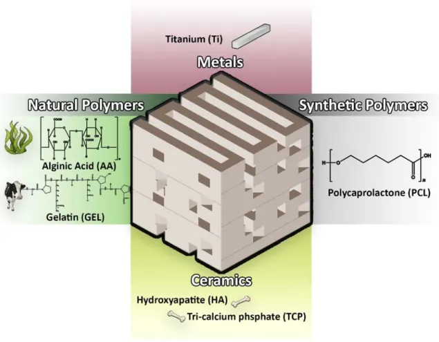
1.6.2. Biomaterials used for scaffold production
The selection of the most appropriate biomaterial for the production of scaffolds aimed for bone tissue engineering applications is a crucial step since its properties will determine the final properties of the scaffold 21, 42, 50. Various materials have been used, so far, for the production
of scaffolds that meet the requirements mentioned above 39, 50. Generally, due to their chemical
composition the materials can be divided in to four main groups: ceramics, polymers, metals and composites 21.
1.6.2.1. Ceramics
Ceramic scaffolds are typically characterized by their high mechanical stiffness, very low elasticity and a hard surface 36. Generally, bone ceramic scaffolds are produced with materials
containing calcium, such as hydroxyapatite (HA) and tricalcium phosphate (TCP) 13, 21, 36, 50. These
ceramics are valuable options for bone tissue engineering since they exhibit excellent biodegradability and biocompatibility behavior, due to their chemical and structural resemblance to the mineral phase of native bone 21, 42, 51. Moreover, ceramics can promote
interactions with osteogenic cells, enhancing osteoblasts differentiation, proliferation and consequently improve the bone healing process 13, 52. In addition, it is described that these
bioactive ceramics are capable of inducing mineralization at the surface of the scaffolds, increase their biointegration, and consequently the bone regeneration process 13, 42. However,
Chapter I – Introduction
15 their clinical applications for tissue engineering purposes has been limited due to their brittleness and shape related issues 50.
1.6.2.2. Polymers
Polymers are macromolecules composed of many repeated subunits, that present bioactivity, biodegradability and flexibility properties that are fundamental for their use in bone tissue engineering 3, 38, 42, 50, 53. According to their source, polymers can be categorized as natural and
synthetic 45, 50.
Figure 7. Schematic representation of materials used in bone tissue engineering.
● Natural polymers
Natural polymers (e.g. alginic acid (AA), cellulose, chitosan, collagen, gelatin (GEL), hyaluronic acid and silk) are obtained from natural sources, such as plants and animals 3, 54, 55. These
materials present as main advantages their low immunogenic potential and biocompatible behavior 45, 50, 54. On the other hand, the limitations of naturally derived polymers include weak
mechanical behaviour, fast degradation rates as well as hard processing and purification 45, 53, 54.
Chapter I – Introduction
16 AA is an example of a natural polymer extracted from cell walls of brown algae 56, 57. This
polymer is a polyanion composed of two repeated monomer units: β-d-mannuronate (M) and α-l-guluronate (G) 56. Physical and mechanical properties of AA are highly related to the chain
length and properties of the guluronate present on its composition 56, 58. Alginate, with a high
content of G residues becomes stiff and stable, whereas AA with a low content of G residues is more elastic and less stable 58, 59.
Additionally, GEL is obtained from collagen denaturation, which is gathered from animals 38, 53.
This negatively charged protein contains sequences composed of Arginine, Glycine, and Aspartic Acid (RGD), that improve cell interaction with the biomaterial and, as consequence, enhances the deposition of new bone tissue 45, 5353.
● Synthetic polymers
Synthetic polymers are chemically synthetized polymers that are widely used in the bone tissue engineering field, due to their high versatility and reproducibility 38, 40, 42, 45. In comparison to
natural polymers, they are generally less biocompatible and more resistant. One of the most common polymers used for bone regeneration is Polycaprolactone (PCL) 3, 13. PCL is an aliphatic
linear polyester, that is synthesized through ring-opening polymerization of ε-caprolactone. It is characterized by being biocompatible, bioresorbable and inexpensive 3, 50. PCL degrades
through of hydrolysis of its ester linkages in physiological conditions and, therefore, has been used as a valuable material for tissue engineering applications 38, 40. However, the use of PCL in
tissue engineering is compromised by its hydrophobic nature, resulting in limited cellular adhesion and uncontrolled biological interaction 40.
1.6.2.3. Metals
Metals are characterized by presenting a great compressive strength and fatigue resistance 54.
The most common metal used in scaffolds production is titanium (Ti) 40. Ti implants are widely
used due to their mechanical properties. However, several complications have been associated with these type of implants, such as infections or excess of fatigue loading. Moreover, the use of metallic implants can release toxic ions or particles through corrosion causing inflammation, tissue loss and in severe cases patients dead 40, 60.
1.6.2.4. Composites
Composite scaffolds have in their constitution different materials, like ceramics and polymers
13, 40, 51. These combinations are carried out in order to overcome the disadvantages of single
material 43. This combination allows a creation of structures with an excellent balance between
strength and toughness 13, 43, 51, 52. Specifically, to overcome the brittleness of the ceramics, they
are combined with polymers in order to increase the flexibility of the composite materials, making them more suitable for bone tissue engineering applications 43, 52, 61.
Chapter I – Introduction
17
1.6.3. Antimicrobial functionalization of scaffolds
As described above, bacterial colonization of scaffold’s surface is one of the worst possible events, that can occur when materials are implanted in the human body, leading to an obligatory implant removal from the body 46, 47. S. Aureus is usually responsible for this type of
complications 46.
In order to surpass these drawbacks, the development of scaffolds endowed with antimicrobial activity is fundamental to avoid infections related to biomaterials implantation 47. Many
approaches have already been adopted in order to confer antimicrobial properties to tissue engineering constructs, such as modification of surface charge, incorporation of metallic nanoparticles, quaternary ammonium compounds, halogens, antibiotics or drugs 47, 49, 62.
1.6.3.1. Salicylic acid (SA)
SA is a phenolic compound produced by plants Nicotiana tabacum, Cucumis sativus, and
Arabidopsis thaliana 63, 64. This non-steroidal anti-inflammatory drug plays an important role in
several physiological processes, such as the induction of plant defense responses against pathogen attacks 63. SA owns anti-inflammatory, analgesic, antipyretic, and antimicrobial
properties that can promote bone regeneration 49, 63. Despite of their attractive properties, SA
is not yet widely used in bone tissue engineering applications. Recently, Griffin and collaborators produced SA-derived poly(anhydride-ester) electrospun fibers for the regeneration of the peripheral nervous system. The authors concluded that SA-based polyanhydride fibers may offer great number of advantages not only for nerve regeneration but also for a variety of biomedical applications 65.
1.6.3.2. Silver nanoparticles (AgNPs)
AgNPs are the most studied metallic nanoparticles for the prevention of implant-based infections 66, 67. The antimicrobial activity of AgNPs have been attributed mainly to its oxidized
form (Ag+), which is able to anchor to the bacterial cell wall and to penetrate through it, thereby causing the disruption of cell membrane 66. This metallic compound can also interact
with the thiol groups of many vital enzymes and, subsequently, inactivate them 67. Moreover,
silver ions act on the sulfur and phosphorus components of DNA leading to inhibition of DNA replication 66. Due to their high surface area to volume ratio, AgNPs present enhanced reactivity
against a range of different bacterial strains that have clinical relevance 67.
1.6.4. Techniques used for scaffolds production
To fabricate scaffolds aimed for bone tissue engineering, various approaches can be used 3, 38, 45. Generally, conventional fabrication techniques such as, salt leaching, gas forming, phase
separation and freeze-drying do not enable the precise control of internal scaffold architecture or the fabrication of complex architectures 3, 38, 40, 45, 50. Rapid prototyping (RP) approaches
Chapter I – Introduction
18 comprise 3D printing, elective laser sintering, stereolithography, and fused deposition modeling that allow the production of scaffolds with interconnected pores 38, 50. These techniques have
also a better design repeatability and allow a creation of scaffolds with good mechanical requirements 40, 50.
1.6.4.1. 3D printing using a Fab@Home printer
3D printing is one of the most used RP technique in the area of tissue engineering. In particular, 3D printing using a Fab@Home printer (Figure 8) allows the use of a wide range of materials, such as composites compromising ceramics and polymers 38, 50. The deposition of successive
layers to produce the final 3D model, enables a better control of pore sizes, morphology, and overall matrix porosity in comparison to other fabrication methods. During the fabrication process, the syringe content is compressed in order to extrude the 3D structure onto a platform, according to the CAD (computer-aided design) file 40. This feasible, cheap and reproducible
technique allows the creation of complex 3D structures with high resolution and with a controlled internal architecture 50, 51. Moreover, structures produced by 3D printing present
regular morphology and improved mechanical properties according to the demands of the damaged bone 50, 51.
Figure 8. Schematic representation of the Fab@Home printer used to produce 3D scaffolds used in this study.
However, this technique possesses limitations like other techniques. The scaffolds produced by 3D printing are not fully capable of reproducing the ECM of bone tissue, thus compromising the bone healing process 68.
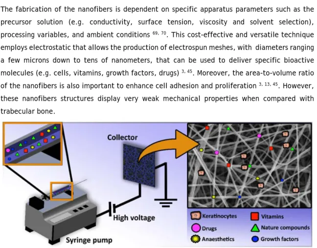
1.6.4.2. Electrospinning (ES)
In order to mimic the ECM and enhance the bone healing process, ES arise as a feasible alternative. The term ES was proposed by Reneker and co-workers in the 1990s based on the
Chapter I – Introduction
19 earlier used term, ‘electrostatic spinning’ 69-71. A basic ES system (Figure 9) is usually composed
by four main components, namely, a syringe pump (responsible to control the flow rate of polymers), a voltage power supply (provides the force to stretch the charged polymer solution into fibers), a needle (which shifts the solution into the high electric field) and a collector (where electrospun nanofibers are collected) 45, 71. During the ES process, the syringe pump is
used to force the content through a needle attached to the syringe with a controlled flow rate
69, 72. When a high voltage is applied at the tip of the capillary needle, it induces repulsive
electrical forces on the droplet surface. The charge causes the elongation of the droplet into a cone, known as the Taylor Cone 69. Once the voltage reaches a critical value the repulsive
electrostatic forces overcome the surface tension forces and a liquid jet emerges from Taylor Cone, reaching the collector in few nanoseconds 70. While the jet moves toward the collector,
the solvent evaporates and ultra-thin dry fibers are collected on the metallic collector 70.
The fabrication of the nanofibers is dependent on specific apparatus parameters such as the precursor solution (e.g. conductivity, surface tension, viscosity and solvent selection), processing variables, and ambient conditions 69, 70. This cost-effective and versatile technique
employs electrostatic that allows the production of electrospun meshes, with diameters ranging a few microns down to tens of nanometers, that can be used to deliver specific bioactive molecules (e.g. cells, vitamins, growth factors, drugs) 3, 45. Moreover, the area-to-volume ratio
of the nanofibers is also important to enhance cell adhesion and proliferation 3, 13, 45. However,
these nanofibers structures display very weak mechanical properties when compared with trabecular bone.
Figure 9. Schematic representation of the electrospinning apparatus used for the production of nanofibrous meshes that coated the 3D scaffolds.
To surpass such constraint, researchers are currently developing new 3D structures that combine different production methods, like 3D Printing and ES, in order to produce constructs that can be used in bone tissue engineering. Recently, Yeo and Kim fabricated PCL/β-TCP composite scaffolds by 3D printing and coated them with collagen/hydroxyapatite nanofibrous meshes 73.
Chapter I – Introduction
20 The hybrid scaffolds produced in this study showed improved mechanical properties and enhanced cell adhesion and proliferation, features that are crucial for a faster and effective bone repair process be attained 73.
Chapter I – Introduction
21
1.7. Aims
The overall aim of the present thesis was to develop and characterize new 3D hybrid scaffolds coated with nanofibers containing antimicrobial agents that can be used in bone tissue engineering.
The specific aims of this study were:
● Optimization of the viscosity of the solutions ● Design and production of 3D hybrid scaffolds
● Study the antimicrobial activity of the 3D hybrid scaffolds produced
● Evaluation of the mechanical, physicochemical and biological properties of the produced 3D hybrid scaffolds
Chapter II – Materials and Methods
23
2. Materials and Methods
2.1. Materials
3-(4,5-dimethylthiazol-2-yl)-2,5-diphenyltetrazolium bromide (MTT) was purchased from Alfa Aesar (Ward Hill, USA). Trifluoroethanol (TFE) was purchased from Acros Organics (New Jersey, USA). Alginic acid (AA), Amphotericin B, Calcium Chloride (CaCl2), Dulbecco’s modified Eagle’s
medium (DMEM-F12), Ethylenediaminetetraacetic acid (EDTA), Gelatin 180 bloom (GEL), gentamicin, kanamycin, LB Broth, Phosphate-buffered saline solution (PBS), Polycaprolactone (PCL) (molecular weight 80,000 g mol-1), Sodium borohydride (NaBH
4) and Trypsin were
purchased from Sigma-Aldrich (Sintra, Portugal). Dimethyl sulfoxide 99.9% (DMSO) was obtained from Thermo Fisher Scientific (Rockford, IL,USA). Fetal bovine serum (FBS) (free from any antibiotic) was acquired from Biochrom AG (Berlin, Germany). Normal human osteoblast (hOB) (406-05f) cryopreserved cells were purchased from Cell Applications, Inc. (San Diego, USA). Polyvinylpyrrolidone (PVP) (molecular weight 44,000 g mol-1) was obtained from BDH Chemicals
Ltd (Poole, UK). Silver nitrate (AgNO3), Sodium hydrogen carbonate (NaHCO3) and Tricalcium
phosphate (TCP) were bought from Panreac (Barcelona, Spain).
2.2. Methods
2.2.1. Preparation of TCP/AA based scaffolds
To produce 3D scaffolds with a proportion of 50/50 (w/w) of TCP/AA, a 15% (w/v) of AA solution was prepared by dissolving the polymer in double deionized and filtered water (obtained using a Milli-Q Advantage A10 ultrapure Water Purification System, resistivity=18.2 MΩ/cm, at 25°C) with overnight agitation. The solution was then homogenized using a X10/25 Ultra-turrax® (Ystral, Germany) for 30 min. Then, TCP powder was added to the AA solution in a 50/50 ratio followed by homogenization. After the dissolution of all the components, 5% (w/w) CaCl2
(crosslinking agent) was added to the composite mixture until a ratio of 1:2 of CaCl2:AA was
obtained. Alginate polymer chains got crosslinked (calcium ions replace the sodium ions of AA) leading to an increase of the solution’s viscosity that is fundamental for scaffolds production. Lastly, a syringe (10 cc Luer Lock) was filled with the CaCl2:AA solution in order to proceed to
solution extrusion using a Fab@Home (see figure 1A for further details). After their production, the scaffolds were immersed in a 5% CaCl2 solution for 24 h to achieve a complete crosslinking
of the printed structure and clear-cut. Afterward, scaffolds were air-dried at room temperature (RT) for 48 h.
Chapter II – Materials and Methods
24
2.2.2. Production and characterization of silver nanoparticles
AgNPs were produced by performing the chemical reduction of silver, as previously described by Silva and colleagues 74. Briefly, AgNPs were produced using AgNO
3 as a metal precursor and
NaBH4 as reducing agent. Initially, 5 mL of AgNO3 solution (4 mM) were added dropwise to 30
mL of NaBH4 solution (4 mM), under constant stirring at RT, until an AgNO3:NaBH4 molar ratio of
1:5 was achieved. The production of AgNPs was confirmed by the change in the solution color (from colorless to yellow). Subsequently, 5 mL of 0.2 % PVP solution, one of the most commonly used metal stabilizers, were added to the AgNPs already produced in order to avoid nanoparticles aggregation 74. All procedures were performed in the dark in order to prevent the
photodecomposition of AgNO3.
The hydrodynamic diameter of the produced nanoparticles was determined by dynamic light scattering (DLS) analysis, using a Zetasizer Nano ZS particle analyzer (Malvern Instruments, Worcestershire, UK).
2.2.3. Production of the electrospun nanofibers meshes
Different polymeric solutions composed of GEL/PCL loaded with AgNPs and GEL/PCL loaded with SA were produced. Briefly, 8% GEL and 6% PCL (combined at 1:1 w/w ratio) were dissolved in 80% TFE (w/v). After the dissolution, AgNPs (0.04 M) (w/v) were added to the polymeric blend. For GEL/PCL loaded with SA production, 8% GEL, 6% PCL and 8% SA (combined at 1:1:1 w/w ratio) were dissolved in TFE 80 % (w/v). Each solution was then electrospun at constant flow rate of 2.8 mL/h for 20 min, at 18 kV, with 15% humidity, and at a temperature of 26°C in order to coat TCP/AA scaffold’ surface using a rotative collector (Figure 10). The system setup was comprised of a high voltage source (Spellman CZE1000R, 0–30 kV), a syringe pump (KDS-100), a plastic syringe with a stainless steel needle (21 Gauge) and an aluminum disk connected to a copper collector, placed at a working distance of 10 cm.
Chapter II – Materials and Methods
25
Figure 10. Schematic representation of the process used to produce 3D hybrid scaffolds. Scaffolds were initially produced using a RP technique (A) and subsequently coated with an electrospun mesh loaded with AgNPs or SA, respectively (B).
Chapter II – Materials and Methods
26
2.2.4 Physicochemical and morphological characterization of the scaffolds
2.2.4.1. Attenuated Total Reflectance-Fourier Transform Infrared Spectroscopy analysisTo characterize the composition of the produced scaffolds, Attenuated Total Reflectance-Fourier Transform Infrared Spectroscopy (ATR-FTIR) analysis was performed. The spectra acquired for the different samples represent an average of 128 scans, between 400 and 4000 cm-1, with a spectral resolution of 4 cm-1. All the samples were crushed to powder, before being
analyzed, mounted on a diamond window, and the spectra were recorded using a Nicolet iS10 FTIR spectrophotometer (Thermo Scientific, Waltham, MA, USA). Moreover, all raw materials used for scaffold and nanofibrous mesh production were analyzed, in order to compare the spectra of individual components with those of the produced scaffolds 51, 75, 76.
2.2.4.2. Energy dispersive spectroscopic analysis
In order to perform the elementary analysis of the samples, an energy dispersive spectroscopic (EDS) technique was used. The different samples were placed on aluminum stubs, and then analyzed using a XFlash Detector 5010 (Bruker Nano, Germany).
2.2.4.3. Characterization of the mechanical properties of the scaffolds
The mechanical behavior of the scaffolds, in wet and dry conditions, was evaluated through compression assays as previously described elsewhere 76. All the measurements were performed
at RT using a Shimadzu AG-X Tensile Testing Machine (Tokyo, Japan), with a crosshead speed of 2 mm/min and a load cell of 10 kN. Five specimens of each sample, in the dry and wet state, were used in each assay 76-78.
The compressive strength (Cs) of the scaffolds was calculated according to equation (1). Cs = w ×F l (1)
Where F corresponds to the load at the time of fracture, w and l represent the width and length of the scaffold, respectively.
The Young Modulus (YM) was calculated through equation (2). YM = HCs
d (2)
Where Hd stands for the height deformation at maximum load, and Cs is the scaffold compressive
strength obtained from equation 1.
2.2.4.4. Evaluation of scaffolds’ porosity
The porosity of the different scaffolds was determined using a liquid displacement method, adapted from Torres and collaborators 78. Briefly, 3 specimens of each formulation were