Journal of Experimental Botany, Vol. 71, No. 11 pp. 3340–3349, 2020 doi:10.1093/jxb/eraa066 Advance Access Publication 4 February 2020
This paper is available online free of all access charges (see https://academic.oup.com/jxb/pages/openaccess for further details)
Abbreviations: AGMPF, algal growth and morphogenesis-promoting factor; DMSP, dimethylsulfoniopropionate; MICP, metal isotope-coded profiling; UCM,
Ulva culture medium; UHPLC, ultra-high performance liquid chromatography
© The Author(s) 2020. Published by Oxford University Press on behalf of the Society for Experimental Biology.
This is an Open Access article distributed under the terms of the Creative Commons Attribution License (http://creativecommons.org/licenses/by/4.0/), which permits unrestricted reuse, distribution, and reproduction in any medium, provided the original work is properly cited.
RESEARCH PAPER
Macroalgal–bacterial interactions: identification and
role of thallusin in morphogenesis of the seaweed Ulva
(Chlorophyta)
Taghreed Alsufyani1,2,*, Gianmaria Califano1,*, Michael Deicke1,*, Jan Grueneberg1,3,*, Anne Weiss1,3,*, Aschwin H. Engelen4, Michiel Kwantes1, Jan Frieder Mohr1,3, Johann F. Ulrich1 and Thomas Wichard1,3,†, 1 Institute for Inorganic and Analytical Chemistry, Friedrich Schiller University Jena, Lessingstr. 8, D-07743 Jena, Germany 2 Algal Research Laboratory, Chemistry Department, Science Faculty, Taif University, Taif 26571, Saudi Arabia
3 Jena School for Microbial Communication, D-07743 Jena, Germany
4 Centre for Marine Sciences (CCMAR), University of Algarve, Campus de Gambelas, 8005-139 Faro, Portugal * These authors contributed equally to this work.
† Correspondence: [email protected]
Received 20 October 2019; Editorial decision 28 January 2020; Accepted 1 February 2020 Editor: Andreas Holzinger, University of Innsbruck, Austria
Abstract
Macroalgal microbiomes have core functions related to biofilm formation, growth, and morphogenesis of seaweeds. In particular, the growth and development of the sea lettuce Ulva spp. (Chlorophyta) depend on bacteria releasing morphogenetic compounds. Under axenic conditions, the macroalga Ulva mutabilis develops a callus-like phenotype with cell wall protrusions. However, co-culturing with Roseovarius sp. (MS2) and Maribacter sp. (MS6), which produce various stimulatory chemical mediators, completely recovers morphogenesis. This ecological reconstruction forms a tripartite community which can be further studied for its role in cross-kingdom interactions. Hence, our study sought to identify algal growth- and morphogenesis-promoting factors (AGMPFs) capable of phenocopying the activity of Maribacter spp. We performed bioassay-guided solid-phase extraction in water samples collected from U. mutabilis aquaculture systems. We uncovered novel ecophysiological functions of thallusin, a sesquiterpenoid morphogen, identified for the first time in algal aquaculture. Thallusin, released by Maribacter sp., induced rhizoid and cell wall formation at a concentration of 11 pmol l−1. We demonstrated that gametes acquired the iron complex of thallusin, thereby linking morphogenetic processes with intracellular iron homeostasis. Understanding macroalgae–bacteria interactions permits further elucidation of the evolution of multicellularity and cellular differentiation, and develop-ment of new applications in microbiome-mediated aquaculture systems.
Keywords: Algal growth, cell wall, cross-kingdom interaction, morphogenesis, morphogenesis-promoting factor,
phytohormone, rhizoid, seaweed, siderophore.
Introduction
The genus Ulva (Ulvales, Chlorophyta) comprises a group of green macroalgae which grows predominantly in inter-tidal zones. Eutrophication of coastal waters results in rapid
growth of some macroalgal species, significantly increasing
their biomass and thus forming, e.g. green tides (Fletcher, 1996;
Smetacek and Zingone, 2013; Zhang et al., 2019). Ulva species
are characterized by either a tubular (‘enteromorpha’) or a
flat-tened form (‘sea lettuces’) (Blomster et al., 2002; Hayden et al.,
2003), but both morphotypes can also appear concomitantly
in some species, such as Ulva compressa and Ulva mutabilis (Tan
et al., 1999; Steinhagen et al., 2019a, b). Remarkably, the growth, cell differentiation, and morphogenesis of Ulva species depend on their interaction with specifically associated bacteria and
the chemical mediators these bacteria produce (Goecke et al.,
2010; Egan et al., 2013; Wichard, 2015). Given that multicel-lularity evolved independently in the lineage preceding Ulva
evolution (Coates et al., 2014), it is interesting to decipher
which molecular mechanism regulates morphogenesis and, in particular, the contribution of bacteria to this regulation pro-cess. Furthermore, the unraveling of these pathways may be facilitated by the rather simple construction of the vegetative thallus of Ulva, which consists of only three cell types: rhizoid, stem, and blade cells. Interestingly, the fast-growing and nat-urally occurring ribbon-shaped U. mutabilis mutant, ‘slender’, lacks stem cells and thus develops only primary rhizoids com-pared with the stronger holdfast of the flattened wild type (Spoerner et al., 2012).
The activity of various algal growth- and morphogenesis-promoting factors (AGMPFs) released by bacteria has been previously determined in coastal waters and land-based algal
aquaculture systems (Grueneberg et al., 2016; Ghaderiardakani
et al., 2019). Accumulated data have suggested that microbial action may contribute plant hormone-like substances to natural
waters (Matsuo et al., 2005; Marshall et al., 2006; Singh et al.,
2011; Spoerner et al., 2012). It is thus possible that the coastal zone, which favors microbial growth due to land drainage, is rich in AGMPFs that are potentially associated with
substan-tial economic implications (Provasoli, 1958). Identification of
these chemical mediators has been under discussion for nearly 70 years, since the phycologist Provasoli stated, ‘To resolve these issues, [there] are not only extensive pure culture studies needed but also convenient sensitive methods for assaying plant
hormones in seawater’ (Provasoli, 1958).
Given that the life cycle of U. mutabilis can be controlled
entirely under laboratory conditions (Stratmann et al., 1996),
its gametes can develop parthenogenetically (Løvlie and
Bryhni, 1978), and axenic cultures of this species are available (Spoerner et al., 2012), the development of cell types can thus be tested through the application of an engineered microbiome (Wichard, 2015). Therefore, U. mutabilis has been utilized as a model organism for investigating the development of
multicel-lularity and morphogenesis (Wichard et al., 2015; De Clerck
et al., 2018) (Fig. 1A).
Under axenic conditions, U. mutabilis zoids or gametes de-velop into callus-like structures that appear as pincushion morphotypes characterized by atypical cell wall formation with protrusions, absence of cell differentiation and rhizoid
formation, and slow growth (Spoerner et al., 2012). Similarly,
axenic gametes inoculated with Roseobacter clade bacteria de-velop into dark green distorted germlings; these propagules do not develop rhizoids and become entirely covered with
protrusions (Fig. 1B) (Spoerner et al., 2012; Ghaderiardakani
et al., 2017). Therefore, the phenotype of U. mutabilis associated with Roseobacter bacteria has often been incorrectly described
as axenic. However, when Roseovarius sp. (MS2) and Maribacter sp. (MS6) were re-seeded with axenic gametes, forming a tri-partite community, all growth and developmental deficiencies associated with the thallus were entirely abolished through
the release of AGMPFs (Fig. 1A). Known phytohormones
cannot replace these factors at naturally relevant
concentra-tions (Spoerner et al., 2012; De Clerck et al., 2018). Currently,
thallusin is the only identified algal morphogenesis inducer (morphogen) isolated from an epiphytic marine bacterium strain [YM2-23 (GenBank AB073558)], which belongs to the Cytophaga–Flavobacterium–Bacteroides group and was isolated from the green macroalga Monostroma oxyspermum
(Ulvales, Chlorophyta) (Matsuo et al., 2005). Other strains with
similar morphogenetic activity were further clustered into a
clade comprising Cytophaga sp. or Zobellia uliginosa (Matsuo
et al., 2003).
Matsuo et al. (2005) described the effect of thallusin at low
concentrations (0.001–1 pg l−1) as a trigger for the development
of the thallus in M. oxyspermum. In addition, they reported that thallusin partially promotes the formation of distromatic thalli in Ulva pertusa. However, the described effects devi-ated from our observations in axenic cultures of U. mutabilis or Ulva intestinalis, which require at least two bacteria with differing properties to affect their development and
morpho-genesis (Spoerner et al., 2012; Ghaderiardakani et al., 2017). In
Ulva, AGMPFs can be categorized as follows: the Roseovarius-released factor resembles a cytokinin functions by promoting cell division, whereas the Maribacter-released factor acts simi-larly to auxins (e.g. indole-3-acetic acid) by promoting rhizoid
initiation and cell wall formation (Spoerner et al., 2012).
Herein we sought to identify AGMPFs, such as thallusin, located in the phycosphere of Ulva that are capable of phenocopying the activity of Maribacter-released factors in
complementation assays with Roseovarius sp. (Fig. 1A, B:
Supplementary Fig. S1 at JXB online). As Matsuo et al. (2005)
previously identified thallusin-producing bacteria, local-ized to a subset of Bacteroidetes within a clade that includes Flavobacteria, we hypothesized that thallusin was the primary AGMPF released by Maribacter spp. into the chemosphere of Ulva spp.
Therefore, in this study, we attempted to determine the role of thallusin in the U. mutabilis (morphotype ‘slender’) tripar-tite community within aquacultures (Faro, Portugal). Overall, our approach demonstrated that ecologically reconstructed macroalgal–bacterial interactions can contribute to the current understanding of the evolution of cellular differentiation and multicellularity.
Materials and methods
Algal aquaculture
Haploid gametophytes from Ulva mutabilis Føyn (sl-G[mt+]; morphotype ‘slender’; locus typicus: Ria Formosa, Portugal) were used in all bioassays and aquacultures. The laboratory strains are direct descendants of the ori-ginal isolates collected by B. Føyn on the south coast of Portugal in 1952 (Føyn, 1958; Løvlie, 1964). The isolate was recently phylogenetically re-classified and renamed Ulva compressa ‘mutabilis’ (Steinhagen et al., 2019a). For isolation of AGMPFs, U. mutabilis aquaculture (V=200 liters) was
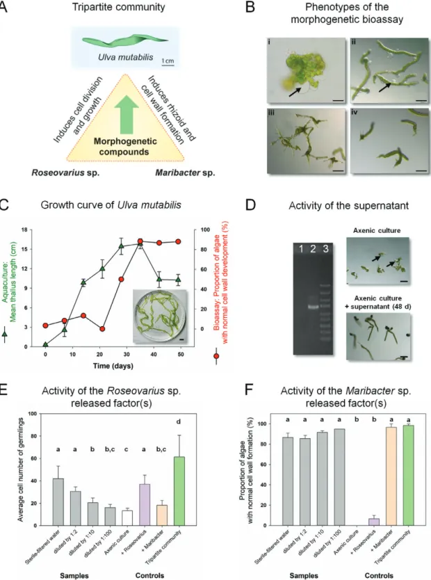
Fig. 1. Ulva mutabilis as a model organism for bacteria-induced morphogenesis (A) U. mutabilis (‘slender’) and two essential bacteria which release
morphogenetic factors establish a tripartite community. (B) The axenic callus-like morphotype of U. mutabilis was compared with axenic cultures (i) inoculated with bacteria of the Roseovarius sp. only (ii), bacteria of the Maribacter sp. only (iii), or both bacterial strains (iv). Arrows indicate the typical colorless protrusions from the exterior cell walls due to lack of morphogens released by Maribacter sp. Scale bar=500 µm (i); 100 µm (ii–iv); image (iii) with permission adapted from Wichard (2015). (C) Growth curve of U. mutabilis in a land-based tank system (error bars: ±SD, n=3). The morphogenetic activity of AGMPFs in sterile-filtered water samples is given as a portion of normally developed cell walls using the ‘Ulva bioassay array’ (inset: algae in stationary phase, scale bar=1 cm). (D) Demonstration of axenicity by PCR amplification of a part of the 16S rDNA gene extracted from the supernatant of the purified gamete stock solution (lane 1) and the non-axenic culture of U. mutabilis (lane 2). Lane 3 shows the GeneRuler DNA Express Ladder (Thermo Fisher Scientific) (scale bar=100 µm). (E, F) The AGMPFs in sterile-filtered water samples derived from aquacultures on day 48 were determined using two different bioassays. Roseovarius-released factor(s) were distinguished (E) from Maribacter-released factor(s) (F) by performing a dilution series. Data were analyzed by one-way ANOVA with Tukey’s post-hoc test (P<0.01, error bars: SE, n=60). Mean values accompanied by differing letters differed significantly. Axenic cultures (white) were inoculated with sterile-filtered water (gray) or Roseovarius sp. (purple), Maribacter sp. (orange), or both (green).
conducted at the Ramalhete Station of the Centro de Ciências do Mar (CCMAR) in Faro (Portugal). Conical tanks, made of polyester resin re-inforced with fiberglass, were used for cultivation to minimize the accu-mulation of leached chemicals that may interfere with MS. Before usage, all tanks were washed with a 10% HCl water solution (v/v) and subse-quently with commercially available bleach. After complete removal of bleach by washing with pure water, tanks were filled with 200 liters of 10 µm filtered artificial seawater (33.2 g l−1 of Instant Ocean obtained from the Institute for Inorganic and Analytical Chemistry, Aquarium Systems, Sarrebough, France). Trace metals, FeIII-EDTA, and vitamins were added based on the final concentrations of the standardized Ulva culture medium (UCM) (Stratmann et al., 1996; Califano and Wichard, 2018). Tanks were continuously aerated to avoid algal accumulation at the bottom, which would affect algal growth. Air was filtered through HEPA-Vent filters (Ø 50 mm, Whatman, Germany) and entered into the tanks from the bottom through a 6 mm diameter polyethylene hose culminating in a glass Pasteur pipette. All tanks were covered with Tygon film. For the starter cultures, axenic gametes were prepared and inocu-lated with the Roseovarius sp. strain MS2 and Maribacter sp. strain MS6. Germlings (0.5 cm length) were used for aquaculture inoculation (50 germlings l−1). Final optical density (OD
620) of the Roseovarius sp. and Maribacter sp. was adapted to 0.0001.
Under standardized laboratory conditions, U. mutabilis was cultivated in UCM, applying a light/dark regime of 17/7 h with light intensity set at 60–120 µmol photons m−2 s−1 (Stratmann et al., 1996; Califano and Wichard, 2018). To monitor the growth of U. mutabilis in the aquacultures, the length of the tubular thallus was measured in triplicate (Løvlie, 1964).
Bacterial cultures
Maribacter sp. strain MS6 (GenBank EU359911) and Roseovarius sp. strain MS2 (GenBank EU359909) were cultivated in marine broth-enriched UCM (50%, w/v; Roth, Karlsruhe, Germany) for inoculation of Ulva cultures. To determine AGMPFs in the bacterial supernatant, Maribacter sp. strain MS6 was cultivated in UCM supplemented with marine broth (10%, w/v, Roth) in sterile Nalgene® polycarbonate bottles (Thermo Fisher Scientific, Schwerte, Germany). A 10 ml aliquot of an exponen-tially growing Maribacter MS6 starter culture (OD620=1.0) was used for the inoculation of 10 liters of marine broth-supplemented medium. The culture was grown at 20–22 °C, stirred continuously, and supplied with sterile air through a HEPA-VENT-filter system (Whatman™; Thermo Fisher Scientific). Subsequently, the culture was harvested at OD620=1.0, centrifuged at 3000 g, and filtered through Whatman™ GF/F filters (Thermo Fisher Scientific).
Preparation of axenic gametes and survey for morphogenetic activity in bioassays
Gametogenesis of mature U. mutabilis was triggered by mincing the thallus and draining of sporulation inhibitors from algal fragments (Stratmann
et al., 1996, Vesty et al., 2015). Following the removal of a swarming in-hibitor through an additional medium exchange, gametes were released 3 d later. Released gametes were purified from bacteria using sterile equip-ment in a laminar flow cabinet by exploiting the positive phototaxis of gametes. The stock solution of axenic gametes was diluted with UCM or the respective sterile-filtered aquaculture water to reach a final density of 20–40 settled gametes per well (200 μl) conducted in 96-well microarray plates (Grueneberg et al., 2016). Axenicity of the purified gamete stock was confirmed via PCR using primers and cycling conditions previously described (Califano and Wichard, 2018). Following inoculation, gametes settled within 24 h in the darkness and were used in the ‘Ulva bioassay array’ (Grueneberg et al., 2016).
Determination of growth- and morphogenesis-promoting factors To survey the prevalence of morphogenesis-inducing AGMPFs under controlled conditions, the ‘Ulva bioassay array’ was performed. All water samples derived from algal or bacterial cultures were passed twice through 0.2 μm polyethersulfone filters (Roth) and inoculated in
96-well plates containing axenic gametes. The bioassay array was per-formed with a dilution series of the sterile-filtered samples using UCM to avoid nutrient deficiency during the experiment. A dilution step refers to the total volume (x/[x+y]), where x indicates the parts of the col-lected aquaculture water, and y denotes the parts of UCM (Grueneberg
et al., 2016). In total, three biological replicates with 20 individuals each were carried out. Qualitative features, including the presence of longi-tudinal growth (number of cells), differentiated rhizoids, and appearance of cell walls with protrusions, were inspected under an inverted micro-scope. After 7–10 d, and upon the initial appearance of malformed cell walls in the negative (axenic) control, the average cell number of the growing germlings and the percentage of thalli with entirely normal cell walls were determined. The log-transformed data set passed normality (Shapiro–Wilk), and an equal variance test (Brown–Forsythe) prior to one-way ANOVA with Tukey’s post-hoc test was performed (Minitab 18, Additive GmbH, Friedrichsdorf, Germany; SigmaPlot 13, Systat Software, Erkrath, Germany).
Preparation of extracts containing AGMPFs
Sterile-filtered supernatants of algal (V=10 liters) or bacterial cultures (V=1 liter) were loaded on a solid-phase cartridge (Sep-Pak C18 Plus Long Cartridge; Waters™, Manchester, UK) conditioned with 15 ml of methanol. By applying a stepwise elution gradient (25, 75, and 100% MeOH/H2O, v/v), 15 ml of each fraction was collected, evaporated under a nitrogen stream, and resuspended in 0.5 ml of UCM for utilization in the ‘Ulva bioassay array’. The procedure was repeated several times to process 50 liters of aquaculture medium or 10 liters of Maribacter sp. MS6 bacterial growth medium. The bioassay is based on complementation of the Roseovarius factor(s) within the tripartite community following re-placement of the Maribacter sp. by the tested extract containing AGMPFs (Weiss et al., 2017). The activity of the extracts was compared with the synthetic reference standard (±)-thallusin (Nishizawa et al., 2007), and model compounds, such as picolinic acid and sclareol as negative control.
Ultra-high performance liquid chromatography (UHPLC) coupled with electrospray ionization (ESI) high-resolution mass spectrometry (HRMS) measurements
UHPLC coupled with high-resolution Orbitrap MS was carried out using an UltiMate HPG-3400 RS binary pump (Thermo Fisher Scientific). The Kinetex® C-18 RP (50×2.1 mm; 1.7 µm; Phenomenex, Aschaffenburg, Germany) chromatography column was maintained at 25 °C within the TCC-3200 column compartment. Eluent A contained water with 2% acetonitrile and 0.1% formic acid. Eluent B was acetonitrile with 0.1% formic acid. The gradient applied was as follows: initial condition, 0.2 min, 0% B; 8.0 min, 100% B; 8.0–9.1 min, 100% B; 9.1–10 min, 0% B. All measurements were performed with a constant flow rate of 0.4 ml min−1. Samples were injected via the WPS-3000 autosampler equipped with a 25 µl injection syringe under temperature-controlled conditions at 10 °C. Mass spectra were recorded using the Q Exactive™ hybrid quadrupole-Orbitrap mass spectrometer (Thermo Fisher Scientific) coupled to a heated ESI source. To reduce source contamination, solvent delay was set to 0.2 min. For analyzing thallusin, targeted selective ion monitoring (tSIM) in the positive ionization mode was used with the following instrument parameters: [M + H]+ (458.21±0.2 m/z); resolution (280 000); AGC target (5×10⁴); maximum IT (254 ms); acquisition time frame (5.0–6.0 min). A simultaneous full scan was set to m/z=100–600; resolution (70 000); AGC target (5×10⁶); and maximum IT (254 ms). Further, the sheath gas flow rate was set to 40; aux gas flow rate 15; sweep gas flow rate 0; discharge current (8.0 µA); capillary temperature (350 °C); S-lens RF level (33); and vaporizer temperature (360 °C). MS/MS meas-urements were conducted with 30 eV collision energy.
Metal isotope-coded profiling (MICP)
Methanolic subsamples of solid-phase extracts derived from either bacterial or algal growth medium were used for analyzing the iron
complexation ability of thallusin. The MICP approach enables the identification of metallophores in complex matrices by using the stable isotopes, 54Fe/58Fe (Deicke et al., 2014). To ensure the stability of the iron–thallusin complexes, UHPLC-ESI-HRMS measurements were performed as described above, but using neutral eluents. Obtained chro-matograms were screened for an artificial isotopic pattern with a mass difference of Δ3.994 m/z (Deicke et al., 2014). Synthetic (±)-thallusin was used as a reference standard for the UHPLC-ESI-HRMS measure-ments (Nishizawa et al., 2007).
Iron uptake experiments in gametes
Gametogenesis was induced to obtain gametes for the uptake experi-ments. Gametes were discharged in iron-free UCM under sterile condi-tions. Gametes were counted by flow cytometry (Califano and Wichard, 2018), and subsequently mobile gametes were used directly. Aliquots of 2 ml of iron-free culture medium, each containing 3.5×10⁷ gametes, were transferred to 5 ml plastic tubes (Eppendorf, Hamburg, Germany), and 58FeCl
3, 58Fe-EDTA, or 58Fe–thallusin was added to these tubes. Iron complexes were prepared in advance by mixing isotopically pure 58Fe as ferric chloride with a 10-fold excess of EDTA or thallusin, respectively. After 2 min and 10 min of continuous shaking, gametes were filtered (nitrocellulose filter, pore size 0.4 µm, Ø 25 mm, Sarstedt, Hildesheim, Germany) and washed with 15 ml of an aqueous solution containing Na-EDTA (0.05 mol l−1) and sodium oxalate (1 mol l−1), followed by a wash with 15 ml of an aqueous NaCl solution (0.45 mol l−1). Gametes were filtered and washed without the addition of iron species. Additional blanks (without gametes) were prepared by adding 58Fe and 58Fe-EDTA to the filters to correct for potential background absorbance. All treat-ments were conducted in triplicate. All filters were digested with 1 ml of ultrapure HNO3 (65%, v/v; Thermo Fisher Scientific) for 30 min at 70 °C. Clear solutions were diluted by transferring 150 µl to 4.35 ml of ultrapure water and 0.5 ml of yttrium solution (final concentration: 1 µg l−1). The 58Fe content was determined using an Agilent 7500c ICP-MS system (Agilent, Santa Clara, CA, USA) equipped with an ASX-500 autosampler (CETAC Technologies Inc., Omaha, NE, USA), a Babington nebulizer, a Scott spray chamber (cooled to 2 °C), and a Fassel torch (Agilent). For calibration, five solutions with varying concentrations of pure 58Fe in the range between 1 µg l−1 and 10 µg l−1 were prepared. Yttrium was measured as an internal standard.
Results and discussion
To identify specific AGMPFs related to cell differentiation processes in U. mutabilis, we employed a functional comple-mentation assay. Specifically, the assay performed was designed to assess the activity of solid-phase extracts from the super-natant of Ulva aquacultures and Maribacter sp. cultures. If the extract can exert morphogenetic activity, complementary to the Roseovarius-released factor, complete morphogenesis of U. mutabilis would be restored.
Monitoring algal growth and morphogenesis-promoting factors during cultivation of Ulva mutabilis Following inoculation with gametes, within 7 weeks, U. mutabilis developed normally in the presence of its microbiome in a closed aquaculture system with thalli of 15±1.8 cm in length. In addition, nutrients and AGMPFs were present in sufficient amounts. During the stationary phase, growth continued;
how-ever, large thalli broke into smaller parts (Fig. 1C).
The ‘Ulva bioassay array’ was employed to determine the morphogenetic activity of the sterile-filtered growth medium; we revealed that this medium induced complete morphogenesis
recovery in U. mutabilis under axenic conditions (Fig. 1D).
Hereby, it was further apparent that the morphogenetic ac-tivity of the sterile-filtered medium in aquaculture increased in
parallel with the adult growth of U. mutabilis (Fig. 1C). During
the early stages, when only weak morphogenetic activity was observed in the sterile-filtered water, algae developed as germlings, indicating the presence of a small amount of mor-phogens produced by very few bacteria. Subsequently, a rapid turnover of morphogenetic activity occurred, correlating with an increase in the growth of U. mutabilis and the depletion of the AGMPFs in the medium during this early period of
culti-vation (Fig. 1C).
Interestingly, following dilution of Roseovarius-released factor(s) below a concentration threshold of 1:100, the germling cell number was no longer distinguishable from
that of axenic control cultures (Fig. 1E). Consequently, the
Maribacter-released factor(s) solely controlled the development
of U. mutabilis (Fig. 1F). Although no thallus was formed,
rhi-zoid and cell wall developed normally. Further, the morpho-genetic activity of the supernatant, even at a 100× dilution, did not significantly differ from that of the axenic gametes
inocu-lated with the Maribacter sp. positive control (Fig. 1F).
Finally, the entire medium was harvested in the stationary phase, at which point the activity was found to be the highest, and examined for AGMPF activity as well as for purification of
the Maribacter-released factor(s) (Fig. 1C). Solid-phase
extrac-tion and step gradient eluextrac-tion from up to 50 liters of aquaculture water resulted in enrichment of the factor that phenocopied Maribacter sp. activity by 106, thus permitting MS analysis. The
induction of rhizoid and cell wall formation activity was iden-tified in the eluent ranging from 25% to 75% methanol (v/v), which did not contain Roseobacter-released morphogens; thus, these morphogens appeared to be more polar. These results confirmed earlier observations in coastal water and integrated
multitrophic aquaculture systems (Grueneberg et al., 2016;
Ghaderiardakani et al., 2019).
Identification of thallusin and its iron complexes in the chemosphere of Ulva species
Whereas Roseovarius sp. can be replaced by many Alpha- and Gamma-proteobacteria releasing unknown morphogens with
similar functions (Grueneberg et al., 2016), the specificity of
the macroalgal–bacterial interactions is defined by few genera
of the Bacteroidetes, such as the Maribacter species (Weiss et al.,
2017). The solid-phase extract of 50 liters of aquaculture water
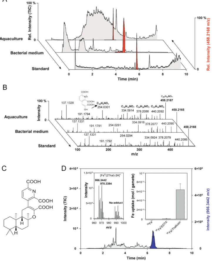
was analyzed by UHLPC coupled with ESI-HRMS, which did
not indicate a prominent peak in the total ion current (Fig. 2A).
However, the monitoring of select ions with 458.2167 m/z
[M+H]+ identified the morphogen thallusin as the AGMPF in
the chemosphere of U. mutabilis. Thallusin identification was subsequently confirmed by comparison with a synthesized racemate reference standard and MS/MS spectra identity (Fig. 2B, C). Thallusin production could be attributed to the Maribacter sp., as it was also identified in the bacterial growth
medium (Fig. 2A). However, it was not obtained from the
me-dium used for Roseovarius sp. growth. Subsequently, thallusin was found in all examined Maribacter and Zobellia cultures
Fig. 2. Identification of thallusin in the chemosphere of Ulva mutabilis (A) Algal morphogenesis-inducing fractions were analyzed by
UHPLC-ESI-HRMS and compared with the reference standard of thallusin (red: extracted ion chromatograms of 458.2168 m/z). Samples were obtained from the supernatant of algal aquacultures and the bacterial growth medium of Maribacter sp. by solid-phase extraction. (B) MS/MS experiments of 458.2168
m/z indicate the substance identity of red-labeled peaks to coincide with that of (C) (–)-thallusin. Fragment 254.0301 m/z belongs to
2,6-dicarboxy-3-(2-carboxy-2-hydroxyvinyl)pyridine-1-ium. (D) The Fe complex of thallusin (966.3442 m/z) was identified in the extracted ion chromatogram (purple) using metal isotope-coded profiling. The artificial isotopic signature (54Fe/58Fe) of the complex is shown (inset, left). U. mutabilis gametes easily acquired Fe–
thallusin compared with iron hydroxides and Fe-EDTA during the short-term uptake experiments (inset, right, error bars: SD).
(Supplementary Fig. S2), except for Maribacter polysiphoniae, in accordance with its lack of morphogenesis-inducing activity
reported previously by Weiss et al. (2017).
Thallusin harbors free carboxylic residues and picolinic acid moieties that may support iron complexation and contribute to iron acquisition. Organisms often release low molecular weight molecules (i.e. siderophores), even in the chemosphere
of Ulva spp. Wichard (2016), to recruit iron under limiting
con-ditions. Thus, we performed MICP using pairs of 54Fe and 58Fe
to identify the FeIII–thallusin complex [FeIII(2Thal-2H+)]+ in
the obtained mass spectrum of the analyzed extracts derived
from U. mutabilis cultures (Fig. 2D, inset). Moreover,
short-term uptake experiments indicated that gametes effectively in-corporated iron through thallusin. However, they were unable
to acquire it from FeIII-EDTA or free FeIII (Fig. 2D, inset),
which forms oxyhydroxids at pH>3 and is mostly unavailable
in the marine environment (Butler, 1998). In addition to iron
acquisition, it was previously demonstrated that siderophores
can regulate gene expression in bacteria (Mettrick et al., 2009).
Hence, gene expression analyses in future studies of Ulva spp. may serve to elucidate whether thallusin mediates morpho-genesis and iron homeostasis, which is essential for many iron-dependent metabolic processes in algae such as photosynthesis (Geider et al, 1993).
Thallusin promoted rhizoid and cell wall formation in Ulva spp.
Although the previously identified thallusin-producing strain
YM2-23 (Matsuo et al., 2003, 2005) has been reported to
be closely related to Maribacter species (Weiss et al., 2017), its
ecophysiological functions appear to vary in studied biological systems. Therefore, we tested the hypothesis that the multiple functions of thallusin induced the observed phenomena de-pending on the recipient’s underlying mechanisms.
Indeed, application of exogenous thallusin to axenic gam-etes induced the development of basal rhizoids and healthy
cell wall formation (Fig. 3; Supplementary Fig. S3). Further,
control experiments with Roseovarius sp. exhibited only
typ-ical cell divisions (Fig. 3A, B), whereas thallusin was
essen-tial to complete morphogenesis of U. mutabilis (Fig. 3C,
D). Thallusin also stimulated protrusion elongation at the
tip, causing separation from the stem cell by a septum and
forming stretched primary rhizoid cells (Fig. 3D, inset).
Rhizoids are important protuberances extending from the lower epidermal cells which establish the rhizoidal zone in Ulva spp. They similarly function as root hairs of vascular land plants by attaching algae to hard substrates. Importantly, indole-3-acetic acid and other examined phytohormones (zeatin, abscisic acid, gibberellins, and jasmonic acid) did not affect the development of germlings under our standardized
conditions (De Clerck et al., 2018). Given that the primary
rhizoid of ‘slender’ individuals was not strong, growing algae detached from the substrate and floated in the aquaculture
water (Fig. 1C), even under controlled conditions in the
UCM after 2 weeks (Fig. 3F). Indeed, it was previously
re-ported that a weak primary rhizoid characterizes the ‘slender’
morphotype (Løvlie, 1964).
We observed that the lowest effective concentration of
thallusin was 1.1×10⁻¹¹ mol l−1 (Supplementary Fig. S3),
con-sidering that (–)-thallusin has, thus far, proven to be the only biologically active enantiomer identified for M. oxyspermum (Gao et al., 2006; Yamamoto et al., 2014). Sclareol and picolinic acid, which are characterized by typical structural moieties similar to thallusin, exhibited no activity, and thus functioned
as negative controls (Supplementary Fig. S4). It is noteworthy
that thallusin previously triggered thallus formation (‘thallusin’)
in M. oxyspermum (Matsuo et al., 2005; Yamamoto et al., 2014,
2018), whereas the same compound promoted cell wall and
rhi-zoid formation (‘rhizoin’) in U. mutabilis in the current study. Taken together, these results indicate that thallusin will promote algal growth through healthy cell wall formation in Ulva, even in those algae which are drifting in aquacultures or coastal waters. Therefore, we concluded that thallusin functions as an essen-tial chemical mediator for algal development; however, like plant hormones, thallusin possesses distinct functions in algal develop-ment depending on the receiver, and works synergistically with unknown Roseovarius-released factors as an algal morphogenesis inducer in Ulva spp. Moreover, thallusin seemed to be essential throughout the life cycle of Ulva, as protrusions of the cell wall appeared again in fast-growing algal cultures, indicating the de-pletion of the morphogen in the absence of Maribacter sp. Cross-kingdom interactions: setting up symbiosis through morphogens
To explore the mechanisms associated with macroalgae–mi-crobe communication and to demonstrate that thallusin could form an essential endogenous morphogen, analytical chemistry was combined with specific bioassays. The observed interaction represents a further example of a more general ecological phe-nomenon in which bacteria are attracted to general signals and promote algal growth or morphogenesis in the phycosphere
following recruitment (Amin et al., 2015; Seymour et al., 2017).
In summary, the development of Ulva spp. is initiated by the settlement of mobile germ cells and resorbing of their
fla-gella (Løvlie, 1964). We previously reported that Roseovarius
sp. is chemotactically attracted to dimethylsulfoniopropionate
(DMSP) and rapidly incorporates the metabolite (Fig. 4),
al-though DMSP does not promote bacterial growth (Kessler
et al., 2018). However, U. mutabilis provides glycerol as a carbon source for Roseovarius sp., thus supporting biofilm formation. Additionally, bacteria have been reported to attract zoids via
secretion of N-acyl homoserine lactones (Joint et al., 2002; Tait
et al., 2005; Callow and Callow, 2011).
Following establishment of these initial interactions, an un-known morphogenetic compound stimulates cell divisions in U. mutabilis (Roseovarius factor) which promotes additional
glycerol production (Kessler et al., 2018), whereas thallusin
(Maribacter factor) enables further mutualistic interaction through the formation of the rhizoidal zone and immobiliza-tion of the algae. Thus, thallusin-mediated rhizoid formaimmobiliza-tion is of particular importance. Our results, in conjunction with
those of Løvlie (1964), have revealed that natural development
of the ‘slender’ mutant permits formation of only one (in some cases up to three) primary rhizoid. In this study, ‘slender’ was
preferentially investigated given its fast growth and simplicity. Primary rhizoid cells are formed in the first stage of germling development following asymmetrical division of zoids, gam-etes, or zygotes. At the 10-cell stage, lateral protuberances of stem cells, located next to the primary rhizoid, are formed, leading to secondary and additional rhizoids which only
de-velop in the wild type (Fig. 4; Løvlie, 1964). The tubular
primary and secondary rhizoid cells predominantly grow
uni-directionally, similar to a fungal mycelium (Spoerner et al.,
2012). In any case, Maribacter sp. induces the specific rhizoid
formation of both morphotypes, ‘slender’ and wild type, ac-cording to their intrinsic program, as demonstrated by previous
experiments (Spoerner et al., 2012; Vesty et al., 2015). Further
transcriptomics studies will reveal the mechanisms underlying thallusin orchestration of rhizoid development, resulting in a primary rhizoid (‘slender’) or a rhizoidal zone (wild type).
In conclusion, thallusin acts as an essential hormone-like compound for algal growth and development by controlling the differentiation and cell wall formation of rhizoid and blade cells and triggering rhizoidal zone formation in U. mutabilis. This finding has important implications for advancing our cur-rent understanding of mutualistic interactions; further, they could enable successful deciphering of regulatory mechanisms of multicellularity-related genes via the activity of exogenous
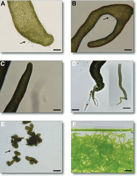
Fig. 3. Thallusin complements the activity of Roseovarius sp. Axenic gametes of Ulva mutabilis were inoculated with the bacteria, Roseovarius sp.,
alone (A, B), or with thallusin (1.1×10⁻¹⁰ mol l−1; C, D). Arrows indicate the colorless protrusions from the exterior cell walls in the absence of thallusin.
While no apical or basal end was recognized in the presence of Roseovarius sp. only, both the apical end (C) and the rhizoid (D) were clearly developed in the presence of thallusin. Axenic gametes developed into a callus [E, negative control; A–E, scale bar=500 µm; 200 µm (inset)]. In the presence of
Roseovarius sp. and thallusin, U. mutabilis developed into the adult algae (scale bar=1 cm) (F).
factors that have contributed to evolution in the green algae lineage. Further elucidation of these mechanisms is critical, es-pecially given that the transition from unicellular to multicel-lular organisms consisting of different cell types was one of the greatest achievements in the evolution of complex life forms (Pires and Dolan, 2012). Therefore, understanding of the mo-lecular and genetic processes triggered by thallusin will probably provide novel insights into the evolution of multicellularity.
Supplementary data
Fig. S1. Cultivation of axenic Ulva mutabilis alone and in the presence of Roseovarius sp. or Maribacter sp. and Roseovarius sp. microbiome (tripartite community).
Fig. S2. Phylogenetic tree based on 16S rRNA gene sequences of Maribacter and relative strains.
Fig. S3. Axenic gametes, cultivated in Ulva mutabilis culture medium, spiked with thallusin.
Fig. S4. Ulva mutabilis morphogenesis bioassay of thallusin precursor molecules.
Acknowledgements
The Deutsche Forschungsgemeinschaft funded this project through the Collaborative Research Centre CRC1127 ChemBioSys (to MK, JFM,
MD, and TW), the Jena School for Microbial Communication (to AW, JG, and TW), and the European Union’s Horizon 2020 research and in-novation program under the Marie Sklodowska-Curie grant agreement no. 642575 - The ALgal Microbiome: Friends and Foes (to GC and TW). ASSEMBLE (no. 227799) supported the early stages of the project (TW, TA and JG). Our study received Portuguese national funds from Fundacao para a Ciencia e a Tecnologia (FCT) through the UID/Multi/04326/2019 and UIDB/04326/2020 projects. AE was supported by CCMAR/ ID/16/2018, within CEECINST/00114/2018, financed by FCT. The au-thors would like to acknowledge networking support by the COST Action FA1406 ‘Phycomorph’, and Georg Pohnert (Friedrich Schiller University Jena, IAAC, Germany) for his great support throughout this project. Maximilian Walter (IAAC) contributed to the development of the bio-assays. We thank Yoshihide Matsuo (Japan) for generously providing us with thallusin as reference material, and François Thomas (Station Biologique de Roscoff, France) for providing the Zobellia spp. strains. We thank João Reis for his great support at the Ramalhete Station (Portugal). The authors de-clare that they have no competing interests.
References
Alsufyani T, Weiss A, Wichard T. 2017. Time course exo-metabolomic
profiling in the green marine macroalga Ulva (Chlorophyta) for identification of growth phase-dependent biomarkers. Marine Drugs 15, 14.
Amin SA, Hmelo LR, van Tol HM, et al. 2015. Interaction and signalling
between a cosmopolitan phytoplankton and associated bacteria. Nature
522, 98–101.
Blomster J, Bäck S, Fewer DP, Kiirikki M, Lehvo A, Maggs CA, Stanhope MJ. 2002. Novel morphology in Enteromorpha (Ulvophyceae)
forming green tides. American Journal of Botany 89, 1756–1763. Fig. 4. Chemical mediators in cross-kingdom interactions of Ulva mutabilis and associated bacteria. Ulva mutabilis provide a glycerol boundary layer as
a carbon source for Roseovarius sp., thus supporting biofilm formation (Alsufyani et al., 2017). U. mutabilis releases DMSP to attract Roseovarius sp. (Kessler et al., 2018), which then promote cell division and growth through the secretion of unknown and partially purified morphogens [Roseovarius-released factor(s), red circle] (Spoerner et al., 2012). N-Acyl homoserine lactone (AHL)-producing bacteria of the Roseobacter clade can also attract zoids of U. mutabilis through quorum-sensing signals (Joint et al., 2002). AHLs differ in the length of their R-group side chain. Following completion of the first interactions, thallusin from Maribacter sp. promotes rhizoid and cell wall formation, thus finalizing the formation of the tripartite community. Top image: germling with bacteria (scale bar=10 µm). Bottom image: both morphotypes ‘slender (sl)’ and ‘wild type (wt)’ are shown (scale bar=1 cm).
Butler A. 1998. Acquisition and utilization of transition metal ions by marine
organisms. Science 281, 207–210.
Califano G, Wichard T. 2018. Preparation of axenic cultures in Ulva
(Chlorophyta). In: Charrier B, Wichard T, Reddy C, eds. Protocols for macroalgae research. Boca Raton, FL: CRC Press, 159–173.
Callow JA, Callow ME. 2011. Trends in the development of environmentally
friendly fouling-resistant marine coatings. Nature Communications 2, 244. Coates JC, Umm-E-Aiman, Charrier B. 2014. Understanding ‘green’
multicellularity: do seaweeds hold the key? Frontiers in Plant Science 5, 737. De Clerck O, Kao SM, Bogaert KA, et al. 2018. Insights into the
evo-lution of multicellularity from the sea lettuce genome. Current Biology 28,
2921–2933.e5.
Deicke M, Mohr JF, Bellenger JP, Wichard T. 2014. Metallophore
map-ping in complex matrices by metal isotope coded profiling of organic lig-ands. The Analyst 139, 6096–6099.
Egan S, Harder T, Burke C, Steinberg P, Kjelleberg S, Thomas T.
2013. The seaweed holobiont: understanding seaweed–bacteria inter-actions. FEMS Microbiology Reviews 37, 462–476.
Fletcher RL. 1996. The occurrence of green tides—a review. In: Schramm W,
Nienhuis PH, eds. Marine benthic vegetation. New York: Springer, 7–43.
Føyn B. 1958. Über die Sexualität und den Generationswechsel von Ulva
mutabilis. Archiv für Protistenkunde 102, 473–480.
Gao X, Matsuo Y, Snider BB. 2006. Synthesis of ent-thallusin. Organic
Letters 8, 2123–2126.
Ghaderiardakani F, Califano G, Mohr JF, Abreu MH, Coates JC, Wichard T. 2019. Analysis of algal growth- and morphogenesis-promoting
factors in an integrated multi-trophic aquaculture system for farming Ulva spp. Aquaculture Environment Interactions 11, 375–391.
Ghaderiardakani F, Coates JC, Wichard T. 2017. Bacteria-induced
mor-phogenesis of Ulva intestinalis and Ulva mutabilis (Chlorophyta): a contribu-tion to the lottery theory. FEMS Microbiology Ecology 93, fix094.
Geider RJ, Laroche J, Greene RM, Olaizola M. 1993. Response of the
photosynthetic apparatus of Phaeodactylum tricornutum (Bacillariophyceae) to nitrate, phosphate, or iron starvation. Journal of Phycology 29, 755–766. Goecke F, Labes A, Wiese J, Imhoff JF. 2010. Chemical interactions between
marine macroalgae and bacteria. Marine Ecology Progress Series 409, 267–299. Grueneberg J, Engelen AH, Costa R, Wichard T. 2016. Macroalgal
morphogenesis induced by waterborne compounds and bacteria in coastal seawater. PLoS One 11, e0146307.
Hayden HS, Blomster J, Maggs CA, Silva PC, Stanhope MJ, Waaland JR. 2003. Linnaeus was right all along: Ulva and Enteromorpha
are not distinct genera. European Journal of Phycology 38, 277–294. Joint I, Tait K, Callow ME, Callow JA, Milton D, Williams P, Cámara M.
2002. Cell-to-cell communication across the prokaryote–eukaryote boundary. Science 298, 1207.
Kessler RW, Weiss A, Kuegler S, Hermes C, Wichard T. 2018.
Macroalgal–bacterial interactions: role of dimethylsulfoniopropionate in mi-crobial gardening by Ulva (Chlorophyta). Molecular Ecology 27, 1808–1819. Løvlie A. 1964. Genetic control of division rate and morphogenesis in Ulva
mutabilis Føyn. Comptes Rendus des Travaux du Laboratoire Carlsberg 34,
77–168.
Løvlie A, Bryhni E. 1978. On the relation between sexual and
partheno-genetic reproduction in haplo-diplontic algae. Botanica Marina 21, 155–163. Marshall K, Joint I, Callow ME, Callow JA. 2006. Effect of marine
bac-terial isolates on the growth and morphology of axenic plantlets of the green alga Ulva linza. Microbial Ecology 52, 302–310.
Matsuo Y, Imagawa H, Nishizawa M, Shizuri Y. 2005. Isolation of an
algal morphogenesis inducer from a marine bacterium. Science 307, 1598. Matsuo Y, Suzuki M, Kasai H, Shizuri Y, Harayama S. 2003. Isolation
and phylogenetic characterization of bacteria capable of inducing dif-ferentiation in the green alga Monostroma oxyspermum. Environmental Microbiology 5, 25–35.
Mettrick KA, Lamont IL. 2009. Different roles for anti-sigma factors in
siderophore signalling pathways of Pseudomonas aeruginosa. Molecular Microbiology 74, 1257–1271.
Nishizawa M, Iyenaga T, Kurisaki T, Yamamoto H, Sharfuddin M, Namba K, Imagawa H, Shizuri Y, Matsuo Y. 2007. Total synthesis
and morphogenesis-inducing activity of (±)-thallusin and its analogues. Tetrahedron Letters 48, 4229–4233.
Pires ND, Dolan L. 2012. Morphological evolution in land plants: new
de-signs with old genes. Philosophical Transactions of the Royal Society B: Biological Sciences 367, 508–518.
Provasoli L. 1958. Effect of plant hormones on Ulva. The Biological Bulletin 114, 375–384.
Seymour JR, Amin SA, Raina JB, Stocker R. 2017. Zooming in on the
phycosphere: the ecological interface for phytoplankton–bacteria relation-ships. Nature Microbiology 2, 17065.
Singh RP, Mantri VA, Reddy CRK, Jha B. 2011. Isolation of
seaweed-associated bacteria and their morphogenesis-inducing capability in axenic cultures of the green alga Ulva fasciata. Aquatic Biology 12, 13–21. Smetacek V, Zingone A. 2013. Green and golden seaweed tides on the
rise. Nature 504, 84–88.
Spoerner M, Wichard T, Bachhuber T, Stratmann J, Oertel W. 2012.
Growth and thallus morphogenesis of Ulva mutabilis (Chlorophyta) depends on a combination of two bacterial species excreting regulatory factors. Journal of Phycology 48, 1433–1447.
Steinhagen S, Barco A, Wichard T, Weinberger F. 2019a. Conspecificity
of the model organism Ulva mutabilis and Ulva compressa (Ulvophyceae, Chlorophyta). Journal of Phycology 55, 25–36.
Steinhagen S, Weinberger F, Karez R. 2019b. Molecular analysis of Ulva
compressa (Chlorophyta, Ulvales) reveals its morphological plasticity,
dis-tribution and potential invasiveness on German North Sea and Baltic Sea coasts. European Journal of Phycology 54, 102–114.
Stratmann J, Paputsoglu G, Oertel W. 1996. Differentiation of Ulva
mutabilis (Chlorophyta) gametangia and gamete release are controlled by
extracellular inhibitors. Journal of Phycology 32, 1009–1021.
Tait K, Joint I, Daykin M, Milton DL, Williams P, Cámara M. 2005.
Disruption of quorum sensing in seawater abolishes attraction of zoospores of the green alga Ulva to bacterial biofilms. Environmental Microbiology 7,
229–240.
Tan IH, Blomster J, Hansen G, Leskinen E, Maggs CA, Mann DG, Sluiman HJ, Stanhope MJ. 1999. Molecular phylogenetic evidence for a
reversible morphogenetic switch controlling the gross morphology of two common genera of green seaweeds, Ulva and Enteromorpha. Molecular Biology and Evolution 16, 1011–1018.
Vesty EF, Kessler RW, Wichard T, Coates JC. 2015. Regulation of
gam-etogenesis and zoosporogenesis in Ulva linza (Chlorophyta): comparison with Ulva mutabilis and potential for laboratory culture. Frontiers in Plant Science 6, 15.
Weiss A, Costa R, Wichard T. 2017. Morphogenesis of Ulva
mutabilis (Chlorophyta) induced by Maribacter species (Bacteroidetes,
Flavobacteriaceae). Botanica Marina 60, 197–206.
Wichard T. 2015. Exploring bacteria-induced growth and
morphogen-esis in the green macroalga order Ulvales (Chlorophyta). Frontiers in Plant Science 6, 86.
Wichard T. 2016. Identification of metallophores and organic ligands in the
chemosphere of the marine macroalga Ulva (Chlorophyta) and at land-sea interfaces. Frontiers in Marine Science 3, 131.
Wichard T, Charrier B, Mineur F, Bothwell JH, Clerck OD, Coates JC.
2015. The green seaweed Ulva: a model system to study morphogenesis. Frontiers in Plant Science 6, 72.
Yamamoto H, Takagi Y, Oshiro T, et al. 2014. Total synthesis of
(–)-thallusin: utilization of enzymatic hydrolysis resolution. Journal of Organic Chemistry 79, 8850–8855.
Yamamoto H, Takagi Y, Yamasaki N, Mitsuyama T, Kasai Y, Imagawa H, Kinoshita Y, Oka N, Hiraoka M. 2018. Syntheses of thallusin
analogues and their algal morphogenesis-inducing activities. Tetrahedron
74, 7173–7178.
Zhang Y, He P, Li H, et al. 2019. Ulva prolifera green-tide outbreaks
and their environmental impact in the Yellow Sea, China. National Science Review 6, 825.