marine drugs
ArticleA New Dihydrochromone Dimer and Other
Secondary Metabolites from Cultures of the Marine
Sponge-Associated Fungi Neosartorya fennelliae
KUFA 0811 and Neosartorya tsunodae KUFC 9213
Decha Kumla1,2,† ID, Tin Shine Aung1,2,† ID, Suradet Buttachon1,2 ID, Tida Dethoup3,
Luís Gales1,4, José A. Pereira1,2 ID, Ângela Inácio2, Paulo M. Costa1,2 ID, Michael Lee5,
Nazim Sekeroglu6, Artur M. S. Silva7 ID, Madalena M. M. Pinto2,8 ID and Anake Kijjoa1,2,* ID 1 ICBAS-Instituto de Ciências Biomédicas Abel Salazar, Rua de Jorge Viterbo Ferreira, 228,
4050-313 Porto, Portugal; decha1987@hotmail.com (D.K.); tinshineaung@gmail.com (T.S.A.); nokrari_209@hotmail.com (S.B.); lgales@ibmc.up.pt (L.G.); jpereira@icbas.up.pt (J.A.P.); pmcosta@icbas.up.pt (P.M.C.)
2 Interdisciplinary Centre of Marine and Environmental Research (CIIMAR), Terminal de Cruzeiros do Porto de Lexões, Av. General Norton de Matos s/n,
4450-208 Matosinhos, Portugal; angelainacio@gmail.com (A.I.); madalena@ff.up.pt (M.M.M.P.) 3 Department of Plant Pathology, Faculty of Agriculture, Kasetsart University, Bangkok 10240, Thailand;
tdethoup@yahoo.com
4 Instituto de Biologia Molecular e Celular (i3S-IBMC), Universidade do Porto, Rua de Jorge Viterbo Ferreira, 228, 4050-313 Porto, Portugal
5 Department of Chemistry, University of Leicester, University Road, Leicester LE1 7RH, UK; ml34@leicester.ac.uk
6 Medicinal and Aromatic Plant Programme, Plant and Animal Sciences Department, Vocational School, Kilis 7 Aralık University, Kilis 79000, Turkey; nsekeroglu@gmail.com
7 Departamento de Química & QOPNA, Universidade de Aveiro, 3810-193 Aveiro, Portugal; artur.silva@ua.pt
8 Laboratório de Química Orgânica, Departamento de Ciências Químicas, Faculdade de Farmácia, Universidade do Porto, Rua de Jorge Viterbo Ferreira, 228, 4050-313 Porto, Portugal
* Correspondence: ankijjoa@icbas.up.pt; Tel.: +351-22-0428331; Fax: +351-22-2062232 † These authors contributed equally to this work.
Received: 4 November 2017; Accepted: 24 November 2017; Published: 1 December 2017
Abstract:A previously unreported dihydrochromone dimer, paecilin E (1), was isolated, together with eleven known compounds: β-sitostenone, ergosta-4,6,8 (14), 22-tetraen-3-one, cyathisterone, byssochlamic acid, dehydromevalonic acid lactone, chevalone B, aszonalenin, dankasterone A (2), helvolic acid, secalonic acid A and fellutanine A, from the culture filtrate extract of the marine sponge-associated fungus Neosartorya fennelliae KUFA 0811. Nine previously reported metabolites, including a chromanol derivative (3), (3β, 5α, 22E), 3,5-dihydroxyergosta-7,22-dien-6-one (4), byssochlamic acid, hopan-3β,22-diol, chevalone C, sartorypyrone B, helvolic acid, lumichrome and the alkaloid harmane were isolated from the culture of the marine-sponge associated fungus Neosartorya tsunodae KUFC 9213. Paecilin E (1), dankasterone A (2), a chromanol derivative (3), (3β, 5α, 22E)-3,5-dihydroxyergosta-7,22-dien-6-one (4), hopan-3β,22-diol (5), lumichrome (6), and harmane (7) were tested for their antibacterial activity against Gram-positive and Gram-negative reference and multidrug-resistant strains isolated from the environment. While paecilin E (1) was active against S. aureus ATCC 29213 and E. faecalis ATCC 29212, dankastetrone A (2) was only effective against E. faecalis ATCC 29212 and the multidrug-resistant VRE E. faecalis A5/102. Both compounds neither inhibit biofilm mass production in any of the strains at the concentrations tested nor exhibit synergistic association with antibiotics.
Mar. Drugs 2017, 15, 375 2 of 17
Keywords: Neosartorya fennelliae; Neosartorya tsunodae; Trichocomaceae; dihydrochromone dimer; paecilin E; dankasterone A; chromanol derivative; marine sponge-associated fungi; antibacterial activity
1. Introduction
In the past decade, marine-derived fungi have increasingly become an important source of bioactive marine natural products, since many consider them among the world’s greatest resources for unprecedented biodiversity and chemodiversity. Moreover, with established methods of cultivation, they can produce quantity of compounds with potential for medicinal chemistry development, clinical trials and marketing [1]. The fungi belonging to the genus Neosartorya (Trichocomaceae) have been revealed to be an important source of interesting bioactive metabolites such as polyketides, isocoumarins, ergosterol analogs, meroditerpenes, pyripyropenes, benzoic acid derivatives, prenylated indole derivatives, tryptoquivalines, fiscalins, phenylalanine-derived alkaloids and cyclopeptides [2]. Marine-derived fungi are also known to produce a myriad of structurally unique metabolites not produced by their terrestrial counterparts [3]. Our group has recently isolated and identified meroditerpene analogs and the indole alkaloids, from some marine-derived fungi from the genus Neosartorya, with interesting antibacterial activity against Gram-positive bacteria (S. aureus and B. subtillis) and multidrug-resistant isolates from the environment (MRSA and VRE). Some of these compounds also had synergistic effects with antibiotics to which the bacteria are resistant. Some of these compounds also inhibit biofilm formation at MIC [4].
In our ongoing search for new natural antibiotics from marine-derived fungi, we have investigated secondary metabolites from the culture of Neosartorya fennelliae KUFA 0811, isolated from the marine sponge Clathria reinwardtii, collected from Samaesan Island in the Gulf of Thailand. Previously, we only isolated two compounds from the marine sponge-associated N. tsunodae KUFC 9213 [5], therefore we have cultured this fungus to reexamine its secondary metabolites.
Chromatographic fractionation and further purification of the ethyl acetate extract of N. fennelliae KUFA 0811, yielded a previously undescribed 2,3-dihydro-4H-chromen-4-one dimer which we have named paecilin E (1), in addition to the previously described dehydromevalonic acid lactone [6], byssochlamic acid [7], β-sitostenone [8], ergosta-4,6,8 (14), 22-tetraen-3-one [9], cyathisterone [10], dankasterone A (2) [11], chevalone B [12], helvolic acid [5], aszonalenin [13], secalonic acid A [14] and fellutanine A [13]. The ethyl acetate extract of N. tsunodae KUFC 9213 furnished, besides sartorypyrone B and helvolic acid which were previous isolated in our first study [5], byssochlamic acid [7], hopan-3β,22-diol (5) [15], chevalone C [16], a chromanol derivative (3) [17,18], (3β,5α,22E)-3,5-dihydroxyergosta-7,22-dien-6-one (4) [19], the alkaloid harmane (7) [20], and lumichrome (6) [21].
Paecilin E (1), dankasterone A (2), a chromanol derivative (3), (3β,5α,22E)-3,5-dihydroxyergosta-7,22-dien-6-one (4), hopan-3β,22-diol (5), lumichrome (6) and harmane (7), (Figure1) were tested for their growth inhibitory activity against two Gram-positive (Staphylococcus aureus ATCC 29213 and Enterococcus faecalis ATCC 29212), two Gram-negative (Escherichia coli ATCC 25922 and Pseudomonas aeruginosa ATCC 27853) bacteria, a clinical isolate sensitive to the most commonly used antibiotic families, and four multidrug-resistant isolates from the environment. Paecilin E (1) and dankasterone A (2) were also investigated for their capacity to inhibit biofilm formation in the four reference strains. The potential synergism between these two compounds and the clinically used antibiotics was also investigated against multidrug-resistant isolates from the environment.
Mar. Drugs 2017, 15, 375 3 of 16
Figure 1. Structures of paecilin E (1) and dankasterone A (2), a chromanol derivative (3), (3β,5α,22E),
3,5-dihydroxyergosta-7,22-dien-6-one (4), hopan-3β,22-diol (5), lumichrome (6), harmane (7). 2. Results and Discussion
The structures of byssochlamic acid [7], hopan-3β,22-diol (5) [15], chevalone B [12], chevalone C [16], sartorypyrone B [5], helvolic acid [5], lumichrome (6) [21], harmane (7) [20], β-sitostenone [8], ergosta-4,6,8 [14] 22-tetraen-3-one [19], cyathisterone [10], dehydromevalonic acid lactone [6], aszonalenin [13], secalonic acid A [14] and fellutanine A [13] (Figure 1 and Supplementary Materials, Figure S1) were elucidated by analysis of their 1H, 13C NMR spectra and HRMS data, as well as by comparison of their spectral data to those reported in the literature (Supplementary Materials, Figures S2–S31).
The molecular formula of 1, a white crystal (mp 203–205 °C), was established as C32H30O14 on the basis of its (+)-HRESIMS m/z 639.1718 [M + H]+, (calculated 639.1712 for C32H31O14), which indicated eighteen degrees of unsaturation. The IR spectrum showed absorption bands for hydroxyl (3443 cm−1), conjugated ketone carbonyl (1645 cm−1), ester carbonyl (1790 cm−1), lactone carbonyl (1738 cm−1), and aromatic (1470 cm−1) groups. The 13C NMR spectrum (Table 1, Supplementary
25 1 8 4 5 6 7 1 2 3 4 23 24 10 5 6 7 9 8 11 26 12 27 13 14 15 16 17 18 19 20 21 22 29 30 28 4 3 2 1 1 5 6 7 8 9 2 3 4 5 6 7 9 1 3 10' 8' 9' 14' 6' 7' 2 1 2 11' 12' 13' 5' 11 12 13 17 8 10 14 4' 4'a 1 1' 2' 3' 15 20 21 14 17 16 22 25 27 28 23 24 26 6 7 8 3 4 5 9 12 18 13 10 19 11 6 2 3 4 6 1 5 9 11 12 6 7 8 10 4 5 2 3 11 15 12 13 14 7 8 9 26 1 1 25 28 27 2 8a 9 5 3 4 4a 16 15 18 13 14 17 10 22 23 24 19 21 20
Figure 1.Structures of paecilin E (1) and dankasterone A (2), a chromanol derivative (3), (3β,5α,22E), 3,5-dihydroxyergosta-7,22-dien-6-one (4), hopan-3β,22-diol (5), lumichrome (6), harmane (7). 2. Results and Discussion
The structures of byssochlamic acid [7], hopan-3β,22-diol (5) [15], chevalone B [12], chevalone C [16], sartorypyrone B [5], helvolic acid [5], lumichrome (6) [21], harmane (7) [20], β-sitostenone [8], ergosta-4,6,8 [14] 22-tetraen-3-one [19], cyathisterone [10], dehydromevalonic acid lactone [6], aszonalenin [13], secalonic acid A [14] and fellutanine A [13] (Figure1and Supplementary Materials, Figure S1) were elucidated by analysis of their1H, 13C NMR spectra and HRMS data, as well as by comparison of their spectral data to those reported in the literature (Supplementary Materials, Figures S2–S31).
The molecular formula of 1, a white crystal (mp 203–205◦C), was established as C32H30O14on the basis of its (+)-HRESIMS m/z 639.1718 [M + H]+, (calculated 639.1712 for C32H31O14), which indicated
Mar. Drugs 2017, 15, 375 4 of 17
eighteen degrees of unsaturation. The IR spectrum showed absorption bands for hydroxyl (3443 cm−1), conjugated ketone carbonyl (1645 cm−1), ester carbonyl (1790 cm−1), lactone carbonyl (1738 cm−1), and aromatic (1470 cm−1) groups. The13C NMR spectrum (Table1, Supplementary Materials, Figure S33) displayed thirty two carbon signals which, based on DEPT and HSQC spectrum (Supplementary Materials, Figure S35), can be classified as two conjugated ketone carbonyl (δC 195.3 and 194.9), four ester carbonyl (δC175.5, 174.9, 169.3 and 168.9), eight quaternary sp2(δC160.4, 158.2, 158.1, 156.1, 116.6, 114.8, 107.5 and 107.0), four methine sp2(δC140.9, 140.6, 109.2 and 107.4), two oxyquaternary sp3 (δC85.4 and 83.9), two oxymethine sp3(δC81.9 and 81.7), two methoxyl (δC53.3 and 53.3), two methine sp3(δC32.9 and 32.5), four methylene sp3(δC39.2, 39,2, 36.9, 35.3) and two methyl (δC14.6 and 14.1) groups. Based on the type and values of their chemical shifts, these carbons were suspected to arise from two structurally similar moieties within compound 1.
Table 1.1H and13C Nuclear magnetic resonance (NMR) (DMSO, 500 and 125 MHz) and Heteronuclear Multiple Bond Correlation (HMBC) assignment for 1.
Position δC, Type δH, (J in Hz) COSY HMBC
2 85.4, C -3α 32.9, CH2 3.58, d (17.4) H-3β C-2, 4, 9, 10 β 3.05, d (17.4) H-3α C-4, 4a 4 194.9, CO -5 107.5, C -6 109.2, CH 6.61, d (8.6) H-7 C-4a, 8 7 140.9, CH 7.50, d (8.6) H-6 C-5, 8a 8 114.6, C -9 168.9, CO (Ac) -OMe-9 53.3, CH3 3.70, s C-9 10 81.7, CH 4.85, d (7.3) H-11 C-2, 3, 12, 13, 14 11 32.9, CH 2.85, m H-10, H2-12, Me-14 C-2, 13, 14 12α 35.3, CH2 1.75, dd (17.0, 9.9) H-11, 12β C-10, 13, 14 β 2.41, dd (17.0, 8.4) H-11, 12 α C-10, 13, 14 13 174.9, CO -14 14.1, CH3 1.06, d (7.1) H-11 C-10, 11, 12 20 83.9, C -30α 39.2, CH2 3.57, d (17.4) H-30β C-20, 40, 90, 100 β 3.09, d (17.4) H-30α C-40, 40a 40 195.3, CO -40a 107.0, C -50 158.1, C -60 116.6, C -70 140.6, CH 7.61, d (8.6) H-80 C-50, 80a, 8 80 107.4, CH 6.60, d (8.6) H-70 C-60, 80a 80a 158.2, C -90 169.3, CO (Ac) -OMe-90 53.3, CH3 3.69, s C-90 100 81.9, CH 4.97, d (6.7) H-110 C-30, 110, 130, 140 110 32.5, CH 2.97, m H-10 0, 110, 120a, 120β C-2 0, 130, 140 120α 36.9, CH2 2.33, dd (17.0, 5.4) H-110, 120β C-100, 130, 140 β 2.86, dd (17.0, 8.1) H-110, 120α C-100, 130, 140 130 175.5, CO -140 14.6, CH3 1.17, d (7.1) H-110 C-100, 110, 120 OH-5 - 11.56, s C-4a, 5, 6 OH-50 - 11.83, s C-40a, 50, 60
The1H NMR and COSY spectra (Table1, Supplementary Materials, Figures S32 and S34) exhibited two singlets of the hydrogen-bonded phenolic hydroxyl groups at δH 11.56 and 11.83, two pairs of ortho-coupled aromatic protons at δH7.50, d (J = 8.6 Hz)/6.61, d (J = 8.6 Hz) and 7.61, d (J = 8.6 Hz)/6.60,
d (J = 8.6 Hz), a pair of doublets at δH 4.85, d (J = 7.3 Hz) and δH4.97, d (J = 6.7 Hz), two pairs of mutually coupled methylene protons at δH 3.58, d (J = 17.4 Hz)/3.05, d (J = 17.4 Hz); δH 3.57, d (J = 17.4 Hz)/3.09, d (J = 17.4 Hz) and δH2.41, dd (J = 17.0, 8.4 Hz)/1.75 dd (J = 17.0, 9.9 Hz); δH2.86, dd (J = 17.0, 8.1 Hz)/2.33, dd (J = 17.0, 5.4 Hz), two methyl singlets at δH 1.06, d (J = 7.1 Hz) and δH1.17, d (J = 7.1 Hz) and two methoxyl singlets at δH3.70 and 3.69.
The existence of a 2,2,8-trisubstituted 5-hydroxy-2,3-dihydro-4H-chromen-4-one moiety was substantiated by COSY correlations from H-6 (δH 6.61, d, J = 8.6 Hz; δC 109.2) to H-7 (δH 7.50, d, J = 8.6 Hz; δC140.9), and HMBC correlations (Supplementary Materials, Figure S36) from H-6 to C-4a (δC107.5) and C-8 (δC114.6), H-7 to C-5 (δC160.4) and C-8a (δC156.1), OH-5 (δH11.56, s) to C-4a, C-5, C-6 (δC109.2), H-3α (δH3.58, d, J = 17.4 Hz; δC39.2) to C-2 (δC85.4) and C-4 (δC194.9), H-3β (δH3.05, d, J = 17.4 Hz; δC 39.2) to C-4 and C-4a. One of the substituents on C-2 was deduced as a methyl formate since both H-3α and the methoxyl singlet (δH3.70) exhibited HMBC cross peaks to the ester carbonyl at δC168.9 (C-9). Another substituent was 4-methyldihydrofuran-2-(3H)-one, which linked through C-10, was substantiated by COSY correlations from H-10 (δH4.85, d, J = 7.3 Hz)/H-11 (δH2.85, m)/H2-12 (δH1.75, dd, J = 17.0, 9.9 Hz and 2.41, dd, J = 17.0, 8.4 Hz), and from H-11 to Me-14 (δH1.06, d, J = 7.1 Hz) as well as by HMBC correlations from H-10 to C-3, C-11 (δC32.9), C-12 (δC35.3) and C-13 (δC174.9), H2-12 to C-10 (δC81.7), C-13, and Me-14 (δC14.1) as well as from H-3α to C-10. However, this first monomer constituted only half of the molecular formula, i.e., C16H15O7and still lacked the substituent on C-8.
The second monomer also consisted of a 5-hydroxy-2,3-dihydro-4H-chromen-4-one core, but it was 2,2,6-trisubstituted as can be corroborated by COSY correlations from H-70(δH7.61, d, J = 8.6 Hz; δC140.6) to H-80(δH6.60, d, J = 8.6 Hz; δC107.4) as well as by HMBC correlations from H-70to C-50 (δC158.1), C-80a (δC158.2), H-80to C-60(δC116.6) and C-40a (δC107.0), OH-50(δH11.83, s) to C-50, C-40a and C-60, H-30β(δH3.09, d, J = 17.4 Hz, δC39.2) to C-40(δC195.3) and C-40a, and H-30α(δH3.57, dd, J = 17.4 Hz; δC39.2) to C-40and C-20(δC83.9). Similarly, the substituents on C-20were methyl formate and 4-methyldihydrofuran-2-(3H)-one, through C-100, which were based on HMBC correlations from H-30αto C-90(δC169.3), C-100(δC81.9), H-100(δH4.97, d, J = 6.4 Hz) to C-20, C-30, C-110(δC32.5), C-130 (δC175.5) and Me-140(δC14.6) as well as the coupling system, as observed in the COSY spectrum, from H-100, through H-110(δH2.97, m) and H2-120(δH2.33, dd, J = 17.0, 5.4 Hz and 2.86, dd, J = 17.0, 8.1 Hz), and from H-110to Me-140. Like the first monomer, the second monomer also had C16H15O7, and still also lacked the substituent on C-60. That the two monomers were connected through C-8 and C-60was supported by HMBC correlations from H-7 to C-60as well as from H-70to C-8.
A literature search revealed that both monomers and dimers of 5-hydroxy-2,3-dihydro-4H-chromen-4-one with the methyl formate and γ-lactone substituents on C-2 have been previously reported. Guo et al. [22] reported the isolation of a 8-80dimer (paecilin A) and its monomer (paecilin B) of 5-hydroxy-2,3-dihydro-4H-chromen-4-one with the methyl formate and γ-lactone substituents on C-2 from the crude extract of mycelium of the endophytic fungus Paeciliomyces sp. (tree 1–7), which was isolated from mangrove bark from Xiamen, China. However, the authors did not determine the stereochemistry of both compounds. Bao et al. [23] reported the isolation, among others, of another 8-80dimer whose1H and13C NMR chemical shift values of the 4-methyldihydrofuran-2-(3H)-one moiety were slightly different from those of paecilin A. Through the NOESY correlations, they postulated that the compound might be an epimer of paecilin A, and thus named it paecilin C. However, only the relative configurations of the stereogenic carbons of the methyl γ-lactone rings were established. El-Elimat et al. [24] mentioned the isolation of paecilin D using a bioactivity-guided fractionation of the organic extract of an unidentified fungus (MSX 45109). However, the structure of paecilin D was published later with the name 11-deoxyblennolide D [25], another monomer of 5-hydroxy-2,3-dihydro-4H-chromen-4-one with the methyl formate and γ-lactone substituents on C-2.
Since 1 was obtained as a suitable crystal, its X-ray analysis was carried out. The ORTEP view, shown in Figure 2, not only confirmed the proposed structure of 1 as a 6-8 dimer of 5-hydroxy-2,3-dihydro-4H-chromen-4-one with the methyl formate and γ-lactone substituents on C-2,
Mar. Drugs 2017, 15, 375 6 of 17
but also determined unequivocally the absolute configurations of C-2, C-20, C-10, C-100, C-11, C-110 as 2R, 20R, 10S, 100S, 11R and 110R. Literature search revealed that 1 has never been previously reported and therefore named paecilin E. It is worth mentioning that this is the first dimer of 5-hydroxy-2,3-dihydro-4H-chromen-4-one with the methyl formate and γ-lactone substituents on C-2 with complete assignment of the absolute configurations of the stereogenic carbons of both 2,3-dihydropyrone and hydroxyl-γ-lactone rings.
Mar. Drugs 2017, 15, 375 6 of 16
assignment of the absolute configurations of the stereogenic carbons of both 2,3-dihydropyrone and hydroxyl-γ-lactone rings.
Figure 2. Ortep view of paecilin E (1).
Analysis of the (+)-HRESIMS, 1H, 13C NMR, COSY, HSQC and HMBC and X-ray crystallographic data of compound 2 (Supplementary Materials, Table S2, Figures S37–S40 and S49) revealed that it was dankasterone A. This compound was first reported as dankasterone, a cytotoxic steroid, isolated from a marine-derived fungus Gymnascella dankaliensis (Castellani) Currah OUPS-N 134, by Amagata et al. [26]. However, the stereochemistry of C-24 was incorrectly assigned. Later on, Amagata and coworkers [11] published the structure of dankasterone, together with other analogs, but inverted the stereochemistry of C-24 and renamed it dankasterone A.
Analysis of the 1H, 13C NMR, COSY, HSQC, HMBC, NOESY (Table 2, Supplementary Materials, Figures S41–S46) and (+)-HRESIMS data of 3, revealed that it has the same planar structure as that of one of the highly substituted chromanols, isolated from cultures of Aspergillus duricaulis [17]. However, there were no details of the 1H and 13C NMR data of the isolated compounds. The authors have proposed that the compound was a mixture of two diastereoisomers, differing in the absolute configurations at C-1, due to a ring-chain tautomerism of the hydroxyphthalide. Moreover, the authors have found that this compound did not show any optical rotation or a Cotton effect [17] and there was no indication of the determination of the absolute configurations of any stereogenic carbons of the isolated chromanol derivatives.
Later on, the same group [18] described the same compound as colorless oil which contained a mixture of the epimers and reported two sets of 1H and 13C NMR data (in deuterated acetone) for both epimers in the mixture but without assignment of the stereochemistry of C-1. On the contrary, compound 3 is optically active (levorotatory), with [α]20
D −80 (c 0.05, CHCl3), and exhibited only one
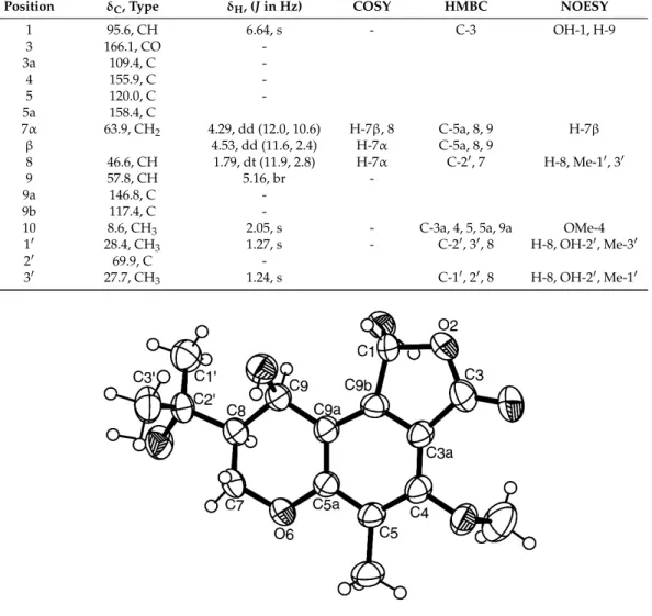
set of the 1H and 13C NMR data (Table 2). Therefore, we concluded that 3 was a pure compound and not a mixture of the epimers as described by Archenbach et al. [17,18]. This prompted us to investigate the absolute configurations of the stereogenic carbons in 3. Since 3 could be obtained in a suitable crystal (mp 223–224 °C), its X-ray analysis was carried out and the ORTEP view is shown in Figure 3. Therefore, 3 was identified as (1R, 8S, 9R)-1,9-dihydroxy-8-(2-hydroxypropan-2-yl)-4-methoxy-5-methyl-1,7,8,9-tetrahydro-3H-furo[3,4-f]chromen-3-one.
Figure 2.Ortep view of paecilin E (1).
Analysis of the (+)-HRESIMS,1H,13C NMR, COSY, HSQC and HMBC and X-ray crystallographic data of compound 2 (Supplementary Materials, Table S2, Figures S37–S40 and S49) revealed that it was dankasterone A. This compound was first reported as dankasterone, a cytotoxic steroid, isolated from a marine-derived fungus Gymnascella dankaliensis (Castellani) Currah OUPS-N 134, by Amagata et al. [26]. However, the stereochemistry of C-24 was incorrectly assigned. Later on, Amagata and coworkers [11] published the structure of dankasterone, together with other analogs, but inverted the stereochemistry of C-24 and renamed it dankasterone A.
Analysis of the1H,13C NMR, COSY, HSQC, HMBC, NOESY (Table2, Supplementary Materials, Figures S41–S46) and (+)-HRESIMS data of 3, revealed that it has the same planar structure as that of one of the highly substituted chromanols, isolated from cultures of Aspergillus duricaulis [17]. However, there were no details of the1H and13C NMR data of the isolated compounds. The authors have proposed that the compound was a mixture of two diastereoisomers, differing in the absolute configurations at C-1, due to a ring-chain tautomerism of the hydroxyphthalide. Moreover, the authors have found that this compound did not show any optical rotation or a Cotton effect [17] and there was no indication of the determination of the absolute configurations of any stereogenic carbons of the isolated chromanol derivatives.
Later on, the same group [18] described the same compound as colorless oil which contained a mixture of the epimers and reported two sets of1H and13C NMR data (in deuterated acetone) for both epimers in the mixture but without assignment of the stereochemistry of C-1. On the contrary, compound 3 is optically active (levorotatory), with [[α]25D −80 (c 0.05, CHCl3), and exhibited only one set of the1H and 13C NMR data (Table2). Therefore, we concluded that 3 was a pure compound and not a mixture of the epimers as described by Archenbach et al. [17,18]. This prompted us to investigate the absolute configurations of the stereogenic carbons in 3. Since 3 could be obtained in a suitable crystal (mp 223–224◦C), its X-ray analysis was carried out and the ORTEP view is shown in Figure3. Therefore, 3 was identified as (1R, 8S, 9R)-1,9-dihydroxy-8-(2-hydroxypropan-2-yl)-4-methoxy-5-methyl-1,7,8,9-tetrahydro-3H-furo[3,4-f]chromen-3-one.
Mar. Drugs 2017, 15, 375 7 of 17
Table 2.1H and13C NMR (CDCl3, 300 MHz and 75 MHz) and HMBC assignment for 3.
Position δC, Type δH, (J in Hz) COSY HMBC NOESY
1 95.6, CH 6.64, s - C-3 OH-1, H-9 3 166.1, CO -3a 109.4, C -4 155.9, C -5 120.0, C -5a 158.4, C 7α 63.9, CH2 4.29, dd (12.0, 10.6) H-7β, 8 C-5a, 8, 9 H-7β β 4.53, dd (11.6, 2.4) H-7α C-5a, 8, 9 8 46.6, CH 1.79, dt (11.9, 2.8) H-7α C-20, 7 H-8, Me-10, 30 9 57.8, CH 5.16, br -9a 146.8, C -9b 117.4, C
-10 8.6, CH3 2.05, s - C-3a, 4, 5, 5a, 9a OMe-4
10 28.4, CH3 1.27, s - C-20, 30, 8 H-8, OH-20, Me-30
20 69.9, C
-30 27.7, CH3 1.24, s C-10, 20, 8 H-8, OH-20, Me-10
Table 2. 1H and 13C NMR (CDCl3, 300 MHz and 75 MHz) and HMBC assignment for 3.
Position δC, Type δH, (J in Hz) COSY HMBC NOESY
1 95.6, CH 6.64, s - C-3 OH-1, H-9 3 166.1, CO - 3a 109.4, C - 4 155.9, C - 5 120.0, C - 5a 158.4, C 7α 63.9, CH2 4.29, dd (12.0, 10.6) H-7β, 8 C-5a, 8, 9 H-7β β 4.53, dd (11.6, 2.4) H-7α C-5a, 8, 9 8 46.6, CH 1.79, dt (11.9, 2.8) H-7α C-2′, 7 H-8, Me-1′, 3′ 9 57.8, CH 5.16, br - 9a 146.8, C - 9b 117.4, C -
10 8.6, CH3 2.05, s - C-3a, 4, 5, 5a, 9a OMe-4
1′ 28.4, CH3 1.27, s - C-2′, 3′, 8 H-8, OH-2′, Me-3′
2′ 69.9, C -
3′ 27.7, CH3 1.24, s C-1′, 2′, 8 H-8, OH-2′, Me-1′
Figure 3. Ortep view of 3.
Analysis of the (+)-HRESIMS, 1H, 13C NMR, COSY, HSQC and HMBC data of 4 revealed that it was (3β,22E)-3,5-dihydroxyergosta-7,22-dien-6-one (Supplementary Materials, Table S2, Figures S47 and S48). However, from a survey of the literature, the stereochemistry of C-5 remained elusive. Aiello et al. [27] first described the isolation of 24-methylcholesta-7,22E-dien-3β,5α-diol-6-one and suggested that, due to the low field chemical shift of H-3 (δH 4.03, m), the hydroxyl group on C-5 was in the α position. However, no optical rotation of this compound was reported. Later on, Ishizuka et al. [28] reported the isolation of 3β,5α-dihydroxy (22E, 24R)-ergosta-7,22-dien-6-one from the fruit bodies of an edible mushroom Grifola frondosa (Fr.) S.F. Gray (Polyporaceae). Interestingly, the optical rotation of this compound was reported as dextrorotatory, [α]25
D +9.1 (CHCl3, 0.1). Finally,
the authors confirmed the structure of this compound by chemical transformation of ergosterol acetate by treatment with Na2Cr2O7, followed by deprotection of 3-acetoxy group. Recently, Fangkratok et al. [19] reported the isolation of (3β,5α22E)-3,5-dihydroxyergosta-7,22-dien-6-one from the extract of the mycelia of Lentinus polychrous, a Thai local edible mushroom. The 1H and 13C NMR data of this compound were very similar to those of 4 except for the chemical shift value of C-10. Furthermore, the sign of the optical rotation reported by Fangkratok et al. was levorotatory, [α]20
D −4.37 (EtOH, 0.01), which is opposite to that of 4, i.e., [α] 20
D +60 (CHCl3, 0.05).
In order to clarify the controversy and to determine unequivocally the position of the hydroxyl group on C-5 of 4, the absolute configuration of C-5 was determined by comparison of the experimental electronic circular dichroism (ECD) spectrum with the calculated ECD spectra.
Figure 3.Ortep view of 3.
Analysis of the (+)-HRESIMS,1H,13C NMR, COSY, HSQC and HMBC data of 4 revealed that it was (3β,22E)-3,5-dihydroxyergosta-7,22-dien-6-one (Supplementary Materials, Table S2, Figures S47 and S48). However, from a survey of the literature, the stereochemistry of C-5 remained elusive. Aiello et al. [27] first described the isolation of 24-methylcholesta-7,22E-dien-3β,5α-diol-6-one and suggested that, due to the low field chemical shift of H-3 (δH 4.03, m), the hydroxyl group on C-5 was in the α position. However, no optical rotation of this compound was reported. Later on, Ishizuka et al. [28] reported the isolation of 3β,5α-dihydroxy (22E, 24R)-ergosta-7,22-dien-6-one from the fruit bodies of an edible mushroom Grifola frondosa (Fr.) S.F. Gray (Polyporaceae). Interestingly, the optical rotation of this compound was reported as dextrorotatory,[α]25D +9.1 (CHCl3, 0.1). Finally, the authors confirmed the structure of this compound by chemical transformation of ergosterol acetate by treatment with Na2Cr2O7, followed by deprotection of 3-acetoxy group. Recently, Fangkratok et al. [19] reported the isolation of (3β,5α22E)-3,5-dihydroxyergosta-7,22-dien-6-one from the extract of the mycelia of Lentinus polychrous, a Thai local edible mushroom. The1H and13C NMR data of this compound were very similar to those of 4 except for the chemical shift value of C-10. Furthermore, the sign of the optical rotation reported by Fangkratok et al. was levorotatory,[α]20D −4.37 (EtOH, 0.01), which is opposite to that of 4, i.e.,[α]20D +60 (CHCl3, 0.05).
In order to clarify the controversy and to determine unequivocally the position of the hydroxyl group on C-5 of 4, the absolute configuration of C-5 was determined by comparison of the experimental electronic circular dichroism (ECD) spectrum with the calculated ECD spectra. Conformational
Mar. Drugs 2017, 15, 375 8 of 17
analysis of the C-5S and C-5R diastereoisomers of 4 by molecular mechanics (MMFF95 force field) resulted in similar lowest energy conformations for both compounds, with rings A and C with chair conformation (Figure4).
Mar. Drugs 2017, 15, 375 8 of 16
Conformational analysis of the C-5S and C-5R diastereoisomers of 4 by molecular mechanics (MMFF95 force field) resulted in similar lowest energy conformations for both compounds, with rings A and C with chair conformation (Figure 4).
Figure 4. Most stable conformation of 4 (C-5R). Rings A and C have chair conformation.
However, both model’s conformational energies were further minimized by a DFT (density functional theory) method starting with ring A in chair conformation and also in boat conformation. This was considered necessary because rings A and B house the main low energy UV and ECD chromophore groups, which may engage in intramolecular hydrogen bonds, depending on the particular conformation of ring A. The DFT minimization showed that the amount of energy released by the formation of intramolecular hydrogen bonds is not enough to stabilize the boat conformations. The chair conformations are more stable than its boat counterparts in excess of 2 kcal/mol (Gibbs energy in methanol), making it overwhelmingly predominant. As such, ECD spectra were calculated for the A-chair C-5S and C-5R diastereoisomers of 4, using a TD-DFT method. Figure 5 compares these spectra and shows how the calculated spectrum for the C-5R isomer fits the experimental data much better, providing enough evidence to conclude that compound 4 is the C-5R diastereoisomer, rather than the C-5S.
Figure 5. Experimental Electronic Circular Dichroism (ECD) spectrum (solid lines, left axes) of 4 in
methanol (equal on both sides). Simulated ECD spectra (dotted lines, right axes) for both configurations.
Paecilin E (1), dankasterone A (2), a chromanol derivative (3), (3β,5α,22E)-3,5-dihydroxyergosta-7,22-dien-6-one (4), hopan-3β,22-diol (5), lumichrome (6) and harmane (7) (Figure 1) were tested for their antibacterial activity against positive and Gram-negative bacteria, including four reference strains, a clinical isolate sensitive to the most commonly used antibiotic families, and four multidrug-resistant isolates from the environment. In the range of concentrations tested, none of the compounds were active against Gram-negative bacteria. Paecilin E
Figure 4.Most stable conformation of 4 (C-5R). Rings A and C have chair conformation.
However, both model’s conformational energies were further minimized by a DFT (density functional theory) method starting with ring A in chair conformation and also in boat conformation. This was considered necessary because rings A and B house the main low energy UV and ECD chromophore groups, which may engage in intramolecular hydrogen bonds, depending on the particular conformation of ring A. The DFT minimization showed that the amount of energy released by the formation of intramolecular hydrogen bonds is not enough to stabilize the boat conformations. The chair conformations are more stable than its boat counterparts in excess of 2 kcal/mol (Gibbs energy in methanol), making it overwhelmingly predominant. As such, ECD spectra were calculated for the A-chair C-5S and C-5R diastereoisomers of 4, using a TD-DFT method. Figure5compares these spectra and shows how the calculated spectrum for the C-5R isomer fits the experimental data much better, providing enough evidence to conclude that compound 4 is the C-5R diastereoisomer, rather than the C-5S.
Mar. Drugs 2017, 15, 375 8 of 16
Conformational analysis of the C-5S and C-5R diastereoisomers of 4 by molecular mechanics (MMFF95 force field) resulted in similar lowest energy conformations for both compounds, with rings A and C with chair conformation (Figure 4).
Figure 4. Most stable conformation of 4 (C-5R). Rings A and C have chair conformation.
However, both model’s conformational energies were further minimized by a DFT (density functional theory) method starting with ring A in chair conformation and also in boat conformation. This was considered necessary because rings A and B house the main low energy UV and ECD chromophore groups, which may engage in intramolecular hydrogen bonds, depending on the particular conformation of ring A. The DFT minimization showed that the amount of energy released by the formation of intramolecular hydrogen bonds is not enough to stabilize the boat conformations. The chair conformations are more stable than its boat counterparts in excess of 2 kcal/mol (Gibbs energy in methanol), making it overwhelmingly predominant. As such, ECD spectra were calculated for the A-chair C-5S and C-5R diastereoisomers of 4, using a TD-DFT method. Figure 5 compares these spectra and shows how the calculated spectrum for the C-5R isomer fits the experimental data much better, providing enough evidence to conclude that compound 4 is the C-5R diastereoisomer, rather than the C-5S.
Figure 5. Experimental Electronic Circular Dichroism (ECD) spectrum (solid lines, left axes) of 4 in methanol (equal on both sides). Simulated ECD spectra (dotted lines, right axes) for both configurations.
Paecilin E (1), dankasterone A (2), a chromanol derivative (3), (3β,5α,22E)-3,5-dihydroxyergosta-7,22-dien-6-one (4), hopan-3β,22-diol (5), lumichrome (6) and harmane (7) (Figure 1) were tested for their antibacterial activity against positive and Gram-negative bacteria, including four reference strains, a clinical isolate sensitive to the most commonly used antibiotic families, and four multidrug-resistant isolates from the environment. In the range of concentrations tested, none of the compounds were active against Gram-negative bacteria. Paecilin E
Figure 5. Experimental Electronic Circular Dichroism (ECD) spectrum (solid lines, left axes)
of 4 in methanol (equal on both sides). Simulated ECD spectra (dotted lines, right axes) for both configurations.
Paecilin E (1), dankasterone A (2), a chromanol derivative (3), (3β,5α,22E)-3,5-dihydroxyergosta-7,22-dien-6-one (4), hopan-3β,22-diol (5), lumichrome (6) and harmane (7) (Figure1) were tested for their antibacterial activity against Gram-positive and Gram-negative bacteria, including four
reference strains, a clinical isolate sensitive to the most commonly used antibiotic families, and four multidrug-resistant isolates from the environment. In the range of concentrations tested, none of the compounds were active against Gram-negative bacteria. Paecilin E (1) exhibited an inhibitory effect on both Staphylococcus aureus ATCC 29213 and Enterococcus faecalis ATCC 29212 (Table3), with MIC values of 32 µg/mL and 16 µg/mL, respectively. However, when tested in a vancomycin-resistant (VRE) strain that was sensitive to ampicillin (E. faecalis A5/102), the MIC obtained was higher than that of the reference strain (64 µg/mL as opposed to 16 µg/mL). In the range of concentration tested, paecilin E (1) was ineffective against a VRE strain which was also resistant to ampicillin (E. faecalis B3/101). In the case of S. aureus strains isolated from the environment, paecilin E (1) was incapable of inhibiting the growth of the bacterial strain sensitive to the most commonly used antibiotic families (S. aureus 40/61/24) as well as of MRSA S. aureus 66/1. However, dankasterone A (2) was only effective against E. faecalis ATCC 29212 and VRE E. faecalis A5/102, with MIC of 32 µg/mL and 64 µg/mL, respectively.
Table 3.Antibacterial activity of paecilin E (1) and dankasterone A (2). MIC and MBC are expressed in µg/mL.
Paecilin E (1) Dankasterone A (2)
Bacterial strain MIC MBC MIC MBC
E.coli ATCC 25922 >64 >64 >64 >64
E.coli SA/2 (ESBL) >64 >64 >64 >64
P. aeruginosa ATCC 27853 >64 >64 >64 >64
E. faecalis ATCC29212 16 >64 32 >64
E. faecalis A5/102 (VRE) 64 >64 64 >64
E. faecalis B3/101 (VRE) >64 >64 >64 >64
S. aureus ATCC 29213 32 >64 >64 >64
S. aureus 40/61/24 >64 >64 >64 >64
S. aureus 66/1 (MRSA) >64 >64 >64 >64
MIC = mininmum inhibitory concentration; MBC = minimum batericidal concentration.
The effect of paecilin E (1) and dankasterone A (2) on biofilm formation was also assessed in four reference strains and neither of them revealed an inhibitory effect on biomass production in any of the strains at the concentration tested. Regarding the screening for potential synergies between the test compounds and clinical relevant antibiotics, none of the compounds revealed a synergistic association with antibiotics, as determined by the different methodologies used.
3. Experimental Section
3.1. General Experimental Procedures
Melting points were determined on a Bock monoscope and are uncorrected. Optical rotations were measured on an ADP410 Polarimeter (Bellingham + Stanley Ltd., Tunbridge Wells, Kent, UK). Infrared spectra were recorded in a KBr microplate in a FTIR spectrometer Nicolet iS10 from Thermo Scientific (Waltham, MA, USA) with Smart OMNI-Transmission accessory (Software 188 OMNIC 8.3). 1H and13C NMR spectra were recorded at ambient temperature on a Bruker AMC instrument (Bruker Biosciences Corporation, Billerica, MA, USA) operating at 300 or 500 and 75 or 125 MHz, respectively. High resolution mass spectra were measured with a Waters Xevo QToF mass spectrometer (Waters Corporations, Milford, MA, USA) coupled to a Waters Aquity UPLC system. A Merck (Darmstadt, Germany) silica gel GF254was used for preparative TLC, and a Merck Si gel 60 (0.2–0.5 mm) was used for column chromatography.
3.2. Fungal Material
The fungal strains, KUFC 9213 and KUFA 0811, were isolated from the marine sponges Aka coralliphaga, collected at the coral reef of Similan Islands, Phang Nga Provice (altitude 8◦3905.39” N
Mar. Drugs 2017, 15, 375 10 of 17
97◦38016.19” E), in April 2010 and Clathria reinwardtii, collected from Samaesan Island, Amphur Sattahip, Chonburi Province, Thailand (altitude 12◦34030.61” N 100◦5705.56” E) in February 2015, respectively. The sponge samples were washed with 0.06% sodium hypochlorite solution for 1 min, followed by sterilized seawater three times and dried on sterile filter papers under aseptic conditions. The sponges were cut into small pieces (5 × 5 mm) and placed on Petri dish plates containing 15 mL malt extract agar (MEA) medium containing 70% seawater, and incubated at 28 ◦C for 5–7 days. Hyphal tips emerged from sponge pieces were individually transferred onto MEA slant for further identification.
The fungi were identified to species level, based on morphological characteristics such as colony growth rate and growth pattern on standard media, namely Czapek’s agar (CZA), Czapek yeast autolysate agar (CYA), MEA and microscopic characteristics including size, shape, ornamentation of ascospores under light and scanning electron microscopes. The fungi were further identified by molecular techniques using ITS primers. DNA was extracted from young mycelia following a modified Murray and Thompson method [29]. Primer pairs ITS1 and ITS4 [30] were used for ITS gene amplification. PCR reactions were conducted on Thermal Cycler and the amplification process consisted of initial denaturation at 95◦C for 5 min, 34 cycles at 95◦C for 1 min (denaturation), at 55◦C for 1 min (annealing) and at 72◦C for 1.5 min (extension), followed by final extension at 72◦C for 10 min. PCR products were examined by Agarose gel electrophoresis (1% agarose with 1×TBE buffer) and visualized under UV light after staining with ethidium bromide. DNA sequencing analyses were sequenced using dideoxyribonucleotide chain termination method [31] by Macrogen Inc. (Seoul, Korea).
The DNA sequences were edited using FinchTV software and submitted into BLAST program for alignment and compared with that of fungal species in the NCBI database (http://www.ncbi.nlm.nih. gov/). The strain KUFC 9213 and KUFA 0811 were identified as Neosartorya tsunodae Yaguchi, Abliz and Y. Horie and N. fennelliae Kwon-Chung and S.J. Kim, respectively, and their ITS gene sequences were deposited in GenBank with accession numbers KT201524 for KUFC 9213 and KU955859 for KUFA 0811. The pure cultures were maintained at Department of Plant Pathology, Faculty of Agriculture, Kasetsart University, Bangkok, Thailand.
3.3. Extraction and Isolation
Each fungus was cultured for one week at 28◦C in separate Petri dish plates containing 20 mL of potato dextrose agar medium per dish. Five mycelium plugs (5 mm in diam.) of each fungus were transferred into separate 500 mL Erlenmeyer flasks containing 200 mL of potato dextrose broth and incubated on a rotary shaker at 120 rpm for one week at 28◦C to prepare mycelial suspension. Fifty 1000 mL Erlenmeyer flasks (for each fungus), each containing 300 g of cooked rice, were autoclaved at 121◦C for 15 min, and when they were cooled to room temperature, 20 mL of mycelial suspension of a fungus were inoculated per flask, and incubated at 28◦C for 30 days. Then, 500 mL of ethyl acetate was added to each moldy flask and macerated for 7 days and then filtered with Whatman No. 1 filter paper. The organic solutions were combined and evaporated under reduced pressure to furnish the crude ethyl acetate extracts of N. tsunodae KUFC 9213 (105 g) and N. fennelliae KUFA 0811 (135 g).
The crude ethyl acetate of N. fennelliae KUFA 0811 (135 g) was washed with H2O and extracted with CHCl3in the same manner. The crude chloroform extract (85 g) was applied on a column of silica gel (420 g), and eluted with mixtures of petrol-CHCl3, CHCl3-Me2CO and CHCl3-MeOH, wherein 250 mL fractions were collected as follows: Frs 1–30 (petrol-CHCl3, 1:1), 31–86 (petrol-CHCl3, 3:7), 87–202 (petrol-CHCl3, 1:9), 203–436 (CHCl3), 437–579 (CHCl3-Me2CO, 9:1), 580–690 (CHCl3-Me2CO, 7:3). Frs 31–60 were combined (6.12 g) and purified by TLC (Silica gel G254, Petrol-CHCl3-EtOAc-HCO2H, 1:8:1:0.01) to give 16.4 mg of β-sitostenone [8] and 10.5 mg of ergosta-4,6,8 (14), 22-tetraen-3-one [9]. Frs 106–135 were combined (254 g) and purified by TLC (Silica gel G254, Petrol-CHCl3-Petrol-HCO2H, 9:1:0.01) to give 93 mg of yellow viscous liquid which was applied on a Sephadex LH-20 column (10 g) and eluted with MeOH and a 1:1 mixture of MeOH:CH2Cl2wherein 1 mL 30 sfrs were collected.
Sfrs 16–30 were combined and crystallized in a mixture of CHCl3 and MeOH to give 12.5 mg of dehydromevalonic acid lactone [6]. Frs 211–255 were combined (201 mg) and crystalized in a mixture of CHCl3and petrol to give 12.3 mg of byssochlamic acid. The mother liquor was combined with the combined frs 136–165 (546 mg) and the combined frs 226–255 (700 mg), and applied on a a column of silica gel (35 g), and eluted with mixtures of petrol-CHCl3, wherein 250 mL sfrs were collected as follows: Sfrs 1–77 (petrol-CHCl3, 1:1), 78–142 (petrol-CHCl3, 3:7), 143–220 (petrol-CHCl3, 1:9), 221–255 (CHCl3). Sfrs 51–63 were combined (50 mg) and crystalized in a mixture of CHCl3and petrol to give 26 mg of byssochlamic acid. Sfrs 125–220 were combined (160 mg) and crystalized in a mixture of CHCl3and petrol to give 120 mg of cyathisterone [10]. Frs 361–420 were combined (312 mg) and purified by TLC (Silica gel G254, petrol-CHCl3-EtOAc-HCO2H, 1:8:1:0.01) to give 9 mg of byssochlamic acid and 20.3 mg of dankasterone A (2) [11]. The combined frs 256–360 (1.33 g) and 421–443 (4.9 g) were joined together and applied on a column of silica gel (65 g), and eluted with mixtures of petrol-CHCl3and CHCl3-Me2CO, wherein 250 mL sfrs were collected as follows: Sfrs 1–250 (petrol-CHCl3, 1:1), 251–386 (petrol-CHCl3, 3:7), 387–605 (petrol-CHCl3, 1:9), 606–858 (CHCl3), 859–915 (CHCl3-Me2CO, 9:1). Sfrs 316–365 were combined (35 mg) and purified by TLC (Silica gel G254, petrol-CHCl3-EtOAc-HCO2H, 1:8:1:0.01) to give 10.5 mg of chevalone B [12] and 4 mg of dankasterone A (2). Sfrs 418–480 were combined (11.3 mg) and crystallized in MeOH to give 7 mg of aszonalenin [13] Fr 449 (736 mg) was crystallized in MeOH to give 138 mg of secalonic acid A [14]. Frs 450–452 were combined (1.7 g) and applied on a column of silica gel (100 g), and eluted with mixtures of petrol-CHCl3andCHCl3-Me2CO, wherein 250 mL sfrs were collected as follows: Sfrs 1–23 (petrol-CHCl3, 1:1), 24–58 (petrol-CHCl3, 3:7), 59–150 (petrol-CHCl3, 1:9), 151–594 (CHCl3), 595–649 (CHCl3-Me2CO, 19:1), 650–735 (CHCl3-Me2CO, 9:1), 736–955 (CHCl3-Me2CO, 9:1). Sfrs 601–602 were combined and crystalized in MeOH to give 10.5 mg of paecilin E (1). Frs 453–457 were combined (1.49 g) and crystalized in MeOH to give 118 mg of secalonic acid A. The mother liquor was applied on a column of Sephadex LH-20 (10 g) and eluted with a 1:1 mixture of MeOH-CH2Cl2, wherein 20 sfrs of 10 mL were collected. Sfrs 10–12 were combined (10.6 mg) and crystalized in MeOH to give another 8.7 mg of helvoloic acid. Frs 617–623 were combined (39 mg) and applied on a column of Sephadex LH-20 (10 g) and eluted with a 1:1 mixture of MeOH: CH2Cl2, wherein 30 sfrs of 3 mL were collected. Sfrs 17–30 were combined and crystalized in MeOH to give 4.5 mg of fellutanine A [13]. Frs 631–675 were combined (3.61 g) and crystallized in MeOH to give further 68.3 mg of secalonic acid A. The crude ethyl acetate extract of N. tsunodae KUFC 9213 was dissolved in 500 mL of CHCl3, and then washed with H2O (3 × 500 mL). The organic layers were combined and dried with anhydrous Na2SO4, filtered and evaporated under reduced pressure to give 60 g of the crude chloroform extract, which was applied on a column of silica gel (410 g), and eluted with mixtures of petrol-CHCl3, CHCl3-Me2CO and CHCl3-MeOH , wherein 250 mL fractions were collected as follows: Frs 1–99 (petrol-CHCl3, 1:1), 100–201 (petrol-CHCl3, 3:7), 202–219 (petrol-CHCl3, 1:9), 220–349 (CHCl3-Me2CO, 9:1), 350–391 (CHCl3-Me2CO, 7:3), 392–437 (CHCl3-MeOH, 9:1), 438–455 (CHCl3-MeOH, 7:3) and 456–459 (MeOH). Frs 134–196 were combined (2.0 g) and purified by TLC (Silica gel G254, CHCl3-petrol-HCO2H, 14:5:1) to give 40.5 mg of byssochlamic acid [7]. Frs 226–234 were combined (4.0 g) and applied on a column of silica gel (33 g), and eluted with mixtures of petrol-CHCl3, CHCl3, and CHCl3-Me2CO, wherein 100 mL subfractions (sfrs) were collected as follows: Sfrs 1–5 (petrol-CHCl3, 7:3), 6–18 (petrol-CHCl3, 3:2), 19–20 (petrol-CHCl3, 1:1), 21–34 (petrol-CHCl3, 3:7), 25–30 (petrol-CHCl3, 9:1), 31–42 (CHCl3) and 43–48 (CHCl3-Me2CO, 9:1). Sfrs 24–30 were combined (211 mg) and crystallized in MeOH to give 64 mg of byssochlamic acid and 35 mg of hopan-3β,22 diol [15]. Sfrs 31–42 were combined (174 mg) and crystallized in MeOH to give further 23.4 mg of byssochlamic acid. Frs 235–244 were combined (1.75 g) and applied on a column of silica gel (45 g), and eluted with mixtures of petrol-CHCl3and CHCl3, wherein 100 mL sfrs were collected as follows: Sfrs 1–9 (petrol-CHCl3, 7:3), 20–32 (petrol-CHCl3, 3:2), 33–45 (petrol-CHCl3, 1:1), 46–60 (petrol-CHCl3, 3:7), 61–112 (petrol-CHCl3, 1:9) and 113–115 (CHCl3). Sfrs 1–5 were combined and purified by TLC (Silica gel G254, CHCl3-Me2CO-HCO2H, 97:3:0.1) to give 4.6 mg of byssochlamic acid
Mar. Drugs 2017, 15, 375 12 of 17
and 12.4 mg of chevalone C [16]. Sfrs 6–75 were combined (91 mg) and crystalized in MeOH to give further 15 mg of byssochlamic acid. Sfrs 76–114 were combined (863 mg) and purified by TLC (Silica gel G254, CHCl3-Me2CO-HCO2H, 97:3:0.1) to give an additional 15.7 mg of byssochlamic acid, 22.4 mg of chevalone C and 39.3 mg of sartorypyrone B [5]. Frs 245–263 were combined (1.53 g) and applied on a column of silica gel (45 g), and eluted with mixtures of petrol-CHCl3, CHCl3, CHCl3-Me2CO, and Me2CO, wherein 100 mL sfrs were collected as follows: Sfrs 1–12 (petrol-CHCl3, 7:3), 13–20 (petrol-CHCl3, 3:2), 21–40 (petrol-CHCl3, 1:1), 41–50 (petrol-CHCl3, 2:3), 51–68 (petrol-CHCl3, 3:7), 69–85 (petrol-CHCl3, 1:4), 86–100 (petrol-CHCl3, 1:9), 101–122 (CHCl3), 123–148 (CHCl3-Me2CO, 9:1), 149–158 (Me2CO). Sfrs 23–123 were combined (57 mg) and crystalized in MeOH to give 12 mg of byssochlamic acid and 7.1 mg of sartorypyrone B. Frs 264–312 were combined (1.12 g) and applied on a column of silica gel (18 g), and eluted with mixtures of petrol-CHCl3and CHCl3, wherein 100 mL sfrs were collected as follows: Sfrs 1–17 (petrol-CHCl3, 7:3), 18–48 (petrol-CHCl3, 3:2), 49–72 (petrol-CHCl3, 1:1), 73–76 (petrol-CHCl3, 2:3), 77–90 (petrol-CHCl3, 3:7), 91–100 (petrol-CHCl3, 1:9), 116 (CHCl3). Sfrs 16–68 were combined (93 mg) and crystalized in MeOH to give 33 mg of byssochlamic acid. Sfrs 69–115 were combined (711 mg) and purified by TLC (Silica gel G254, CHCl3-Me2CO-HCO2H, 4:1:0.01) to give to 14.1 mg of lumichrome [21] and 8.0 mg of helvolic acid [5]. Frs. 313–352 were combined (487 mg) and applied on a Sephadex LH-20 column (10 g) and eluted with MeOH, wherein 20 mL of 42 sfrs were collected. Sfrs 15–42 were combined (104 mg) and purified by TLC (Silica gel G254, CHCl3-Me2CO-HCO2H, 4:1:0.01) to give 10 mg of byssochlamic acid, 7.8 mg of helvolic acid, 4.7 mg of lumichrome, 10.6 mg of (3β,5α,22E)-3,5-dihydroxyergosta-7,22-dien-6-one (4) [28] and 21.6 mg of chromanol (3). Fractions 400–420 were combined (1.47 g) and applied on a Sephadex LH-20 column (20 g) and eluted with MeOH, wherein 20 mL of 42 sfrs were collected. Sfr 23–42 were combined (306 mg) and purified by TLC (Silica gel G254, CHCl3-Me2CO-HCO2H, 9:1:0.01) to give to 25.4 mg of byssochlamic acid and 5.3 mg of harmane [20]. Frs 421–440 were combined (1.33 g) and applied on a Sephadex LH-20 column (20 g) and eluted with MeOH, wherein 20 mL of 33 sfrs were collected. Sfrs 18–33 were combined (126 mg) and crystalized in MeOH to give additional 42.2 mg of harmane. 3.3.1. Paecilin E (1)
White crystal; mp 203–204◦C.[α]20D +154 (c 0.03, MeOH); IR (KBr) υmax3444, 2959, 2920, 1790, 1738, 1645, 1470, 1261cm−1. For1H and13C spectroscopic data (DMSO, 500 and 125 MHz), see Table2; (+)-HRESIMS m/z 639.1718 (M + H)+(calcd. for C32H31O14, 639.1714).
3.3.2. Dankasterone (2)
White crystal; mp 135–137◦C.[α]20D +166 (c 0.04, CHCl3); IR (KBr) υmax2959, 2924, 1727, 1710, 1536, 1462 cm−1. For1H and13C spectroscopic data (CDCl3, 500.13 and 125.8 MHz), see Table S1; (+)-HRESIMS m/z 347.1111 (M + Na)+(calcd. for C16H20O7Na, 341.1107). (+)-HRESIMS m/z 425.3054 (M + H)+(calcd. for C28H41O3, 425.3056).
3.3.3. (1R, 8S, 9R)-1,9-Dihydroxy-8-(2-hydroxypropan-2-yl)-4-methoxy-5-methyl-1,7,8,9-tetrahydro-3H-furo[3,4-f]chromen-3-one (3)
White crystal; mp 223–224◦C.[α]20D –80 (c 0.05, CHCl3); IR (KBr) υmax3467, 3434, 3018, 2969, 1743, 1597, 1507, 1262 cm−1. For1H and13C spectroscopic data (DMSO, 300.13 and 75.4 MHz), see Table2; (+)-HRESIMS m/z 347.1111 (M + Na)+(calcd. for C16H20O7Na, 341.1107).
3.3.4. (3β,5α,22E)-3,5-Dihydroxyergosta-7,22-dien-6-one (4)
White amorphous solid; [α]20D +60 (c 0.05, CHCl3); For 1H and 13C spectroscopic data (CDCl3, 500.13 and 125.8 MHz), see Table S2. (+)-HRESIMS m/z 429.3388 (M + H)+ (calcd. for C28H45O3, 429.3369).
3.4. Electronic Circular Dichroism (ECD)
The ECD spectrum of 4 (1.6 mM in methanol) was obtained in a Jasco J-815 CD spectropolarimeter with a 0.01 mm cuvette and eight accumulations. Dihedral driver and MMFF95 minimizations were done in Chem3D Ultra (Perkin-Elmer Inc., Waltham, MA, USA). All DFT minimizations and ECD spectral calculations (TD-DFT) were performed with Gaussian 09W (Gaussian Inc., Wallingford, CT, USA) using the APFD/6-311+G (2d, p) method/basis set [32] with IEFPCM solvation model of methanol. The simulated spectral lines (Figure4) were obtained by summation of Gaussian curves, as recommended in Stephens and Harada [33]. A line broadening of 0.4 eV was applied to all transitions to generate the calculated spectral lines.
3.5. X-ray Crystal Structure of 1 and 3
Diffraction data were collected with a Gemini PX Ultra equipped with CuKα radiation (λ = 1.54184 Å). The structures were solved by direct methods using SHELXS-97 and refined with SHELXL-97 [34]. Carbon, oxygen and sulfur atoms were refined anisotropically. Hydrogen atoms were either placed at their idealized positions using appropriate HFIX instructions in SHELXL, and included in subsequent refinement cycles, or were directly found from difference Fourier maps and were refined freely with isotropic displacement parameters. Full details of the data collection and refinement and tables of atomic coordinates, bond lengths and angles, and torsion angles have been deposited with the Cambridge Crystallographic Data Centre.
Paecilin E (1). Crystals were monoclinic, space group P21, cell volume 1487.9(2) Å3and unit cell dimensions a = 13.5112(7) Å, b = 8.1824(11) Å and c = 14.7531(9) Å and β = 114.179(7)◦(uncertainties in parentheses). The refinement converged to R (all data) = 5.27% and wR2 (all data) = 10.31%. The absolute structure was established with confidence (flack x parameter 0.0(2)). Diffraction data were collected at 148 K. CCDC 1579859.
(1R, 8S, 9R)-1,9-Dihydroxy-8-(2-hydroxypropan-2-yl)-4-methoxy-5-methyl-1,7,8,9-tetrahydro-3H-furo[3,4-f]chromen-3-one (3). Crystals were triclinic, space group P1, cell volume 773.78(18) Å3and unit cell dimensions a = 9.1295(12) Å, b = 9.2537(14) Å and c = 10.4317(12) Å and angles α = 94.622(11)◦, β= 104.310(11)◦ and γ = 112.486(13)◦ (uncertainties in parentheses). The refinement converged to R (all data) = 14.12% and wR2 (all data) = 29.88%. Diffraction data were collected at 291 K. CCDC 1579876.
3.6. Antibacterial Activity Bioassays
3.6.1. Bacterial Strains and Growth Conditions
For reference, a clinical isolate sensitive to the most commonly used antibiotic families, and four multidrug-resistant bacterial strains were used in this study. The Gram-positive bacteria comprised Staphylococcus aureus ATCC 29213, Enterococcus faecalis ATCC 29212, a clinical isolate S. aureus 40/61/24, MRSA S. aureus 66/1 isolated from public buses [35], and VRE E. faecalis A5/102 and VRE E. faecalis B3/101 isolated from river water [36]. The Gram-negative bacteria used were Escherichia coli ATCC 25922, Pseudomonas aeruginosa ATCC 27853, and a clinical isolate ESBL E. coli SA/2. Frozen stocks of all strains were grown in Mueller-Hinton agar (MH-BioKar diagnostics, Allone, France) at 37◦C. All bacterial strains were sub-cultured in MH agar and incubated overnight at 37◦C before each assay. 3.6.2. Antimicrobial Susceptibility Testing
The minimum inhibitory concentration (MIC), which was used for determining the antibacterial activity of each compound, was determined according to the method described previously by May Zin et al. [37].
Mar. Drugs 2017, 15, 375 14 of 17
3.6.3. Biofilm Formation Inhibition Assay
The effect of the compounds on biofilm formation was assessed using crystal violet staining as previously described by May Zin et al. [37].
3.6.4. Antibiotic Synergy Testing
Evaluation of the combined effect of the compounds and clinical relevant antimicrobial drugs was performed according to the method previously described by May Zin et al. [37].
4. Conclusions
Chemical investigation of the culture of the marine-derived fungus Neosartorya fennelliae KUFA 0811, isolated from the marine sponge Clathria reinwardtii, resulted in the isolation of the previously undescribed 6-8 dimer of substituted 3,5-dihydrochromone which we have named paecilin E (1), and the previously reported metabolites including β-sitostenone, ergosta-4,6,8 (14), 22-tetraen-3-one, cyathisterone, byssochlamic acid, dehydromevalonic acid lactone, chevalone B, aszonalenin, dankasterone A (2), helvolic acid, secalonic acid A and fellutanine A. Re-examination of the culture of N. tsunodae KUFC 9213, led to the isolation of the chromanol derivative (3), in addition to sartorypyrone B and helvolic which were previously isolated from this fungus, and other known compounds including byssochlamic acid, hopan-3β,22-diol (5), chevalone C, (3β,5α,22E)-3,5-dihydroxyergosta-7,22-dien-6-one (4), the alkaloid harmane (7) and lumichrome (6). The absolute configurations of the stereogenic carbons of the previously undescribed paecilin E (1) and the chromanol derivative (3) were unambiguously established by X-ray analysis. Although (3β,5α,22E)-3,5-dihydroxyergosta-7,22-dien-6-one (4) has been reported from several sources, the absolute configuration of its C-5 had never been determined unambiguously by any modern techniques. By comparison of the experimental and calculated ECD spectra, we determined conclusively the absolute configuration of C-5 as 5R. Paecilin E (1), dankasterone A (2), the chromanol derivative (3) and some of the isolated compounds which have not been previously tested for antibacterial activity, i.e., (3β,5α,22E)-3,5-dihydroxyergosta-7,22-dien-6-one (4), hopan-3β,22-diol (5), lumichrome (6) and harmane (7) were tested for their antibacterial activity against Gram-positive and Gram-negative bacteria of four reference strains, a clinical isolate sensitive to the most commonly used antibiotic families, and four multidrug-resistant isolates from the environment. Only paecilin E (1) and dankasterone A (2) were able to inhibit growth of Gram-positive bacteria. While paecilin E (1) exhibited an inhibitory effect on both S. aureus ATCC 29213 and E. faecalis ATCC 29212 with MIC values of 32 µg/mL and 16 µg/mL, respectively, dankasterone (2) was only effective against E. faecalis ATCC 29212 and VRE E. faecalis A5/102, with MIC of 32 µg/mL and 64 µg/mL, respectively. Despite a great structural diversity of the secondary metabolites produced by these two marine-derived species of Neosartorya, a majority of them did not possess the antibacterial activity. Nevertheless, it does not mean that they do not have other interesting biological activities. Therefore, more biological assays will be performed in the future.
Supplementary Materials: The following are available online atwww.mdpi.com/1660-3397/15/12/375/s1,
Figure S1: Structures of metabolites isolated from Neosartorya tsunodae KUFC 9231 and N. fennelliae KUFA 0811, Figures S2–S48: 1D and 2D NMR spectra of isolated compounds, Figure S49: Ortep view of dankasterone A (2), Table S1:1H and13C NMR (CDCl3, 500 MHz and 125 MHz) and HMBC assignment for 2, Table S2:1H and13C NMR (CDCl3, 500 MHz and 125 MHz) of 4.
Acknowledgments: This work was partially supported through national funds provided by FCT/
MCTES—Foundation for Science and Technology from the Minister of Science, Technology and Higher Education (PIDDAC) and European Regional Development Fund (ERDF) through the COMPETE—Programa Operacional Factores de Competitividade (POFC) programme, under the project PTDC/MAR-BIO/4694/2014 (reference POCI-01-0145-FEDER-016790; Project 3599—Promover a Produção Científica e Desenvolvimento Tecnológico e a Constituição de Redes Temáticas (3599-PPCDT)) in the framework of the programme PT2020 as well as by the project INNOVMAR—Innovation and Sustainability in the Management and Exploitation of Marine Resources (reference NORTE-01-0145-FEDER-000035, within Research Line NOVELMAR), supported by North Portugal Regional Operational Programme (NORTE 2020), under the PORTUGAL 2020 Partnership Agreement, through the European Regional Development Fund (ERDF). Decha Kumla and Tin Shine Aung thank the Alfabet and
Lotus Unlimited Projects of the Erasmus Mundus for scholarships. We thank Júlia Bessa and Sara Cravo for technical support.
Author Contributions: Anake Kijjoa and Madalena M. M. Pinto conceived, designed the experimental and
elaborated the manuscript; Decha Kumla and Tin Shine Aung performed isolation and purification of the compounds; Suradet Buttachon assisted in purification and determination of physical data of the compounds; Tida Dethoup collected, isolated, identified and culture the fungi; Luís Gales performed X-ray analysis; José A. Pereira performed calculations and measurement of ECD spectra. Paulo M. Costa and Ângela Inácio performed and interpreted the results of antibacterial assays; Nazim Sekeroglu assisted elaboration of the manuscript; Michael Lee provided HRMS; Artur M. S. Silva provided NMR spectra.
Conflicts of Interest:The authors declare no conflict of interest. References
1. Prompanya, C.; Fernandes, C.; Cravo, S.; Pinto, M.M.M.; Dethoup, T.; Silva, A.M.S.; Kijjoa, A. A new cyclic hexapeptide and a new isocoumarin derivative from the marine sponge-associated fungus Aspergillus similanensis KUFA 0013. Mar. Drugs 2015, 13, 1432–1450. [CrossRef] [PubMed]
2. Zin, W.W.; Prompanya, C.; Buttachon, S.; Kijjoa, A. Bioactive secondary metabolites from a Thai collection of soil and marine-derived fungi of the genera Neosartorya and Aspergillus. Curr. Drug Deliv. 2016, 13, 378–388. [PubMed]
3. Bugni, T.S.; Ireland, C.M. Marine-derived fungi: A chemically and biologically diverse group of microorganisms. Nat. Prod. Rep. 2004, 21, 143–163. [CrossRef] [PubMed]
4. Gomes, N.M.; Bessa, L.J.; Buttachon, B.; Costa, P.M.; Buaruang, J.; Dethoup, T.; Silva, A.M.S.; Kijjoa, A. Antibacterial and antibiofilm activity of tryptoquivalines and meroditerpenes from marine-derived fungi Neosartorya paulistensis, N. laciniosa, N. tsunodae, and the soil fungi N. fischeri and N. siamensis. Mar. Drugs
2014, 12, 822–839. [CrossRef] [PubMed]
5. Eamvijarn, A.; Gomes, N.M.; Dethoup, T.; Buaruang, J.; Manoch, L.; Silva, A.; Pedro, M.; Marini, I.; Roussis, V.; Kijjoa, A. Bioactive meroditerpenes and indole alkaloids from the soil fungus Neosartorya fischeri (KUFC 6344), and the marine-derived fungi Neosartorya laciniosa (KUFC 7896) and Neosartorya tsunodae (KUFC 9213). Tetrahedron 2013, 69, 8583–8591. [CrossRef]
6. Krings, U.; Zelena, K.; Wu, S.; Berger, R.G. Thin-layer high-vacuum distillation to isolate volatile flavour compounds of cocoa powder. Eur. Food Res. Technol. 2006, 223, 675–681. [CrossRef]
7. Szwalbe, A.J.; Williams, K.; O’Flynn, D.E.; Bailey, A.M.; Mulholland, N.P.; Vincent, J.L.; Willis, C.L.; Cox, R.J.; Simpson, T.J. Novel nonadride, heptadride and maleic acid metabolites from the byssochlamic acid producer Byssochlamys fulva IMI 40021—An insight into the biosynthesis of maleidrides. Chem. Commun. 2015, 51, 17088–17091. [CrossRef] [PubMed]
8. Prachayasittikul, S.; Suphapong, S.; Worachartcheewan, A.; Lawung, R.; Ruchirawat, S.; Prachayasittikul, V. Bioactive metabolites from Spilanthes acmella Murr. Molecules 2009, 14, 850–867. [CrossRef] [PubMed] 9. Kobayashi, M.; Krishna, M.M.; Ishida, K.; Anjaneyulu, V. Marine sterols. XXII. Occurrence of
3-oxo-4,6,8(14)-triunsaturated steroids in the sponge Dysidea herbacea. Chem. Pharm. Bull. 1992, 40, 72–74. [CrossRef]
10. Kawahara, K.; Sekita, S.; Satake, M. Steroids from Calvatia cyathiformis. Phytochemistry 1994, 37, 213–215. [CrossRef]
11. Amagata, T.; Tanaka, M.; Yamada, T.; Doi, M.; Minoura, K.; Ohishi, H.; Yamori, T.; Numata, A. Variation in cytostatic constituents of a sponge-derived Gymnascella dankaliensis by manipulating the carbon source. J. Nat. Prod. 2007, 70, 1731–1740. [CrossRef] [PubMed]
12. Zin, W.W.; Buttachon, S.; Buaruang, J.; Gales, L.; Pereira, J.A.; Pinto, M.M.M.; Silva, A.M.S.; Kijjoa, A. A new meroditerpene and a new tryptoquivaline from the algicolous fungus Neosartorya takakii KUFC 7898. Mar. Drugs 2015, 13, 3776–3790. [CrossRef] [PubMed]
13. Zin, W.W.; Buttachon, S.; Dethoup, T.; Fernandes, C.; Cravo, S.; Pinto, M.M.M.; Gales, L.; Pereira, J.A.; Silva, A.M.S.; Sekeroglu, N.; et al. New cyclotetrapeptides and a new diketopiperzine derivative from the marine sponge-associated fungus Neosartorya glabra KUFA 0702. Mar. Drugs 2016, 14, 136. [CrossRef] [PubMed]
Mar. Drugs 2017, 15, 375 16 of 17
14. Noinart, J.; Buttachon, S.; Dethoup, T.; Gales, L.; Pereira, J.A.; Urbatzka, R.; Freitas, S.; Lee, M.; Silva, A.M.S.; Pinto, M.M.M.; et al. A new ergosterol analog, a new bis-anthraquinone and anti-obesity activity of anthraquinones from the marine sponge-associated fungus Talaromyces stipitatus KUFA 0207. Mar. Drugs
2017, 15, 139. [CrossRef] [PubMed]
15. Tanaka, R.; Matsunaga, S. Saturated hopane and gammacerane triterpene-duils from the stem bark of Abies veitchii. Phytochemisty 1992, 31, 3535–3539. [CrossRef]
16. Prata-Sena, M.; Ramos, A.A.; Buttachon, S.; Castro-Carvalho, B.; Marques, P.; Dethoup, T.; Kijjoa, A.; Rocha, E. Cytotoxic activity of secondary metabolites from marine-derived fungus Neosartorya siamensis in human cancer cells. Phytother. Res. 2016, 30, 1862–1971. [CrossRef] [PubMed]
17. Achenbach, H.; Mülhlenfeld, A.; Weber, B.; Brillinger, G.U. Highly substituted chromanols from culture of Aspergillus duricaulis. Tetrahedron Lett. 1982, 45, 4659–4660. [CrossRef]
18. Achenbach, H.; Mülhlenfeld, A.; Brillinger, G.U. Stiffwechselprodukte von mikroorganismen, XXX. Phthalide und chromanole aus Aspergillus duricaulis. Liebigs Ann. Chem. 1985, 1985, 1596–1628. [CrossRef]
19. Fangkrathok, N.; Sripanidkulchai, B.; Umehara, K.; Noguchi, H. Bioactive ergostanoids and a new polyhydroxyoctane from Lentinus polychrous mycelia and their inhibitory effects on E2-enhanced cell proliferation of T47D cells. Nat. Prod. Res. 2013, 27, 1611–1619. [CrossRef] [PubMed]
20. Kodani, S.; Imoto, A.; Mitsutani, A.; Murakami, M. Isolation and identification of the antialgal compound, harmane (1-methyl-β-carboline), produced by the algicidal bacterium, Pseudomonas sp. K44-1. J. Appl. Phycol.
2002, 14, 109–114. [CrossRef]
21. Sasaki, M.; Takamatsu, H.; Oshita, K.; Yukio Kaneko, Y.; Yokotsuka, T. Isolation of lumichrome from the culture filtrate of Aspergillus oniki 1784. Nippon N¯ogeikagaku Kaishi 1974, 48, 569–571. [CrossRef]
22. Guo, Z.; She, Z.; Shao, C.; Wen, L.; Liu, F.; Zheng, Z.; Lin, Y.1H and13C NMR signal assignments of paecilin A and B, two new chromone derivatives from mangrove endophytic fungus Paeciliomyces sp. (tree 1–7). Magn. Reson. Chem. 2007, 45, 777–780. [CrossRef] [PubMed]
23. Bao, J.; Sun, Y.-L.; Zhang, X.-Y.; Han, Z.; Gao, H.-C.; He, F.; Qian, P.Y.; Qi, S.-H. Antifouling and antibacterial polyketides from marine gorgonian coral-associated fungus Penicillium sp. SCSDAF 0023. J. Antibiot. 2013, 66, 219–223. [CrossRef] [PubMed]
24. El-Elimat, T.; Figueroa, M.; Adcock, A.F.; Kroll, D.J.; Swanson, S.M.; Wani, M.C.; Pearce, C.J.; Oberlies, N.H. Cytotoxic polyketides from an unidentified fungus (MSX 45109). Planta Med. 2013, 79-PL13. [CrossRef] 25. El-Elimat, T.; Figueroa, M.; Raja, H.A.; Graf, T.N.; Swanson, S.M.; Falkinham, J.O., III; Wani, M.; Pearce, C.J.;
Oberlies, N.H. Biosythetically distinct cytotoxic polyketides from Setophoma terrestris. Eur. J. Org. Chem. 2015, 2015, 109–121. [CrossRef] [PubMed]
26. Amagata, T.; Doi, M.; Tohgo, M.; Minoura, K.; Numanta, A. Dankasterone, a new class of cytotoxic steroid produced by Gymnascella species from a marine sponge. Chem. Commun. 1999, 30, 1321–1322. [CrossRef] 27. Aiello, A.; Fattorusso, E.; Magno, S.; Menna, M. Isolation of five new 5α-hydroxy-6-keto-∆7sterols from the
marine sponge Oscarella lobularis. Steroids 1991, 56, 337–340. [CrossRef]
28. Ishizuka, T.; Yaoita, Y.; Kikuchi, M. Sterol constituents from the fruit bodies of Grifola frondosa (Fr.) S.F. Gray. Chem. Pharm. Bull. 1997, 45, 1756–1760. [CrossRef]
29. Murray, M.G.; Thompson, W.F. Rapid isolation of high molecular weight plant DNA. Nucleic Acids Res. 1980, 8, 4321–4325. [CrossRef] [PubMed]
30. White, T.J.; Bruns, T.; Lee, S.; Taylor, J. Amplification and direct sequencing of fungal ribosomal RNA genes for phylogenetics. In PCR Protocols: A Guide to Methods and Applications; Innis, M.A., Gelfand, D.H., Sninsky, J.J., White, T.J., Eds.; Academic Press: New York, NY, USA, 1990; pp. 315–322.
31. Sanger, F.; Nicklen, S.; Coulson, A.R. DNA sequencing with chain-terminating inhibitors. Proc. Natl. Acad. Sci. USA 1977, 72, 5463–5467. [CrossRef]
32. Austin, A.; Petersson, G.A.; Frixch, M.J.; Dobek, F.J.; Scalmani, G.; Throssel, K. A density functional with spherical atom dispersion terms. J. Chem. Theory Comput. 2012, 8, 4989–5007. [CrossRef] [PubMed]
33. Stephens, P.J.; Harada, N. ECD Cotton effect approximated by the Gaussian curve and other methods. Chirality 2010, 22, 229–233. [CrossRef] [PubMed]
34. Sheldrick, G.M. A short story of SHELX. Acta Crystallogr. A 2008, 64, 112–122. [CrossRef] [PubMed] 35. Simões, R.R.; Aires-de-Sousa, M.; Conceicao, T.; Antunes, F.; da Costa, P.M.; de Lencastre, H. High prevalence
of EMRSA-15 in Portuguese public buses: A worrisome finding. PLoS ONE 2011, 6, e17630. [CrossRef] [PubMed]
36. Bessa, L.J.; Barbosa-Vasconcelos, A.; Mendes, A.; Vaz-Pires, P.; Martins da Costa, P. High prevalence of multidrug-resistant Escherichia coli and Enterococcus spp. in river water, upstream and downstream of a wastewater treatment plant. J. Water Health 2014, 12, 426–435. [CrossRef] [PubMed]
37. Zin, W.W.; Buttachon, S.; Dethoup, T.; Pereira, J.A.; Gales, L.; Inácio, A.; Costa, P.M.; Lee, M.; Sekeroglu, N.; Silva, A.M.S.; et al. Antibacterial and antibiofilm activities of the metabolites isolated from the culture of the mangrove-derived endophytic fungus Eurotium chevalieri KUFA 0006. Phytochemistry 2017, 141, 86–97. [CrossRef] [PubMed]
© 2017 by the authors. Licensee MDPI, Basel, Switzerland. This article is an open access article distributed under the terms and conditions of the Creative Commons Attribution (CC BY) license (http://creativecommons.org/licenses/by/4.0/).