J. Braz. Chem. Soc., Vol. 15, No. 3, 441-444, 2004. Printed in Brazil - ©2004 Sociedade Brasileira de Química 0103 - 5053 $6.00+0.00
Short Report
* e-mail: [email protected]
Dipeptide Metabolites from the Marine Derived Bacterium
Streptomyces acrymicini
Isara L. C. Hernándeza, Mario L. Macedoa, Roberto G. S. Berlinck*,a, Antonio G. Ferreirab and Mirna J. L. Godinhoc
a
Instituto de Química de São Carlos, Universidade de São Paulo, CP 780, 13560-970 São Carlos - SP, Brazil
b
Departamento de Química, Universidade Federal de São Carlos, CP 676, 13565-905 São Carlos - SP, Brazil
c
Departamento de Ecologia e Biologia Evolutiva, Universidade Federal de São Carlos, CP 676, 13565-905 São Carlos - SP, Brazil
A investigação química do extrato bruto obtido a partir do meio de crescimento do actinomiceto
Streptomyces acrimycini isolado de sedimentos marinhos levou ao isolamento de dois dipeptídeos: a 8-amino-[1,4]diazonano-2,5-diona (1) e leucil-4-hidroxiprolina (3). Os compostos foram isolados a partir do seu meio de crescimento por uma série de etapas cromatográficas e identificados pela análise de seus dados espectroscópicos. O esqueleto macrocíclico da 8-amino-[1,4]diazonano-2,5-diona foi descrito apenas uma vez nas marinobactinas, sideróforos isolados a partir de uma bactéria marinha do gênero Marinobacter.
The chemical investigation of the crude extract obtained from the growth media of the marine-derived actinomycete Streptomyces acrimycini isolated from marine sediments led to the isolation of two new dipeptide derivatives: 8-amino-[1,4]diazonane-2,5-dione (1) and leucyl-4-hydroxyproline (3). The dipeptides were isolated from the growth media by a series of chromatographic steps, and identified by analysis of spectroscopic data. The macrocyclic carbon backbone of 8-amino-[1,4]diazonane-2,5-dione has been previously reported only once in marinobactins, siderophores isolated from the marine bacterium Marinobacter sp.
Keywords: 8-amino-[1,4]diazonane-2,5-dione, leucyl-4-hydroxyproline, Streptomyces acrimycini
Introduction
Marine microorganisms constitute the latest and yet poorly explored source of new biologically active secondary metabolites, mainly because it is assumed that marine microbiology is still a starting research field, and that microbiology methods should be improved in order to enable the isolation and growing of marine microorganisms
in artificial media.1,2 We have recently started a program
aimed to investigate the secondary metabolites of
marine-derived microbial strains.3 Several fungi and bacteria were
isolated from sediments, algae and invertebrates, and were grown in different media in order to evaluate their production of secondary metabolites. Crude extracts obtained from the culture media were subjected to different chemical and
biological screenings. Based on 1H NMR analysis, the crude
extract of the actinomycete Streptomyces acrimycini was
selected for a chemical investigation. We report here the
isolation and structure determination of two new dipeptide
metabolites isolated from S. acrimycini:
8-amino-[1,4]dia-zonane-2,5-dione (1) and leucyl-4-hydroxyproline (3).
Experimental
General experimental procedures
442 Hernández et al. J. Braz. Chem. Soc.
Bruker AC-4.7 Tesla spectrometer, on a Bruker ARX 9.4 Tesla instrument, or on a Bruker AMX500 11.75 Tesla
instrument. All the NMR spectra were obtained at 25 oC
using tetramethylsilane as internal reference. High resolution EI mass spectra were obtained on a VG-7070 equipment. Solvents employed for extraction and column chromatography were glass distilled prior to use. TLC analysis were performed with Aldrich precoated TLC sheets of silica gel on polyester with 254 nm fluorescent indicator eluting with two eluents: hexanes-ethyl acetate 1:1 and
CH2Cl2-MeOH 9:1. Plates were developed by observing at
254 nm and subsequently by spraying with ninhydrin in ethanol and further heating at 120º.
Microorganism collection, isolation and growth. S.
acrimycini was isolated from samples of sediments
collected with a Kojak apparatus, at depths between 12 and 15 m in the São Sebastião channel (north coastline of São Paulo state, Brazil). Samples of sediments were immediately processed at the Marine Biology Station of the Universidade de São Paulo. Aliquots of sediments were inoculated in Petri dishes containing different culture media. Marine streptomycetes were selected using standard growing conditions (g/L): soluble starch 10.0, casein 1.0, agar 15.0, pH 7.0-7.5. Spread plates were incubated at 25 oC
during two weeks, then single colonies were harvested and restreaked for purity.
Streptomyces acrimycini was identified by
morphological and physiological analyses (carbon sources, sugars fermentation and enzymatic assays) and by chemotaxonomic analyses (cellular wall amino acids, fatty acids composition, rDNA 16S sequencing). A voucher sample is deposited at the Deutsche Sammlung von Mikroorganismen und Zellkulturen, under the code 02-042 SS99BA-4.
Isolation of 1, 3 and 4 from Streptomyces acrimycini
S. acrimycini was grown in 10 L of marine broth 2216
(Difco) with 1% soluble starch, at 25 oC during 5 days at
200 rpm. Culture media was processed as follows: after filtration on Whatman paper #1, ethyl acetate was added to the culture medium and left overnight under magnetic stirring. The mycelium was extracted with MeOH. The ethyl acetate was separated from the culture medium aqueous phase by liquid-liquid partition. The EtOAc and MeOH extracts were pooled and concentrated to 300 mL of a
MeOH-H2O suspension which was partitioned against hexanes. The
MeOH-H2O layer was concentrated, dissolved in MeOH and
subjected to chromatography on Sephadex LH-20 (MeOH),
yielding eight fractions which were analysed by 1H-NMR
and by thin-layer chromatography. Based on the chemical
and spectroscopic analyses, the fourth fraction was selected
for further fractionation by HPLC (µBondapak C18,
MeOH-H2O 3:7), yielding 1.2 mg of a mixture of N-acetyltyramine
(4) and leucyl-4-hydroxyproline (3), as well as 1.0 mg of
pure N-acetyltyramine (4). The fifth fraction arising from the Sephadex LH-20 fractionation was further fractionated
by a series of HPLC separations on a µBondapak C18 column
(MeOH-H2O 3:7, then H2O-acetonitrile 6:4) and finally
purified on an CSC-Inertsil ODS-2 column (H2O-MeOH 9:1),
to yield 1.4 mg of 8-amino-[1,4]diazonane-2,5-dione (1).
6-amino-[1,4]diazonane-2,5-dione (1). Glassy solid,
[α]D –6.8o (c 0.08, MeOH). IR (film) ν
max/ cm -1: 3332
(br, νN-H), 2925 (νC-H), 1668 (νC=O, amide I), 1456 (νC-N),
1309, 1108, 1002. HREIMS m/z 154.07392 (calc. for
C7H10N2O2, 154.07423) corresponding to [M-NH3]+. EIMS
m/z (rel. intens.): 154 (78) [M-NH3]+, 111 (100), 98 (31),
84 (77), 83 (96), 70 (77), 66 (83). 1H and 13C NMR data: see
table 1.
Leucyl-4-hydroxyproline (3) and N-acetyltyramine (4). Glassy solid. LC-ESIMS m/z (rel. intens.): 245 (2) [M+H]+,
229 [M-CH4]+ (60), 202 (100). Negative-mode HRFABMS
m/z 243.13476 (calc. for C11H19N2O4, 243.13448) corres-ponding to [M-H+]-. 1H and 13C NMR data: see Table 1.
N-acetyltyramine (4). Glassy solid. Spectroscopic data
in agreement with literature values.7
Results and Discussion
Streptomyces acrimycini was grown in 10 x 1L
erlenmeyer flasks, each one containing 500 mL of enriched marine broth (see Experimental), during 10 days at 180
rpm and 27 oC. Chromatography of the organic crude
extract on Sephadex LH-20 (MeOH) yielded several fractions which were further purified by reversed phase
chromatography (µBondapak C18, MeOH-H2O 3:7). The
dipeptide 8-amino-[1,4]diazonane-2,5-dione (1) was
obtained as a glassy solid, [α]D –6.8o (c 0.08, MeOH), with
a formula C7H13N3O2 established by HREIMS at [M-NH3]+
(observed: 154.07392; calculated: 154.07423) and analysis
of the NMR spectra. The infrared spectrum of 1 presented
bands at 3332 (broad, νN-H) 2925 (νC-H), 1668 (νC=O, amide
I) and 1456 cm-1 (ν
C-N), indicating the presence of amide
bonds. No significant absorption was observed in its UV
spectrum. Analysis of the 1H, 13C and gHMQC NMR spectra
obtained in DMSO-d6 indicated the presence of one
methine, four methylenes and two carbonyl quaternary carbons, strongly suggested a dipeptide structure. The methine was assigned to a typical alpha amino acid moiety
(CH-8, 4.23, brt, 9 Hz) (13C 58.4), which showed a coupling
in the 1H-1H COSY spectrum to CH
2-7 at δ 2.34 (m) and
1.99 (m) (13C 28.2). The CH
443 Dipeptide Metabolites from the Marine Derived Bacterium Streptomyces acrymicini
Vol. 15, No. 3, 2004
coupled to CH2-6 at δ 2.02 (m) and 1.97 (m) (13C 22.4),
which was in turn coupled to the CH2-5 methylene group
at δ 3.53 (m) (13C 45.1). Both 1H and 13C chemical shifts of
CH2-5 agreed with a N-substitution. These data were
indicative of an ornithine residue, confirmed by analysis of the gHMBC spectrum. The remaining NMR data indicated the presence of a glycine residue, with a single
methylene group at δ 3.99 (d, 17 Hz) and 4.11 (d, 17 Hz)
(13C 46.2) coupled with two carbonyl carbons at δ 169.9
(C-3) and 164.4 (C-9). We first considered that the structure of this compound corresponded to the diketopiperazine
cyclo[Gly-Orn] (2).4 However, analysis of the NMR data in
DMSO-d6 disfavoured this hypothesis, since a 1H coupling
in the 1H-1H COSY spectrum between CH
2-5 at δ 3.53 with
the NH-4 amide exchangeable proton at δ 8.10 was
observed. Additionally, both CH2-5 at δ 3.53 and NH-4
proton at δ 8.10 showed long-range couplings to the
carbonyl group at δ 169.9. Therefore, in order to account
for the NMR data obtained in DMSO-d6, the structure
should have a lactam group including the ornithine δ
-amino group and the glycine acid group, corresponding
to 8-amino-[1,4]diazonane-2,5-dione (1). We have been
unable to establish the stereochemistry of the ornithine
residue in 1 due to the small amount of compound isolated.
To the best of our knowledge, the structure of 1 is totally
unprecedent among dipeptides. A related macrocyclic dipeptide has been recently reported as part of the macrobactins, a group of amphiphillic siderophores recently isolated from Marinobacter sp.5,6
The second dipeptide isolated from S. acrimycini was
the linear leucyl-4-hydroxyproline (3), isolated in a mixture with N-acetyltyramine (4). Due to the small quantity of the mixture (~1 mg), we have not attempted to separate both
compounds in order to avoid any loss of material. Since we have been also able to isolate a pure sample of 4,7 we could
analyse the MS and NMR data of 3 and 4, in order to assign
the 1H (at 500 MHz) and 13C (at 125 MHz) signals of both
compounds. Analysis of the gHSQC spectrum enabled us to
assign all hydrogen bearing carbons of 3. The position of
the hydroxyl group in the 4-hydroxyproline moiety was
established by analysis of the 1H-1H COSY spectrum, which
showed sequencial couplings from H-5 (δ 4.38) to H-4a
(δ 1.93) and H-4b (δ 2.02), from these two hydrogens to
H-3 (δ 4.27), which was in turn coupled with both H-2a
(δ 3.21, overlapped by the H2O signal in DMSO-d6) and
H-2b (δ 3.47). Since the spectra were obtained in DMSO-d6, we have been able to observe a vicinal coupling between H-3 and the hydroxyl proton at δ 5.08 (d, J 3 Hz). Further
support to the 4-hydroxyproline moiety of 3 was obtained
by analysis of the gHMBC spectrum, which showed
couplings between H-5 and C-4 (δ 36.6), between H-5 and
the carbonyl group C-6 at δ 166.6, between H-3 and C-5
(δ 57.1), between H-4b and C-2 (δ 53.7) as well as between
H-4b and C-3 (δ 67.0), between H-4a and C-5, and finally
between H-2a and C-3. The amide bond between the two amino acid residues was established as the carbonyl group C-6, which chemical shift at δ 166.6 typically resonates at a higher field than the carbonyl group chemical shift of a free carboxylic acid group. Additionally, analysis of the gHMBC spectrum showed long-range couplings between the exchangeable amide N-H proton at δ 7.98 and C-6 (δ 166.6), between the N-H proton and C-5 of 4-hydroxyproline (δ 57.1), as well as between H-8 of leucine (δ 4.04) and C-6
of 4-hydroxyproline. Analysis of the gHMQC, 1H-1H COSY
and gHMBC spectra defined all hydrogen and carbons of
the leucine moiety. 1H-1H couplings were observed between
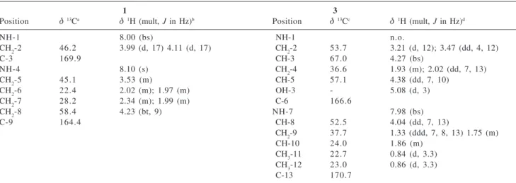
Table 1.1H and 13C NMR assignments of compounds 1 and 3
1 3
Position δ13Ca δ1H (mult, J in Hz)b Position δ13Cc δ1H (mult, J in Hz)d
NH-1 8.00 (bs) NH-1 n.o.
CH2-2 46.2 3.99 (d, 17) 4.11 (d, 17) CH2-2 53.7 3.21 (d, 12); 3.47 (dd, 4, 12)
C-3 169.9 CH-3 67.0 4.27 (bs)
NH-4 8.10 (s) CH2-4 36.6 1.93 (m); 2.02 (dd, 7, 13)
CH2-5 45.1 3.53 (m) CH-5 57.1 4.38 (dd, 7, 10)
CH2-6 22.4 2.02 (m); 1.97 (m) OH-3 - 5.08 (d, 3)
CH2-7 28.2 2.34 (m); 1.99 (m) C-6 166.6
CH2-8 58.4 4.23 (bt, 9) NH-7 7.98 (bs)
C-9 164.4 CH-8 52.5 4.04 (dd, 7, 13)
CH2-9 37.7 1.33 (ddd, 7, 8, 13) 1.75 (m)
CH-10 24.0 1.86 (m)
CH3-11 22.7 0.84 (d, 3.3) CH3-12 23.0 0.86 (d, 3.3)
C-13 170.7
aData obtained at 100 MHz, in DMSO-d 6;
bData obtained at 400 MHz, in DMSO-d 6;
cData obtained at 125 MHz in DMSO-d 6;
444 Hernández et al. J. Braz. Chem. Soc.
H-8 and H-9a (δ 1.33) and H-9b (δ 1.75). Further hydrogen
couplings were observed between both methyl groups CH3-11 at δ 0.84 (d, 3.3 Hz) and CH3-12 at δ 0.86 (d, 3.3 Hz) and H-10 at δ 1.86 (m). 1H-13C Long-range couplings were
observed between H-8 and C-9 (δ 37.7), C-10 (δ 24.0),
between both H-9a and H-9b and C-8, with the carbonyl group at δ 166.6 (C-6 of 4-hydroxyproline, a 4J long-range
correlation) and with H3C-12, between H-10 and C-8 (δ 52.5) and both methyl groups, as well as between the hydrogens of both methyl groups with C-10 and C-9. The LC-ESIMS of the mixture of 3 and 4 displayed a ion at m/z 229, sugesting
the loss of CH4 from the leucyl-4-hydroxyproline molecular
ion of small intensity at m/z 245. Negative-mode HRFABMS
indicated a parent ion peak [M-H+]– at m/z 243.13476 (calcd.
243.13448, ∆mu +2.8), confirming the molecular formula
C11H20N2O4 for 3. Since we obtained only a tiny amount of this mixture, no attempt has been made in order to further
purify the dipeptide leucyl-4-hydroxyproline (3), and the
stereochemistry of the stereogenic centers were not determined. To the best of our knowledge, this is the first report on the identification of the linear dipeptide
leucyl-4-hydroxyproline (3). No chemical investigation of the S.
acrimycini secondary metabolites has been previously reported, but only genetic and morphological des-criptions.8-11
Although there is abundant literature data about
dipeptides isolated from microbial sources,12,16 their true
origin remains controversial. Some authors suggest that dipeptides are fermentation artifacts, generated by
hydrolysis of proteins present in the growth media.16,17
However, several dipeptides isolated from microorganisms display relevant biological activities.14,15 Recently, it has
been demonstrated that several dipeptides present an important role as chemical mediators of bacterial
quorum-sensing signalling systems.19 The isolation of unusual
metabolites from the previously unstudied marine-derived
S.acrimycini reinforce the importance of continuing studies on marine microbiology and on marine microorganisms secondary metabolism.
Acknowledgments
The authors thank to Dr. David E. Williams and Raymond J. Andersen (Departments of Chemistry and Earth and Ocean Sciences, University of British Columbia, Vancouver, Canada) as well as Dr. Brent Copp (Department of Chemistry, University of Auckland) for help in obtaining NMR and mass spectra, Fundação de Amparo à Pesquisa do Estado de São Paulo for a scholarship to ILCH and financial support, and to the technical staff of the Centro de Biologia Marinha of the Universidade de São Paulo for
providing technical assistance during the sediments collection and logistic support.
References
1. Hyde, K. D.; Pointing, S. B.; Marine Mycology: A Practical Approach, 1st ed., Fungal Diversity Research Series l: Hong Kong, 2002.
2. Paul, J. H.; Marine Microbiology. Methods in Microbiology, Academic Press: New York, 2001, vol. 30.
3. Hernandez, I.L.C.; Godinho, M.J.L.; Magalhães, A.; Schefer, A.B.; Ferreira, A.G.; Berlinck, R.G.S.; J. Nat. Prod.2000, 63, 664.
4. Ishibashi, N.; Kouge, K.; Shinoda, I.; Kaneshisa, H.;Okai, H.;
Agr. Biol. Chem. Tokyo1988, 52, 819.
5. Martinez, J. S.; Zhang, G. P.; Holt, P. D.; Jung, H.-T.; Carrano, C. J.; Haygood, M. G.; Butler, A.; Science2000, 287, 1245. 6. Xu, G. F.; Martinez, J. S.; Groves, J. T.;Butler, A.; J. Am.
Chem. Soc. 2002, 124, 13408.
7. Comin, V. J.; Keller-Schierlein, W.; Helv. Chim. Acta1959,
42, 1730.
8. Asturias, J. A.; Martin, J. F.; Liras, P.; J. Ind. Microbiol. 1994,
13, 183.
9. Murray, I.A.; Gil, J. A.; Hopwood, D. A.; Shaw, W. V.; Gene
1989, 85, 283.
10. Gil, J. A.; Kieser, H. M.; Hopwood, D. A.; Gene1985, 38, 1. 11. Wright, H. M.; Hopwood, D. A.; J. Gen. Microbiol. 1977,
102, 417.
12. Kanzaki, H.; Yanagisawa, S.; Nitoda, T.; J. Antibiotics2000,
53, 1257.
13. Obata, Y.; Mizutani, J.; Bull. Agr. Soc. Japan1958, 22, 14. 14. Yang, L.; Ren-xiang, T.; Wang, Q.; Huang, W.; Yin, Y.;
Tetrahedron Lett. 2002, 43, 6545.
15. Otsuka, T.; Shibata, T.; Tsurumi, Y.; Takase, S.; Okuhara, M.; Terano, H.; Kohsaka, M.; Imanaka, H.; J. Antibiotics1992,
45, 348.
16. Prasad, C.; Peptides1995, 16, 151.
17. Son, B. W.; Jensen, P. R.; Kauffman, C. A.; Fenical, W.; Nat. Prod. Lett. 1999, 13, 213.
18. Turner, W. B.; Aldridge, D. C.; Fungal Metabolites, 1st ed., Academic Press: London, 1983.
19. Holden, M.T.G.; Chabra, S.R.; de Nys, R.; Stead, P.; Bainton, N.J.; Hill, P.J.; Manefield, M.; Kumar, N.; Labatte, M.; England, D.; Rice, S.; Givskov, M.; Salmond, G.P.C.; Stewart, G.S.A.B.; Bycroft, B.W.; Kjelleberg, S.A.; Williams, P.; Mol. Microbiol.
1999, 33, 1254.
Received: August 19, 2003 Published on the web: May 10, 2004