Contents lists available atScienceDirect
IJP: Parasites and Wildlife
journal homepage:www.elsevier.com/locate/ijppawHu
ffmanela lusitana sp. n. (Nematoda: Trichosomoididae) infecting pouting,
Trisopterus luscus (Teleostei: Gadidae) o
ff the Atlantic coast of Portugal
Paula Ramos
a,∗, Raquel Carvalho
b, Fernanda Rosa
c, Graça Alexandre-Pires
d, Fernanda Seixas
b,
Alexandra Esteves
b, David Hu
ffman
eaPathology Laboratory of Aquatic Animals, Portuguese Institute for the Sea and Atmosphere, I.P. (IPMA), Rua Doutor Alfredo Magalhães Ramalho, 6, 1495-165 Algés,
Portugal
bCentre of Studies in Animal and Veterinary Science (CECAV), University of Trás-os-Montes e Alto Douro (UTAD), Apartado 1013, 5001-801, Vila Real, Portugal cInstituto Superior de Agronomia, Universidade de Lisboa, Tapada da Ajuda, 1349-017 Lisboa; Centro de Estudos do Ambiente e do Mar, LA, Faculdade de Ciências,
Universidade de Lisboa, Campo Grande, 1749-016 Lisboa, Portugal
dCIISA-FCT-UID/CVT/00276/2013 - Faculty of Veterinary Medicine, University of Lisbon, Avenida da Universidade Técnica, 1300-477, Lisboa, Portugal eDepartment of Biology. 212 Freeman Aquatic Biology Bldg, Texas State University, San Marcos, TX, 78666, USA
A R T I C L E I N F O Keywords: Trisopterus luscus Dark muscle Huffmanela lusitana sp. n. Nematode Portuguese coast Marketed infected food-fish
A B S T R A C T
Some pouting caught off the Atlantic coast of Portugal are discarded as unmarketable due to a dark dis-colouration of the skin and muscle. This study investigates the cause of this condition, describes the new parasite species responsible, and highlights the importance of educating those in charge of premarket inspection of food fish in order to reduce likelihood that consumers will eat infected fish. Macroscopically, infected fish showed considerable heterogeneity in darkening of the skin and hypaxial and epaxial muscles. Microscopical observation revealed bipolar nematode eggs in varying stages of development arranged in a linear pattern along muscle fibers. Histopathology confirmed the presence of eggs of a nematode of the genus Huffmanela Moravec, 1987 as the cause of muscle darkening and established a relationship between infection intensity and consequent dar-kened appearance of the tissues. The eggs are oval or barrel-shaped, with a smooth surface and polar plugs at opposite ends. The thin outer vitelline membrane is smooth and lacks ornamentation. Under light microscopy, the main eggshell of older eggs exhibits the outermost delicate and smooth vitelline membrane, and a thicker layer, correspondent to chitinous and chondroitin proteoglycan layers. Scanning electron microscopy of eggs confirmed light microscopic studies, namely the presence of a smooth vitelline membrane surrounding the egg. Microscopic and ultrastructural characteristics of eggs, and a new host family in a new geographic area, all suggest that a new species, herein named Huffmanela lusitana sp. n. is involved.
1. Introduction
Fish consumption in Portugal is reported as 62 kg/year/person (FAO, 2010), which makes Portugal the largest consumer offish in the European Union (EU). Pouting, Trisopterus luscus (Linnaeus, 1758) (Gadiformes: Gadidae) is a common marine food fish caught off the Atlantic coast of Portugal. In the European Union,fishery workers op-erate under detailed rules relating to the detection of parasites infish products destined for human consumption (Commission Regulation EC No. 2074/,2005), and thefindings herein may help to provide a fra-mework for the development of new guidelines appropriate for grading of commercially caught pouting.
In Portugal, the presence of Huffmanela sp. Moravec (1987)
(Trichinelloidea: Trichosomoididae) nematodes infecting muscles of pouting was reported by Ramos (IPMA Report, 2002; unpublished data). Thisfirst data was presented in a scientific meeting (Mendes et al., 2005) and afterwards resulted in an MSc thesis by Mendes (2006), who described a“range of colour change” of infected pouting to support veterinarian control offishery products. Later, this Huffmanela infection was revisited (Esteves et al., 2009), but the species remained unidentified. More recently, another population of Huffmanela sp. was discovered (Esteves et al., 2016) in afish from a new host and family, Microchirus azevia (Pleuronectiformes: Soleidae) caught off the Portu-guese coast, which possibly represents yet another species.
The genus HuffmanelaMoravec, 1987comprises 20 nominal species (Ruiz and Bullard, 2013;Ruiz et al., 2013;Justine and Iwaki, 2014)
https://doi.org/10.1016/j.ijppaw.2019.05.010
Received 28 February 2019; Received in revised form 28 May 2019; Accepted 29 May 2019
∗Corresponding author. Pathology Laboratory of Aquatic Animals, Portuguese Institute for the Sea and Atmosphere, I.P. (IPMA), Rua Doutor Alfredo Magalhães Ramalho, 6, 1495-165 Algés, Portugal.
E-mail address:pramos@ipma.pt(P. Ramos).
IJP: Parasites and Wildlife 9 (2019) 266–273
2213-2244/ © 2019 The Authors. Published by Elsevier Ltd on behalf of Australian Society for Parasitology. This is an open access article under the CC BY license (http://creativecommons.org/licenses/BY/4.0/).
with hosts representing variousfish families, but prior to our report, no Huffmanela population had been reported from a gadiform fish.
Most diagnosed Huffmanela species are classified mainly based only on morphological characteristics of their eggs, and life-cycle features such as host family and targeted tissue (Bullard et al., 2012;Ruiz et al., 2013; Justine and Iwaki, 2014). Exceptions are H. balista Jean-Lou Justine, 2007, H. canadensisMoravec et al. (2005), H. huffmaniMoravec (1987), H. longa Jean-LouJustine, 2007and H. moraveciCarballo and Navone (2007), for which the adult worms have also been described.
The aim of this parasitological study of pouting caught off the Portuguese coast is to describe the new Huffmanela species involved and the lesions it causes in order to provide commercial foodfish operators and also veterinary and food-safety authorities with new knowledge necessary to establish models for risk assessment and risk management. 2. Material and methods
The study was carried out from December 2011 to January 2012 at thefish auction in Figueira da Foz. Eleven specimens of pouting cap-tured along the Atlantic Coast of Portugal between Figueira da Foz and Cabo da Roca using bottom trawls and gillnets, were selected based on their dark colour. Ten pouting were preserved in 10% neutral buffered formalin, while one was refrigerated and transported to the Pathology Laboratory of Aquatic Animals (Portuguese Institute of Sea and Atmosphere, IPMA). The study also included another refrigerated pouting without any change in skin colour for comparative purposes. Fish weight and length were measured and a database of photographs was built using a digital camera (Samsung ES15-®).
The intensity of darkening of theflesh was observed after removing the skin. Theflesh of sample fish was graded subjectively as Grade 1 (normal colouration with no darkening), Grade 2 (with slight to mod-erate darkening), and Grade 3 (with intense darkening). Some muscle samples of all specimens were dehydrated in a graded series of ethanol, embedded in paraffin, sectioned at 3 μm and stained with Harris hae-matoxylin and eosin (H&E), according to standard methods. The number of brown eggs was counted infive random fields on histological slides from each pouting and data were recorded as number of eggs followed by the mean.
Morphological and ultrastructural studies of the eggs were per-formed on tissues from the refrigerated pouting. Samples of darkened musculature were examined with a stereomicroscope on wet-mounted slides without coverslip pressure in order to assess the presence of eggs, larvae or adult nematodes. The eggs (n = 187) were photographed and measured (length with and without plugs, and width) using a Leitz Laborlux K light microscope (LM) connected to a Leica DFC 420 camera and using the measurement software LAS (Leica Application Suite, 2009). Measurements are reported in micrometers (μm) in the form mean (standard deviation; minimum – maximum). Polar plug mea-surements represent the axial distance between external membrane of the embryo and the outer vitelline membrane.
For scanning electron microscopy (SEM), darkened muscle and isolated eggs were rinsed by pipetting and routinely processed. Fresh tissues with eggs were post-fixed in 2.5% glutaraldehyde in 0.1M ca-codylate buffer (pH 7.4) at 4 °C (overnight) and then washed in buffer twice. The specimens were dehydrated through a graded ethanol series and dried using the critical point method. They were then sputter-coated with gold and mounted on metal stubs. SEM photographs were taken with a JEOL5200-LV electron microscope.
Eggs morphology and measurements were used to classify eggs into six presumed developmental stages from the least to the most devel-oped as follows: Stage 1 (less develdevel-oped eggs with no polar plugs, probably unfertilized); Stage 2 (clear-shelled with incipient polar plugs); Stage 3 (amber-shelled); Stage 4 (advanced brown-shelled but with no larvae); Stage 5 (advanced brown-shelled with larvae); and Stage 6 (fully developed brown-shelled with larvae near hatching).
Huffmanela species identification was based on the characteristics of
the advanced brown-shelled eggs with larvae and their comparison with previous descriptions (Bullard et al., 2012; Ruiz et al., 2013; Justine and Iwaki, 2014).
3. Results
3.1. Huffmanela lusitana sp. n
Type host-: Pouting, Trisopterus luscus (Linnaeus, 1758) (Fig. 1) Site of infection: Epaxial and hypaxial musculature.
Type-locality: Atlantic coast of Portugal (between Figueira da Foz and Cabo da Roca). No available coordinates or water depth but caught by bottom trawl and gill net.
Deposition of type specimens: Syntypes, Collection of Pathology at the Portuguese Institute of Sea and Atmosphere (Lisbon, Portugal), under the code PAT/PEIXES/16/2012 to PAT/PEIXES/25A/2012 (10 infected hosts preserved in 10% neutral buffered formalin and one in-fected pouting refrigerated) (GBIF,https://doi.org/10.15468/x0z0xw). Etymology: The name was related to the region where it was found and described which refers to the ancient name of the Iberian Peninsula area where the Lusitanian people lived before Roman invasions, and mainly refers to the Portugal mainland.
Adults: Unknown.
Description: The eggs present the typical trichosomoidid shape, with a markedly thick eggshell surrounded by a thin, smooth, trans-parent vitelline membrane, closely appressed to the underlying chit-inous layer. Most egg development occurred in host tissues after release from the female at an early stage and was characterized by the thick-ening and darkthick-ening of the shell layers, consistent changes in the polar plugs, and a consistent pattern of dimensional variation. The photos in Fig. 2display six apparently successional stages of egg development. Measurements that follow are reported inμm and listed as Mean (SD; Min-Max).
Stage 1 eggs (apparently unfertilized) (n = 21,Fig. 2a, a’) contained a small central cluster of cellular material, apparently surrounded only by the vitelline membrane with no noticeable shell layers or polar plugs, and measured 48 (5.5; 39.7–59.1) in length and 30 (3.78; 22.0–36.8) in width; stage 2 eggs (n = 56,Fig. 2b, b’) were colourless with clear bilayered shell and well-defined polar plugs consisting of two layers, and measured 84 (3.5; 77.2–86.7) in length and 40 (1.2; 39,0–42,0) in width with polar plugs measuring 13.8; Stage 3 eggs (n = 6, Fig. 2c, c’) were amber-shelled and measured 79 (8.3; 65.3–86.3) in length and 39 (3.5; 33.6–42.0) in width with polar plugs measuring 14.2 (2.84; 10.8–15.1); Stage 4 eggs (n = 34,Fig. 2d, d’) brown-shelled with no evident larva and measured 83 (2.9; 76.1–88.3) in length and 40 (1.4; 36.9–42.9) in width, with polar plugs measuring 9.96 (1.6; 6.7–13.4); Stage 5 eggs (n = 50,Fig. 2e, e’- f, f’) were lar-vated brown-shelled and measured 75 (3.0; 69.1–82.9) in length and 42 (1.7; 36.9–45.7) in width with polar plugs smaller than in stage 4, and measuring 4.1 (1.9; 0.7–8.6); Stage 6 eggs (n = 20,Fig. 2g, g’) were dark brown fully developed larvae and no polar protruding plugs and Fig. 1. Pouting, Trisopterus luscus after removing the skin to show intense darkening offlesh caused by millions of very dark eggs of Huffmanela lusitana sp. n.
measured 73 (2.6; 67.6–77.2) in length and 41 (2.4; 36.1–47.1) in width.
Eggs were oval or barrel-shaped, with a smooth surface and polar plugs. A thin and smooth vitelline membrane was present and there was no eggshell ornamentation (spines orfilamentous structures). By light microscopy the main eggshell appeared bilayered; the outer layer was translucent and the inner layer was typically dark or optically dense (Fig. 2b’-c’) with a thickness ranging from 1.7 to 2.6 (amber-shelled eggs) to 2.0–3.0 (advanced brown-shelled eggs). In wet-mounted eggs, the larvae in the advanced brown-shelled eggs were tightly folded (Fig. 2g’). When subjected to coverslip pressure they emerged from one polar opening and when fully extended they werefiliform and 0.37 mm in length (Fig. 2h, h’). SEM study revealed Huffmanela eggs arranged in rows along muscle fibres (Fig. 3a). These eggs showed a smooth
vitelline membrane surrounding the eggshell (Fig. 3b and c).
The nematode larvae in the eggs show regularly spaced, transverse cuticular ridges on the body, which are clearly evident in SEM (Fig. 3d) but indistinct in LM (Fig. 2h’).
All attempts tofind adult nematodes were unsuccessful. 3.2. Remarks on infection
The infected pouting (n = 10) preserved in formalin weighed (wt ± SD) 137 ± 42.6 g and were 23 ± 2.7 cm in length. The corre-sponding values for refrigerated pouting were 230 g and 29.3 cm (Fig. 4).
After removing the skin from infectedfish, we observed hetero-geneous dark areas with more consistent darkness in hypaxial Fig. 2. Presumptive developmental stages of eggs and larvae of Huffmanela lusitana sp. n. from pouting (a, a’) Stage 1: apparent unfertilized egg surrounded only by vitelline membrane with no evidence of polar plugs. (b, b’) Stage 2: clear-shelled egg with polar plugs consisting of an outer and inner layer. (c, c’) Stage 3: amber-shelled egg with bilayer eggshell ob-servable; outer layer translucent and innermost ty-pically dark (circle). (d, d’) Stage 4: brown-shelled egg. (e, e’, f, f’) Stage 5: advanced brown-shelled egg with larva and outer layer of polar plugs. (g, g’) Stage 6: fully developed brown-shelled egg; outer layer of polar“plugs” missing; egg apparently ready to hatch. (h, h’) Freshly hatched larva expressed from egg under coverslip pressure. (For interpretation of the references to colour in thisfigure legend, the reader is referred to the Web version of this article.)
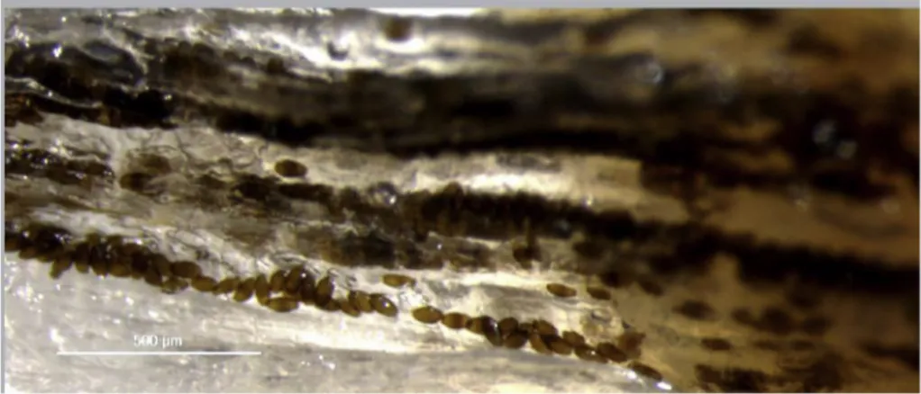
musculature in comparison with epaxial musculature which often had large areas with no darkenedflesh. Although macroscopic examination often revealed homogenous dark patches (Fig. 1), eggs in wet mounts were arranged in rows or clusters along the musclefibres, creating a linear pattern (Fig. 4). Eggs occurring in a group were sometimes of a uniform stage of development and sometimes of mixed stages.
Histological sections show the presence of eggs in different stages of development, arranged linearly between muscle fibers, isolated and dispersed in muscle tissue, and also with intracellular location. Through histological observation it was possible to establish a relationship be-tween the intensity of darkening of the muscle and the number of brown-shelled eggs infive random fields of sectioned tissue, reported as min to max (mean): Grade-1 pouting with no darkening, no eggs ob-served; Grade-2 pouting with slight to moderate darkening, 10 to 25 (χ = 17.5) eggs and Grade-3 pouting with intense darkening, 26 to 74 (χ = 47.6) eggs (Fig. 5). A range of development stages of eggs was observed in individualfish, from early stage clear-shelled eggs with a hyaline wall and basophilic content, to advanced, late-stage eggs with brown-shelled eggs containing basophilic larvae and with eosinophilic polar plugs (Fig. 6) in individualfish. Nematodes, probably 3rd or 4th stage larvae, were observed in some histological sections, and could be clearly seen to occur in both intercellular and intracellular locations.
In sections of infected muscle, an inflammatory reaction and dif-fused multifocal granulomas involving both immature eggs (with hya-line shells) and/or brown-shelled eggs were also observed, sometimes even infish with only slight darkening of flesh. Curiously, there seemed to be more granulomas infish with minor darkening of flesh than in fish with heavy darkening of flesh, although this would require larger sample size to verify. No necrotic tissue was observed surrounding eggs in the granulomatous lesions. In somefields there were degenerative and dystrophic calcifications of the tissue and in others hypertrophied musclefibres with circular and regular vacuous spaces (Fig. 7), pre-sumed to have been previously occupied by migrating Huffmanela worms.
4. Discussion
Given that each species of Huffmanela deposits its eggs in a specific organ of the host (Worsham et al., 2016) and considering egg size, superficial envelope characteristics, host taxa and localities of 20 nominal species (Ruiz et al., 2013;Ruiz and Bullard, 2013;Justine and Iwaki, 2014), only four are known to infect the musculature. These are (with reported egg length and width, host taxonomy, and localities): H. japonica Moravec et al. (1998) (58–69 × 26–30 μm, Perciformes: Fig. 3. Scanning electron microscopy of Huffmanela lusitana sp. n. eggs in muscle of pouting. Eggs ar-ranged in rows among muscle fibers (150x) (A); Huffmanela egg on the surface of the muscle fibers (m) with eggshell (sh) surrounded by a smooth vi-telline membrane (*) (1 500x) (B), except at the polar plugs (arrow) (1 500x) (C); Egg partially cru-shed. The cuticle of the larva (l) shows regularly spaced, transverse cuticular ridges (3 500x) (D).
Mullidae: Upeneus bensasi, Inland Sea of Japan); H. shikokuensis Moravec et al. (1998) (78–90 × 36–45 μm, Tetraodontiformes: Mon-acanthidae: Stephanolepis cirrhifer, Japan); H. hamo Justine and Iwaki (2014) (66–77 × 33–38 μm, Anguilliformes: Muraenesocidae: Mur-aenesox cinereus, Japan); and H. banningi Moravec (1987) (99–108 × 42–45 μm, Pleuronectiformes: Cynoglossidae: Cynoglossus browni, Atlantic Ocean off Senegal). To this group of muscle-infecting worms can now be added H. lusitana sp. n. (73–94 × 40–59 μm, Gadi-formes: Gadidae: Trisopterus luscus, Portugal) as previously reported (Esteves et al., 2009) and described herein.
All the other named, muscle infecting Huffmanela species are dis-tinct from H. lusitana sp. n. based on one or more of: size and mor-phology of eggs, host, taxonomy, infected tissue, and geographic lo-cality. Considering measurements, the eggs from our specimens are smaller than those of H. shikokuensis and H. banning; and larger than those of H. hamo and H. japonica. With respect to morphology, in H. banningi the vitelline membrane is spinous, but it is smooth in H. lusi-tana sp. n. The vitelline membrane of H. japonica is also smooth, but the underlying eggshell presents with protuberances, while the latter is smooth in H. lusitana sp. n. Regarding the host species, pouting occurs Fig. 5. Hypaxial muscle of pouting containing eggs of Huffmanela lusitana sp. n. Longitudinal sections dis-played at same scale and showing the relationship between darkness rating offlesh and degree of egg infection. Muscle section fromfish graded as normal had 0 eggs (A); muscle section fromfish graded as slight to moderate darkening had about 19 eggs (B); muscle section fromfish graded as intense darkening had about 89 eggs (C). H&E. Bar: 100μm.
Fig. 6. Hypaxial muscle of pouting infected with eggs of H. lusitana sp. n. (H&E stain). Longitudinal sec-tions: (A) immature eggs (1) and advanced brown-shelled eggs (2) with a linear distribution; (B) clear-shelled (1) and brown-clear-shelled eggs (2). Cross sec-tions: (C) eggs in different stages of development and intense inflammatory lesions and muscle destruction (note the eosinophilic color of polar plugs at arrows); (D) eggs and nematodes in cross section [(1, 4, 5) eggs with intercellular location; (2) worm with in-tracellular location, probably 3rd or 4th stage larva; (3) apical view of a polar plug]. H&E. Bar: 100μm. (For interpretation of the references to colour in this figure legend, the reader is referred to the Web ver-sion of this article.)
in the Eastern Atlantic from the British Isles and Skagerrak to the African coast, including offshore islands, and also in the western Mediterranean (Froese and Pauly, 2014). None of the other nominal species, or even the multiple reported innominate populations, has been reported from pouting or any other gadiform, and none occurs in the Northeast Atlantic. In previous studies of Huffmanela cf. lusitana sp. n. from pouting,Esteves et al. (2009)obtained similar egg measurements (73–94 × 40–59 μm). The slight discrepancy in egg size was probably due, at least in part, to the different methods used to process studied eggs - artificial digestion in the previous study and refrigeration of the samples in this study. Thus, data obtained from morphometric, biolo-gical and ultrastructural studies of the Huffmanela population infecting pouting from the Atlantic coast of Portugal suggest that it represents an undescribed species.
Among the causes offish rejection for the commercial fish market, pouting infected by Huffmanela lusitana sp. n. is considered negligible. Only the specimens that exhibit intense darkening of skin colour were likely to be discarded. All the infected pouting examined in this study had the recommended weight for marketing and did not reveal poor condition such as that observed byMendes (2006), and infected spe-cimens with slight colour change would have been cleared for human consumption. However, the skin of pouting can camouflage the pre-sence of Huffmanela infections that, if passed along through the supply chain, would probably result in rejection of theflesh during preparation by the end consumer (Fig. 8). End consumers would probably see the darkenedflesh as black mould, and the consumer's consequent rejection of fish as either parasitized or spoiled, will often have a long-lasting effect on that consumer's willingness to return to that market as a
source of food. If the timings in the life cycle of H. lusitana sp. n. are similar to those revealed byWorsham et al. (2016)for H. huffmani, then the initial infection event of a pouting would be followed by a year or more of the larvae and adult worms wandering through the muscu-lature of thefish before there are enough dark eggs to detect macro-scopically using standard inspection protocols. Suchfish in early stages of infection might not be rejected by the consumer preparing thefish for consumption, but the histological damage done by the worms mi-grating in and out of the muscle cells could conceivably cause thefish to be rejected on the dish due to unexpected irregularities in texture. Thus, while the rejection of darkenedfish by the fishermen might seem fi-nancially insignificant, allowing fish with poor-quality flesh to move along to the consumer might have a previously unnoticed but poten-tially substantial depressing effect on the end market – a cost that may be difficult to quantify, but nonetheless important enough to consider. The zoonotic impact of marketing food-fish infected with Huffmanela eggs is unknown. However, since all known species of Huffmanela are parasites of fishes (as adults) and the only known host for larvae hatched from eggs is amphipods, it is unlikely that con-sumption of infected pouting by humans (or any other mammal) could ever result in an infection with Huffmanela – although such eggs, later determined to be eggs of Huffmanela spp., have twice been reported in human stools after consumption of infectedfish (Schouten et al., 1968; Gállego et al., 1993). With that said, it is quite likely that the rugged chitinous shells of H. lusitana sp. n. in infected pouting could pass un-harmed through the gut of any piscivorous mammal, and that a stool examination could result in a false diagnosis of trichocephalid para-sitism. So, it might be prudent to encourage medical and veterinary Fig. 7. Hypaxial muscle of pouting infected with eggs of H. lusitana sp. n. (H&E stain). Cross sections: (A) diffuse inflammatory reaction involving eggs at dif-ferent stages of development; (B) granuloma with six eggs. (C) calcification of muscle tissue (arrow); (D) degenerative changes with regular cystic structures [of presumed parasitic nature (arrows)]. H&E. Bars: A, B, D, 100μm; C, 50 μm.
practitioners having patients with stools positive for bipolar eggs to inquire about recentfish consumption.
The number of eggs obtained in the different grades of colour change seems to be directly linked to the intensity of the darkening observed macroscopically, which is related to the presence of advanced brown-shelled eggs. However, pouting with slightly darkened colour presented with normal tissue interspersed with other patches having high oviposition intensity.
Macroscopically, when eggs occur in local masses in infected pouting, there is a corresponding darkening of surrounding muscu-lature, as well as the formation of dark spots in the overlying skin. The darkened skin of infected pouting does not exhibit any peculiar pattern or lesions, in contrast to other hosts where Huffmanela species infect the skin. H. markgracei eggs in Atlantic sharpnose shark causes sinuous tracks occupying a swathe of the skin on the basihyal, branchial arches and the dorsal surface of the buccal cavity (Ruiz and Bullard, 2013). H. oleumimica eggs in red snapper, Lutjanus campechanus, are deposited in dense patches or in scribble-like tracks in the skin (Ruiz et al., 2013). Rockfish (Sebastes spp.) infected with Huffmanela eggs are associated with grossly visible skin lesions (Moravec et al., 2005). Differential diagnosis is essential to distinguish black pigmentation spots on the axillary skin of seabream, Pagellus acarne (Risso) from Huffmanela in-fection. The affected seabream occurs in the same geographic area as that of the studied pouting, but the diagnosis was spontaneous mela-notic lesions (Ramos et al., 2013).
In wet mounts, pouting eggs are arranged in rows, probably re-sulting from the female dropping eggs as it moves along musclefibres. As observed in other species - H. huffmani (Huffman and Moravec, 1988), H. japonica and H. shikokuensis (Moravec et al., 1998), H. ca-nadensis (Moravec et al., 2005) and H. paronai (Moravec and Garibaldi, 2000) - eggs are apparently deposited in an early stage as colourless and unlarvated eggs. As the larva gradually develops, the egg grows in size and the chitinous layer becomes thicker and gradually turns from col-ourless to brown. This is also in agreement with the observations on H. oleumimica infection (Ruiz et al., 2013).
Eggs at different stages of development were found within the same wet mount preparation and histologicalfield. This is probably the result of mixing of different generations of eggs, due to repeated migration of several females through the same path at different tissues (Bullard et al., 2012). Since the tenure of a laying Huffmanela female is appar-ently ephemeral (Worsham et al., 2016), the observed mixing of eggs of obviously differing ages is probably indicative of separate infection events.
The eosinophilic colour of the polar plugs contrasts with the brown coloration of the rest of the advanced brown-shelled eggs. The absence of chitin in the polar plugs explains the absence of darkening of the plugs. In the Huffmanela huffmani egg shell, the polar plug sits in a collar formed by the chitinous layer and consists of an electron-lucid matrix with electron-densefibrils (Žd'árská et al., 2001).
Žd'árská et al. (2001)also referred to three layers in the Huffmanela huffmani egg shell: an external vitelline layer, a middle chitinous layer, and an inner “lipid” layer. More recently,Olson et al. (2012), in a comprehensive study of the egg shell of the model nematode Cae-norhabditis elegans, determined that the third internal layer is com-posed, not of lipid, but of chondroitin proteoglycan, and recommended that this third layer of the nematode egg shell be referred to as the CPG layer instead of the falsely suggestive“lipid” layer. In our LM studies of the H. lusitana sp. n. shell, we detected the outermost delicate and smooth vitelline membrane, and a thicker layer of the shell wall, ap-parently correspond to the combined outer chitinous and inner CPG layers.
In the SEM image ofFig. 3B, one can clearly see where the vitelline membrane has been torn partially away to expose the smooth outer surface of the chitinous layer beneath. In the LM images ofFig. 2b’-g’, one can, with some study, discern a translucent outer layer to the wall that presumably corresponds to the chitinous layer, and a darker inner
layer that presumably corresponds to the CPG layer.
During egg development of Huffmanela lusitana sp. n. the vitelline membrane apparently completely surrounds the egg in early stages. In Fig. 2b’-e’, the part of the membrane covering the polar plugs appears to be turgid as iffilled with a clear fluid under pressure. InFig. 2f’, the membrane over the plug appears wrinkled andflacid, and inFig. 2g’, it appears to have ruptured or to have been partially torn away. The inner layer of the plug proper is probably still intact, since the larva has not emerged. Indeed, the SEM inFig. 3C is of an egg with the polar section of the vitelline layer removed, exposing the core of the plug beneath. Thus, it is probably inappropriate to consider the bulging portion of the polar “plug” to be part of what actually keeps the larva in the egg. Appleton and White (1989), in a study of the polar plugs of a distant relative (Trichuris trichiura), observed that the bulge is apparent in LM studies, but disappears in air-dried and critical-point dried eggs, and speculated that this bulging protrusion is a“weak-spot” that may be important in initiating the hatching process.
The cuticle of a larva that was exposed when an egg was broken showed regularly spaced, transverse ridges, which were also described for larvae of H. oleumimica (Ruiz et al., 2013).
Muscle tissue changes and cellular responses of the host to the presence of these histozoic parasites were observed. The muscular fi-brosis described in infected pouting byEsteves et al. (2009)was not observed in this study. Granulomatous reactions are sometimes formed as the host's response to the presence of persistent foreign structures. The presence of higher numbers of granulomas infish with slight dark colour allows us to highlight the possibility that there may be parasitic granulomas in marketedfish, potentially resulting in rejection of fish on the plate due to textural irregularities. Nevertheless, the presence of cavities (of presumed parasitic origin) in muscle cells is indicative of severe cellular damage, and the areas of dystrophic calcification ob-served are probably indicative of prior muscle necrosis. All these al-terations contribute to reduced quality of the parasitizedfish as food, and may result in rejection offish on the plate, with consequent re-luctance of the consumer to return to the same market.
No encapsulated nematode bodies were found in histological sec-tions of infected pouting inside the muscle cells, as previously reported byEsteves et al. (2009)in pouting and by Moravec et al. (1998)in Stephanolepis cirrhifer in Japan. It was unclear if the nematode bodies we did observe in histological sections were late larvae or adults. Moravec et al. (1998)speculated that the larvae of H. shikokuensis occur encapsulated in the hosts’ musculature, whereas adults, after the rup-ture of infected muscle cells, migrate, copulate and lay eggs in the in-tercellular space.
Little is known about the life-cycles of Huffmanela spp., although the life cycle of the only reported freshwater species, Huffmanela huffmani Moravec (1987), was experimentally completed by Worsham et al. (2016). Apparently, the life-cycle of Huffmanela species infecting in-ternal organs may require passage through the digestive tract of a predatoryfish (or death and decay of the host) before they can infect the intermediate host.
This paper describes and highlights the morphological, ultra-structural and histological features of Huffmanela lusitana sp. n. infec-tion in pouting from the Atlantic Coast of Portugal, emphasizing the importance of this parasitic disease in the poutingfishery. In reality, the quantity of pouting discarded due to dark colour is negligible according to Portuguese official records, although the condition has been known since 2002 (first record observed in IPMA Laboratory). At present we continue to identify the existence of Huffmanela eggs in commercialized pouting. According to the Regulation 2074/2005 Annex II, Section I, Chapter I, the parasites in this study cannot be considered as“visible parasites” clearly distinguishable from the fish tissues and so, it isn't an “obviously” contaminated fish with parasites which must not be placed on the market for human consumption (Regulation 853/2004 EC. Annex III, Section VIII, Chapter V, Part D). On the other hand, the re-cent description of another commercialized fish infected with
Huffmanela sp. (Esteves et al., 2016) from the same geographic area raises the question of whether or not these populations are conspecific and how important they are in the wild stocks.
Declarations of interest None.
Acknowledgments
This research was funded by IPMA, I.P., Projecto SANAQUA - Saúde e Bem-Estar Animal das Populações Aquícolas, Grant Nº 16-02-05-FMP – Programa MAR 2020-Eixo2. The authors are grateful to Adolfo Franco as an end consumer who rejected an infected pouting we include in this study; and to Ken McKenzie for his kind comments and review of the manuscript.
References
Appleton, C.C., White, B.J., 1989. The structure of the shell and polar plugs of the egg of the whipworm, Trichuris trichiura (Nematoda: Trichuridae) from the Samango monkey (Cercopithecus albogularis). Onderstepoort J. Vet. Res. 56, 219–221.
Bullard, S.A., Ruiz, C.F., McElwain, A., Murray, M.J., Borucinska, J.D., Benz, G.W., 2012. Huffmanela CF. Carcharhini (nematoda, Trichosomoididae, huffmanelinae) from skin of a sandbar shark, Carcharhinus plumbeus, in the pacific ocean. J. Parasitol. 49, 333–340.
Carballo, M.C., Navone, G.T., 2007. A new Huffmanela species (nematoda:
Trichosomoididae) parasitizing atherinidfishes in north patagonian Gulfs, Argentina. J. Parasitol. 93, 377–382.
Commission Regulation (EC) No. 2074/2005 (EC 853/2004 rev) of 5 December 2005 laying down implementing measures for certain products under Regulation (EC) No 853/2004, Regulation (EC) No 854/2004 and Regulation (EC) No 882/2004, dero-gating from Regulation (EC) No 852/2004 and amending Regulations (EC) No 853/ 2004 and (EC) No 854/2004. Off. J. Eur. Union, 27-59.
Esteves, A., Seixas, F., Carvalho, S., Nazário, N., Mendes, M., Martins, C., 2009. Huffmanela sp. (Nematoda: Trichosomoididae) muscular parasite from Trisopterus luscus captured off the Portuguese coast. Dis. Aquat. Org. 84, 251–255.
Esteves, A., Oliveira, I., Ramos, P., Carvalho, A., Nazário, N., Seixas, F., 2016. Huffmanela spp. (Nematoda, Trichosomoididae) from Microchirus azevia: tissue location and correspondence of host muscle discoloration with parasite burden. J. Fish. Aquat. Sci. 11, 304–310.
FAO, Food and Agriculture Organization, 2010. Fishery and Aquaculture Statistics 2008. Statistics and Information Service of the Fisheries and Aquaculture Department. FAO Yearbook. FAO Fisheries and Aquaculture Department, Rome available at: http:// www.fao.org/docrep/015/ba0058t/ba0058t.pdf.
Froese, R., Pauly, D. (Eds.), 2014. FishBase. World Wide Web electronic publication.
www.fishbase.org,version.
Gállego, J., Riera, C., Portús, M., 1993. Huffmanela sp. eggs (Nematoda:
Trichosomoididae), as a human spurious parasite in a child from Barcelona (Spain). Folia Parasitol. 40, 208–210.
Huffman, D.G., Moravec, F., 1988. First description of adult Huffmanela huffmani Moravec, 1987 (nematode: Trichosomoididae) from the swimbladder of centrarchid fishes of the upper San Marcos River, Central Texas. Folia Parasitol. 35, 227–234.
Justine, J.-L., 2007. Huffmanela spp. (Nematoda, Trichosomoididae) parasites in coral reeffishes off New Caledonia, with descriptions of H. balista n. sp. and H. longa n. sp. Zootaxa 1628, 23–41.
Justine, J.-L., Iwaki, T., 2014. Huffmanela hamo sp. n. (Nematoda: Trichosomoididae: Huffmanelinae) from the dagger-tooth pike conger Muraenesox cinereus off Japan. Folia Parasitol. 61, 267–271.
Mendes, M., 2006. Huffmanela sp. (Nematoda, Trichosomoididae) parasita muscular da faneca– Trisopterus luscus (Linnaeus, 1758) – na Costa Portuguesa. MSc Thesis. Universidade Técnica de Lisboa, Portugal.
Mendes, M., Madeira de Carvalho, L.M., Ramos, P., Fazendeiro, I., Afonso-Roque, M.M., 2005. Presence of Huffmanela eggs (nematoda, Trichosomoididae) in the faneca muscle - Trisopterus luscus (Linnaeus, 1758) of the coast of Portugal. Acta Parasitológica Portuguesa 12, 334.
Moravec, F., 1987. Revision of Capillariid Nematodes (Subfamily Capillariinae) Parasitic in Fishes. Studies CSAV No. 3. Academia, Praha.
Moravec, F., Garibaldi, F., 2000. Huffmanela paronai sp. n. (nematoda:
Trichosomoididae), a new parasite from the skin of swordfish Xiphias gladius in the Ligurian Sea (western Mediterranean). Folia Parasitol. 47, 309–313.
Moravec, F., Koudela, B., Ogawa, K., Nagasawa, K., 1998. Two new Huffmanela species, H. japonica n. sp. and H. shikokuensis n. sp. (Nematoda: Trichosomoididae) from marinefishes in Japan. J. Parasitol. 84, 589–593.
Moravec, F., Conboy, G.A., Speare, D., 2005. A new trichosomoidid from the skin of Sebastes spp. (Pisces) from British Columbia, Canada. J. Parasitol. 91, 411–414.
Olson, S.K., Greenan, G., Desai, A., Muller-Reichert, T., Oeguma, K., 2012. Hierarchical assembly of the eggshell and permeability barrier in C. elegans. J. Cell Biol. 198 (4), 731–748.
Ramos, P., Victor, P., Branco, S., 2013. Spontaneous melanotic lesions in axillary seab-ream, Pagellus acarne (Risso, 1827). J. Fish Dis. 36, 769–777.
Regulation (EC) No 853/2004 of the European parliament and of the council of 29 april 2004. Off. J. Eur. Union, 22-82.
Ruiz, C.F., Bullard, S.A., 2013. Huffmanela markgracei sp. n. (Nematoda:
Trichosomoididae) from buccal cavity of Atlantic sharpnose shark, Rhizoprionodon terraenovae (Carcharhiniformes: carcharhinidae), in the northwestern Gulf of México off Texas. Folia Parasitol. 60, 353–358.
Ruiz, C.F., Ray, C.L., Cook, M., Grace, M.A., Bullard, S.A., 2013. A new species of Trichosomoididae (Nematoda) from skin of red snapper, Lutjanus campechanus (Perciformes: Lutjanidae), on the Texas-Louisiana shelf, northern Gulf of Mexico. J. Parasitol. 99, 318–326.
Schouten, H., Suriel-Smeets, R.M., Kibbelaar, M.A., 1968. The simultaneous occurrence of ova resembling Dicrocoelium dendriticum or Capillaria hepatica in the stools of in-habitants of Curaçao. Trop. Geogr. Med. 20, 271–275.
Worsham, M.L.D., Huffman, D.G., Moravec, F., Gibson, J.R., 2016. The life cycle of Huffmanela huffmani Moravec, 1987 (Nematoda: Trichosomoididae), an endemic marine-relict parasite of Centrarchidae from a Central Texas spring. Folia Parasitol. 63 020-234.
Žd’árská, Z., Huffman, D.G., Moravec, F., Nebesářová, J., 2001. Egg shell ultrastructure of thefish nematode Huffmanela huffmani (Trichosomoididae). Folia Parasitol. 48, 231–234.