Departamento de Física
Development of new biomaterials based
on liquid crystalline phases of cellulose
derivatives
Cláudio Miguel Dionísio dos Santos
Dissertação apresentada na Faculdade de Ciências
e Tecnologia da Universidade Nova de Lisboa para
obtenção do Grau de Mestre em Engenharia
Biomédica
Orientadores:
Prof. Dr. João Paulo Borges, DCM
–
FCT/UNL
Dr. Pedro Granja, INEB
–
Instituto de Engenharia
Biomédica, Universidade do Porto
Cláudio Santos Dissertação de Mestrado em Engenharia Biomédica i
This thesis was supervised by:
→ Prof. Dr. João Paulo Borges (supervisor)
DCM – Departamento de Ciências dos Materiais, Faculdade de Ciências e Tecnologia – Universidade Nova de Lisboa.
→ Dr. Pedro Granja (co-supervisor)
NEWTherapies Group, Biomaterials Division, INEB – Instituto de Engenharia Biomédica, Universidade do Porto.
The research described in this thesis was conducted at:
→ DCM – Departamento de Ciências dos Materiais, Faculdade de Ciências e Tecnologia – Universidade Nova de Lisboa.
Cláudio Santos Dissertação de Mestrado em Engenharia Biomédica ii
Acknowledgements
This is perhaps the hardest section I have to write, but I will, nonetheless, try. I would like to thank to all the ones who have contributed to the accomplishment of this thesis.
First of all I would like to acknowledge to my supervisors Prof. Dr. João Paulo Borges and Dr. Pedro Granja for the help, orientation and transmitted knowledge they gave me. I want to express my gratitude for the opportunity and the privilege of working in a renowned institution, Instituto de Engenharia Biomédica (INEB, Universidade do Porto), that hosted me in an enthusiastic way. I am grateful to several persons who supported and encouraged my research work at Biomaterials Division of INEB and at Departamento de Ciências dos Materiais of Faculdade de Ciências e Tecnologia (DCM, FCT – Universidade Nova de Lisboa). I want to send thanks to all my remarkable friends and colleagues at FCT and INEB with whom I had the privilege of working with. They helped me along these months and were always there to listen and share their experience and knowledge.
At INEB, I would like to give a special thank to Cristina Barrias, who patiently answered all my questions, tracked down papers, guided me during my work and was always there. She was my advisor and taught me numerous things, enabling me to face the hard work with a smile. The scientific discussions and encouragement made by her made the difference to the progress of this work. I also want to thank to the people who worked with me at INEB and FCT for their collaboration and help in several situations at the lab. In particular, the collaboration and advice of Ricardo Vidal Silva and Manuela Brás were fundamental. I can’t forget to mention Ana Filipa Lourenço that is the best bench colleague that someone could have. Thank you for all the hours spent with me in that lab. At CEMUP (Centro de Materiais da Universidade do Porto) I would like to thank to all who helped me in Scanning Electron Microscopy and X-ray Photoelectron Spectroscopy. I am sincerely grateful to all. This work would not have been possible without your help and dedication.
I wish to thank my great friends for their support during this last 5 years, in particular to my girlfriend Ana who helped me to get through the difficult situations. As much as I could write here would never be enough to show how grateful I am to her. I am indebted to my student colleagues for providing a stimulating environment to learn and grow.
My final words go to my family. I wish to thank my entire extended family for providing a lovely environment for me, particularly my parents and my brother. I dedicate this thesis to them, for standing by me in difficult hours, trying to enhance my spoiled weekends, dealing with my bad temper, but above all for their effort and motivation during all my entire life.
Cláudio Santos Dissertação de Mestrado em Engenharia Biomédica iii
Abstract
In the present work, new biomaterials based on liquid crystalline phases of cellulose derivatives were prepared and characterized. The possibility to monitor growth and differentiation of stem cells could be an interesting use for the HPC films studied in this thesis. Additionally, the application of HPC films as an anti-adhesive substratum in biomedical applications could also be interesting.
Cláudio Santos Dissertação de Mestrado em Engenharia Biomédica iv
Resumo
Neste trabalho procedeu-se à preparação e caracterização de novos biomateriais baseados em fases líquido-cristalinas de derivados da celulose. A possibilidade de monitorizar o crescimento e diferenciação de células estaminais poderia ser uma aplicação interessante para os filmes de HPC estudados nesta tese. O seu uso como substrato anti-adesivo em aplicações biomédicas poderia ser igualmente interessante.
Cláudio Santos Dissertação de Mestrado em Engenharia Biomédica v
Aim and structure of the thesis
The aim of this thesis was to develop and characterize cross-linked hydroxypropylcellulose (HPC) films with different properties that can be significant to a variety of potential applications in biology and medicine. The main goal was to explore possible biomedical applications for these biomaterials. It is well known that surface properties influence the adsorption of serum proteins and, hence, cell adhesion. Surface properties such as wettability, charge, topography and chemical composition, among others, play an important role in cell behaviour. The characterization and interaction of these biomaterials with mesenchymal stem cells (MSC) were investigated. The properties of HPCi and HPCa films were compared, as well as the behaviour of MSCs when in contact with them.
It was recently demonstrated that liquid crystals can monitor the growth of embryonic stem cells [1] proving that research in this field could have a bright future. The diversity of biomaterials that reside in living systems and exhibit liquid crystal properties was also demonstrated [2]. It is also known that mesenchymal stem cells are a promising candidate in regenerative medicine and present a huge potential clinical use. Cellulose is the most abundant biopolymer in nature and one of the most commonly used in biomedical applications. Many of its derivatives, like HPC, exhibit chiral nematic liquid crystalline phases. The possibility to monitor growth and differentiation of stem cells could be an interesting use for this biomaterial.
Chapter I gives a general overview of liquid crystals principles and their applications in medicine, describes the current status of regenerative medicine and explains the mechanisms behind cell-material interactions as well the importance of non-fouling biomaterials in biomedical applications.
Currently, the evaluation of biomaterials includes an initial sequence of both non-biological and biological (cell culture) tests. The experimental work is presented along 2 chapters (chapters II
and III).
Cláudio Santos Dissertação de Mestrado em Engenharia Biomédica vi and hydrophilicity). The biological experimental characterization includes cytotoxicity evaluation, cell adhesion, morphology, proliferation and viability assays.
In Chapter III, the procedures used in each technique are described and the results are interpreted and discussed.
Cláudio Santos Dissertação de Mestrado em Engenharia Biomédica vii
List of Symbols
AFM – Atomic Force Microscopy
APC – Acetoxypropylcellulose
ATR – Attenuated total reflectance
BMSC –Bone Marrow Stromal Cells
BPC – n-butyric
CAB – Cellulose Acetate Butyrate
CEL – hydroxypropylmethylcellulose (HPMC)-CMC-coated substratum
CMC – Carboxymethylcellulose
CU – Cuprophan
DMAc –Dimethylacetamide
DMEM – Dulbecco's Modified Eagle Medium
DRS – Diffuse reflectance spectroscopy
E –Young’s modulus
EDS – Energy Dispersive Spectrometer
EGC – Embryonic germ cells
EKA –Electro Kinetic Analysis
ESC – Embryonic stem cells
ESCA – Electron Spectroscopy for Chemical Analysis
ESEM – Environmental Scanning Electron Microscopy
EVA – Ethylene Vinyl Acetate
FACS – Fluorescence-activated cell sorting
FBS – Fetal Bovine Serum
FTIR – Fourier Transform Infrared Spectroscopy
HepPc – Heptanionic acid
hESC – human Embryonic Stem Cells
Cláudio Santos Dissertação de Mestrado em Engenharia Biomédica viii
HPC – Hydroxypropylcellulose
HPCa –Anisotropic 60 wt% chiral nematic cross-linked solution
HPCi – Isotropic 30 wt% cross-linked solution
HPMC– Hydroxypropylmethylcellulose
iBPC – Isobutyric
iVPC – Isovaleric
LC – Liquid Crystal
LCD – Liquid crystal display
MC – Methylcellulose
MSC – Mesenchymal stem cells
NFS – Non-Fouling Surfaces
OCA – Optical Contact Angle
PAAm – Polyacrylamide
PBS – Phosphate Buffered Saline
PDMAA – Poly(N,N-dimethyl methacrylamide)
PEG – Poly(Ethylene Glycol)
pHEMA – Poly(2-hydroxyethyl methacrylate)
PPC – Propionic
SEM – Scanning Electron Microscopy
TAC – Cellulose triacetate
vdW – van der Waals
VPC – Valeric
Cláudio Santos Dissertação de Mestrado em Engenharia Biomédica ix
Contents
Acknowledgments...iiAbstract...iii
Resumo...iv
Aim and outline of this thesis...v
List of Symbols...vii
Contents...ix
Index of Figures...xii
Index of Tables...xv
Chapter I – INTRODUCTION...1
1. Liquid crystals...1
1.1. A brief review...1
1.2. Liquid crystals in biological systems...2
1.3. Liquid crystals in medical applications...3
1.4. Liquid crystals based on cellulose derivatives...6
1.4.1. Hydroxypropylcellulose as a biomaterial...7
2. Non-fouling biomaterials...9
2.1. A brief review...9
2.2. Surface modulation of biological behaviour...12
2.3. Protein adsorption and cell adhesion on biomaterial surfaces...14
2.4. Cellulose as anti-adhesive substratum in biomedical applications...15
3. Stem Cells and Regenerative Medicine...16
3.1. Principles of Regenerative Medicine ...16
3.2. Stem cells...16
3.2.1. Mesenchymal stem cells...18
3.2.1.1. Biological characterization, techniques for isolation and expansion...19
3.2.1.2. A promising candidate in regenerative medicine, clinical applications and trials...21
Cláudio Santos Dissertação de Mestrado em Engenharia Biomédica x
Chapter II – MATHERIALS AND METHODS...25
1. Materials Preparation...25
1.1. Production of HPC cross-linked films...25
2. Structural and Physico-Chemical Characterization...26
2.1. Optical polarizing microscopy...28
2.2. Determination of the gel fraction...28
2.3. Mechanical properties of the dried and wet films...28
2.4. Contact Angle Measurements...29
2.5. Chemical characterization of surfaces...33
2.5.1. Fourier Transform Infrared (FTIR) Spectroscopy...33
2.5.2. X-ray Photoelectron Spectroscopy (XPS)...35
2.6. Scanning Electron Microscopy (SEM)...37
2.7. Atomic Force Microscopy (AFM)...39
3. Sterilization...40
4. Biological characterization...40
4.1. Cell cultures...40
4.2. Cytotoxicity...41
4.3. Cell Attachment...41
4.4. Cell Morphology...42
4.5. Cell Proliferation...42
4.6. Cell viability...43
4.7. Environmental Scanning Electron Microscopy...43
5. Statistical verification of results...43
Chapter III – RESULTS AND DISCUTION...44
1. Characterization...44
1.1. Optical polarizing microscopy...44
1.2. Determination of the gel fraction...45
1.3. Mechanical properties of the dried and wet films...45
1.4. Contact Angle Measurements...48
1.5. Chemical characterization of surfaces...49
1.5.1. Fourier Transform Infrared (FTIR) Spectroscopy...49
1.5.2. X-ray Photoelectron Spectroscopy (XPS)...51
1.6. Scanning Electron Microscopy (SEM)...56
Cláudio Santos Dissertação de Mestrado em Engenharia Biomédica xi
2. Biological characterization...59
2.1. Cytotoxicity Assay...59
2.2. Cell Attachment...61
2.3. Cell Morphology...63
2.4. Cell Proliferation...68
2.5. Cell viability...69
2.6. Environmental Scanning Electron Microscopy...70
Chapter IV – CONCLUSION...71
Chapter V – FUTURE DIRECTIONS...73
References...75
Cláudio Santos Dissertação de Mestrado em Engenharia Biomédica xii
Index of Figures
Chapter I Figure 1.1 – Schematic representation of the collective arrangement of the rod-like molecules in the (a) nematic, (b) smectic A and (c) cholesteric phases.…………...……….…...…2Figure 1.2 –Liquid crystal thermometer………...………..4
Figure 1.3 – Thermographic pictures of (a) peripheral neuropathy, (b) rheumatoid arthritis and (c) breast cancer………...………..…….5
Figure 1.4 – Ideal structure of HPC. DE = 2.5 (Degree of Eterification); ME = 3 (Molar Eterification)………...…….7
Figure 1.5 – Top view image of the amplitude scan of the free surface of sheared HPC films prepared at a shear rate of v = 5 mm/s from solutions with HPC/water ratio 65% w/w………...…..8
Figure 1.6 – Schematic representation of some methods used to modify surfaces of biomaterials………...…………13
Figure 1.7 –Interaction of cells with foreign surfaces………...………….14
Figure 1.8 – Hierarchy of stem cells...…...17
Chapter II Figure 2.1 – Visual aspect of an HPCa solution in DMAC showing characteristic iridescent colours resultant of the reflection of the light...25
Figure 2.2 – Schematically representation of the cross-linking reaction…...………….………26
Figure 2.3 – Schematic representation of HPC films preparation………...………26
Figure 2.4 – Photograph of the (a) tensile tests equipment and (b) detail of the film sample placed between the clamps………...…29
Figure 2.5 – Contact angle with associated forces………...……….30
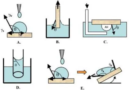
Figure 2.6 – Different contact angle measurements: A. Sessile drop method; B. Wilhemly plate method; C. Captive air bubble method; D. Capillary rise method; E. Tilted-drop method…...…...31
Figure 2.7 – Optical Contact Angle Device OCA 15..………...……….32
Figure 2.8 – Optical layout of a typical FTIR spectrometer...…...34
Figure 2.9 – Schematic diagram illustrating basic components of a XPS system………...36
Figure 2.10 – Schematic representation of how does a SEM work………...…38
Cláudio Santos Dissertação de Mestrado em Engenharia Biomédica xiii
Chapter III
Figure 3.1 – (a) HPCi film and (b) HPCa film. Arrow indicates the direction of films spreading...44
Figure 3.2 – Schematic representation of the directions used to measure the mechanical behaviour of HPC films...45
Figure 3.3 – Typical stress-strain curves of HPC solid films recorded at 25ºC. (a) dry conditions; (b) wet conditions………...46
Figure 3.4 – Measurement of the water contact angle on a HPC film: (a) pendant drop, (b) water drop wetting the HPC surface………...…….48
Figure 3.5 – Contact angle measurements on HPCi and HPCa films. (* means statistically significant difference; p<=0.05)………...48
Figure 3.6 – FTIR spectra of (a) HPC and (b) diisocyanatohexane provided by the manufacturer……….…...50
Figure 3.7 – FTIR spectra of (a) HPCa and (b) HPCi films, evidencing the presence of the N-H groups resultant from the cross-linking of HPC molecules...……….….…….51
Figure 3.8 – XPS spectra of (a) HPCi crosslinked film and (b) HPCa crosslinked film...……….….…...52
Figure 3.9 – Elemental compositions of HPCi and HPCa films, as determined by XPS...…….…...…53
Figure 3.10 – spectra of (a) HPCi and (b) HPCa films...……….………...….54
Figure 3.11 – spectra of (a) HPCi and (b) HPCa films...……….…….……54
Figure 3.12 – spectra of (a) HPCi and (b) HPCa films...……….….……55
Figure 3.13 – (a) HPCi film (magnification: 1000x); (b) HPCi film (magnification: 10000x); (c) HPCa film (magnification: 1000x; arrow represents shear force direction); HPCa film (magnification: 5000x; arrow represents shear force direction)...………..………..56
Figure 3.14 – 3D topography image ( ) of the HPCa surface...………..……..57
Figure 3.15 – Top view image of the HPCa surface...……….…..…..58
Figure 3.16 – Top view image of 60% HPC and height profile analysis at the cross sections AA’...………..…..58
Figure 3.17 – Tests for in vitro cytotoxicity after (a) 24h and (b) 72h (where fDMED means fresh DMEM and eDMEM means extracted DMEM)...……….….……60
Figure 3.18 – Percentage of cell attachment on HPCi and HPCa films...……….61
Cláudio Santos Dissertação de Mestrado em Engenharia Biomédica xiv
Figure 3.20 –Cell morphology 4 h post-seeding on (a) HPCi film, (b) HPCa film, (c) coverslip and (d) pHEMA film...64
Figure 3.21 – Cell morphology 12 h post-seeding on (a) HPCi film, (b) HPCa film, (c) coverslip and (d) polyHEMA film...……….….……..64
Figure 3.22 – Cell morphology 24 h post-seeding on (a) HPCi film, (b) HPCa film, (c) coverslip and (d) polyHEMA film...……….….……..65
Figure 3.23 – Cell morphology 48 h post-seeding on (a) HPCi film, (b) HPCa film, (c) coverslip and (d) polyHEMA film...……….….…..66
Figure 3.24 – Cell morphology 12 h post-seeding on (a-d) HPCa films; (e-g) HPCa films, after fluorescent staining of actin micro filaments and DNA; (h) coverslips, after fluorescent staining of actin micro filaments and DNA...……….…….67
Figure 3.25 – Cell proliferation indexes on HPCi film, HPCa film, polyHEMA and coverslip after 24 and 48 h. Cell proliferation corresponds to N/N0 ratio, where N is the total number of cells at T = 24 h or T =48 h and N0 is the number at T = 0. The blue line corresponds to the ratio of 1 representing the cell number at seeding...………..69
Cláudio Santos Dissertação de Mestrado em Engenharia Biomédica xv
Index of Tables
Chapter I
Table 1.1 – Thermodynamics of protein adsorption...……….10
Table 1.2 – Non-fouling biomaterial surface compositions...……….12
Table 1.3 – Clinical trials using mesenchymal stem cells...………..21
Chapter II
Table 2.1 – Common methods used to characterize biomaterial surfaces...………..27
Chapter III
Table 3.1 –Average values for the Young’s modulus obtained for HPC cross-linked films for various HPC concentration ratios and different directions...……….47
Table 3.2 – Contact angle measurements on HPCi and HPCa films...……….….49
Table 3.3 – Elemental compositions of HPCi and HPCa films, as determined by XPS...……….53
Table 3.4 – AFM measurements obtained for 60% HPC cross-linked films...……….59
Table 3.5 – Cell proliferation assessed on HPCi and HPCa films, coverslips and polyHEMA films at 24 h and 48 h post-seeding...……….68
Cláudio Santos Dissertação de Mestrado em Engenharia Biomédica 1
Chapter I
–
INTRODUCTION
1. Liquid crystals
1.1. A brief review
The first observation of liquid crystalline behaviour was made by an Austrian botanist named Friedrich Reinitzer who, about a hundred years ago, was investigating a material known as cholesteryl benzoate that had two distinct melting points [3]. Since this discovery, thousands of compounds have been identified to exhibit that new phase of matter — the mesogenic or liquid crystalline phase. The term liquid crystal (LC) describes a state of matter that is intermediate between an isotropic liquid and a crystalline solid state. LCs can be further divided into two main families: thermotropic and lyotropic. These types of LCs are distinguished by the mechanisms that drive their self-organization. Thermotropic LCs exhibit mesophases as function of temperature. On the other hand, lyotropic LCs are obtained when an appropriate concentration of a material is dissolved in some solvent. The most important variable is the amount of solvent (or concentration), under certain conditions of temperature and pressure.
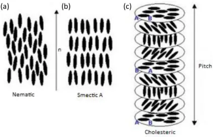
Cláudio Santos Dissertação de Mestrado em Engenharia Biomédica 2 normal layer [4-6]. The collective arrangement of the rod-like molecules in the nematic, cholesteric and smectic A phases are schematically represented in the Fig. 1.1.
Figure 1.1 – Schematic representation of the collective arrangement of the rod-like molecules in the (a) nematic, (b) smectic A and (c) cholesteric phases. Adapted from [7].
Liquid crystals are found to be birefringent due to their anisotropic nature. It means that they demonstrate double refraction, having two indices of refraction. This effect occurs because the structure of the material is anisotropic and the light polarized parallel to the director has a different index of refraction than the light polarized perpendicularly to the director.
1.2. Liquid crystals in biological systems
Within the biological systems several liquid crystal structures can be found. This idea was firstly proposed by Lehmann [8]. Afterwards, several liquid crystalline materials, which are found in biological systems, have been studied by several researchers. Most liquid crystals are derived from natural sources including cellular membranes [2], hemoglobin [9], chromosomes [9], bile [10], carapace of some insects [9], concentrated solutions of biomolecules, such as proteins, [9, 11-13], and solutions of cellulose and its derivatives [9]. Many liquid crystal biomaterials are found in living systems, which means that several structures have properties that are dependent on the liquid crystallinity for their function. Even materials such as DNA can exhibit mesomorphic behaviour [2].
The structures of cell walls that surround all cells can be described by the fluid mosaic model, where integral proteins float in a two-dimensional lipid sea. Cell membranes contain a variety of
Cláudio Santos Dissertação de Mestrado em Engenharia Biomédica 3 biological molecules, especially lipids and proteins. Each of the membrane lipid classes (phospholipid, glycolipid and cholesterol) and related derivatives has been shown to possess mesomorphic properties and some of them have been widely studied both in their lyotropic, as well as their thermotropic states [2]. For example, aliphatic derivatives of cholesterol were among the first thermotropic liquid crystals to be investigated. Cerebrosides, a class of glycolipids derived from the sphingosine were also found to be potentially thermotropic or lyotropic liquid crystals. J. W. Goodby [2] showed that cells could use liquid crystal technology, typical of materials for display device applications, for modifying their behaviour and/or properties (e.g. modify melting behaviour and to fine tune viscoelastic properties). Goodby [2] also discussed the liquid crystal properties of other bio-materials including aliphatic derivatives of vitamin C and solanin witch exhibit thermotropic smectic A phases in certain conditions. It can be easily concluded that there is a great variety of bio-materials that exhibit mesomorphic behaviour. Some of them are certainly involved in bio-processes and applications (e.g., inter-cellular recognition and surfactants) or could have attractive properties (e.g., pharmaceuticals) [2].
Brown [14] also presented several analogies between the properties of liquid crystals and those of supramolecular arrangements in biology. Brown studies stimulated more probing investigation of liquid crystals in biology over the last 30 years. The importance and the role of liquid crystals properties in and to living systems were demonstrated [14]. Their role in sensory systems, in cellular shape and in the transmission of information were some of the topics discussed.
It is believed that the field of liquid crystals could have a bright future and will play an important role in the understanding of biological process.
1.3. Liquid crystals in medical applications
Cláudio Santos Dissertação de Mestrado em Engenharia Biomédica 4 director to point in a specific direction by introducing an outside agent to the system [15]. The most frequent application of liquid crystal technology is the liquid crystal displays (LCD). LCDs are used in several applications like calculators, laptop computer screens, watches and airplanes [9, 16-19].
Temperature changes can influence the colour of a liquid crystal, which makes them useful in medical applications, for instance, to measure body temperature (see Fig. 1.2). Chiral nematic (cholesteric) liquid crystals reflect light with a wavelength equal to the pitch. Since the pitch (distance that the liquid crystals twist over) is temperature dependent, the colour reflected is also temperature dependent, which allows the use of liquid crystals as temperature sensors, just by looking at the colour that indicates different temperatures. When the pitch equals half of the wavelength of red light the liquid crystal will reflect red light. It is possible to build a device for practically any temperature range by mixing diverse compounds. The resolution of liquid crystal sensors is in the 0.1°C range. LC thermometers are economical, simple to install, practically unbreakable and there is no risk of toxic elements being released [19, 20].
Figure 1.2 – Liquid crystal thermometer. Adapted from [21].
Cláudio Santos Dissertação de Mestrado em Engenharia Biomédica 5 precise, has high resolution and does not involve ionizing radiation. The thermographic pictures of some affected areas is shown in Fig. 1.3 [18, 22].
Figure 1.3 – Thermographic pictures of (a) peripheral neuropathy, (b) rheumatoid arthritis and (c) breast cancer. Adapted from [21].
Liquid crystals may also be used in ergonomic applications, for example for detection of occupational joint stress (response inflammation) as a result of physical effort. Other medical application is the measurement of the transient pressure transmitted by a walking foot to the ground. Several techniques have been developed to improve liquid crystal temperature measurements [23, 24].
In 2007, Atyabi and co-workers [25] have studied the utilization of liquid crystal embedded cellulose membranes as a mechanism of thermoresponsive drug permeation. The main objective was to modify the control of permeability and pore variability due to its thermotropic characteristics. As a result, under different temperatures, the permeability can be altered, allowing, for example, the release of drugs only in hyperthermia sites, as desired in the application of chemotherapeutics [25]. Liquid crystals can significantly change the release rate of drugs, increasing solubility, absorption and bioavailability of control. They may also alter the pharmacokinetics, decreasing toxicity and improving clinical effectiveness [26].
Recently, some studies in which synthetic LCs were put in contact with cells have been reported [1, 27, 28]. The main goal of these studies was to investigate the ordering of LCs in contact with cells as a potential tool to infer about the interactions of cells with their environment. Recently, the orientation of the nematic liquid crystal 4’-pentyl-4-cyanobiphenyl (5CB) on fixed (dead) cells attached to surfaces was investigated by Fang and co-workers [28]. A subsequent study showed that this material cause cell death when in contact with live cells [27]. Luk and co-workers [27] found,
Cláudio Santos Dissertação de Mestrado em Engenharia Biomédica 6 during this study, numerous liquid crystals that present no toxicity to live mammalian cells [27]. More recently, Lockwood and co-workers [1] confirmed that it was possible to culture human embryonic stem cells (hESC) on the surface of a film of a nematic LC TL205 (a mixture of cyclohexane-fluorinated biphenyls and fluorinated terphenyls) that were decorated with thin films of the extracellular matrix, Matrigel. This study showed that hESC could survive for approximately two weeks on liquid-crystal substrates without visible signs of toxicity. It was also demonstrated that the ordering of the LC was influenced by the extracellular matrix of the cells and that the reorganization of the extracellular matrix by the stem cells led to ordering transitions in the LCs [1].
It is also known that liquid crystals exhibit electro-optic effects [29], which make them an attractive tool for a variety of applications including fast, compact, and tunable spectral filters, phase modulators, polarization controllers and optical shutters [30]. LCs have been used in a wide range of applications including computer monitors, television, gaming devices, aircraft cockpit displays, instrument panels, video players, calculators and telephones, among others. However, their application in the field of optical imaging just started to emerge. LC devices can also have a great potential in biomedical optical imaging systems and other biomedical applications. According to Abdulhalim et al. [30], using a collection of tunable phase retarders one can perform:
Stokes parameters imaging for skin and eye polarimetric imaging;
Tunable filtering to be used for hyperspectral imaging, fluorescence microscopy, and frequency domain optical coherence tomography;
Adaptive optical imaging and eye aberrations correction;
Phase shift interferometric imaging;
Variable frequency structured illumination microscopy.
New properties and applications of LCs in medicine are being investigated. These materials have an enormous potential and more industrial and scientific applications involving LCs will certainly be developed in the near future.
1.4. Liquid crystals based on cellulose derivatives
Cláudio Santos Dissertação de Mestrado em Engenharia Biomédica 7
1.4.1. Hydroxypropylcellulose as a biomaterial
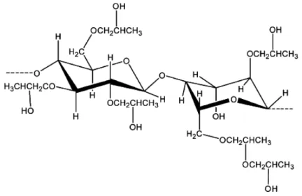
HPC is an organic polymer of high solubility in water and alcohols, due to the hydrophilic OH and COC groups as well as hydrophobic organic groups. The chemical structure of HPC can be seen in Fig. 1.4.
Figure 1.4 – Ideal structure of HPC. DE = 2.5 (Degree of Eterification); ME = 3 (Molar Eterification).
HPC is one of the most studied ether cellulose derivatives. However, it has been poorly studied in the biomedical field.
The first lyotropic cholesteric phase of a cellulose derivative was obtained from this ether, for high concentrations of polymer in water [35]. It has been shown that HPC can originate lyotropic phases when used in some concentrations and can form solid films that can act as barriers to air and moisture. It was also used in coatings, even at high humidity and as a packaging material [36, 37]. Its electro-optical properties have also been the subject of several theoretical and experimental studies [29, 38]. The use of HPC solid films as the matrix of a polymer/liquid crystal composite system, with applications in the production of electro-optical displays, was also investigated [38].
The preparation of various esters of HPC can be found in the literature [35, 39]. The first ester derivative of HPC presented in the literature, as having a liquid crystalline thermotropic phase, was the acetoxypropylcellulose (APC). Subsequently, other esters of HPC, which can generate lyotropic and thermotropic mesophases [40, 41], were prepared and characterized. Propionic (PPC),
Cláudio Santos Dissertação de Mestrado em Engenharia Biomédica 8 their optical properties were studied [39]. The mesophases of APC, PPC, iBPC, VPC, and iVPC exhibit reflection bands in the visible region, at wavelengths that depend on temperature [42], moisture content, size and number of substituents [42], and degree of polymerization [39]. The pitch of the cholesteric helical structure can be controlled through different processes, including increasing the side chain length and the temperature [29, 39]. Due to the ability of their molecules to spontaneous self-assemble in helicoidal arrangements, light can be reflected selectively, depending on some parameters, like the chain length and the temperature [43-46]. Many research works have been contributing to improve the use of cellulose derivatives for electro-optical applications [39, 47].
Aharoni [48] reported in 1979 that solutions of liquid crystalline polymers could form banded textures in thin-film samples. The film surface features of HPC/water (50–65% w/w) were recently investigated by atomic force microscopy studies [49]. It was demonstrated in this study that the surface of HPC films prepared from liquid crystalline aqueous solutions shows two periodic structures.
Figure 1.5 – Top view image of the amplitude scan of the free surface of sheared HPC films prepared at a shear rate of v = 5 mm/s from solutions with HPC/water ratio 65% w/w [49].
Cláudio Santos Dissertação de Mestrado em Engenharia Biomédica 9
2. Non-fouling biomaterials
2.1. A brief review
Surfaces that resist to the adsorption of proteins and/or adhesion of cells are usually known as non-fouling surfaces (NFS). It is generally accepted that surfaces that strongly adsorb proteins will probably bind cells and that surfaces that resist protein adsorption will resist cell adhesion [50]. Apart from a few exceptions it was also established that hydrophilic surfaces seem to be more resistant to protein adsorption, while hydrophobic surfaces usually tend to adsorb a monolayer of tightly adsorbed protein [50]. NFSs have several medical and biotechnological applications, including blood compatible materials, biosensors, implantable devices, microchannel flow devices, urinary catheters, diagnostic assays, affinity separators, intravenous syringes and tubing as well as nonmedical applications, including biofouling-resistant heat exchangers and ship bottoms [50]. The use of blood contacting devices, including dialyzers, vascular grafts, oxygenators, blood containers and catheters, requires prevention of protein adsorption and NFSs may resist to fibrinogen adsorption and platelet adhesion [51]. The adsorption of serum proteins is the first event that takes place when a material surface is placed in contact with blood. This will lead to the formation of thrombus or blood clots which could cause serious clinical problems. NFSs have been the subject of many investigations, because they offer important experimental insights into one of the most important phenomenon in biomaterials science, i.e., protein adsorption. Ongoing investigation aims at developing protein-resistant surfaces in order to overcome these issues [51]. The production of anti-fouling films is an important element in the development of biomedical materials, for applications such as medical devices, implants and in vitro tests [52]. Such coatings favour the biological integration of these tools by limiting the interactions between the implants and physiological fluids [53, 54]. Until now, various water-soluble polymers have been used for surface grafting. They include nonionic, hydrophilic polymers such as polyacrylamide (PAAm), poly(N,N-dimethyl methacrylamide) (PDMAA), poly(ethylene glycol) (PEG), ethylene vinyl acetate (EVA) and Poly(2-hydroxyethyl methacrylate) (pHEMA) [51].
The protein and cell interactions with biomaterial surfaces have been the subject of several investigations in recent decades and some considerations have been established [50], namely:
Cláudio Santos Dissertação de Mestrado em Engenharia Biomédica 10 release of many hydrophobically structured water molecules from the interface, leading to a large entropy gain for the system [57].
At low ionic strengths cationic proteins bind to anionic surfaces and anionic proteins bind to cationic surfaces [57, 58]. The major thermodynamic driving force for these actions is a combination of ion–ion coulombic interactions, accompanied by an entropy gain due to the release of counter ions along with their waters of hydration [58].
Proteins tend to adsorb in monolayers, which means that they do not adsorb non-specifically onto their own monolayers [59]. The retention of hydration water by the adsorbed protein molecules, preventing close interactions of the protein molecules in solution with the adsorbed protein molecules is the probable cause for it.
Studies about surfaces coated with physically or chemically immobilized PEG concluded that its molecular weight should be above a minimum of ca. 2000 in order to provide good protein repulsion [60-62]. The mechanism of protein resistance by the PEG surfaces involves the resistance of the polymer coil to compression due to its ability to retain the volume of a random coil (called “entropic repulsion” or “elastic network” resistance) and involves the resistance of the PEG molecule to release both bound and free water from within the hydrated coil (called “osmotic repulsion”) [62, 63].
The thermodynamic principles governing the adsorption of proteins onto surfaces involve a number of enthalpic and entropic factors favouring adsorption or favouring resistance to protein adsorption (Table 1.1).
Table 1.1 – Thermodynamics of protein adsorption. Adapted from [50].
Favouring adsorption
( ) van der Waals (VdW) interactions (short-range) ( ) ion–ion interactions (long-range)
Cláudio Santos Dissertação de Mestrado em Engenharia Biomédica 11
Opposing adsorption
( ) dehydration (interface between surface and protein) ( ) chain compression (PEO)
( ) unfolding of protein
( ) protein hydrophobic exposure ( ) chain compression (PEO) ( ) adsorption of protein ( ) osmotic repulsion (PEO)
The main factors favouring adsorption are the entropic gain of released water and the enthalpy loss due to cation–anion attractive interactions between ionic protein groups and surface groups. The major factors favouring resistance to protein adsorption are the retention of bound water and, in the case of an immobilized hydrophilic polymer, entropic and osmotic repulsion of the polymer coils.
The mechanism behind the action of non-fouling surfaces has been the subject of several studies. Although it seems difficult to ascertain, the principal factor favouring resistance to protein adsorption seems to be the retention of bound water by the surface molecules. In the case of the immobilized hydrophilic polymer, entropic and osmotic repulsion by the polymer coils seems to be also related. Taking these observations into account, one can conclude that the most common approaches used in non-fouling surfaces research, consists in making them more hydrophilic with the aim of reducing protein adsorption and cell binding. The behaviour of non-fouling surfaces in vivo is still largely unknown and the stability and longevity of non-fouling biomaterials remains unclear. Biomaterials researchers keep trying to develop non-fouling surfaces. Their specific applications, biological environments and intended life services must be taken into account in their development.
Cláudio Santos Dissertação de Mestrado em Engenharia Biomédica 12
Table 1.2 – Non-fouling biomaterial surface compositions. Adapted from [50].
Synthetic Hydrophilic Surfaces Natural Hydrophilic Surfaces
► PEG polymers and surfactants
► Neutral polymers
- Poly(2-hydroxyethyl methacrylate)
- Polyacrylamide
- Poly(N-vinyl-2-pyrrolidone)
- Poly(N-isopropyl acrylamide) (below 31◦C)
► Anionic polymers
► Phosphoryl choline polymers
► Gas discharge-deposited coatings (especially from PEG-like monomers)
► Self-assembled n-alkyl molecules with oligo-PEG head groups
► Self-assembled n-alkyl molecules with other polar head groups
► Passivating proteins (e.g., albumin and casein)
► Polysaccharides (e.g., hyaluronic acid, cellulose and its derivatives)
► Liposaccharides
► Phospholipid bilayers
► Glycoproteins (e.g., mucin)
2.2. Surface modulation of biological behaviour
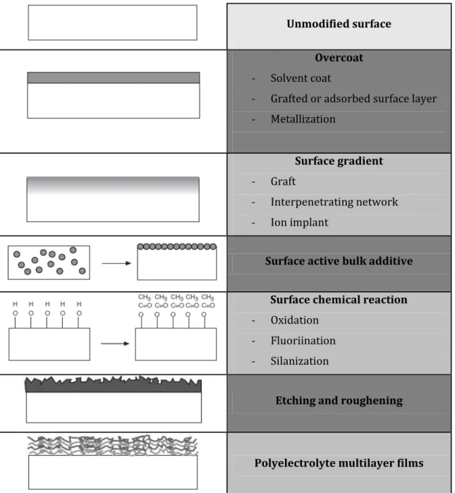
The design and synthesis of biomaterials and devices has been the target of several biomedical investigations to ensure appropriate durability, mechanical properties and in vivo
Cláudio Santos Dissertação de Mestrado em Engenharia Biomédica 13 Unmodified surface
Overcoat
- Solvent coat
- Grafted or adsorbed surface layer
- Metallization
Surface gradient
- Graft
- Interpenetrating network
- Ion implant
Surface active bulk additive
Surface chemical reaction
- Oxidation
- Fluoriination
- Silanization
Etching and roughening
Polyelectrolyte multilayer films
Figure 1.6 – Schematic representation of some methods used to modify surfaces of biomaterials. Adapted from [50].
Cláudio Santos Dissertação de Mestrado em Engenharia Biomédica 14
2.3. Protein adsorption and cell adhesion on biomaterial surfaces
The body is able to recognize and respond to implanted biomaterials. Clotting of blood and foreign body reaction are typical biological responses. This response starts with the adsorption of adhesion proteins to the surface of the biomaterials. Afterwards, the interaction of cells with foreign surfaces is mediated by integrin receptors with the adsorbed adhesion proteins. Fig. 1.7 is a scheme which demonstrates how this process works. Proteins are represented by circles, squares and triangles, while the cell is represented by the circular space with a bilayer membrane in which the adhesion receptor protein molecules are partly embedded. The cell will only adhere to the surface-bound form of one protein (represented by a solid circle in Fig. 1.7) that is recognized by the receptor proteins.
Figure 1.7 – Interaction of cells with foreign surfaces. Adapted from [50].
It is established that adsorbed adhesion proteins influence cellular interactions. An important factor which influences the adhesiveness of cells is the preadsorption of adhesion proteins onto a substrate. It occurs because cell receptors on cell membranes bind specifically to these proteins. It has been proven that albumin preadsorption prevents adhesion of fibroblasts, while fibronectin preadsorption increases it [50]. Several experiments were performed with a variety of cells and adhesion proteins to measure the percentage of adherent cells on different surfaces [50].
Cláudio Santos Dissertação de Mestrado em Engenharia Biomédica 15 biomaterials is to add specific inhibitors of their function. As explained above, the integrin receptors present in cells specifically recognize the adhesion protein binding to it. By adding an antibody that binds to the receptor, the access of a cell to the adhesion protein is blocked and the reaction is inhibited [50].
2.4. Cellulose as anti-adhesive substratum in biomedical applications
As described above, cell attachment to surfaces may be influenced by a diverse range of factors, which modulate the cell responses, interfacial chemical conditions and protein adsorption [64-70]. Most cells are anchorage-dependent and adhere to the extracellular matrix in order to grow and survive. Cells unable to spread are deleted by apoptosis, a biochemical cascade that leads to characteristic cell changes and death without triggering an inflammatory response [71, 72]. When adherent cells are cultured onto anti-adhesive surfaces they are not able to find anchorage sites and aggregate [73, 74].
Polysaccharides, like cellulose, are a group of polymers widely used in biomedical applications. Their properties, including nontoxicity, stability to temperature and pH variations, and water solubility or high swelling ability by simple chemical modification are very interesting for applications in therapy [75]. Several cellulose derivatives have been used as anti-adhesive substrata
Cláudio Santos Dissertação de Mestrado em Engenharia Biomédica 16 hemodialysis membrane [88, 89]. It was demonstrated that CU promotes poor adsorption of serum adhesive proteins and causes the aggregation of Swiss 3T3 fibroblasts [90]. Cellulose triacetate (TAC) is another membrane frequently used in hemodialysis. Several other ongoing investigations aim at developing non-fouling cellulose-based substrates trying to understand the processes that regulate their behaviour and to assess their efficiency [91, 92].
3. Stem Cells and Regenerative Medicine
The aim of this section is to present scientific knowledge about stem cells and the state of art in regenerative medicine.
3.1.Principles of Regenerative Medicine
Regenerative medicine is a promising branch of medicine whose main goal is to repair, regenerate or replace organs and tissue functions, damaged by chemical or physical injuries or as result of infectious or congenital anomalies, using a biologic approach that involves the delivery of cells, signalling molecules and support structures [93]. Congestive heart failure, osteoporosis, Alzheimer’s and Parkinson’s diseases, severe burns, spinal cord injuries and birth defects are some areas of critical need that have, currently, few accepted treatments or no cures and kill millions of patients all over the world. The short number of organs available for donation, an aging population and a growing crisis in organ transplantation led to an incredible hope in the therapeutic potential of regenerative medicine in general and stem cells in particular. Perry [94] demonstrated that 128 million people could benefit with the potential of stem cells only in the United States, being the cardiovascular disorders, autoimmune diseases and diabetes on the top of injuries that may be helped by stem cell research [94]. Regenerative medicine covers a wide range of clinical applications. Neural applications in regenerative medicine include trauma (such as spinal cord injuries) and diseases (such as Parkinson’s). Cardiovascular applications involve blood vessel substitutes, vascular replacements and myocardial repair [93].
3.2. Stem cells
Cláudio Santos Dissertação de Mestrado em Engenharia Biomédica 17 are we from using stem cells as a tool in regenerative medicine? Could stem cells be the solution to some untreated diseases? These are some issues that have aroused the curiosity of the scientific community and society in general [93].
Broadly speaking, a stem cell is an unspecialized cell that can self-renew and differentiate
into a wide range of specialized cells of embryonic or adult tissues. Stem cells can originate from embryonic, fetal or adult tissue. Embryonic stem cells (ESC) are commonly derived from the inner
mass of a blastocyst, an early (4-5 days) stage of the embryo. Embryonic germ cells (EGC) are isolated
from the gonadal ridge of a fetus (5-10 week) and adult stem cells differ from ESCs and EGCs and can
also be found in tissues after birth [93]. A schematic picture of the hierarchy of stem cells can be
seen in Fig. 1.8.
Figure 1.8 – Hierarchy of stem cells. Adapted from [95].
Cláudio Santos Dissertação de Mestrado em Engenharia Biomédica 18 ESCs appear to be the most versatile stem cell type for application in regenerative medicine, although their isolation from the inner cell mass of a blastocyst results in the destruction of the pre-implantation embryo. While the use of ESCs in cell-based therapies is under ethical debate and present social, political and legal consequences, adult stem cells are not seen as an ethical problem and allow for the use of autologous cells for individually customized therapeutic applications, avoiding immunological troubles. The ethical issues surrounding the use of ESCs, the lack of understanding about how to specifically regulate ESCs differentiation and the tumorigenicity reported [96] have driven the researchers to use adult stem cells that lack these side effects [93].
Adult stem cells are able to self-renew and yield differentiated cell types. They have been derived from a wide range of adult tissues, including the human bone marrow [97], blood [98], brain [99], fat [100], liver [101], muscle [102], pancreas [103], umbilical cord blood [104], heart, lungs, kidney and spleen [93, 95]. Adult stem cells have been used in some cell-based therapeutical trials. The success in several clinical trials and the progression in the understanding of the mechanisms by which adult stem cells exert their seemingly favourable effects encouraged their use in clinical applications [93].
3.2.1. Mesenchymal stem cells
Mesenchymal stem cells (MSC) are undifferentiated multipotent cells originating from the mesodermal germ layer, which reside in various human tissues and have potential to differentiate. MSCs have been described to give rise to diverse cells including osteoblasts, chondrocytes, adipocytes, myocytes, fibroblasts and other cells from tissues of mesenchymal origin [93, 95]. At the beginning, MSCs were isolated from the bone marrow and from the stroma of spleen and thymus. However, many other tissues in the human body were found to harbor MSC populations and their isolation has been reported from several tissues including cartilage, lung, trabecular bone synovial membrane, adipose tissue, dermis, liver, amniotic fluid, placenta, dental pulp, muscle, periosteum, umbilical cord blood and other skeletal sites, suggesting that MSCs are diversely distributed in vivo
Cláudio Santos Dissertação de Mestrado em Engenharia Biomédica 19 capacity of differentiation showed by MSCs derived from different tissues, make the comparison of the existing data difficult [95, 105].
MSCs represent only between 0.001 and 0.01% of the total nucleated cells within isolated bone marrow aspirates [95]. However, there still remains a great interest in these cells because they can be easily isolated and expanded in a few weeks. The International Society for Cellular Therapy has provided the minimum criteria for defining multipotent human mesenchymal stromal cells [106], namely:
Plastic-adherent under standard culture conditions;
Positive for expression of CD105, CD73, and CD90, and absent for expression of the hematopoietic cell surface markers CD34, CD45, CD11a, CD19, and HLA-DR;
Multipotent differentiation potential: under specific stimulus, MSCs should differentiate into osteocytes, adipocytes, and chondrocytes in vitro.
The easy isolation, culture-expansion potential in vitro, plasticity, immunosuppressive properties, use in allogeneic transplantation, paracrine-mediated effects, homing and migratory behaviour to sites of tissue injury, inflammation and tumours and absence of ethical considerations are some factors that make MSCs a promising candidate in a wide range of clinical applications.
3.2.1.1. Biological characterization, techniques for isolation and expansion
MSCs have high proliferation potential and can be easily manipulated permitting differentiation before implantation. There are a wide number of procedures for the isolation of MSCs. The simplest method was identified more than 30 years ago by Friedenstein et al. [107-109] and involves the adherence properties of MSCs. In this study, Friedenstein put whole bone marrow in plastic culture dishes and the non-adherent cells were washed out after 4 hours. It was observed that cells remained dormant for 2-4 days and then proliferated rapidly.
Cláudio Santos Dissertação de Mestrado em Engenharia Biomédica 20 is the magnetic bead sorting technique, which uses epitopes positive for MSCs, which are labeled with antibody-coated magnetic beads. After that, an external magnetic field is applied separating the positive from the negative labelled cells [93, 107]. The second method developed was fluorescence-activated cell sorting (FACS), in which a heterogeneous population of cells (e.g. blood, bone marrow, etc) are characterized and separated based on the intensity of the fluorescence they emit while passing through an illuminated volume. In order to isolate these cells from solid tissues like bone, cartilage or fat, enzymatic treatment with collagenases is required. Collagenases are enzymes that are able to cleave the peptide bonds in the triple helical collagen molecule. Cells are released from the tissue and collected by wash and centrifugation [93, 107].
Cláudio Santos Dissertação de Mestrado em Engenharia Biomédica 21
3.2.1.2. A promising candidate in regenerative medicine, clinical applications and trials
As previously mentioned, MSCs reside in the connective tissues of most organs, can differentiate into multiple mesenchymal lineages including adipose tissues, bone and cartilage, possess the ability to trans-differentiate into other tissue cells types, can migrate to sites of injury, inflammation and tumours and can alter the tissue microenvironment via secretion of soluble factors. Based on these unique properties, intense research work focus on MSCs has been developed, in recent years, to explore their potential for therapeutic applications. Several phase I/II and III clinical trials have explored the therapeutic potential of MSCs in tissue repair and regeneration. Clinical trials are biomedical or health-related research studies in human beings that follow a pre-defined protocol [112]. Approximately 120 clinical trials were completed or are currently exploring the application of MSCs. At the moment, clinical trials include both interventional and observational studies. Interventional studies are those whose research subjects are assigned by the investigator to a treatment or other intervention and their outcomes are measured. In observational studies, individuals are observed and their outcomes are measured by the investigators. Their beneficial properties were already demonstrated in a diverse range of conditions including renal pathologies, hematologic pathologies, such as graft-versus-host disease [112], osteogenesis imperfect [113], amyotrophic lateral sclerosis [112], Hurler syndrome [114], metachromatic leukodystrophy [114], Crohn’s disease [112], fracture [115], ischemic cerebral disease [116, 117], cardiovascular disease [118] and spinal cord injury [119]. After the expansion and in vivo
administration, MSCs home and engraft to injured tissues and modulate an inflammatory response. Table 1.3 provides a general update on clinical trials involving MSC-based therapies.
Table 1.3 – Clinical trials using mesenchymal stem cells [112].
Clinical Trial Disease Cell Type/Source Status Location/Sponsor
Prochymal™ Adult
Human Mesenchymal Stem Cells for Treatment
of Moderate-to-severe Crohn's Disease Crohn's Disease Bone Marrow derived allogenic MSC (Prochymal)
Completed Osiris Therapeutics, U.S
Allogeneic Mesenchymal Stem Cells Transplantation for Systemic Sclerosis (SSc)
Cláudio Santos Dissertação de Mestrado em Engenharia Biomédica 22
The Use of Autologous Bone Marrow Mesenchymal Stem Cells
in the Treatment of Articular Cartilage
Defects
Degenerative
Arthritis; Chondral
Defects; Osteochondral Defects Autologous Bone Marrow Mesenchymal Stem Cells
Recruiting Cairo University
Autologous Mesenchymal Stem Cell (MSC) Transplantation in Multiple Sclerosis Relapsing-Remitting Multiple Sclerosis; Secondary Progressive Multiple Sclerosis; Progressive Relapsing Multiple Sclerosis Autologous mesenchymal stem cell Not yet recruiting The Cleveland Clinic
Mesenchymal Stem Cells and Myocardial Ischemia
Chronic Myocardial
Ischemia;
Left Ventricular
Dysfunction
Mesenchymal
Stem Cells Recruiting
University
Hospital, Toulouse
Cord Blood Expansion on Mesenchymal Stem Cells
Myelodysplastic
Syndrome;
Leukemia
Mesenchymal
stem cells Recruiting
M.D. Anderson
Cancer Center
Autologous Transplantation of Mesenchymal Stem Cells for Treatment of Patients With Onset of Type 1
Diabetes
Autologous
Transplantation;
Type 1 Diabetes
Mellitus
Autologous bone
marrow
mesenchymal
stem cells
Recruiting Third Military Medical University
Mesenchymal Stem Cells Transplantation for Refractory Systemic Lupus Erythematosus
(SLE)
Refractory Systemic
Lupus Erythematosus Allogeneic MSC Recruiting
Nanjing Medical
University
Mesenchymal Stem Cells Under Basiliximab/Low
Dose RATG to Induce Renal Transplant
Tolerance
Kidney Transplant Mesenchymal
stem cells Recruiting
Mario Negri
Institute for
Pharmacological
Research
Cláudio Santos Dissertação de Mestrado em Engenharia Biomédica 23
Marrow Stem Cells Stimulated by Proteins Scaffold to Heal Defects Articular Cartilage of the
Knee Knee Arthrosis; Osteochondral Defect; Osteochondritis Dissecans; Osteonecrosis
Stem Cells Marseille
Treatment of Severe Osteogenesis Imperfecta
by Allogeneic Bone Marrow Transplantation
Osteogenesis
Imperfecta
Allogeneic Bone
Marrow Completed
St. Jude Children's
Research Hospital
Bone Mesenchymal Stem Cell (BMSC) Transplantation in Liver Cirrhosis Via Portal Vein
Liver Cirrhosis Autologous BMSCs
Active, not
recruiting
Sun Yat-sen
University
Stem Cell Therapy for Vasculogenesis in Patients With Severe Myocardial Ischemia
Myocardial Ischemia;
Coronary Heart
Disease
Bone Marrow
derived MSC Completed
Rigshospitalet,
Denmark
Cell Transplant in Spinal Cord Injury Patients
Chronic Spinal Cord
Injury
Autologous Bone
Marrow
Mesenchymal
Stem Cells
Completed Cairo University
Using Mesenchymal Stem Cells to Fill Bone Void Defects in Patients With
Benign Bone Lesions
Bone Neoplasms Mesenchymal Stem Cells
Not yet
recruiting Emory University
Autologous Mesenchymal Stem Cell Transplant for
Parkinson's Disease
Parkinson's Disease
Autologous Bone
marrow derived
stem cells
Recruiting Jaslok Hospital and Research Centre
Safety and Efficacy of Prochymal for the Salvage of Treatment-Refractory Acute GVHD
Patients
Graft Versus Host
Disease
Bone Marrow
derived allogenic
MSC (Prochymal)
Completed Osiris Therapeutics
Long-Term Follow-up of
Liver Failure Patients Liver Failure
Mesenchymal
Stem Cells Recruiting
Sun Yat-sen
Cláudio Santos Dissertação de Mestrado em Engenharia Biomédica 24
Who Received Autologous Mesenchymal
Stem Cells (MSCs) Transplantation
3.3. Liquid Crystal-Based Stem Cell Therapies
As shown in the previous section, a large number of investigations and clinical trials have demonstrated the enormous potential of MSCs. The absence of adverse side effects and the seemingly beneficial effects of MSCs have encouraged the field of MSC therapy. Despite this area presents controversial data due to the variation of the techniques followed in each study, several trials employing MSCs as therapeutic agents are still under way. It is believed that MSCs are a promising candidate for regenerative medicine in the near future and the optimization of the techniques will certainly provide a better understanding about MSCs allowing new therapeutic advances.
Cláudio Santos Dissertação de Mestrado em Engenharia Biomédica 25
Chapter II
–
MATHERIALS AND METHODS
1. Materials Preparation
Hydroxypropylcellulose (Mw ~100,000 g.mol-1) and the crosslinking agent 1,6-diisocyanatohexane (98%) were purchased from Aldrich. Dimethylacetamide (DMAc – Aldrich) was used as solvent to prepare the HPC solutions.
1.1. Production of HPC crosslinked films
To produce the solid films, two solutions of HPC (30 and 60 wt%) in DMAc were prepared. The synthesis of HPC solutions was performed by carefully adding HPC to DMAc at room temperature. An isotropic 30 wt% solution (HPCi) and an anisotropic 60 wt% chiral nematic solution (HPCa) were obtained and their content was allowed to homogenize for several weeks. A few days after this procedure the solutions had a similar aspect to that shown in the Fig. 2.1.
Figure 2.1 – Visual aspect of an HPCa solution in DMAC showing characteristic iridescent colours resultant of the reflection of the light. Kindly provided by Prof. Helena Godinho.
The concentration of HPC used to prepare those solutions was chosen taking into account the studies, performed by Gray [120], on lyotropic phases of HPC in various solvents, including DMAc, in order to have two extreme concentrations.
Cláudio Santos Dissertação de Mestrado em Engenharia Biomédica 26
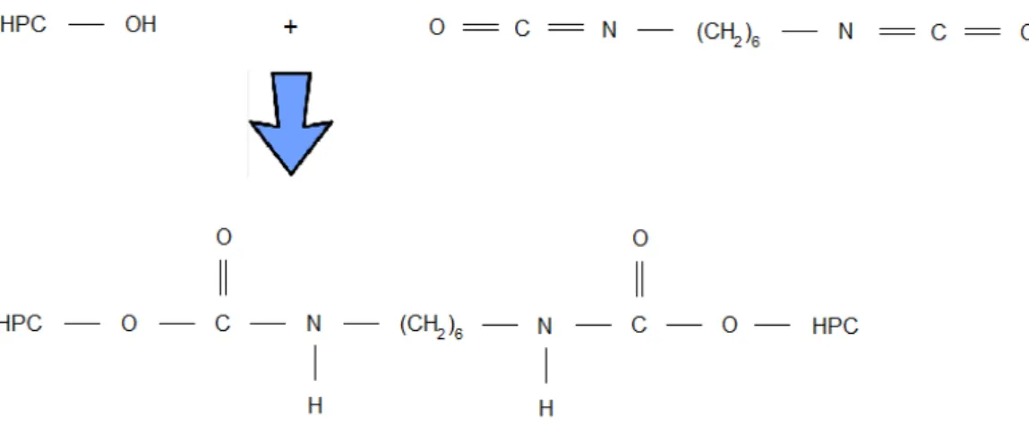
Figure 2.2 – Schematically representation of the cross-linking reaction.
HPC cross-linked solution was then deposited on parafilm, with the help of a calibrated ruler moving at a controlled rate (v = 6 mm.s-1), at room temperature, as shown in Fig. 2.3. The solvent was allowed to evaporate in a laboratory atmosphere at room temperature.
Figure 2.3 – Schematic representation of HPC films preparation. Kindly provided by Prof. Helena Godinho.
The average thickness of the dried films was measured using a digital micrometer (from Mitutoyo, Japan) and was approximately for HPCi films and for HPCa films.
2. Structural and Physico-Chemical Characterization
Cláudio Santos Dissertação de Mestrado em Engenharia Biomédica 27 Surface characterization requires particular techniques and has an important role in biomaterials characterization because the atoms that make up the outermost surface of a biomaterial drive many biological reactions that occur in response to the foreign body, like protein adsorption, cell adhesion, cell growth and blood compatibility, among others. [50] The following table resumes some methods used in this work to characterize film surfaces.
Table 2.1 – Common methods used to characterize biomaterial surfaces. Adapted from Ratner [50].
Method Principle Depth
analyzed
Spatial resolution
Contact angle
Liquid wetting of surfaces is used to estimate the
energy of surfaces
3–20 Å 1 mm
ESCA (XPS)
Electron Spectroscopy for Chemical Analysis (X-ray
photoelectron spectroscopy)
X-rays induce the emission of electrons of characteristic energy
10–250 Å 10–150 μm
FTIR
Fourier transform infrared spectroscopy
IR radiation is adsorbed and excites molecular
vibrations
1–5 μm 10 μm
SEM
Scanning Electron Microscopy
Secondary electron emission induced by a focused electron beam is
spatially imaged
5 Å 40 Å, typically
Cláudio Santos Dissertação de Mestrado em Engenharia Biomédica 28
2.1. Optical polarizing microscopy
It has been previously described [49] that thin solid films prepared from lyotropic solutions of cellulose derivatives develop a characteristic banded texture perpendicular to the shear direction.
The optical anisotropy of the HPC films was inspected by optical polarizing microscopy. The optical properties of thin films were observed using an Olympus (Model BH2) microscope equipped with cross polarizers. Optical microphotographs were taken using a microscope equipped with a camera.
2.2. Determination of the gel fraction
Gel fractions of the HPC films were obtained as a function of HPC concentration used to prepare the solid films and were calculated as the ratio of the weight of the extracted film to that of the non-extracted one. The soluble material of the solid films was extracted in water for 24h using a Soxhlet apparatus. The extracted film was dried in vacuum, at 60ºC.
2.3. Mechanical properties of the dried and wet films
HPC films were submitted to tensile tests performed under dry and wet conditions to determine the influence of HPC composition and of the anisotropy in mechanical properties (measured by the difference in mechanical properties between the direction of the film spreading and the perpendicular to this direction). The mechanical properties of the HPC films are compared. These properties are extremely important to characterize biomaterials and to prevent future failures of materials in biomedical applications.
Cláudio Santos Dissertação de Mestrado em Engenharia Biomédica 29 Load extension graphs were obtained during this test and converted to stress-strain curves applying the following equations:
Equation 1
where is the applied force and is the cross sectional area.
Equation 2
where and are the length at the time of data collection and original length (between clamps), respectively.
The measurements were performed with dry and wet films at room temperature.
Figure 2.4 – Photograph of the (a) tensile tests equipment and (b) detail of the film sample placed between the clamps.
The Young’s modulus or elastic modulus (E) is a mechanical parameter that gives information about material stiffness and it is defined as the slope of the stress-strain curve (E=stress/strain) in the elastic deformation region.
2.4. Contact Angle Measurements

![Table 1.1 – Thermodynamics of protein adsorption. Adapted from [50].](https://thumb-eu.123doks.com/thumbv2/123dok_br/16544678.736885/26.892.180.700.920.1077/table-thermodynamics-protein-adsorption-adapted.webp)
![Figure 1.8 – Hierarchy of stem cells. Adapted from [95].](https://thumb-eu.123doks.com/thumbv2/123dok_br/16544678.736885/33.892.123.770.466.872/figure-hierarchy-stem-cells-adapted.webp)