THE ROLE OF GLIPTINS ON ADIPOGENESIS
Marta Maria Vieira Matutino Falcão Estrada
Dissertação elaborada com vista à obtenção do Grau de Mestre em Biotecnologia
Orientadores:
Professora Doutora Cláudia Cavadas Doutora Joana Rosmaninho-Salgado
THE ROLE OF GLIPTINS ON ADIPOGENESIS
Marta Maria Vieira Matutino Falcão Estrada
Dissertação elaborada com vista à obtenção do Grau de Mestre em Biotecnologia
Orientadores:
Professora Doutora Cláudia Cavadas Doutora Joana Rosmaninho-Salgado
III
This work was performed at the Neuroendocrinology and Neurogenesis Group at the Center for Neurosciences and Cell Biology of the University of Coimbra, and financed by FCT (
V
AgradecimentosAo Centro de Neurociências o meu obrigada pela disponibilidade demonstrada em me acolher e por me ter proporcionado todas as condições necessárias ao desenvolvimento deste trabalho de investigaçãoo.
À Doutora Joana Rosmaninho-Salgado agradeço a vontade excepcional de ensinar ao longo de todo o ano; a amizade que sempre demonstrou, dentro e fora do laboratório; a disponibilidade constante para ajudar no que fosse necessário, sem com isto descuidar aquilo que me competia fazer; puxou sempre por mim, estando sempre atenta ao meu limite. Obrigada Joana por todo o carinho e amizade. Obrigada por teres sido uma excelente Professora e por seres uma excelente amiga.
À Professora Doutora Cláudia Cavadas, agradeço a disponibilidade e vontade que demonstrou em me aceitar e por me ter acompanhado neste desafio e orientado ao longo deste trabalho.
À Dra. Patrícia Marques agradeço todo o empenho demonstrado, do inicio ao fim, em me ensinar tudo o que sabia; todos os momentos passados dentro e fora do laboratório, tanto os momentos de stress em que tínhamos imenso trabalho para fazer, como os momentos mais calmos em que também éramos capazes de relaxar. Obrigada por me teres acompanhado todo o ano com uma amizade incondicional, por sempre teres tido a coragem de apontar o que estava bem e o que estava errado. Obrigada por teres tornado este ano de tese muito mais leve e partilhado.
À Dra. Fábia Vicente agradeço toda a amizade e gargalhadas constantes; obrigada por tornares todos os momentos de pausa especiais e divertidíssimos; obrigada por estares sempre lá!
À Doutora Célia Aveleira agradeço especialmente a disponibilidade a qualquer momento para ensinar, ajudar e partilhar tudo, especialmente as receitas para as soluções; obrigada por aquele sorriso matinal sempre presente e cheio de energia; obrigada pela atenção e espírito de equipa constante.
À Dra. Magda Santana agradeço a boa disposição constante e a disponibilidade para ajudar em tudo, mesmo fora do laboratório; obrigada pelos fins de semana enfiados no laboratório, sempre divertidíssimos; obrigada pela companhia e partilha nas idas ao CUMN e a todas as beatices.
À Dra. Vera Cortez, ao Dr. Jorge Pascoal, à Dra. InêsMorte, ao Doutor. BrunoCarreira, à Dra. Mariana Botelho, à Dra. Ana Carvalho, ao Dr. Gabriel Costa, obrigada por toda a amizade, companheirismo e boa disposição.
Aos meus pais obrigada pelas oportunidades inigualáveis que me proporcionaram até agora em termos de formação e de vida; obrigada por nunca me ter faltado nada; por estarem sempre atentos a tudo; por serem os melhores amigos que tenho e por me terem apoiado incondicionalmente em todos os momentos, especialmente nos de stress; obrigada por toda a paciência e vontade de dar tudo e mais alguma coisa sem nunca descuidar na educação; obrigada por me estimularem a ser mais e melhor e especialmente a pensar além dos próximos meses de vida.
À minha irmã Mafalda obrigada por toda a paciência demonstrada neste ano de muito stress e ausência; por ter sempre ajudado no que fosse preciso; por ter sido uma amiga em todos os momentos de todos os dias.
À Dra. Luísa Martins agradeço ter sido uma Mãe durante este ano, sempre atenta e disponível para tudo, nunca deixou que me faltasse nada e fez-me sentir em sua casa como se estivesse em minha; obrigada pela companhia excepcional, por todos os almoços e jantares, pelas dormidas, pelas boleias, pelas idas à oficina, pelos convites e, acima de tudo, pela amizade incondicional.
VI
VII
AbstractDipeptidyl-peptidase IV (DPPIV) (E.C.3.4.14.5.) is a 110 KDa peptidase expressed in almost every tissue of the human body. DPPIV expression is altered in obese conditions. Preliminary studies in our laboratory showed that this enzyme stimulates pre-adipocyte differentiation and lipid accumulation using a murine pre-adipocyte cell line. Vildagliptin, sitagliptin and saxagliptin are a new class of DPPIV selective inhibitors used in diabetes to increase the half-life of some insulin-stimulating hormones. The aim of this study is to evaluate, the effect of gliptins on the adipose tissue formation (adipogenesis and lipolysis). Using a pre-adipocyte murine cell line, 3T3-L1 we analysed the effect of gliptins on lipid accumulation. Our results showed that gliptins reduced both basal and stimulated lipid accumulation. We further evaluated if gliptins could modulate lipolysis or adipogenesis. Our studies show that gliptins do not induce lipolysis but play an inhibitory role on adipogenesis. This inhibition is achieved by inhibiting the expression of a transcription factor crucial for adipocyte differentiation, PPARγ. It was also observed that gliptins inhibit lipid accumulation through PKA pathway.
Neuropeptide Y (NPY) is a DPPIV substrate that acts through six G-protein-coupled receptors: Y1, Y2, Y3, Y4, Y5 and y6. Previous studies in our lab showed that DPPIV stimulates lipid accumulation through cleavage of NPY1-36 into NPY3-36. The cleaved peptide stimulates lipid accumulation through Y2 receptor activation. The second aim of our study was to evaluate the effect of gliptins on NPY-stimulated lipid accumulation. Our results show that gliptins inhibit NPY-induced lipid accumulation. We also analysed the intracellular mechanism of action of NPY and conclude that NPY stimulates lipid accumulation through modulation of PKA.
The present study suggests that gliptins can be used as new putative pharmacological strategies to prevent adipose tissue increase without the risk of dyslipidemia.
IX
ResumoA Dipeptidyl-peptidase IV (DPPIV) (E.C.3.4.14.5.) é uma peptidase de 110 KDa expressa em quase todos os tecidos do corpo humano. A sua expressão encontra-se alterada em situações de obesidade. Estudos anteriores, realizados no nosso laboratório, demonstraram que a DPPIV estimula a diferenciação de adipócitos e a acumulação lipídica nos adipócitos. A vildagliptina, a sitagliptina e a saxagliptina fazem parte de uma nova classe de inibidores selectivos da DPPIV cuja função principal é aumentar o tempo de meia vida de algumas hormonas responsáveis pelo estímulo da produção de insulina. O objectivo principal deste trabalho consiste em avaliar o efeito que as gliptinas têm na formação do tecido adiposo (adipogénese e lipólise). Usando uma linha celular de pre-adipócitos de murganho (3T3-L1), foi testado o efeito das gliptinas na acumulação lipídica. Os resultados obtidos demonstram que as gliptinas reduzem tanto a acumulação lipídica basal como a acumulação lipídica induzida pela insulina. De seguida avaliámos se as gliptinas exerciam efeito na lipólise ou na adipogénese. Verificámos que apesar de as gliptinas não provocarem lipólise, têm um efeito inibitório na adipogénese. Esta inibição ocorre pela inibição da expressão de um factor de transcrição, PPARγ, crucial para a diferenciação dos adipócitos. Observámos ainda que as gliptinas inibem a acumulação lipídica via PKA.
O neuropéptido Y (NPY) é um conhecido substrato da DPPIV cuja acção ocorre pela activação de seis receptores acoplados à proteína G: Y1, Y2, Y3, Y4, Y5 e y6. Estudos anteriores realizados no nosso laboratório demonstraram que a DPPIV estimula a acumulação lipídica através da clivagem do NPY1-36 em NPY 3-36. O peptídeo clivado estimula a acumulação lipídica através da activação do receptor Y2. O segundo objectivo deste trabalho consistiu na avaliação do efeito das gliptinas na acumulação lipídica induzida pelo NPY. Verificamos que as gliptinas reduzem esta acumulação lipídica induzida pelo NPY, e que este mecanismo era mediado pela acção da PKA.
O presente estudo sugere que os inibidores selectivos da DPPIV são possíveis estratégias farmacológicas para prevenir o aumento do tecido adiposo sem o risco de dislipidémia.
XI
Table of contents:Abstract VII
Resumo IX
Table of contents XI
Index of figures XV
Index of tables XVII
Abreviations XIX
CHAPTER 1 – INTRODUCTION
1.1 Adipose tissue 3
1.2 Adipogenesis 3
1.2.1 Adipogenic transduction pathways 5
1.3 Metabolic function 6
1.3.1 Lipid droplets formation 7
1.3.1.1 Perilipin 7
1.3.2 Lipolysis 8
1.4 Endocrine function 9
1.5 Dipeptidyl peptidase IV 12
1.5.1 Structure and localization 12
1.5.2 Function and substrates 12
1.5.2.1 DPPIV substrates and their functions in the adipose tissue 14
1.6 Neuropeptide Y 15
1.6.1 NPY synthesis 15
1.6.2 NPY receptors 16
1.6.2.1 NPY Y1 receptor 17
1.6.2.2 NPY Y2 receptor 17
1.6.2.3 NPY Y3 receptor 17
1.6.2.4 NPYY4 receptor 17
1.6.2.5 NPY Y5 receptor 18
1.6.2.6 NPY y6 receptor 18
1.6.3 NPY and NPY receptors in the adipose tissue 18
1.6.3.1 NPY role on lipid accumulation 18
1.7 Dipeptidyl peptidase IV inhibitors 19
1.7.1 Vildagliptin 20
1.7.2 Sitagliptin 20
XIII
1.8 DPPIV and gliptins in the adipose tissue 22
1.9 Objectives of the present study 24
CHAPTER 2 – MATERIALS AND METHODS
2.1 Material 27
2.2 Methods 27
2.2.1 Cell culture 27
2.2.2 Cell differentiation conditions 27
2.2.3 Oil red-O staining 28
2.2.4 Immunocytochemistry 28
2.2.5 Total Protein extracts and Quantification 28
2.2.6 Western blotting 29
2.2.7 Lipolysis Assay Kit (Glycerol Quantification) 29
2.2.8 Statistical analysis 29
CHAPTER 3 - RESULTS
3.1 The role of gliptins on lipid accumulation 33
3.1.1 The role of gliptins on DPPIV-induced lipid accumulation 33 3.1.2 The role of gliptins on basal lipid accumulation 33 3.1.3 The role of gliptins on lipid accumulation induced by insulin 34
3.2 Effect of gliptins on lipolysis 35
3.3 The role of gliptins on adipogenesis 38
3.4 The role of gliptins on NPY-induced lipid accumulation 42
CHAPTER 4 – DISCUSSION 44
CHAPTER 5 – CONCLUSIONS 51
XV
Index of Figures:Figure 1.1 Pre-adipocyte differentiation into mature adipocytes 4 Figure 1.2 Progression of 3T3-L1 pre-adipocyte differentiation 4
Figure 1.3 Adipogenic transduction pathways 6
Figure 1.4 Lipid metabolism in adipocytes 7
Figure 1.5 Lipolysis transduction pathways in a human adipocyte 9 Figure 1.6 Molecules secreted by the adipose tissue with varied effect on glucose
homeostasis 10
Figure 1.7 Synthesis and post-transactional modifications of neuropeptide Y 16 Figure 1.8 Representation of the amino acid sequence of the G-protein coupled Y1
receptor 16
Figure 3.1 Gliptins decrease rDPP IV-induced lipid accumulation 33
Figure 3.2 The role of gliptins on basal lipid accumulation 34
Figure 3.3 Gliptins reduce insulin-stimulated lipid accumulation 34
Figure 3.4 Gliptins do not affect basal glycerol release 35
Figure 3.5 Gliptins do not affect perilipin levels 36
Figure 3.6 Gliptins do not change perilipin location 36
Figure 3.7 DPPIV does not affect glycerol release 37
Figure 3.8 DPPIV does not change perilipin levels 38
Figure 3.9 Gliptins reduce are reducing PPARγ levels 39
Figure 3.10 Gliptins are reducing insulin induced PPARγ levels 40
Figure 3.11 Gliptins inhibit lipid accumulation through PKA 41
Figure 3.12 Vildagliptin inhibits insulin- stimulated lipid accumulation through PKA 41
Figure 3.13 Gliptins decrease NPY-induced lipid accumulation 42
Figure 3.14 NPY stimulates lipid accumulation through PKA 43
XVII
Index of tablesTable 1: Examples of some factors secreted in the adipose tissue and respective function 12
Table 2: DPPIV substrates 14
XIX
Abbreviations59-AMP 59-adenosine monophosphate 7TM seven-transmembrane-helix a2-AR a2-adrenoreceptor
AC Adenylate cyclase
ACS acyl-coenzyme A synthetase ADA adenosine deaminase
ADRP adipose differentiation-related protein ALBP Adipocyte Lipid binding protein AMPK AMP-activated protein kinase aP2 adipocyte fatty acid binding protein AR adrenergic receptor
ATGL adipocyte triglyceride lípase AT adipose tissue
ATP adenosine triphosphate
β-AR β -adrenoceptor BAT brown adipose tissue BSA bovine serum albumin
C/EBP CCAAT/enhancer binding protein cAMP AMP cyclic
CPON C-flanking peptide of NPY
CREB cAMP-responsive element-binding protein
DEX dexamethasone
DPPIV Dipeptidyl-peptidase IV
ERK extracellular signal-regulated kinase
FA fatty acids
XX
FDA food and Drug Administration FFA free fatty acidGC guanyl cylase
GHRF growth-hormone releasing factor Gi inhibitory GTP-binding protein GIP gastric inhibitory peptide GLP-1 glucagon-like peptide-1 GLUT4 glucose transporter type 4 glycerol 3-P glycerol 3-phosphate GRP gastrin-releasing peptide Gs stimulatory GTP-binding protein HSL hormone-sensitive lipase IBMX 3-isobutyl-1-methylxanthine
IC50 half maximal inhibitory concentration IL-6 Interleukin-6
IR insulin receptor
IRS insulin receptor substrate LD lipid droplets
LPL lipoprotein lipase
MAPK mitogen activated protein kinase MCE mitotic clonal expansion
MEF mouse embryo fibroblast MGL monoglyceride lipase
MIG monokine induced by gamma interferon MIX methylisobutylxanthine
MSC mesenquimal stem cells NK natural killer
NPY neuropeptide Y
XXI
PBS phosphate buffered salinePDE-3B phosphodiesterase 3B
PI3-K phosphatidylinositol-3-phosphate kinase PKA protein kinase A
PKB protein kinase B PKG protein kinase G POMC proopiomelanocortin PP pancreatic polypeptide
PPARγ peroxisome proliferator-activated receptor γ
PYY peptide YY
RBP 4 retinol-binding Protein 4 RXRα retinoid receptor α
SEM standard error of the mean
SP substance P
T2DM Type 2 Diabetes Mellitus
TAG triglyceride molecules
TNF – α tumor Necrosis Factor – α
TZDs thiazolidinediones UCP-1 uncoupling protein – 1 VIP vasoactive intestinal peptide VLDL very low density lipoproteins WAT white adipose tissue
Introduction
3
1.1 Adipose tissueThe adipose tissue (AT) is a mesodermal tissue composed by several types of cells [1, 2]. One third are mature adipocytes whereas the other two thirds are small blood vessels, nerve tissue, fibroblasts and pre-adipocytes [3]. In mammals, the adipose tissue is divided in two types of tissue: brown adipose tissue (BAT) and white adipose tissue (WAT), the former being responsible for energy dissipation in the form of heat, through non-shivering thermogenesis, and the later being involved in energy accumulation, mainly in the form of triglycerides [2, 4, 5].
Brown adipose tissue (BAT) contains predominantly adipocytes, which are rich in mitochondria and possess small multilocular lipid droplets (LD) [1, 6]. In humans, this tissue is present in neonates and new-born children, and is distributed through several areas of the body: pancreas, kidneys, adrenal glands, interscapular region (shoulder), muscles in the neck, in the axillae, trachea, esophagus and surrounding blood vessels [2]. Brown adipocytes main function is to transform energy from food into heat, through oxidative phosphorylation by uncoupling protein – 1 (UCP-1), present in the inner membrane of the mitochondria. In most of the tissues, where UCP-1 is absent, protons are pumped out of the mitochondrial matrix into the intermembrane space, generating an electrochemical gradient across the membrane. An ATPase would dissipate this gradient by pumping these protons back into the matrix, transforming ADP into ATP. However, when UCP-1 is present proton gradient is dissipated and ATP production is not allowed, thereby generating heat [1, 2, 4, 7, 8].
White adipose tissue (WAT) is found in several anatomically and physiologically distinct depots. There are two main white adipose tissues: the visceral and the subcutaneous adipose tissue. The first can be divided in omental adipose tissue, mesenteric adipose tissue and retroperitoneal adipose tissue [2]; whereas the second can be divided in superficial and deep subcutaneous adipose tissue [2]. WAT contains predominantly spherical white adipocytes that accumulate lipids, in the form of triglycerides, within one large lipid droplet [1, 2]. Besides accumulating triglycerides, the adipose tissue can also secrete molecules with endocrine, paracrine and autocrine functions [9].
1.2 Adipogenesis
Adipogenesis is a process that corresponds to pre-adipocytes proliferation and differentiation into mature adipocytes, see figure 1.1 [1]. Pre-adipocytes have origin in mesenquimal stem cells (MSC) which, besides adipocyte differentiation, are also capable of differentiating into osteoblasts, chondrocytes, myoblasts and connective tissue [1]. During the differentiation process that begins after birth, pre-adipocytes go through four different stages before becoming mature adipocytes: i) growth arrest, ii) clonal expansion, iii) early differentiation and iv) terminal differentiation [1]. This differentiation process is only possible due to the activation of a cascade of transcription factors like peroxisome proliferator-activated receptor γ (PPARγ) and CCAAT enhancer binding protein (C/EBPs) family [1, 2, 10-12]
Introduction
4
[1]. Whereas some are associated with the adipogenesis process, like C/EBPα, C/EBPβ and C/EBPδ, others are related to the inhibition of this process, like CHOP-10 [11].
Figure 1.1: Pre-adipocyte differentiation into mature adipocytes.
Pre-adipocytes differentiation is induced with cAMP-elevating agents. When in the differentiated state, adipocytes are able to accumulate triglycerides inside lipid droplets.
The differentiation process has been extensively studied using cell lines, like 3T3-L1, and primary cultures, like mouse embryo fibroblast (MEF) [12]. This process can only be initiated when pre-adipocytes are post-confluent and growth arrested [1, 10-12]. When these two conditions are accomplished and the differentiation inducers are added to the in vitro culture, pre-adipocytes initiate the mitotic clonal expansion (MCE) with, at least, two rounds of mitosis [13].
Immediately before the initiation of the MCE, C/EBPβ is expressed (see figure 1.2). C/EBPβ plays a role in two different stages of the differentiation process: it initiates the mitotic clonal expansion and activates the expression of C/EBPα and PPARγ[11, 13, 14].
Figure 1.2: Progression of 3T3-L1 pre-adipocyte differentiation
The periods of gene expression during the differentiation programme have the gene name. C/EBP, CCAAT/enhancer binding protein; PPARγ, peroxisome proliferator activated receptor γ. Adapted from [15].
Introduction
5
differentiation phase (figure 1.2). The delayed expression of these two transcription factors is critical, since they both have anti-mitotic activity [12, 13]. Moreover, PPARγ and C/EBPα will function together as transcriptional activators of a large group of genes, like those involved in lipid metabolism that confer the adipocyte phenotype [10, 11, 16]. In addition, they regulate each other to maintain their levels during and after the differentiation process [17].
Although PPARγ and C/EBPα act together during differentiation, PPARγ has a dominant action. This was observed when PPARγ null embryonic stem cells did not differentiate into adipocytes, whereas in C/EBPα null stem cells, PPARγ was able to promote differentiation [1, 18]. PPARγ expression is not restricted to induction of adipogenesis, it is also needed to maintain the differentiated state, otherwise adipocytes lose their ability to accumulate lipids and express their adipocyte markers [1]
. PPARγ is only expressed two days after the beginning of differentiation [15]. Its activation is correlated with the loss of DNA binding activity of E2F/DP, which is involved in cell division [19]. PPARγ is a cis-acting element [19] that promotes gene expression via formation of a heterodimeric DNA-binding complex with the retinoid receptor α (RXRα) [16]. To have adipogenic activity PPARγ needs more than being transcribed, it needs to be activated [15, 20]. Such activators can be micromolecular concentrations of long-chain fatty acids, like linoleic acid [19-21]; synthetic compounds, like thiazolidinediones (TZDs) [19, 20]; or naturally occurring eicosanoids, like prostaglandins [21]. These activators also differentially activate the other PPAR family members, for example, some eicosanoids, like 8(S)- hydroxyeicosatetraenoic acid, only activate PPARα[21].
PPARγ possesses two isoforms, PPARγ1 and PPARγ2, which are generated by alternative splicing and alternative promoter usage [1, 16]. These two isoforms are identical peptides, although PPARγ2 has 30 additional amino acids at the N-terminus [11]. These two isoforms are important for adipogenesis, although PPARγ2 plays an indispensable role [22]. When PPARγ1 and PPARγ2 expression was abolished in 3T3-L1, these cells were unable to differentiate [22]. However, with the addition of exogenous PPARγ2 the differentiation was restored, whereas when the exogenous PPARγ1 was used no differentiation occurred [22]. In another study using PPARγ2 knockout mice, the same conclusion was obtained [23]. These animals showed a decrease in the overall amount of AT, less lipid accumulation, reduced expression of the adipogenic genes and also insulin resistance [11, 23].
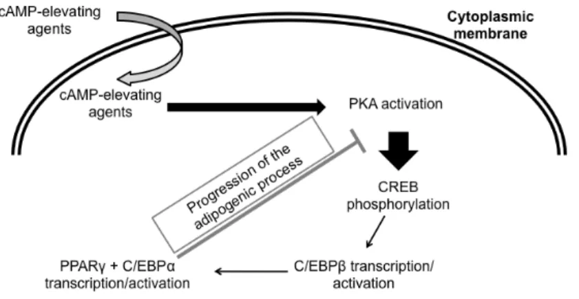
1.2.1 Adipogenic transduction pathways
Introduction
6
CREB [24, 25]. Although PKA seems to have a crucial role during the beginning of the differentiation process, it is described that the continuous activation of PKA is responsible for inhibition of late differentiation. These results were obtained by the demonstration that 72 hours after the beginning of differentiation, cAMP levels and PKA activity are similar to basal [28]. It was also demonstrated that the continuous activation of PKA resulted in blockage of differentiation [28], whereas others demonstrated that PKA inhibitor (H-89) reduced the time span needed for full adipogenesis [29], once again, proving that PKA inhibits adipogenesis.
Figure 1.3: Adipogenic transduction pathways.
PKA activation by the increased cAMP levels leads to CREB phosphorylation. This transcription factor activates the transcription of C/EBPβ, which in turn activates PPARγ and C/EBPα transcription. For the successful progression of adipogenesis PKA needs to be inactivated. PKA, protein kinase A; CREB, cAMP-responsive element-binding protein; c/EBP: CCAAT/enhancer binding protein; PPARγ: peroxisome proliferator-activated receptor γ;
1.3 Metabolic function
WAT is the specialized tissue, of the human body, in accumulating energy in the form of triacylglycerols (see figure 1.4), also called neutral fats or triglycerides [30]. Each molecule of triacylglycerol is constituted by three molecules of esterified fatty acids (FA) and one molecule of glycerol [30].
Introduction
7
Figure 1.4: Lipid metabolism in adipocytes.
ACS, acyl-coenzyme A synthetase; aP2, adipocyte fatty acid binding protein; FATP, fatty acid transporter protein; glycerol 3-P, glycerol 3-phosphate; FFA, free fatty acids;. Adapted from [2].
1.3.1 Lipid droplets formation
In the differentiated state, adipocytes can either synthesize FA de novo or accumulate them from the dietary lipids [2]. To be accumulated from dietary lipids, triacylglycerides are transported through the blood plasma in lipoprotein transport particles, like chylomicrons or very low density lipoproteins (VLDL), to the adipose tissue [31]. In order to enter the cell, they are digested by lipoprotein lipase (LPL) and, as fatty acids, are able to enter the cell through fatty acid transporters [2, 31]. These transporters can be protein CD36 (human homologue to the murine fatty acid translocase, (FAT), fatty acid transport protein (FATP) and fatty acid binding protein (FABPpm) [31]. When in the cytoplasm, FA are carried out by cytoplasmatic binding proteins, like aP2, to acyl CoA synthase reaction site [31]. This enzyme catalyses the reaction of FA with an acetyl-CoA, originating fatty acyl-CoA [32]. Afterwards, this molecule reacts with glycerol-3-P, which is produced during glycolysis, originating the triglyceride molecule [32]. During the fatty acids de novo synthesis, the acyl-CoA molecule comes from an acetyl-CoA molecule that was produced from pyruvate in the glycolytic pathway, inside the mitochondria [30]. This acyl-CoA molecule, together with a glycerol-3-P molecule gives rise to a triglyceride molecule [30]. These triglyceride molecules (TAG) are kept inside lipid droplets protected by proteins called perilipin [33]
.
1.3.1.1 Perilipin
Introduction
8
isoforms: perilipin A and perilipin B, that result from mRNA splicing of a single perilipin gene [33]. Perilipin A has six phosphorylation sites, whereas perilipin B only has three phosphorylation sites [34]. Perilipin A is found in the outer surface of the lipid droplet and is a critical component of a scaffold that stabilizes the lipid droplet [35]. This distribution at the surface of the lipid droplets is the main mechanism that prevents lipases from reaching TAG, thus inhibiting lipolysis [35].
1.3.2 Lipolysis
Lipolysis is the controlled process of hydrolysis of TAG, with consequent release of three fatty acid molecules and one glycerol [33, 36]. This process is triggered by hormonal stimulus, like catecholamines; by agents that elevate cAMP levels, like forskolin; or by cAMP analogous [33, 37]. When a catecholamine binds its β-adrenergic receptor, occurs the activation of a stimulatory G-protein (Gαs) that activates adenyl cyclase [33, 37]. Consequently, cAMP levels rise and activate the regulatory subunits of PKA [33, 37]. When in its active form, PKA phosphorylates the two key enzymes of the lipolysis process: perilipin and hormone sensitive lipase (HSL) (see figure 1.5) [33, 37]. As described above, perilipin plays a crucial role in preventing lipolysis [33, 35, 37]. However, it is also a crucial enzyme in “allowing” lipolysis [33, 35]. When this enzyme is not phosphorylated it is anchored to the lipid droplet and contributing to the barrier formation against the action of lipases like HSL. However, when it is phosphorylated by PKA, perilipin plays a different role [35]. When PKA phosphorylates these two proteins, perilipin changes its conformation allowing HSL to translocate to the surface of the lipid droplet [33]. While leaving the surface of the lipid droplet, perilipin facilitates the interaction between HSL and the lipid droplet [35]. HSL is a serine hydrolase regulated by reversible phosphorylation, whose metabolic function is to catalyse hormone-stimulated lipolysis [36]. This enzyme is present in the cytosol of adipocytes in its unphosphorylated form [33, 36]. Although HSL has several phosphorylation sites, the crucial for its activation as a lipase are Ser659 and Ser660[33, 36]. It is important to note that the perilipin-HSL interaction is required, otherwise HSL alone is not able to reach the lipid droplet surface [33]
Introduction
9
Figure 1.5: Lipolysis transduction pathways in a human adipocyte.
Lipolytic pathway is described. PKA, protein kinase A; HSL, hormone sensitive lipase; FFA, free fatty acid; P- phosphorylated; PLP, phosphorylated perilipin. Adapted from [37].
1.4 Endocrine function
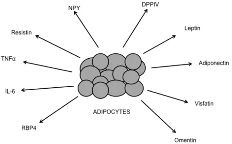
Besides being a specialized organ in accumulating energy, the adipose tissue is also seen as an endocrine, autocrine and paracrine organ [9]. Such action is accomplished by the production of several molecules, also called adipocytokines or adipokines [39], whose actions influence not only the metabolic activity of the adipose tissue but also other tissues like the brain, muscles and liver [9, 40]. As outlined in table 1, adipocytokines have effects on multiple biological systems like energy homeostasis (lipid and carbohydrate metabolism, appetite, thermogenesis), immune system, reproductive function, hemostasia/coagulation, blood pressure and angiogenesis, see revision [41]. As referred before, the adipose tissue possesses other cell types besides adipocytes that can be the source of some of these adipocytokines [39].
Some of these molecules are leptin, adiponectin, visfatin, omentin, resistin, retinol-binding protein 4 (RBP 4), tumor necrosis factor – α (TNF-α), Interleukin-6 (IL-6) and other cytokines, see revision [42]. The former four molecules have anti-diabetic functions, while the later four molecules tend to raise blood glucose levels (see figure 1.6) [40, 43].
Introduction
10
result in improvement of insulin sensitivity in muscle and liver [40]. Animals and humans with mutations in lepin or in leptin receptor became obese, showing the importance of this adipokine in regulating food intake and energy expenditure [6]. Morrison et al., (2005) demonstrated that this effect of leptin mutations is due to increased NPY expression in the arcuate nucleus, which leads to increased food intake and decreased energy expenditure [44, 45]. In the same study, it was also observed that when leptin was injected in leptin deficient mice, NPY mRNA levels were decreased in the arcuate nucleus [44]
. These results suggest that leptin action might also be through modulation of other peptides secretion [44].
Figure 1.6: Examples of molecules secreted by the adipose tissue with varied effect
Leptin, adiponectin, visfatin and omentin are adipokines with anti-hyperglycaemic effects. Resistin, tumour necrosis factor-α (TNFα), interleukin 6 (IL6) and Retinol-binding protein 4 (RBP4) are molecules with pro-hyperglycaemic activity. Neuropeptide Y (NPY) and dipeptidyl-peptidase IV (DPPIV) with adipogenic activity. Adapted from [40].
Adiponectin is a 30 kDa protein also known as apM1, GBP28, AdipoQ and ACRP30 [40]. It has an amino-terminal collagen-like domain and a carboxyl-terminal globular domain that mediates multimerization [40]. Adiponectin is specifically expressed in differentiated adipocytes and suffers posttranslational modification [46]. This adipokine circulates as a trimer, hexamer or as a higher-order multimer that circulates in plasma in such high concentrations that account for 0.01% of all plasma protein [40]. Adiponectin has two different receptors, AdipoR1 and AdipoR2, being the former primarily expressed in muscles, and the latter primarily expressed in the liver [41]. Adiponectin effects depend on circulating levels, on the properties of the different adiponectin isoforms and also on tissue specific expression of adiponectin receptors [46]. Nevertheless, there are some general functions associated with adiponectin, like anti-diabetic and anti-inflammatory functions [47]. The anti-diabetic functions are related with the increased levels of adiponectin when TZDs are administered, resulting in improved insulin sensitivity [40].
Introduction
11
which results in an enhanced glucose uptake [43]. Because its circulating levels are less than 10% compared to insulin levels, these two molecules do not compete for receptor binding [40]. A recent article suggested that visfatin not only activates the insulin receptor, but also regulates insulin secretion and several β-cell function-associated genes in mouse [48].
Omentin is secreted by stromal-vascular cells present in visceral fat [32]. It acts as an insulin sensitizer rather than insulin mimetic, resulting on positive effects in glucose uptake [32]. In humans and macaques it is produced in high quantities, but in mice no production was detected [40]. Although omentin levels decrease with obesity and insulin resistance, the mechanism of action is still unknown [43]
.
Retinol-binding protein 4 (RBP4) is a member of the lipocalin superfamily and its expression is regulated by changes in the glucose transporter type 4 (GLUT4) [40]. High serum levels are associated with insulin resistance in humans with obesity, type 2 diabetes mellitus and in lean non-diabetic people with family history of type 2 diabetes mellitus. RBP4 overexpression in mice, also resulted in impaired insulin action both in muscle and liver [32].
The adipose tissue also produces some cytokines, like interleukin 6 (IL-6), and tumour-necrosis factor α (TNFα) [40]. Although TNFα is secreted in the adipose tissue, it is not secreted by adipocytes, but by macrophages that surround adipose tissue [46]. This adipokine also plays a role in glucose homeostasis, by decreasing insulin sensitivity [39]. Its levels are elevated in obese conditions and in other insulin-resistant cases [39]. Moreover, when TNFα expression was blocked, insulin sensitivity was restored, both in vitro and in vivo[40]. On the other hand, IL-6 is produced by adipocytes and circulates in multiple glycosylated forms [46]. Its expression and circulating levels are positively correlated with obesity, glucose intolerance and insulin resistance [39]. When peripherally administrated, IL-6 reduces the expression of insulin receptor signalling components, decreases adiponectin secretion and inhibits adipogenesis [46].
Resistin, also known as FIZZ3, is a small inflammatory molecule with hyperglycaemic action [40]
. Although some authors indicate that it is secreted by adipocytes [49], it is still controversial since new data suggests that resistin is secreted by macrophages [50] or other stromal cells present in the adipose tissue [51]. Resistin circulates in the plasma in several multimeric forms, but are those with small weight that seem to have an effect at the cellular level [6]. Resistin reduces glucose uptake in muscles and is repressed by TZDs [6]. This molecule can also modulate the secretion of other molecules, such as NPY [52]. This was observed when resistin was centrally administered to mice, resulting in increased NPY production in the arcuate nucleus [52]. Resistin effects on glucose production were blocked in mice lacking NPY [52].
Introduction
12
Table 1: Examples of some factors secreted in the adipose tissue and respective function [6, 32].
Adipokine Biological effect
Leptin Signals to the CNS about the body’s energy stocks
Adiponectin Increases sensitivity to insulin; antiinflammatory; attenuates the progression of atherosclerosis
Visfatin Insulin-mimetic; predominantly produced by visceral fat
Omentin Enhances insulin-stimulated signals and glucose uptake but it is not insulin-mimetic
TNFα Lipolytic; increases energy consumption; reduces sensitivity to insulin IL-6 Proinflammatory, lipolytic, reduces sensitivity to insulin
Resistin Increases insulin resistance
RBP4 Reduces insulin sensitivity; Impaires insulin action in the muscle
1.5 Dipeptidyl peptidase IV
1.5.1 Structure and localization
Dipeptidyl peptidase IV (E.C. 3.4.14.5) is a 110 kDa glycoprotein, also known as DPPIV or CD26 [5]. This enzyme is a ubiquitous and multifunctional molecule that exists in both soluble and membrane bound form. DPPIV is also a homodimer but it functions only while in its dimer form [5, 54-57]. In its membrane form, DPPIV has three regions: a cytoplasmatic domain with 6-amino acids, a hydrophobic transmembranar region with 22-amino acids and an extracellular domain with 738-amino acids [56]. When the hydrophobic N-terminal domain is cleaved [58] by chymotrypsin-like or pepsin-like enzymes [59], DPPIV is released into the plasma, becoming soluble, though maintaining both substrate specificity and susceptibility to inhibitors [57]. While in its soluble form, DPPIV can be found not only in the blood plasma, but also in the cerebrospinal fluid and semen [60, 61]. As a membrane protein, it is widely expressed in various tissues and organs, including the exocrine pancreas [62], kidneys [63], gastrointestinal tract [64], thymus [65], lymph nodes [66], uterus [67], placenta, prostate [68], adrenal [69], sweat [70], salivary and mammary glands [70], endothelia of spleen [71], lungs [63], brain [69], and vessels supplying the liver [61, 72, 73]. In addition, this enzyme is also present in lymphocytes, as the cell-surface CD26 T-cell-activating antigen and also as a membrane antigen of both Natural Killer (NK) and B cells [61, 73-75]
.
1.5.2 Function and substrates
Introduction
13
[78]: (i) cell-extracellular matrix interactions [60] (ii) co-stimulatory role in the immune system [5, 60, 78, 81, 82] and (iii) cleavage of biologically active molecules [60].
The extracellular matrix interactions are characterized by the ability of this enzyme to bind some ligands, like adenosine deaminase (ADA), kidney Na+/H+ ion exchanger 3 [60, 78]. DPPIV is also able to bind fibronectin and collagen and the association of DPPIV with fibronectin, in lung cancer cells, is responsible for growth arrest of these cells [83]. In cancer cells DPPIV has anti-proliferative and anti-oncogenic effects and is down-regulated [68, 78]. Although it is not known the exact mechanism of action, DPPIV down-regulation will create a proteolytic imbalance of the extracellular regulatory proteins within the tumour environment [68, 69, 84] which is characteristic of malignancy [78].
DPPIV also has a very important role in the immune system. This enzyme is not only able to regulate multiple T-cell functions, like maturation, activation, migration and interaction with antigen-presenting cells; but is also able to regulate proliferation and activation of B-cells and NK cells [60, 78]. Furthermore, DPPIV can also cleave several immunoregulating cytokines [78]. When in the surface of cells, the signal transduced by CD26 co-stimulates the cell receptor CD3 pathways, leading to T-cell activation [85]. CD26 activation occurs through caveolin-1 or CD45 and leads to tyrosine phosphorylation which increases phosphorylation of several molecules like p56lkc, p59fyn, zeta associated protein-tyrosine kinase of 70000MW (ZAP-70) and MAPK [5, 60, 78, 86]. Studies also showed that in some autoimmune diseases, like Multiple Sclerosis and Arthritis, the CD26 expression is up-regulated in T-cells, B-cells and NK-cells [5, 78]. Taking all together, it can be concluded that DPPIV inhibitors are useful tools for immune suppression in autoimmune diseases [5, 78, 82].
The DPPIV ability to cleave molecules is restricted to those enzymes having a Xaa-Pro or Xaa-Ala dipeptides, from the N-terminus of polypeptides (where Xaa is any amino acid except Pro) [5, 54, 61, 74]
Introduction
14
Table 2: DPPIV substrates [61, 78, 87, 88]
Neuropeptides Neuropeptide Y
Endomorphin Substance P
Beta-casomorphin
Pituitary adenylate-cyclase-activating polypeptide (PACAP) Vasoactive intestinal peptide (VIP)
Other regulatory
peptides Gastric inhibitory peptide (GIP)
Gastrin-releasing peptide (GRP)
Glucagon-like peptide 1 (GLP-1)
Glucagon-like peptide 2
Growth-hormone releasing factor (GHRF)
Peptide YY (1-26)
Chemokines Eotaxin
Monokine induced by gamma Interferon (MIG)
Interferon-inducible protein-10
Chemokine lingand 5 (RANTES)
Macrophage-derived chemokine
Macrophage inflammatory protein-1beta Monocyte chemotatic proteins 1-3
Some of these substrates have different functions, depending on the tissue where they are produced and on the presence of their receptors [87]. The adipose tissue, has receptors for GLP-1 [89], GIP [90], Substance P (SP) [91-93], Pituitary adenylate-cyclase-activating polypeptide (PACAP) and NPY [94]
.
1.5.2.1 DPPIV substrates and their functions in the adipose tissue
GLP-1 is a peptide released by the enteroendocrine cells (L-cells), in the gut [95, 96]. Besides stimulating the adipose tissue to produce leptin, this peptide also decreases fat storage [89, 97]. GLP-1 enhances insulin-stimulated glucose metabolism in 3T3-L1 adipocytes: one of several potential extrapancreatic sites of GLP-1 action [98]. This process is not accomplished through lipolysis, but via direct modulation of adipocyte metabolism [97]. Nevertheless, this ability to modulate the adipocyte metabolism is not present in obese condition, suggesting an obesity-induced adipocyte resistance to GLP-1 [97].
Introduction
15
SP is released from the enteric nerves, sensory neurons and also from inflammatory cells of the lamina propria during intestinal inflammation [91]. This peptide also plays a role in the adipose tissue by increasing pre-adipocyte viability and proliferation, and also by decreasing apoptosis [91].
PACAP is released upon stimulation of the parasympathetic nerves [101]. By alternative splicing this peptide can be released in two forms, PACAP38 and PACAP27, having the former more affinity to DPPIV [87]. PACAP function in the adipose tissue depends on the presence of insulin, i.e., if insulin is present, this peptide potentiates glucose uptake which leads to lipogenesis; if insulin is absent, PACAP becomes lipolytic [94].
NPY is synthesized both in the hypothalamus and in the adipose tissue [102, 103]. In the adipose tissue, NPY increases expression and activity of lipoprotein lipase, which leads to increased lipogenesis [102]. It also has a very strong anti-lipolytic effect, resulting in increased weight gain [102, 104]. The effect of NPY in the adipose tissue will be discussed later on section 1.6.3.
1.6 Neuropeptide Y
NPY is a 36 amino acid peptide amidated in the C-terminal [105]. It belongs to the same family as peptide YY (PYY) and pancreatic polypeptide (PP), with whom shares 70 and 50% of sequence identity, respectively [105]. This peptide was first discovered in the porcine brain [53] and is the most abundant peptide in the human brain and very well conserved among species [106]. The NPY gene is located in the human chromosome 7 and is expressed mainly in the hypothalamus, more specifically, in the paraventricular nucleus, arcuate nucleus, suprachiasmatic nucleus, median eminence and dorsomedial nucleus [53]. In the sympathetic neurons, NPY is colocalized with norepinephrine [106]. Besides neuron cells, there are also other cells able to secrete NPY like, for example, liver, heart, spleen, endothelial cells of blood vessels and adipose tissue [53, 103].
NPY is a neurotransmitter involved in the regulation of several actions like, control of food intake, regulating the activity of neuroendocrine axes under poor metabolic conditions, vasoconstrictive actions, cardiovascular regulation, blood pressure, catecholamine secretion, energy homeostasis, neuroendrocrine regulation, memory processing, anxiety, body temperature, lipogenesis, among others [53, 105-107].
1.6.1 NPY synthesis
Introduction
16
already in its biologically active form it can be further be cleaved by aminopeptidase P or DPPIV, like described above [106].
Figure 1.7: Synthesis and post-transactional modifications of neuropeptide Y. Adapted from [106].
1.6.2 NPY receptors
NPY acts through six G-protein-coupled receptors: Y1, Y2, Y3, Y4, Y5 and y6 [106]. This type of receptor is associated with a seven-transmembrane-helix (7TM) receptors (see figure 1.8) that, when activated, suffers a conformational change which in turn activates a G protein [30]. NPY receptors are Gi/Go coupled receptors that when activated inhibit the action of adenyl cyclase [108]. This action results in decreased cAMP levels that in turn prevent PKA activation [30]. Besides decreasing cAMP levels, NPY receptors are also able to increase intracellular calcium levels [108, 109].
Figure 1.8: Representation of the amino acid sequence of the G-protein coupled Y1 receptor [110] . PreproNPY ProNPY NPY1-39 NPY1-37 NPY1-36 NPY3-36 Signal peptidase Prohormone convertase Carboxy peptidase Dipeptidyl-peptidase IV 1 97
Introduction
17
1.6.2.1 NPY Y1 receptorNPY Y1 receptor gene is localized in the chromosome 4q(31,3-32) and it is expressed in the brain, heart, kidneys and gastrointestinal tract, see revision [106]. NPY binding activity to this receptor is largely impaired when enzymes, like DPPIV, cleave the NPY N-terminal peptides [53]. However, when the C-terminal peptides are modified, NPY retains its full binding capacity to Y1 receptor, suggesting that this neurotransmitter binds this receptor through its N-terminal region [53]. The main effects of NPY, mediated by Y1 are vasoconstriction [53], increased appetite [111], decreased depression [112] and anxiety [106], activation of the neuroendocrine axes [106], and proliferation of smooth muscle cells [113], progenitor cells of the hippocampus [114], pancreatic β cells [115], Muller cells [116] and tumour cells [117].
1.6.2.2 NPY Y2 receptor
The NPY Y2 receptor is located in the chromosome 4q31, close to the NPY Y1 and Y5 receptor locus [106]. Its expression is found in the central and peripheral nervous system, gut, certain blood vessels and adipose tissue, see revision [53]. This receptor does not require the NPY N-terminal sequence, since it binds NPY3-36 and NPY1-36 with the same affinity [53]. NPY Y2 receptor also plays a role in control of appetite [118] and angiogenesis [113], neurotransmitter release [106].
1.6.2.3 NPY Y3 receptor
NPY Y3 receptor is called the NPY preferring receptor since it shows 10-fold higher affinity for
NPY than for PYY [53]. Although it has never been cloned nor well characterized [106], it can be found in several peripheral tissues, like rat superior cervical ganglia sympathetic neurons [119], rat cardiac ventricular membranes [106] and rat distal colon [120]. In human adrenal medulla mediates NPY-induced secretion of catecholamines [121].
1.6.2.4 NPYY4 receptor
Introduction
18
1.6.2.5 NPY Y5 receptorNPY Y5 receptor gene is located in chromosome 4q32, in the same locus as NPY Y1 receptor, although their transcription is in opposite directions [106]. NPY Y5 receptor is activated by NPY, PYY, PYY analogs and fragments of peptides, such as NPY3-36 and PYY3-36 [126]. It is mainly expressed in the hypothalamus, where it stimulates the appetite [127]; and, at the peripheral level, is present in the intestine, ovary, testis, prostate, spleen, pancreas, kidney, skeletal muscle, liver, placenta and heart, see revision [53].
1.6.2.6 NPY y6 receptor
NPY y6 receptor is localized in the chromosome 5q31 [106] and, like NPY Y4 receptor, shows higher affinity to PP than to NPY or PYY [128, 129]. This receptor is present in mice, rabbits, primates and humans, although its physiological action is not yet known [130-132]. y6 receptor mRNA was also detected in several tissues like hypothalamus, hippocampus, gut and adrenal glands of rabbits; and also in skeletal muscle and hypothalamus of humans [130, 131, 133].
1.6.3 NPY and NPY receptors in the adipose tissue
The presence of NPY and its receptors in several tissues and the important actions played by this neuropeptide, raised the question of whether NPY would also play a role in the adipose tissue. When synthesized in the hypothalamus, NPY has a very potent orexigenic action [134]. When centrally administered, NPY causes a strong increase on food intake (hyperphagia) and a decrease on energy expenditure, resulting in weight gain [135]. Recent studies showed that both NPY and some of its receptors are synthesized in the human [135], pig [136], mouse [94] and rat [135] adipose tissue as well as in a murine pre-adipocyte cell line (3T3-L1) [94], suggesting that this peptide might play a role directly in the adipose cells.
1.6.3.1 NPY role on lipid accumulation
Introduction
19
effect on adipocytes by promoting cell proliferation, lipid accumulation and cell differentiation in a 3T3-L1 cell line [94, 103]. Moreover, when NPY was subcutaneously applied to mice and monkeys, it stimulated growth of the adipose tissue [137]. Although it is still not established if these effects in the adipose tissue are mediated by Y1 or Y2 receptor [103, 137], it is already accepted that NPY up-regulates both lipoprotein lipase and fatty acid synthase expression and activity, two key enzymes in lipogenesis [94, 102]
.
Apart from playing a role in lipogenesis, NPY also has a role in lipolysis [94, 104]. Like it was described above, for lipolysis to occur, cAMP levels must be elevated [34]. However, when NPY receptors are activated, cAMP levels decrease [108]. In fact, this was observed in primary human adipocytes where NPY inhibited lipolysis [94]. This effect was confirmed by adding a NPY antagonist (S.A.0204), resulting in increased lipolysis and consequently the total lipid content decreased [107]. Other study also showed that NPY injection in rats or mice, not only stimulated food intake, but also inhibited lipolysis [53]. In addition, it was also shown that under hyperinsulinemic conditions, NPY increased adipocytes size [102]. The NPY receptor that mediates this action is still controversial. Some groups indicate that NPY’s anti-lipolytic effect is through Y1 receptor [135, 138], proven by binding studies and using antagonists [102, 106, 135, 138, 139]; and others showed evidences of the activation of Y2 receptors [94]
. Nevertheless, it seems that in rat adipose tissue there is a dual control through Y1 and Y2 [135]. Y5 involvement was also proposed since the addition of Y5 receptor agonists stimulated the accumulation of triglycerides by inhibiting lipolysis [140, 141].
1.7 Dipeptidyl peptidase IV inhibitors
Dipeptidyl peptidase IV (DPPIV) inhibitors are a class of drugs mainly used in patients with Type 2 Diabetes Mellitus (T2DM) [61]. This disease is characterized by high circulating glucose levels that resulted from insulin resistance and further impaired insulin secretion by the β-cells in pancreas [142, 143]
.
The main target of DPPIV inhibitors is to increase the half life of some insulin-stimulating hormones, like, GLP-1 and GIP [74]. Inhibition of plasma DPPIV leads to enhanced endogenous GLP-1 and GIP activity, which ultimately results in the potentiation of insulin secretion by pancreatic β -cells and subsequent lowering of blood glucose levels, HbA1c, glucagon secretion and liver glucose production [61, 81, 142]. In addition, GLP-1 and GIP have beneficial effects on pancreatic β-cells, including increased β-cell survival and expansion of β-cell mass [61].
Introduction
20
Several inhibitors can be already found in the market and some are still waiting for Food and Drug Administration (FDA) approval (see table 3).
Because until 2010 Vildagliptin, Sitagliptin and Saxaglipin were the only three gliptins commercialized in Europe and, except from vildagliptin, also approved by FDA, our work will focus on them.
1.7.1 Vildagliptin
Vildagliptin, also known as LAF237 or Galvus®, is commercialized by Novartis and is a selective inhibitor of DPPIV [147]. This drug has a half maximal inhibitory concentration (IC50) of 3,5 nM [61, 148]
and specificity (Ki) of 17 nM [148]. It is normally taken by T2DM patients at a dose of 100 mg, being rapidly absorbed with 85 % of bioavailability [148]. When in the plasma, its half-life is 90 minutes [148]
whereas when in the enzyme-inhibitor complex is 135 minutes [143]. 15-30 minutes after oral administration, DPPIV is inhibited by almost 100 % and maintains more than 80 % of inhibition for 16 hours [147]. This enzyme-inhibitor complex is covalently bound and is reversible [61, 148, 149]. In addition, vildagliptin behaves as a slow-binding inhibitor with slow decline in potency with time, which suggests that it acts as a tight-binding inhibitor [143]. Nevertheless, this inhibitor has some adverse effects like increased risk of infection (nasopharyngitis and urinary tract infection), dizziness and nausea [150].
1.7.2 Sitagliptin
Sitagliptin (MK-0431, Januvia®) is produced by Merck and, like vildagliptin, is a selective inhibitor for DPPIV [147] with an IC50 of 18 nM and a Ki of 9 nM [61, 148]. Although its plasma half-life is 2-fold higher than vildagliptin [61], the enzyme-inhibitor complex only lasts for 80 minutes [143]. Sitagliptin’s selectivity is also higher than vildagliptin’s, however no differences on side effects were found, suggesting that this difference is not of great importance [61]. The recommended dose is 100 mg once daily [150] and DPPIV plasma activity was also inhibited almost 100 % after 15-30 minutes of oral administration and lasted superior to 80 % for more than 16 hours [147]. It has renal excretion, where approximately 80 % of the oral dose is excreted unchanged in the urine [150]. Sitagliptin has a different kind of action, since it is a non-covalent reversible inhibitor [61, 148, 149]. Nevertheless, this gliptin is also a slow binding inhibitor with slow decline in potency with time [143].
Introduction
21
1.7.3 SaxagliptinSaxagliptin (BMS-477118, Onglyza®) is produced by Bristol-Myers Squib/Astra Zeneca/Otsuka Pharma and has an IC50 of 3.37 nM, a Ki of 0.6 nM [148] and the enzyme-inhibitor complex has a half-life of 713 minutes [143]. This inhibitor is also highly selective for DPPIV and the daily dose taken by patients with T2DM is 5 mg [150]. Saxagliptin is well absorbed, has a low plasma protein binding and is metabolized in vivo to form an active metabolite that is 2-fold less potent than the parent molecule [152]. Saxagliptin is also covalently bound to DPPIV in a reversible way [148, 152]. Like vildagliptin and sitagliptin, saxagliptin also behaves as a slow-binding inhibitor with a slow decline in potency with time [143]. This gliptin has a dissociation constant 5-8 fold slower than the dissociation constants of the other inhibitors [143]. Likewise, saxagliptin showed the longer duration of action and the greatest potency of all DPPIV inhibitors [143].
When administrated to T2DM patients, it significantly improved glycaemic control by decreasing fasting plasma glucose and post-prandial glucose, when added with sulphonylureas or TZDs [148]. This inhibitor also has adverse effects like arthralgia, cough, headache, nasopharingitis, nausea, upper respiratory tract infection and urinary tract infection [150].
One of the disadvantages pointed to this type of inhibitors, is its specificity among DPPIV family members. However the IC50 or Ki needed to inhibit these other members are much higher than those used in therapy [148].
Table 3: Examples of DPP IV inhibitors [5, 61, 73, 74, 81, 142, 149, 150, 153-156]
Name Type of Action Status
Vildagliptin (LAF-237) Covalently bound, Reversible inhibitor Approved in Europe; Approved in Portugal
Sitagliptin (MK-0431) Non-Covalently bound, Reversible inhibitor Approved by FDA in 2006; Approved in Portugal
Saxagliptin (BMS-477118) Covalently bound,Reversible inhibitor Approved by FDA in 2009; Approved in Portugal
Linagliptin (BI-1356) Reversible inhibitor Approved by FDA in 2011
Denagliptin (GSK823093C) - Discontinued
Alogliptin (SYR-322) Reversible inhibitor Rejected by FDA in 2009
Dutogliptin (PHX-1149) Reversible inhibitor Phase II trials
Carmegliptin (R-1579) Reversible inhibitor Phase II trials completed
Melogliptin (GRC8200) - -
Isoleucine thiazolidide (P32/98) Non-Covalently bound, Reversible inhibitor Phase II trials
NVP-DPP728 Covalently bound, Reversible inhibitor Discontinued
PSN-9301 Reversible inhibitor Phase II trials
NN-7201 Reversible inhibitor Phase I trials
ALS 2-0426 - Phase I trials
Aminomethylpyridine (R1438) Reversible inhibitor Phase III trials
ABT-279 - Phase II trials
Introduction
22
1.8 DPPIV and gliptins in the adipose tissueInhibition of DPPIV became one of the most promising treatments for T2DM mainly because it lowers glucose levels by regulating GLP-1 and GIP [157]. Later it was discovered that not only this enzyme is also secreted in the adipose tissue [158], but it might also play a role in the modulation of this tissue [74, 158]. T2DM patients treated with sitagliptin have a decrease in the hepatic input of non-esterified fatty acids that was correlated with lower fat depots and a decrease in adipocytes size [151]. These effects were due to an increase in post-prandial lipid mobilization and oxidation [146, 151]. During clinical trials with vildagliptin, sitagliptin and saxagliptin, no significant changes in body weight were observed [144]. However, vildagliptin treatment also showed an increase in post-prandial lactate and glycerol in the adipose tissue, together with a decrease in pyruvate and lactate in skeletal muscle [144]. Taking it together, vildagliptin seems to be promoting lipolysis in the adipose tissue and, at the same time, increasing fatty acid oxidation in skeletal muscle [144].
However, in another study, C57BL/6 mice fed with a high fat diet became obese, but when treated with sitagliptin for 12 weeks it was observed a decrease in body weight which was originated by a decrease in the adipose tissue and a decrease in inflammation in the adipose tissue [159]. The decrease in the amount of adipose tissue was due to a decrease in the number of large adipocytes resulting in increased number of small adipocytes [159]. In addition, GcK+/- mice also fed with high fat diet, became obese with adipocytes hyperthrophy and, when des-fluo-sitagliptin was administred, the hyperthrophy was reduced [160]. Recently, another study was made where C57BL/6 mice were fed with high fat diet for 10 weeks to become obese and, after this period sitagliptin was given [161]. The results showed that this drug prevented the hyperinsulinemia, hyperglycaemia and dyslipidemia found in the non-treated obese animals [161]. Furthermore, WT mice showed accelerated weight gain and hyperinsulinemia, in contrast with DPPIV knockout mice that were resistant to the development of obesity and hyperinsulinemia [162]. This resistance was associated with reduced food intake and increased energy expenditure [162]. Moreover, Fischer 344 mutant (CD26knockout mice) mice were also fed with high fat diet showing similar results: resistance to obesity and decreased food intake [163]. In addition, these animals also showed decreased blood glucose, increased insulin sensitivity and increased plasma levels of GLP-1 [163]. Other study with DPPIV deficient mice also showed similar results. These animals were fed with high fat diets, and showed reduced weight gain, when compared with wild type mice [164]. These differences were attributed to the reduction of intraabdominal fat depots. However, some contrasting results appeared in this study, since DPPIV knockout mice also showed increased NPY levels, which would lead to increased food intake [164]. Nevertheless, these animals still lost weight, suggesting that DPPIV action is directly on intraabdominal fat [164].
Introduction
23
higher levels of plasma DPPIV, when compared with normal diet-fed rats [158]. These authors suggest that the post-prandial glucose elevation, which leads to visceral fat accumulation, may be responsible for the increase in plasma DPPIV [158]. The higher the levels of DPPIV, the lowest the levels of GLP-1 and GIP, resulting in impaired glucose tolerance [158].
Introduction
24
1.9 Objectives of the present study:DPPIV is a peptidase released by the adipose tissue whose expression is increased in obese conditions [158, 165]. Previous studies demonstrated that this enzyme stimulates adipocyte differentiation and lipid accumulation (Ana P Marques; Joana Rosmaninho-Salgado, unpublished data). These evidences suggest that DPPIV is involved in the adipocyte metabolism and that this involvement might lead to an increase in the amount of adipose tissue as well as increased adipocyte diameter. A new class of DPPIV selective inhibitors is being commercialized for the treatment of T2DM [61]. These inhibitors are called gliptins and are associated with low risk of hypoglycaemia and weight loss or neutrality [144]. Three types of gliptins are already in the market: vildagliptin, sitagliptin and saxagliptin. The role played by these drugs in the adipose tissue is not yet well studied. NPY is one of DPPIV substrates and is also produced and secreted in the adipose tissue, among other tissues [94, 102, 103, 135]. Previous studies demonstrated that DPPIV stimulates lipid accumulation through cleavage of NPY1-36 in NPY 3-36, which leads to activation of Y2 receptor (Ana P Marques; Joana Rosmaninho-Salgado, unpublished data). The mechanism by which NPY stimulates lipid accumulation is not yet known, nor the effects of gliptins on NPY-induced lipid accumulation.
Therefore, to understand the role played by gliptins on the adipose tissue, the main goals of this study are:
1) To evaluate the effect of gliptins on lipid accumulation, using a 3T3-L1 murine cell line 2) To investigate whether gliptins can modulate lipolysis or adipogenesis
3) To study the intracellular mechanism of action of gliptins
Chapter 2: Materials and Methods
Material and Methods
27
2.1 Material:3T3-L1 pre-adipocytes were obtained from the American type Culture Collection – LGC Promochem (Barcelona, Spain); Visceral and Epididymal Human pre-adipocytes cells and also the Lipolysis Assay Kit were obtained from Zenbio, Inc.; Dubeccos Modified Medium high glucose was obtained from Gibco (Barcelona, Spain); Cell plates were obtained from Orange Scientific (Geneva, Switzerland); Insulin, 3-isobutyl-1-methylxanthine (IBMX), dexamethasone, Oil red-O staining and isopropanol were obtained from Sigma (St Louis, MO, USA). NPY was obtained from Tocris Bioscience (Bristol, United Kingdom). BCA protein assay kit was obtained from Thermo Scientific Pierce. Antibody anti-PPARγ is from Santa Cruz Biotechnology (Heidelberg, Germany) and antibody anti-perilipin is from Cell Signaling, Inc. (Danvers, MA, USA). Hoechst 33342, antibody anti-rabbit IgG labelled with Alexa 488 were purchased from Molecular Probes (Invitrogen, Paisley, UK). rDPPIV and Vildagliptin (Galvus) were gently provided by Dr. Eric Grouzmann (Lausanne, Switzerlan). Sitagliptin (Januvia) was bought from Merck, and Saxagliptin (Onglyza) from Bristol-Myers Squib and Astra Zeneca.
2.2 Methods:
2.2.1 Cell culture
The murine pre-adipocyte cell line (3T3-L1) was plated in 22.1 cm2 flasks and maintained in a humidified atmosphere of 5 % CO2-95 % air. Cells were grown in Dubeccos Modified Medium (DMEM-F12) high glucose with phenol red and supplemented with 2.5 mM l-glutamine, 4.5 g/L glucose, 1.5 g/L NaHCO3, 10% heat-inactivated fetal bovine serum (FBS) (45ºC, 30min.), 100U/mL penicillin, 100U/mL streptomycin and 0.25 µg/mL amphotericin B. At 80 % confluence, cell culture was splited 1:10 by incubating cells with trypsin solution (37ºC, 3min.) and subcultured in 22.1 cm2 polystyrene culture plates.
2.2.2 Cell differentiation conditions


![Table 1: Examples of some factors secreted in the adipose tissue and respective function [6, 32]](https://thumb-eu.123doks.com/thumbv2/123dok_br/16679625.743113/36.892.142.744.151.420/table-examples-factors-secreted-adipose-tissue-respective-function.webp)
![Table 2: DPPIV substrates [61, 78, 87, 88]](https://thumb-eu.123doks.com/thumbv2/123dok_br/16679625.743113/38.892.146.748.138.609/table-dppiv-substrates.webp)

![Table 3: Examples of DPP IV inhibitors [5, 61, 73, 74, 81, 142, 149, 150, 153-156]](https://thumb-eu.123doks.com/thumbv2/123dok_br/16679625.743113/45.892.121.773.653.1105/table-examples-dpp-iv-inhibitors.webp)

