Tiago André Cunha Reis
Mestre em Engenharia Química e Bioquímica
Three-Dimensional Electrospun
Constructs for Wound Healing
Applications
Dissertação para obtenção do Grau de Doutor em Sistemas de Bioengenharia (MIT-Portugal)
Orientador: Prof.ª Doutora Ana Isabel Nobre Martins Aguiar
Oliveira Ricardo,
Universidade Nova de Lisboa
Co-orientadores: Prof.ª Doutora Paula Therese Hammond,
Massachusetts Institute of Technology
Prof. Doutor Ilídio Joaquim Correia
Universidade da Beira Interior
Presidente: Prof. Doutor Manuel Nunes da Ponte
Arguentes: Prof. Doutor Lino Silva Ferreira
Prof. Doutor Nuno João Meleiro Alves das Neves
Vogais: Prof. Doutora Ana Isabel Nobre Martins Aguiar Oliveira Ricardo
Prof. Doutor João Pedro Conde
III
THREE-DIMENSIONAL ELECTROSPUN CONSTRUCTS FOR WOUND HEALING APPLICA-TIONS
Copyright © Tiago André Cunha Reis, Faculdade de Ciências e Tecnologia, Universidade Nova de Lisboa.
V
VII
Acknowledgements
Family always comes first. Pai, obrigado por ensinares-me os valores da vida e de que nunca devemos desistir. Mãe, obrigado pelo esforço e sacrifício de uma vida, que mesmo assim levou-te a ajudar e muito eslevou-te levou-teu filho. Diogo, thank you for being an awesome and loyal brother that I can rely no matter what or when. Sofia, you are the best part of all this MIT-Portugal experience. Thank you for the support and love that we share.
I would like to thank my supervisor Prof. Ana Aguiar Ricardo (FCT-UNL). I would also like to thank Prof. Paula T. Hammond (MIT) for the unique opportunity to conduct part of my research in her lab. Her co-supervision was key in many moments of this thesis, something that she would always add her smile and outstanding analytical capabilities. Thank you Professor!
I have to say a few words about Prof. João Paulo Borges (FCT-UNL), a man that I deeply respect for his personality and professionalism. Prof. João Paulo Borges is the type of a Professor that inspires his students to be better, and if today I am deeply in love with material science is because of him. Thank you Professor!
In my mind, polymer science is equal to Prof. Ana Ramos (FCT-UNL). I miss so much her lectures and the way that she introduces her students to this fascinating field. Attending the polymer sci-ence course was one of the best decisions in my life. I recall that I had already exceeded the maximum number of courses that one could attend during a master’s program, but I attended her course anyway. Once again, one of the best decisions in my life. Thank you Professor!
There are others that take an important role in my life – not only during my PhD – being role models regarding their ethics, personality and engineering skills. These are my role models: Prof. Pedro Simões (FCT-UNL), Prof. João Paulo Crespo (FCT-UNL), Prof. Rui Oliveira (FCT-UNL) and Prof. Manuel Carrondo (FCT-UNL). Thank you all!
I would also thank Prof. Ana M. Rego (IST-UL) for her support regarding the use and interpretation of the XPS analysis. It was a pleasure to meet you and work with you. Thank you Professor!
Someone that is truly underappreciated by the MIT-Portugal Program is Prof. José Silva Lopes. Prof. José Silva Lopes was outstanding in interacting with the MIT-Portugal Program PhD stu-dents. His dedication is beyond any doubt. Thank you Professor!
I am also thankful to meet people like: Rita Restani and Steven Castleberry, thank you both for the friendship.
VIII
with, the American Red Cross staff was always present to help me. Among previous tears and blood, thank you!
IX
Abstract
This thesis embraces the opportunity to develop a wound dressing substrate that not only at-tends the functional requirements of a wound dressing, but also avoids the need of secondary dressings. Novel electrostatically driven self-assembled fibrous based materials made of poly(ε -caprolactone) are manufactured, resulting in asymmetrical materials with enhanced topogra-phies. Such constructs are characterized by a flat bottom side and a top side populated with fibrous-based microsized protrusions, which have a median inter-protusion distance of 528 µm and a median peak density of 73 peaks per cm2. For the first time, it is provided a full explanation
of the underlying fabrication phenomena, suggesting new routes to other polymers such as gelatin or chitosan. After the characterization of the proposed substrates, such materials are functional-ized by layer-by-layer. Several combinations of polyelectrolytes (chitosan, gelatin, alginate, hya-luronic acid, poly-1, linear polyethyleneimine and dextran sulphate) and layer numbers (n = 1, 3, 5 or 10) are tested regarding the physicochemical properties of the generated multi-layered films, as well as the cellular adhesion on these constructs. It is intended to formulate, test and control, the underlying phenomena that avoids the cellular adhesion and proliferation within the used dressing. As prepared these materials are capable of withstanding (11.0 ± 0.3)×104 kg per m2
after 14 days of hydration. Their unique asymmetry promotes unidirectional liquid uptake (from the top side towards the inner structure of the materials), while being impermeable to potential external liquid-forms of infection at its bottom side. Nevertheless, such constructs also observed the high porosity (89.9%) and high surface area (1.44 m2.g-1) characteristic of traditional
electro-spun mats. The selected coating reduced cellular adhesion on the constructs throughout the gen-eration of a rubbery film layer, which would also provide a means to tailor water vapor transmis-sion and swelling ratio for different wound environments specifications (e.g. ischemic wounds, I/II/III-degree burns, etc.). As a showcase, functionalized wound dressing substrates were able to achieve 90 ± 0.5 % of wound closure within 48 hours.
XI
Resumo
Esta tese abraça a oportunidade de desenvolver uma base para pensos de feridas que não só atende aos requisitos funcionais de um penso, mas que também evita a necessidade de pensos secundários. São fabricados novos materiais fibrosos auto-formados electrostaticamente de poli(ε-caprolactona), os quais resultam em materiais assimétricos com topografias melhoradas. Tais construções caracterizam-se por uma base plana e uma parte superior preenchida com microssaliências fibrosas, as quais têm uma distância inter-saliência média de 528 µm e uma densidade média de picos de 73 picos por cm2. Pela primeira vez, é fornecida uma explicação
completa dos fenómenos da fabricação subjacente, sugerindo novas vias para outros polímeros como a gelatina ou o quitosano. Após a caracterização dos substratos propostos, tais materiais são funcionalizados segundo a técnica camada-a-camada. Várias combinações de polieletrolitos (quitosano, gelatina, alginato, ácido hialurónico, poli-1, polietileneimina linear e sulfato de dex-trano) e números de camada (n = 1, 3, 5 ou 10) são testados quanto às propriedades físico-químicas dos revestimentos em multicamadas gerados, bem como quanto à adesão celular so-bre estas construções. Pretende-se formular, testar e controlar, os fenómenos subjacentes que evitem a adesão celular e proliferação dentro do penso utilizado. Como preparado, estes mate-riais são capazes de suportar (11,0 ± 0,3) × 104 kg / m2 após 14 dias de hidratação. A sua
assi-metria única promove a absorção unidirecional de líquidos (do lado superior em direção à estru-tura interna dos materiais), sendo impermeável a potenciais formas líquidas de infeção externas no seu lado inferior. No entanto, tais construções também observaram uma porosidade elevada (89,9%) e uma área superficial elevada (1,44 m2.g-1), as quais são características de materiais
eletrofiados tradicionais. O revestimento selecionado reduziu a adesão celular nestes materiais, segundo a geração de uma película de viscosa, a qual também fornece um meio adequado à transmissão de vapor de água e absorção de líquidos para diferentes especificações de feridas (feridas isquémicas, por exemplo, feridas, queimaduras I/II/III grau, etc.). Além disso, o penso melhorado foi capaz de alcançar 90 ± 0,5% de oclusão da ferida num período de 48 horas.
XIII
Table of Contents
Acknowledgements……….……….. VII
Abstract ……….……….………..……… IX
Resumo ………..……….. XI
Table of Contents……….………….………. XIII
Index of Figures………..…...……… XV
Index of Tables………..……. XIX
Chapter 1: Wound Pathophysiology and Wound Dressing Conceptualization..….. 1
1.1 Wound Pathophysiology and Repair ……… 3
1.1.1 The human skin anatomy and wound pathophysiology ………. 3
1.1.2 Chronic wound repair ………... 6
1.2 Functional Requirements of a Wound Dressing ………. 10
1.2.1 Biocompatibility, non-antigenicity and non-cytotoxicity ……….. 11
1.2.2 Exudate management ……….. 12
1.2.3 Moisture management ………. 13
1.2.4 pH management ………...……… 14
1.2.5 Gaseous exchange ……….. 14
1.2.6 Prevention and Infection control ………. 15
1.2.7 Odor management ……… 16
1.2.8 Reduce adherence ………... 17
1.2.9 Provision of thermal insulation ……… 18
1.3 Thesis Outline and Main Goals ………...………... 19
1.3.1 Is there an ideal wound dressing? ………. 19
1.3.2 Chapter outline and thesis goals ………...………...………. 20
1.4 Chapter References ………..……….... 22
Chapter 2: Fabrication of Three-Dimensional Electro-spun Constructs……… 31
2.1 Chapter Introduction and Thesis Alignment ………... 33
2.1.1 Chapter Introduction ………. 33
2.1.2 Thesis Alignment ………... 35
2.2 Materials and Methods ………... 35
XIV
2.2.2 Morphological characterization ………... 35
2.2.3 Electrodynamic simulations ………..………. 37
2.3 Results and Discussion ………... 37
2.3.1 3DEC morphology ……… 37
2.3.2 Electrodynamic simulations ………..…….… 37
2.3.3 Experimental validation ……….….. 46
2.4 Concluding Remarks ………... 53
2.5 Chapter References ………... 54
Chapter 3: Three-Dimensional Multilayered Fibrous Constructs for Wound Healing Applications………. 57
3.1 Chapter Introduction and Thesis Alignment ………... 59
3.1.1 Chapter Introduction ………. 59
3.1.2 Thesis Alignment ………... 60
3.2 Materials and Methods ………... 61
3.2.1 Fabrication of three-dimensional multilayered electrospun constructs … 61 3.2.2 Constructs morphology characterization ……….. 62
3.2.3 Constructs chemical characterization ……… 62
3.2.4 In vitro swelling ratio ………. 63
3.2.5 In vitro degradation and mechanical properties ……….. 63
3.2.6 Water Vapor Transmission Rate ……… 64
3.2.7 Thermal Insulation ……… 64
3.2.8 Film thickness and surface characterization ……….... 64
3.2.9 Hyaluronic acid release studies ………...……….. 64
3.2.10 Wound scratch assay ………...………. 65
3.3 Results and Discussion ………... 66
3.3.1 Production and morphological characterization of three-dimensional elec-trospun constructs (3DECs) ………. 66
3.3.2 Fluid uptake directionality and long-term mechanical stability ………….. 72
3.3.3 LbL coating and in vitro assessment of the modified multilayered electro-spuns constructs ………... 75
3.4 Concluding Remarks ………... 83
3.5 Chapter References ………... 83
Chapter 4: Concluding Remarks……… 89
Annex I: Annex A Relative to Chapter 2……….. 97
Annex II: Annex A Relative to Chapter 3………... 97
Annex III: Annex B Relative to Chapter 3……….…... 107
XV
Index of Figures
Figure 1.1Basic skin anatomy ……….. 3
Figure 1.2 Schematics of the five basic models of healing and corresponding wound depth ……….. 4
Figure 1.3 Acute wound healing model and comparison between the acute healing ver-sus the chronic healing processes ……….. 8
Figure 1.4 Wound healing overlapping phases and inter- and intracorrelation. Cell phe-notypes and their effects on acute healing ……….. 10
Figure 1.5 Thesis schematics and wound dressing key features pursued ……… 21
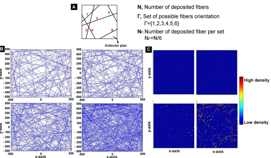
Figure 2.1 Electrospinning apparatus description ……….. 36
Figure 2.2 Micropatterned electrospun membrane ……… 38
Figure 2.3 Surface roughness and morphology comparison of different localized por-tions of the same electrospun membrane (35 wt. % PCL in 40AA/60FA at 1.5 kV.cm-1 with a flow rate of 0.07 mL.h-1) ………... 39 Figure 2.4 Static contact angles assessment ………. 39
Figure 2.5 Electric field simulation before the fibers deposition ……….. 40
Figure 2.6 Pore size at different localized portions of the same electrospun membrane (35 wt. % PCL in 40AA/60FA at 1.5 kV.cm-1 with a flow rate of 0.07 mL.h-1) ……… 40
Figure 2.7 Arbitrary fiber deposition in the collector’s center ……….. 41
Figure 2.8 Electric field simulation during the electrospinning process …..……….. 43
Figure 2.9 Electric field in the presence of one nanofiber ……… 43
Figure 2.10 Photograph of a fibrous PCL-gelatin cluster that turn into complex peaks as time goes by (70 wt. % PCL /30 wt. % gelatin in 40AA/60FA at 1.5 kV.cm-1 with a flow rate of 1.0 mL.h-1) ………... 44 Figure 2.11 Direct-assembly mechanism tested on porous paper-based substrates …. 45 Figure 2.12 Variation of Rmax ………. 46
Figure 2.13 Transition state for self-assembly ………... 47
Figure 2.14 Topographic morphology comparison of equivalent electrospun mem-branes processed under different electric field intensities (35 wt. % PCL in 40AA/60FA with a flow rate of 0.07 mL.h-1 during 24 h, the surrounding environment was set for 20 ºC and a humidity level of 31 ± 1%) ……….. 48
XVI
Figure 2.15 Topographic morphology comparison of equivalent electrospun
mem-branes processed under different temperature and humidity levels ……… 49
Figure 2.16 Evidence of the need to extend the production time of the electrospun
ma-terial (35 wt.% PCL in 40AA/60FA processed at 1.375 kV.cm-1 with a flow rate of 0.07
mL.h-1 during 24 h, the surrounding environment was set for 20 ºC and a humidity level
of 31 ± 1%) for the sake of obtaining closed cells. On the right it is highlighted the smooth
appearance of new MAFs ……… 50
Figure 2.17 Morphology comparison of electrospun membranes processed from
paren-tal polymer solutions with different concentration under the same processing conditions (PCL in 40AA/60FA at 1.5 kV.cm-1 during 24 h with a flow rate of 0.07 mL.h-1, the
sur-rounding environment was set for 21 ºC and a humidity level of 32 ± 1%) ………. 50
Figure 2.18 Example of a dextran electrospun mesh, where the experimental conditions
where defined as follow: 50 wt. % DEX in Distilled Water at 1.667 kV.cm-1 with a flow
rate of 0.2 mL.h-1, in a surrounding environment set for 40 ºC and a humidity level of 24
± 1% ………... 53
Figure 3.1 Conceptualization of three-dimensional multilayered electrospun constructs
(3DMECs) ……….. 61
Figure 3.2 Bioinspired production of 3DMECs ……… 67
Figure 3.3 Fibers self-assembly induced by their in situ polarization leads to the
gener-ation of protrusions ………... 68
Figure 3.4 3DEC topographic characterization ………... 69
Figure 3.5 Phase contrast microscopy images from a 3DEC, including the cross-section and top side at different z-planes, evidencing, simultaneously, multiple protrusions and z-axis built in core characterized by dense fiber regions ……… 70
Figure 3.6 Solvent contamination assessment by 1H-NMR of as-spun 3DECs in CDCl3 71
Figure 3.7 3DECs chemical characterization after plasma treatment ………. 71
Figure 3.8 Physicochemical characterization of 3DECs ………. 72
Figure 3.9 Spreading and imbibition dynamics ……… 74
Figure 3.10 SEM cross-section images of a coated protrusion evidencing the
electro-spun fibers self-assembly. The fiber based network evidences a high tortuosity ……….. 76
Figure 3.11 LbL coated protrusions characterization ………. 76
Figure 3.12 Chitosan and hyaluronic acid incorporation through spray-LbL ………. 77
Figure 3.13 LbL coated protrusions characterization ………. 78
Figure 3.14 SEM images and pseudo-colored SEM of a pulled out coated protrusion …. 78
Figure 3.15 Morphological characterization at the interprotrusion space ……….. 79
Figure 3.16 Phase contrast and fluorescent microscopy images of cell seeded 3DMECs
top side evidencing the LbL film swelling ………. 80
Figure 3.17 Phase contrast and fluorescent microscopy images from a cell seeded
3DEC top side at different z-planes evidencing simultaneously multiple protrusion, z-axis built in formation and parallel fibers between protrusions ………. 81
Figure 3.18 LbL-film incorporation influence in dressing properties ……… 82
XVII
Figure 2A.1 Flow curves of PCL polymer solutions in 40AA/60FA at different
concentra-tions ……… 98
Figure 2A.2 SEM micrographs of poly(ε-caprolactone) and chitosan 95/5 % wt.
poly-meric blend ……… 98
Figure 2A.3 SEM micrographs of poly(ε-caprolactone) and chitosan 90/10 % wt.
poly-meric blend ……… 99
Figure 3A.1 Example of a three-dimensional electrospun constructs within the radio
fre-quency tubular reactor for plasma treatment, placed on a non-porous metallic plate …… 102
Figure 3A.2 3DECs fiber diameter distribution in the bottom side ……… 102
Scheme 3A.1 Resting droplet assay in a 3DEC construct ………. 102
Figure 3B.1 Spray Layer-by-Layer (spray LbL) for surface coating of biomedical relevant
scaffolds ………. 110
Figure 3B.2 Chemical structures of the used polycations (+) and polyanions (-) ………. 112
Figure 3B.3 LbL film characterization ……….. 113
Figure 3B.4 LbL film physicochemical characterization for the (X/Y)n architectures
where, X is Gelatin, Poly-1 or Chitosan, Y is Alginate or Hyaluronic Acid, and n is 5, 3 or
1 layers ………... 115
Figure 3B.5 Cellular proliferation and covering dynamics on the assembled LbL films.
A, Cellular densi-ty up to 10 days ……….. 116
Figure 3B.6 Schematics illustrating the cell circularity determination and on how to
in-terpret the calcu-lated values accordingly with cell shape ………. 117
Figure 3B.7 Cellular shape assessment and LbL films degradation behavior for the
baselayer [(LPEI/DS)10] and (Cht/HA)10 LbL coatings ………. 118
Figure 3B.8 Cellular shape assessment and LbL films degradation behavior for the
(Gel/Alg)n LbL coatings, throughout the same analysis protocol of Figure 3B.7 …………. 119
Figure 3B.9 Phase contrast micrographs of cultured cells on PO substrates (plasma
cleaned glass slides) and (LPEI/DS)10 multilayered coatings up to 10 days ……….. 120
Figure 3B.10 Phase contrast micrographs of cultured cells on (Gel/Alg)n multilayered
coatings up to 10 days, where n is 1, 3, 5 and 10 layers ……….. 121
Figure 3B.11 Phase contrast micrographs of cultured cells on (Poly-1/Alg)n multilayered
coatings up to 10 days, where n is 1, 3, 5 and 10 layers ……….. 122
Figure 3B.12 Phase contrast micrographs of cultured cells on (Cht/Alg)n multilayered
coatings up to 10 days, where n is 1, 3, 5 and 10 layers ……….. 123
Figure 3B.13 Phase contrast micrographs of cultured cells on (Gel/HA)n multilayered
coatings up to 10 days, where n is 1, 3, 5 and 10 layers ……….. 124
Figure 3B.14 Phase contrast micrographs of cultured cells on (Poly-1/HA)n multilayered
coatings up to 10 days, where n is 1, 3, 5 and 10 layers ……….. 125
Figure 3B.15 Phase contrast micrographs of cultured cells on (Cht/HA)n multilayered
XVIII
Figure 3B.16 Dynamic substrate area coverage up to 10 days, represented as the area
percentage according to the initial substrate area, for all the in vitro tested samples plasma cleaned glass slides (PO), used LbL baselayer (LPEI/DS)10 and (X/Y)n LbL
coat-ings, with the corresponding profile curves ………... 127
Figure 3B.17 Flow curves of PCL dissolved in a 40/60 (v/v) solution of acetic and formic
acid at a de-sired concentration of 35 wt.% displaying complex viscosity and shear stress 129
Figure 3B.18 SEM images of three-dimensional (Cht/HA)10 multilayered electrospun
construct ………. 129
Figure 3B.19 MMP-9 inhibitors compatible with (Cht/HA)10……….. 133
Figure 3C.1 Fabrication process of PCL based electrospun mats treated with NaOH …. 138
Figure 3C.2 Contact angles of untreated and NaOH treated wound dressing substrates 140
Figure 3C.3 Inter and intra-correlation between the contact angles and FTIR of untreated
and NaOH treated wound dressing substrates ……… 141
Figure 3C.3 Photographs of wound dressing substrates differently treated to enhance
XIX
Index of Tables
Table 1.1 Differential diagnosis of non-healing wounds ……….. 6
Table 2.1 Hansen solubility polar parameters ………. 52
Table 3.1Ideal specifications of a wound dressing and advantages in the use of poly(ε
-caprolactone) ………. 66
Table 4.1 Hansen solubility polar parameters database for future work ………. 93
Table 3B.1 Inter and intra-comparison of growth rate values for the generated LbL
mul-tilayered coatings ……….. 114
Table 3B.2 Kinetic parameters t* and t0 for the spreading and imbibition stages on the
LbL multilayered coatings tested ……… 116
Table 3B.3 Sigmoid function parameters estimation of the dynamic substrate area
cov-erage behavior for the determination of the kinetic parameter t50……… 128
Table 3C.1 Fiber distribution fitting ………. 139
1
Wound Pathophysiology and Wound
Dressing Conceptualization
SUMMARY
1The wound healing phenomena is a multipart and orchestrated process of bioevents and cell lineages. In addition to its complexity, wound repair is also deeply affected by external conditions that can compromise the healing rate of the injured tissue. The proper designed of a wound dressing is challenging, since such constructs must attend a set of multivariable and dynamic requirements in order to avoid the generation of non-functioning mass of fibrotic tissue or a chronic wound healing stage. Herein, it is reviewed the most important aspects of wound patho-physiology while highlighting the most important requirements to be fulfilled by a wound dressing in a wound dressing.
1 This Chapter was partially submitted for publication (Tiago C. Reis, “Wound Pathophysiology and Wound
Dressing Conceptualization”).
3
1.1 WOUND PATHOPHYSIOLOGY AND REPAIR
1.1.1 The human skin anatomy and wound pathophysiology
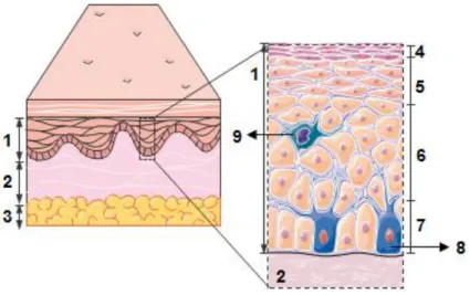
The human skin is the largest organ in the body, consisting of three layers: (i) epidermis; (ii) dermis, and (iii) hypodermis (Figure 1.1). The epidermis is a multi-layered epithelium that starts
from the interface with the dermis and is about 80-100 µm thick, comprising four sublayers: the stratum basalis, the stratum spinosum, the stratum granulosum and the stratum corneum. The epidermis is deprived of extracellular matrix (ECM), with the exception of its stratum basalis, a cellular configuration which its progenitor cells undergo a continuous self-renewal and differenti-ation to keratinocytes. As the keratinocytes migrate towards the surface of the skin, they suffer a terminal differentiation and maturation, resulting in a keratinized layer of dead cells which confers the main barrier properties of the skin in its stratum corneum. The dermis is positioned bellow the epidermis, being a connective tissue that encompasses vascular endothelial cells, ECM, fibro-blasts and skin appendages such as sweat glands, hair follicles, etc., and it is responsible for most of the mechanical properties and resilience of the human skin. Among the several dermis constituents, fibroblasts play an important role on modulating the skin mechanical strength and elasticity, throughout the secretion of collagen and elastin, respectively. The tensile strength of the dermis can range from 3.4 MPa to 68.9 MPa,[1] depending on the type of species, orientation of the skin specimen tested and the location on the body. Finally, underneath the dermis one will find the hypodermis, an adipose like tissue that its main role is to act as an insulator and cush-ioning layer between the skin and skeletal structures as muscles and bones.
Figure 1.1 Basic skin anatomy. 1, Epidermis. 2, Dermis. 3, Hypodermis. 4, Stratum corneum. 5, Stratum granulosum. 6, Stratum spinosum. 7, Stratum basalis. 8, Melanocyte. 9, Langerhans cell.
unre-4
solved medical needs that affect treatment results, quality of life, length of hospital stay and re-imbursement rates in healthcare.[2] A wound is defined as a disruption of normal anatomic struc-ture and function.[3] In the specific case of skin wounds, this category of wounds comprises sur-gical and accidental lacerations, burns, pressure ulcers, diabetic and venous ulcers. Just in the United States, wound treatment and complications thereof surpasses $ 20 billion per year,[4] whereas chronic wounds are especially costly since they require repetitive treatments. In a similar fashion, the National Health Service of the United Kingdom spends annually £ 1 billion in the treatment of wounds, being venous leg ulcerations, pressure wounds and diabetic foot ulcerations the most expensive types of wounds to treat.[5] In the case of Portugal, Furtado et al.[6] estimated that 1.42 out of 1000 Portuguese citizens has a chronic wound, and that number will increase due to an existing correlation between poor healing and an aging society.[7] Furthermore, chronic wounds are predisposed to complications that also have impact on the time of the reparative process. Such complications of chronic wounds include infections, malignant transformation and functional limitations. Cellulitis, osteomyelitis, abscess formation, gangrene and sepsis can result from an infected wound, while some chronic wounds can potentially observe a malignant trans-formation (for example, Marjolin ulcers).[8, 9] In addition, functional limitations include, for in-stance, gait impairment.[10]
5
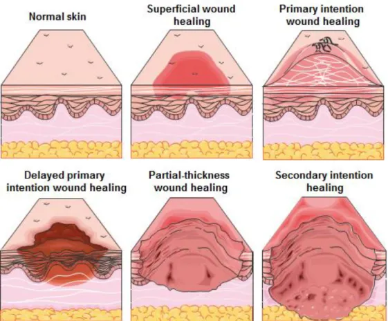
By definition, a chronic wound is a lesion of a tissue that failed to continue through an timely and orderly reparative process – named wound healing– which aims to restore and produce the an-atomic and functional integrity of the injured tissue, leading to a stage of pathologic inflammation. In this way, the healing process is interrupted, delayed and therefore incomplete, resulting in a poor anatomical and functional outocome.[11] Currently, there are five basic models of healing and one can find similarities in all, whereas such similarities guide the development of better wound dressings.[12] The five basic healing models are: (i) superficial wound healing; (ii) primary intention wound healing; (iii) delayed primary intention wound healing; (iv) partial-thickness wound healing, and (v) secondary intention healing (Figure 1.2).
Superficial wound healing: An inflammatory repair process can result as an alteration of the skin’s surface either by pressure (which includes friction and shearing, typically stage I pressure ulcers), contusions and first-degree burns. The involvement of the skin surface may be associated with deep tissue death, and consequently the tissue ruptures and generates a deep cavity, a common problem observed in pressure ulcers and grade I neuropathic ulcers. This wounds usually shows several alterations in the skin such in its temperature (warmth followed by coolness, an imperative indication of tissue death), color, tension and sensation, all of them leading a clinician to conclude tissue congestion. Despite soft tissues heal by themselves over time, medical intervention for this type of wounds (for example, though the use of ultrasounds for reabsorption of hematoma) boosts the recovery over functional activities and mobility.
Primary intention wound healing: This healing model consists in bringing closer the edges of a wound in order to promote closure, a model frequently employed in surgical lacerations. Thus, the usage of this healing model avoids the loss of subcutaneous tissue and the possible contam-ination of the wound bed with microorganisms or debris. The outcome is a minimal residual scar-ring with a full closure within 3-7 days for patients without functional impairments.
Delayed primary intention wound healing: However, when a wound is contaminated with micro-organism or other foreign bodies, or where a significant tissue loss occurred and the employment of the primary intention wound healing model would outcome in unbearable tissue tension, the delayed primary intention wound healing model is preferential. Despite stiches are used in the subcutaneous and fascial layers, in this model the wound is left open. After the risk of infection is mitigated, or tissue loss is partially recovered, the wound will self-close within 5-7 days for patients without functional impairments.
6
Secondary intention healing: This healing model diverges from the previous, since it is only em-ployed in wounds where full-thickness loss of the dermis occurred and such loss may also be extended to the underlying tissues. The secondary intention healing model, or also commonly found in the literature as the contraction healing, results when myofibroblasts generate contractile forces that draw together the wound. This healing approach leads to minimal epitheliazation and to the production of scar tissue, and therefore the skin anatomic functionality and structure is not replaced. Moreover, a wound that heals throughout secondary intention healing present elasticity or tensile strength that does not match the native properties of the surrounding tissue. The con-traction healing is the process commonly observed during the healing of chronic wounds, which can be extended up to 6 months to 2 years.
Since the conception of wound dressings for non-healing partial-thickness wounds and non-heal-ing full-thickness wounds is one of the main goals of this thesis, the reparative process of these wounds it will be properly introduced and detailed in the Section 1.1.2 – Acute wound repair. Moreover, Table 1.1 shows the differential diagnosis of the underlying etiology of a non-healing
wound, whereas 70 % of ulcers are caused by ischemia, secondary to diabetes mellitus, venous stasis, and pressure.[14]
Table 1.1 Differential diagnosis of non-healing wounds.
Chronic wound category Etiology
● Vascular
Arterial Atherosclerosis, arteriovenous malformation
Lymphatic Lymphedema
Mixed (Venous-Arterial)
- Vasculitis Systemic lupus erythematosis, rheumatoid arthritis, scleroderma, polyarteritis nodosa, Wegener’s granulomatosis
- Venous Venous stasis
● Pressure Spinal cord injury, bedbound, elderly
● Neuropathic Diabetes, peripheral neuropathy
● Hematologic Polycythemia rubra vera, sickle cell disease ● Traumatic Burns, cold injury, radiation, factitious
● Neoplastic Basal carcinoma, squamous cell carcinoma, melanoma,
● Others Marjolin’s ulcer, Bowen’s disease Sarcoidosis, obesity, tropical ul-cer, pyoderma gangrenosum, necrobiosis lipoidica diabetecorum
1.1.2 Chronic wound repair
self-7
limiting biochemical cascade of events that aims to remove necrotic tissue, debris, microorgan-isms and theirs contaminates, while it triggers the recruiting and activation of fibroblasts.[15] On the other hand, in the case of chronic wounds, the inflammation process is not self-limiting, i.e., it is responsible to further injure the wounded tissue while continuously reactivating and promoting novel routes of inflammation. For example in chronic wounds, tissue trauma by continuous in situ pressure, leucocyte trapping, bacterial populations or ischemic reperfusion injury, are potential causes for recruiting and keeping neutrophils at the wounded site throughout long periods of time (months up to years). This phenomenon leads to the up-regulation of the inflammatory cascade and consequent abnormal inflammatory profile, while in comparison with acute wounds the neu-trophils are only present in the first 72 h,[16] allowing the continuation of the wound healing cas-cade without further problems. The presence of activated neutrophils at wound bed of a chronic wound triggers the production of local ECM degradative matrix metalloproteinases (MMPs), cal-cium-dependent zinc-containing endopeptidases that are capable of degrading extracellular ma-trix components.[17] While in acute wounds such MMPs are locally inhibited (e.g. by the nonspe-cific proteinase inhibitor α2-macroglobulin) in proper stoichiometric ratios, at non-healing chronic wounds the stoichiometric MMP:inhibitor ratio is unbalanced leading to the wound bed degrada-tion.[17-19] Therefore, the secretion rate of ECM components, such as collagen, by fibroblasts, is lower than the rate of degradation of these components, and consequently these cells are incapable to make progress in depositing ECM.[19] Moreover, the extended inflammatory envi-ronment in chronic wounds contributes to the predominant presence of cytokines (e.g. tumor ne-crosis factor α), important biochemical cues for cell signaling.[20] As a result, it is observed a considerable reduction of endogenous factors that should promote the proliferation of novel epi-dermal cells at the wound edges (e.g. platelet-derived growth factor).[21] Other reported differ-ence between the acute and chronic reparative processes is in the fibroblasts themselves. Brem et al. demonstrated that fibroblasts within a chronic wounded environment have premature se-nescence, which disrupts their normal functioning.[22] As a result, such fibroblasts report impaired migration capacity as well as reduced response to local growth factors. In summary, the patho-logic inflammation of chronic wounds results in more inflammation, turning out in an interrupted, delayed and therefore incomplete healing process, due to intermittent feedback events that con-verge towards an uncontrolled inflammatory positive feedback loop.
So far, it was only introduced the main differences between acute and chronic healing specially in association with local inflammation. Nevertheless, the wound healing process is far more com-plex, it is indeed a complex cascade of biochemical events that is orderly synchronized. The understanding of each stage contributes to the development of better wound dressings, since their imperative requisites become known and therefore can be attended from a dressing con-ception point-of-view. The wound healing process can be divided into 4 overlapping stages: (i)
hemostasis, (ii) inflammation, (iii) proliferation and (iv) remodeling (Figure 1.4). This tissue
8
soluble mediators and infiltrating cooperative cells. Nevertheless, and accordingly to this PhD thesis framework and intent, it is avoided the extensive biological detailed information over these stages, since the main goal at this point it is only to contextualize the reader with the general panorama of the wound healing process and to guarantee future references understanding. Proper literature was selected and quoted in order to provide a source of information for those whom seek more detail.
Figure 1.3 Acute wound healing model and comparison between the acute healing versus the chronic heal-ing processes. A, Multivariable molecular and cellular model of a chronic wound in a chronic inflammatory staged characterized by abundant platelets and polymorphonuclear leucocytes (PMN) and macrophage (Mϕ) infiltration. The presence of abundant inflammatory cells triggers the production of proinflamatory cy-tokines (TNF-α, IL-1 and IL-6) inducing a pro-oxidant milieu, which leads to the degradation of ECM struc-tural proteins (collagen) and to the degradation of excreted growth factors. The attracted leukocytes, mainly neutrophils, are a rich source of reactive oxygen species (H2O2, O2-) whereas those species can actively
9
Tissue injury is often linked to the disruption of blood vessels and consequent extravasation of blood constituents. After wounding, it is observed that the injured blood and lymphatic vessels undergo a process called vasoconstriction, which allows to achieve local hemostasis within few minutes.[23, 24] At this moment, in situ attracted platelets are activated by adhesive matrix pro-teins (e.g., collage and fibronectin) in the vascular wall, while thus adhering and aggregating in order to form a platelet plug. In parallel, platelets are continuously excreting soluble mediators (thromboxane A2, serotonin and adenosine diphosphate) and other adhesive proteins
(thrombos-podin, factor VIII, etc.),[23] causing the stimulation of local thrombin production. It is this serine protease that converts local fibrinogen in fibrin and consequently generates a fibrin clot. Later on, the fibrin clot is plasmin cleaved.[25] The next stage of the wound healing process is inflammation, an important step where chronic wounds accordingly to the above reported features cannot sur-pass. This stage is characterized by the early recruitment of neutrophils and monocytes, which evade from surrounding capillaries into the wound bed. The recruitment process is based in the local present chemotactic factors previously generated by the platelets (e.g., kallikrein), and once in the wounded environment the neutrophils are responsible to kill and phagocyte bacteria.[26] On the other hand, local monocytes differentiate to macrophages and therefore are responsible to kill and phagocyte bacteria, scavenge tissue debris and destroy residual neutrophils.[27] In the absence of infection, it is observed a gradual decrease in the number of local neutrophils.[28, 29] The wound healing process moves towards the execution of the biochemical events associated with the proliferation stage, a phase that comprises the most prominent events of the healing process. This phase tends to occur after 4-5 days of the wounding and may last up to several weeks in patients with acute wounds.[30] The proliferation stage can be subdivided in the follow-ing subsequent sub phases: granulation tissue formation, re-epithelization and ECM
reorganiza-tion. The granulation tissue comprises collagen type III and acts as ground substance for fibro-blasts, macrophages and the newly replicated endothelial cells.[31] At this point, several capillar-ies populate the novel tissue while fibroblasts are continuously producing glycoproteins (fibrin, fibronectin and hyaluronic acid) that will reinforce the novel extracellular matrix. The cooperative roles of fibroblasts and blood vessels allow to support cell in growth and oxygen and nutrient supply, whereas the macrophages are mainly responsible to produce growth factors (such as the platelet-derived growth factor (PDGF) and the transforming growth factor β1 (TGF-β1).) that con-tinuously stimulate fibroplasia and angiogenesis. The provisional extracellular matrix generated by the fibroblast is then hypothetical remodeled by TGF-β1 and/or TGF-β2, ensuring a more sta-ble collagen based matrix.[14, 17, 32] After achieving a viasta-ble cellular scaffold within the wound bed, keratinocytes migrate towards the wound bed and consequently proliferate and differentiate
10
fibroblasts will differentiate into a myofibroblast phenotype that is responsible to compact the con-nective tissue and for further wound contraction. As briefly stated above, TGF-β1 and/or TGF-β2, as well as PDGF, are hypothetically the main growth factors responsible to stimulate tissue re-modeling through the integrin receptors and cross-links between individual bundles of colla-gen.[33, 34] Nevertheless, collagen remodeling from granulation tissue (collagen type III) to scar tissue (collagen type I), depends on the continuous fabrication of collagen by the present sub-population of fibroblasts, as well as on its catabolic metabolization controlled by the local matrix metallic proteinases. In addition, it is also observed in this tissue remodeling phase the proper reorientation of collagen fibers, leading to a partial restoration of the wound original mechanical strength. However, a scar is only 70 % as strong as normal skin.[30]
Figure 1.4 Wound healing overlapping phases and inter- and intracorrelation. Cell phenotypes and their effects on acute healing.
1.2 FUNCTIONAL REQUIREMENTS OF A WOUND DRESSING
manipu-11
lation of the wounded environment by negative pressure[37] or electrical stimulation,[38] admin-istration of small molecules,[39] gene-therapy approaches,[40] and cell-based strategies such as the local administration of stem cells.[41, 42] While all of these strategies have demonstrated their potential benefit on in vitro and in vivo models,[43] many of them are being employed in combi-nation with constructs – named wound dressings– which are conceptualized to partially fulfil the main skin functions. For example, Kim et al.[44] demonstrated that alginate wound dressings can be used to customize negative pressure wound therapy for intractable auricular defects. On the other hand, Shan et al.[45] reported the in vivo use of silk fibroin/gelatin wound dressings func-tionalized with astragaloside IV to elicit anti-scar effects on partial-thickness burn wounds, whereas Wen et al.[46] demonstrated that bacterial cellulose dressing containing uniform silver sulfadiazine nanoparticles for burn wound healing can promote a better epithelialization progress. In addition, Falanga et al.[47] verified that autologous bone marrow–derived cultured mesenchy-mal stem cells delivered in a fibrin dressing, were capable to boost the healing process either in murine and human cutaneous wounds. In all of these examples, the usage of wound dressings provides a protective environment for the injured tissue.[48] Nevertheless, there is a set of func-tional requirements that a wound dressing must attend:
1.2.1 Biocompatibility, non-antigenicity and non-cytotoxicity
12
hydroxyapatite, methacrylate, acrylamides and silicone, have higher plasma concentrations of myeloperoxidase (3.9-fold) and the chitinase-like proteins chitotriosidase (2.5-fold) and YKL-40 (2.0-fold), biomarkers of innate cell-related immune response,[56-58] in comparison with non-filler subjects. On the other hand, hyaluronic acid alone elicited a small immune response. Besides the potential inflammatory and immunogenic behavior of a wound dressing, its cytotoxicity is also a key parameter one must control. Highly toxic wound dressings can lead to bulk tissue necrosis due to apoptotic cell death in the surrounding tissues, which is further stimulated by ongoing autophagy and pyroptosis.[59] These cell death events are highly regulated throughout signaling networks caused by foreign chemical compounds, which can be the end product of the wound dressing degradation process. Typically, their effects increase the production of reactive oxygen species within the cell, leading to the oxidation of protein thiols and a shift in cellular redox sig-naling.[60] Moreover, the effects of such end products are mediated by disruption/modulation of cellular Ca2+ homeostasis. Ca2+ depletion or modifications in the Ca2+ transport systems, are
di-rectly linked to the endoplasmic reticulum stress through the activation of procaspase-12, an en-zyme that acts on effector caspases to induce apoptosis.[61]
1.2.2 Exudate management
13
the wound exudate also comprises several biochemical agents that have a positive effect on the regenerative mechanisms, such as fibroblast growth factors, epithelial growth factors and platelet-derived growth factors.[43] While from one end proper exudate absorption and sequestration of detrimental compounds is advised, on the other end one must guarantee a moist environment and the local provision of the beneficial healing agents already present in the wound exudate. In addition, a dressing absorptive capacity must also be optimized in order to reduce the number of wound dressing changes, enhancing patient comfort and compliance while reducing the needed healthcare services.
1.2.3 Moisture management
The moisture levels at the wound bed result throughout a dynamic balance between wound exu-date production, the dressing absorptive capacity and its permeability for water vapor. According to the wound phenotype, the wound exudate production differs from wound to wound type. For example, in the case of venous leg ulcers, a chronic wound example, the continuous inflammatory stage promotes the wound exudate production through the mechanisms previously reported.[69] On the other hand, in the case of diabetic foot ulcers, wound bed moisture levels are depleted.[70] The absence of proper restoration of the wound bed moisture leads to the adherence of the dressing applied, whereas new blood vessels and granulation tissue can grow into the dressing structure. Therefore, a good moisture management strategy is strictly linked to the underlying wound pathology. Upon the dressing absorptive capacity, commonly designated in the literature as swelling capacity, wound dressings are classified into three classes: moisture absorbing, mois-ture maintaining and moismois-ture donating. Moismois-ture absorbing dressings tend to absorb 15 to 20 times their weight of fluid, being ideally for highly exuding scenarios.[71] In contrast, moisture donating dressings, also designated as hydrocolloids, are characterized by a matrix of insoluble polymers comprising up to 90 % water content, which enables the delivery of water molecules in poorly exuding wounds.[72, 73] Besides the importance of wound pathology in the exudate pro-duction, different wound types have shown different water vapor transmission rates. Water loss at a wounded site arises from the rupturing effect of the semipermeable protein layer in the
stra-tum corneum, which breakdown promotes profuse water loss through the wound. While the der-mis comprises about 80 % water, the stratum corneum comprises about 40 % and about 15 % in its inner and outermost layers.[74] Nevertheless, water has a large latent heat of evaporation of 2.3 MJ.kg-1 [75] and consequently, local temperature increasing leads to water vapor transmission
across the wound interface. Therefore, according to the wound pathophysiology, different wound types represent different needs in terms of moisture balance. For example, granulating wounds show a water vapor transmission rate of 214.1 ± 8.4 g.m-2.h-1, while first, second and third degree
burns show values of 11.6 ± 1.1 g.m-2.h-1,178.1 ± 5.5 g.m-2.h-1 and 143.2 ± 4.5 g.m-2.h-1
14
transmission rate value of the wound dressing. After the absorption of the exudate by the dress-ing, water vapor transmission stills occurring through the dressing itself. Unmatching the wound exudate production rate, the exudate absorption rate and the dressing water vapor transmission rate, can conduct to a subsequent exudate accumulation in the wounded environment or to dry out the wound. For example, it has been suggested[77] for the case of granulating wounds that a value of water vapor transmission rate between 83.3 and 104.2 g.m-2.h-1, would be adequate to
provide a proper moisture environment and avoiding exudate accumulation in granulating wounds.
1.2.4 pH management
The keratinocytes present in the epidermis are continuously secreting organic compounds that acidify the intact skin tissue, turning it to a naturally occurring acidic medium with a pH ranging from 4 to 6, a proactive action to avoid fungi and bacterial development. When the skin is injured, and consequently local blood and lymphatic vessels are disrupted, the wound pH increases to 7.4. According to the wound pathophysiology, acute wounds tend to become acidic while chronic wounds tend to become alkaline.[78] An alkaline pH is responsible to impair healing by favoring bacterial colonization and proliferation (pH > 6), and proteolysis mediated by the in situ host and bacterial proteases kinetically suppresses the healing bioevents.[79] Due to the wound pH role in the healing period, the clinical community have relied in topical formulations and wound dressings to control or modify the wound pH. In parallel with their thermal insulation provision, wound dress-ings are mainly conceptualized to control pH through the prevention of the respiratory alkalosis by preventing the loss of CO2 from the wounded environment, which in turn contributes to lower
the wound pH. Nevertheless, upon on the chosen wound dressing type, the wound pH tends to evolve differently. The wound exudate of chronic wounds under non-permeable dressings is fre-quently observed as more acidic, in comparison with chronic wounds were a permeable dressings is utilized. Wilson et al.[80] studied the clinical outcome of tailored dressings into two groups both suffering from chronic venous leg ulcers, whereas group 1 was set for a pH value 7.3 and group 2 was set for a pH value 6.0. Group 2 reported a rate of healing 3-fold higher than group 1. Regarding the listing process of the functional requirements for a wound dressing, pH manage-ment is a crucial one. In this way, local titration of the wound pH is currently understood as a beneficial feature to accelerate the wound healing process. It is suggested that more acidic wounds heal better, since a low pH value leads to an increase of local oxygen to the cells, a common observation described in literature as the Bohr-effect.[81]
1.2.5 Gaseous exchange
15
around 1 %.[82] Indeed, in wounded skin tissues pO2 values as low as 1 % are still recorded at
the wound surface, suggesting that atmospheric oxygen can oxygenate the wound bed.[83] The generated hypoxia due to the locally damaged skin microvasculature leads the cells to execute anaerobic metabolism, which increases the local production of lactic acid, reduces pH and stim-ulates the up-regulation of vasodilatory factors.[84, 85] Moreover, the pH reduction lowers the hemoglobin affinity for oxygen, further reducing the oxygen delivery.[81] Several studies have linked the oxygen presence as essential for the phagocytosis of external microorganisms,[86, 87] referring the positive effect of an in situ oxygen tension for the overall wound rate closure. Also, the locally generated hypoxia-inducible factors are utilized in many proangiogenic mechanisms through the vascular endothelial growth factor[88] and nitric oxide[89, 90] signaling. In this way, it is being observed an increase of treating modalities that balance the provision of oxygen into the wound bed, such as the hyperbaric oxygen therapy[91, 92] and topical oxygen therapy.[93] On the other hand, the carbon dioxide tension in a wound bed tends to increase as the healing processes moves forward. The partial pressure of carbon dioxide (pCO2) rises from 60 to 75
mmHg at the second week, and such increase is related to higher metabolic activity and deficient diffusion over the damaged vascular network.[82] It has been demonstrated that high levels of local carbon dioxide can down-regulate genes related to innate immunity,[94] while resulting in reduced phagocytic activity due to a lower NFκB cellular activity.[95] Tsuji et al.[96] reported that endothelial cells under hypoxia conditioning were capable to proliferate at high levels of CO2,
suggesting that local pCO2 mediates the cellular events regardless the local pH or oxygen values.
These newly observations over the potential use of CO2 in wound healing have deeply contribute
to new therapeutic approaches. For instance, in a clinical study Brandi et al.[97] treated patients affected by chronic wounds through the subcutaneously administration of CO2, concluding that
comparatively to the control group (no CO2 administration) such patients could observe an
in-crease in microcirculation and in the tissue oxygenation values. Despite their different roles in the wound healing process, oxygen and carbon dioxide must be able to freely permeate a wound dressing, while keeping minimal tension values that can beneficially stimulate the wound regen-eration process.
1.2.6 Prevention and Infection control
16
tend to accumulate at the wound surface, accelerating the directed neutrophil locomotion or in-terfering with cell–matrix interactions.[101] At the wound bed, bacteria produce an exopolysac-charide matrix, designated as biofilm, in order to function as a protective substrate and barrier for the colony. Kierker et al.[102] demonstrated that just in 24 hours, biofilm communities of
Staphy-lococcus aureus significantly increased the apoptosis of human keratinocytes. In addition, Zhao
et al.[103] demonstrated that Pseudomonas aeruginosa biofilm-challenged wounds typically heal in approximately 6 weeks, at least 2 weeks longer than nonbiofilm-challenged normal wounds. Therefore, an ideal wound dressing must not only prevent the initial contamination by endogenous and exogenous bacteria, but it must also eradicate infection during the healing period. While the initial contamination prevention can be mainly achieved by proper impermeability to foreign bod-ies, the clinical community have pursued infection control either by the continuous elution of an-timicrobial agents imbibed in the dressing inner structure[104] or the usage of polymeric materials that are antimicrobial by nature.[105] For example, in a randomized comparative study, 20 pa-tients with ulcers of vascular aetiology, wound duration ≥ 6 months and ankle brachial index > 0.6, where treated with a silver-containing hydrofiber dressing, which promoted a bacterial load reduction of 41.6 %.[106] Alternatively, Moghadas et al.[107] reported that film-like wound dress-ings based on chitosan and biofunctionalized montmorillonite could achieve killing efficiencies over 99.6 % and 99.7 % towards Escherichia coli and Staphylococcus aureus populations re-spectively. Nevertheless, in the absence of drug eluting systems or natural antibiotic matrixes, proper infection control can be achieved by promoting an adverse environment for bacterial growth. For instance, by inducing a hyperosmotic environment, Connel et al.[108] were able to reduce the Escherichia coli and Enterococcus faecalis bioburden levels by 3 logs within 24 hours, values that were similar to wounds treated with the control silver sulfadiazine. Adverse milieus can be alternatively triggered by modulation of the wound environment pH,[109] or by promoting the in situ production of hydrogen peroxide.[110]
1.2.7 Odor management
17
better odor management, odor concealers are also essential to capture and neutralize the gen-erated volatile compounds usually under electrostatic interactions. In an international survey con-ducted by Gethin et al.[114] regarding 1444 clinical professionals, activated charcoal dressings were the most used type of wound dressings to manage a wound’s malodor (84.9 %). Neverthe-less, such constructs present some limitations. For example, these wound dressings are not rec-ommend to be cut or trimmed, otherwise charcoal particles will enter the wound, a constrain that hinders their ideal fit to any wound.[115] Recently, Narayanan et al.[116] disclosed the production
of a poly(ε-caprolactone) (PCL)/β-cyclodextrin functional nanofibrous wound dressings, which were able to absorb and retain high amounts of odor compounds (butyric and propionic acid), while preserving the typical flexibility of electrospun non-woven mats. Regardless the incorpora-tion of antibacterial compounds in some dressings to control a wound infecincorpora-tion, some hydrophilic dressings produce a characteristic odor as they decompose in situ after gelling over the wound.[63] Ideal wound dressings are then not only characterized by an appropriate wound exu-date and moisture management, but also by their ability to retain and neutralize the malodor agents generated by the wound milieu, especially when a reduced dressing change frequency is desirable.
1.2.8 Reduce adherence
18
and solubility at the wound bed take a significant role. For instance in the case of a bloody wound, higher concentrations of fibrinogen, serum proteins and glycoproteins, are present in the wounded microenvironment, and therefore such components diffuse towards the wound dressing ending up to be stabilized at the generated layer of moisture, which significantly increases the tendency for future cellular anchorage. While studying the effect of protein concentration in the in vitro cel-lular adhesion, Koblinski et al.[123] found that the presence of low levels of adhesion proteins (fibronectin), together with high concentrations of non-adhesion proteins (osteonectin), could still promote the cells adhesion on U-bottom 96-well plastic plates, while low levels of adhesion pro-teins by their own were unable to promote cellular adhesion. Another potential driver of protein adsorption is the electrostatic attraction between a charged wound dressing and proteins at the wound site that are bipolar.[124] The moisture layer mediated protein adsorption on a wound dressing is also driven by the wound dressing topography. Despite fibrinogen being recognized as an adhesion protein, Rechendorff et al.[125] observed that the fibrinogen adsorption depends on the construct surface topography that it comes in contact with. The authors reported a rein-forcement of 70 % on cellular adhesion when increasing the substrate roughness from 2.0 to 32.9 nm. In agreement, Damanik et al.[126] also correlated the surface topography and roughness with a better cell attachment, morphology and proliferation, extending their findings to an ob-served enhancement in the expression profile of proinflammatory (IL-1β, IL-6) and antiflammatory cytokines (TGF-β1, IL-10), as well as in the collagen and elastin expression. In summary, the reduced adherence property – ideally complete non-adherence – of a wound dressing is an im-portant functional requirement, since it contributes to an easy and painless removal.[127] In ad-dition, extending the period of non-adherence or avoiding completely the adherence phenomena, is also a cost-saving measurement, since average nursing time and costs per patient are signifi-cantly reduced.[128]
1.2.9 Provision of thermal insulation
Wound overheating has been reported to decrease the proliferative response of local lympho-cytes.[82] Also, if the wound bed temperature is inferior to the core body temperature, it is ob-served a decrease in collagen deposition, slow epithelial repair and the reduction of local fibro-blasts and inflammatory cells.[129] Lower wound bed temperature can locally occur due to im-paired oxygenation and blood supply, and in some types of chronic wounds this wound type is usually 5 ºC bellow comparatively to the normal body temperature.[130] Despite a temperature value of 33 ºC was observed as the minimum viable value for neutrophil, fibroblast and epithelial cell activity,[82, 129] studies have shown that mitotic activity is higher for wound dressings that maintain the wound at the body temperature. For example, Kloth et al.[131, 132]demonstrated that pressure ulcers shown a faster surface reduction rate at 36-38 ºC, in comparison with wounds above or below the normal body temperature. More recently in a cohort study, Kanazawa et
19
the wound bed and periwound skin, 72.7 % of these pressure ulcers had developed an under-mining condition. Several strategies have been utilized to warm wounded tissues at temperatures ~ 37 ºC. Khan et al.[134] utilized topical radiant heating to improve the wound healing rate in split-thickness skin graft donor-site wounds, while Price et al.[135] reported a superior healing rate when radiant heat dressings were used in chronic wounds. In this way, a functional requirement of a wound dressing it will be its capacity to provide thermal insulation, supporting a wounded bed temperature as close as to the core body temperature for proper healing.
1.3 THESIS OULTLINE AND MAIN GOALS
1.3.1 Is there an ideal wound dressing?
As previously introduced, the concept of a unique “ideal” wound dressing is impossible to attend due to the fact that each wound pathophysiology represents a specific set of functional require-ments. For instance, regarding the wound exudate production rate parameter, diabetic foot ulcers are commonly characterized by a poor exudate production, while in contrast third-degree burns exhibit severe exudate production. The hypothesis to develop a wound dressing that can simul-taneously be adequate, for example, for diabetic foot ulcers and third-degree burns, is unrealistic from a clinical point of view. A high absorptive wound dressing can dry out a diabetic foot ulcer promoting tissue adherence, while in the other hand a poor absorptive wound dressing can cause the skin maceration due to locally and excessively accumulated exudate.
Wound dressings are frequently selected according to the following wound aspects: color, depth and exudate. A wound color varies from black (necrotic tissue), to yellow (sloughy), to red (gran-ulation), to pink (epithelization). A wound depth varies from deep cavity, shallow or superficial, while exudation can be high, moderate, minimal or none. Other aspects with a strong influence in choosing a wound dressing are: type of the tissue that surrounds the wound, presence of in-fection, need to apply compression or current skin fragility. In this way, the conceptualization of a wound dressing to attend all the potential aspects of a wounded environment is unpractical. Nev-ertheless, upon the dynamic behavior of the healing environment, the dressing choice can also vary according to the healing stages. A wound healing therapy can be initiated by using a certain type of wound dressing that is the most appropriate at that time point, but accordingly to the healing process evolution a new type of dressing can be used in agreement with the now occur-ring wound characteristics at that time frame.
20 1.3.2 Chapter outline and thesis goals
This thesis embraces the opportunity to develop a wound dressing substrate that not only attends the previously enumerated functional requirements, but also avoids the need of secondary dress-ings. While the pursuit of an ideal wound dressing is technically unadvised, it was the intent of this PhD work plan to elaborate a substrate that could offer the required structural, mechanical and physicochemical characteristics (Figure 1.5). Upon the key pursued features, the author
highlights the achievement of each feature throughout Chapter 2 and 3, while quantifying their contribution to the main goal of this thesis. From a potential technology transfer point of view, econometric parameters such as production cost, production time and suitability score for full-scale industrial processes, were also taken in consideration. In addition, the construct was inten-tionally produced in such manner that could favor its functional modification by common industrial practices, allowing to spin-off several wound dressing types from the proposed substrate. As a showcase of this two-step based process, after the production of the wound dressing substrate, such construct was properly modified to achieve high absorptive capacity and proper delivery of healing promoting compounds. It is further provided modeling suitability of promising pharmaceu-tical compounds with the proposed wound dressing substrate, being strongly encouraged the execution of these leads as a future guidance. This thesis is mainly characterized not only by the acquisition of extensive data sets from lab intensive activities, but also by the continuous provision of theoretical and semi-empirical models proposed by the author. The author looked to validate his models and equations either by the data reported in this document (properly referenced in each chapter), as well as by the data published by others that were not fully understood until this moment.
According to the multi-scientific domains of the present thesis, this document is divided in four chapters, and each chapter goals are now summarized:
CHAPTER 1 – WOUND PATHOPHYSIOLOGY AND WOUND DRESSING
CONCEPTUALIZA-TION
Chapter 1 aims to introduce the reader into the pathophysiology of the wound healing phenom-ena, highlighting the complexity of the comprised biochemical events. It is also aimed to familiar-ize the reader with the current functional requirements of a wound dressing, while in parallel it is established the association of such requirements with the proper biochemical cues needed to attend. This chapter has also as a primary goal to report the wound dressing state of the art, completing such information with their advantages and disadvantages, while further information is provided according to the clinical use of secondary dressings.
CHAPTER 2 – FABRICATION OF THREE-DIMENSIONAL ELECTRO-SPUN CONSTRUCTS
21
Figure 1.5 Thesis schematics and wound dressing key features pursued.
how such technique was used to manufacture asymmetrical substrates according to an electro-static driven self-assembly mechanism. Proper process control through the experimental param-eters is disclosed. Moreover, this chapter also aims to provide a theoretical model that cannot only describe the self-assembly mechanism of the generated constructs, but can also describe the data published by other peers.
CHAPTER 3 – THREE-DIMENSIONAL MULTILAYERED FIBROUS CONSTRUCTS FOR
WOUND HEALING APPLICATIONS
22
characterization techniques, demonstrating therefore the good performance of these wound dressings according to the functional requirements previously introduced.
CHAPTER 4 – CONCLUSION AND FUTURE WORK
Chapter 4 highlights the most important findings within the scope of the wound dressing substrate fabrication and according to its functionalization. Moreover, upon the current state of the art and achieved goals, it is further provided instructions for future work and guidance.
1.4 CHAPTER REFERENCES
[1] Saxena V. 3 - Biomechanics of skin. In: Orgill D, Blanco C, editors. Biomaterials for Treating Skin Loss: Woodhead Publishing; 2009. p. 18-24.
[2] Thomas DR. Prevention and treatment of pressure ulcers. Journal of the American Medical Directors Association 2006;7:46-59.
[3] Atiyeh BS, Ioannovich J, Al-Amm CA, El-Musa KA. Management of acute and chronic open wounds: the importance of moist environment in optimal wound healing. Current pharmaceutical biotechnology 2002;3:179-95.
[4] Braddock M, Campbell CJ, Zuder D. Current therapies for wound healing: electrical stimulation, biological therapeutics, and the potential for gene therapy. International journal of dermatology 1999;38:808-17.
[5] Harding KG, Morris HL, Patel GK. Science, medicine and the future: healing chronic wounds. Bmj 2002;324:160-3.
[6] Furtado A. Úlceras de perna: Tratamento baseado na evidência. Revista Nursing Portuguesa2003. p. 1-9.
[7] Sgonc R, Gruber J. Age-related aspects of cutaneous wound healing: a mini-review. Gerontology 2013;59:159-64.
[8] Eltorai IM, Montroy RE, Kobayashi M, Jakowatz J, Guttierez P. Marjolin's ulcer in patients with spinal cord injury. The journal of spinal cord medicine 2002;25:191-6.
[9] Raffetto JD. Dermal pathology, cellular biology, and inflammation in chronic venous disease. Thrombosis research 2009;123 Suppl 4:S66-71.
[10] Falanga V. Wound healing and its impairment in the diabetic foot. Lancet 2005;366:1736-43. [11] Lazarus GS, Cooper DM, Knighton DR, Margolis DJ, Pecoraro RE, Rodeheaver G, et al. Definitions and guidelines for assessment of wounds and evaluation of healing. Archives of dermatology 1994;130:489-93.
[12] Hanlon MD. Wound Care: A Collaborative Practice Manual for Physical Therapists and Nurses. Nutrition in Clinical Practice 2001;16:371-.
[13] Heng MC. Wound healing in adult skin: aiming for perfect regeneration. International journal of dermatology 2011;50:1058-66.
[14] Menke NB, Ward KR, Witten TM, Bonchev DG, Diegelmann RF. Impaired wound healing. Clinics in dermatology 2007;25:19-25.