Patrícia Isabel da Cruz Morgado Ferreira
Mestre em Ciências Biomédicas
Hydrogel-based asymmetrical membranes for
wound dressing application: manufacture, drug
delivery and wound-healing effects
Dissertação para obtenção do Grau de Doutor em Bioengenharia
Orientadora: Prof.ª Doutora Ana Isabel Nobre Martins
Aguiar de Oliveira Ricardo, Professora Catedrática,
Faculdade de Ciências e Tecnologia da Universidade
NOVA de Lisboa
Orientador: Prof. Doutor Ilídio Joaquim Sobreira Correia,
Professor Auxiliar com Agregação, Faculdade de
Ciências da Saúde da Universidade da Beira Interior
iii
Patrícia Isabel da Cruz Morgado Ferreira
Mestre em Ciências Biomédicas
Hydrogel-based asymmetrical membranes for wound
dressing application: manufacture, drug delivery and
wound-healing effects
Copyright © Patrícia Morgado Ferreira, Faculdade de Ciências e Tecnologia da Universidade NOVA de Lisboa
A Faculdade de Ciências e Tecnologia e a Universidade NOVA de Lisboa têm o direito, perpétuo e sem limites geográficos, de arquivar e publicar esta dissertação através de exemplares impressos reproduzidos em papel ou de forma digital, ou por qualquer outro meio conhecido ou que venha a ser inventado, e de divulgar através de repositórios científicos e de admitir a sua cópia e distribuição com objectivos educacionais ou de investigação, não comerciais, desde que seja dado crédito ao autor e editor.
v
Agradecimentos
Um doutoramento é uma longa viagem e aventura. Chega-se a um local novo e desconhecido, um pouco apreensiva com o que ali vem, mas ao mesmo tempo ansiosa por poder conhecer o desconhecido e aprender aquilo que ainda não foi aprendido. Um misto de momentos de euforia, tristeza, alegria, ansiedade e medos abraçaram esta aventura. Muitos percalços surgiram ao longo desta viagem, que só foram possíveis de ultrapassar porque as pessoas certas estavam ao meu lado, me apoiaram, me deram a mão, me disseram para não desistir e ser mais forte que os meus medos. São essas pessoas, que vou enunciar aqui numa das partes mais importantes desta dissertação, que ficaram no coração e que tornaram possível eu ter chegado ao fim e estar aqui hoje. Porque elas acreditaram em mim!
À minha orientadora Prof.ª Dr.ª Ana Aguiar-Ricardo, que me recebeu de braços abertos no seu laboratório quando decidi ficar por Lisboa. Obrigada pelo seu acolhimento durante estes quatro anos e conhecimento, para mim total desconhecido, sobre fluidos supercríticos, células de alta pressão e afins. Obrigada a si por ter contribuído a sair da minha zona de conforto e a alargar os meus conhecimentos em áreas completamente novas e tão úteis.
Ao meu orientador Prof. Dr. Ilídio J. Correia, que já me acompanha há bastantes anos. Um muito obrigado por todo o conhecimento sobre Tissue Engineering e afins, confiança e
persistência que me transmitiu desde o mestrado.
À Fundação para a Ciência e Tecnologia e programa doutoral MIT-Portugal pela concessão da bolsa de investigação, sem o apoio dos quais este projeto de investigação não teria sido viável.
Ao meu amigo, confidente e irmão da Covilhã, Maximiano Ribeiro, um muito obrigado por todos os ensinamentos em culturas celulares, estudos in vitro e in vivo e desenvolvimento de
hidrogéis. Contigo ri, chorei, desabafei e acredito que onde quer que estejamos a nossa amizade permanecerá. Obrigada à tua esposa Joana Tomás e vosso rebento Maria Inês por todo o carinho com que sempre me receberam.
Aos meus grandes amigos, confidentes e irmãos que arranjei em Lisboa, Vanessa Correia e Pedro Lisboa. Foram sem dúvida o meu suporte e sem o vosso apoio nada disto teria sido possível. Uma amizade cresceu, consolidou e desejo que permaneça assim para todo o sempre, estejam onde estiverem e dê a vida as voltas que tiver de dar. Obrigada pelos vossos conselhos, brainstormings, e partilha de conhecimentos. Desejo-vos o maior sucesso nas
vossas vidas.
Às minhas ladies Anita Lourenço, Márcia Tavares, Vanessa Almeida, Gosia e Ana Paninho por
me deixarem entrar nas vossas vidas e me proporcionarem momentos de pura felicidade. Obrigada por nunca se calarem sempre que queria silêncio naquele gabinete . Um muito obrigado pela vossa amizade e apoio que sempre demonstraram.
À Rita Restani pela partilha de todas as nossas ansiedades e medos e apoio que demos uma à outra em momentos difíceis das nossas vidas.
À Sofia Silva, Mara Gonçalves e Diana Bicho, minhas companheiras desde a licenciatura. Obrigada pela vossa amizade e desejo-vos muito sucesso nas vossas vidas. Sejam felizes!
À Sónia Miguel, minha companheira de investigação na Covilhã. Obrigada pelos testes de citotoxicidade e estudo in vivo e pela simpatia que sempre demonstraste.
À Dona Idalina, Conceição e Maria José, por todo o carinho e simpatia que sempre demonstraram.
À Cláudia Madeira um muito obrigado por todo o suporte e conselhos que me deu. Muito provavelmente sem a sua ajuda e profissionalismo, eu teria descambado e não teria tido forças psicológicas e mentais para prosseguir.
Aos primos Vi e Titi que me ajudaram muito enquanto estive sozinha. Obrigada pela vossa presença sempre que precisava, carinho, amizade e humildade.
Às minhas colegas de casa em Lisboa, Joana Paulo e Suzanna Hopffer. Tive muita sorte em vos ter encontrado.
Aos meus amigos de longa data, Sandrina Moura, Fernando Ribeiro, João Silva, Zé XT, Gonçalo Loureiro, Filipa Martins e o pequeno Migui. Obrigada pela vossa preocupação comigo e em saberem o que andava a fazer mesmo vocês sendo de áreas completamente diferentes. Obrigada por me deixarem fazer parte das vossas vidas, obrigada pela vossa amizade, carinho e que permaneçamos sempre juntinhos e amiguinhos para todo o sempre!
Aos meus sogros, Silvina e Diamantino Ferreira, cunhados, Nuno e Susana Ferreira, e sobrinhos, Nicole e Kevin, por me terem recebido sempre tão bem desde o primeiro dia em que fui apresentada à família e por se interessarem pela minha carreira. Por todo o amor que me dão. Muito obrigada por me permitirem fazer parte da vossa família.
À minha família por todo o suporte que sempre demonstraram nas minhas decisões. Aos meus queridos pais, João e Belinha, sem eles não teria chegado onde cheguei. Obrigada por todas as oportunidades que me deram para investir na minha carreira e não só! Ao meu querido irmão, Pedro, pelas suas maluqueiras e doidices, mas também pelo amor, carinho, amizade, proteção que sempre demonstrou. À minha querida avó Olinda que me criou com tanto amor e carinho e que eu adoro. À minha Titi, madrinha, confidente, amiga e segunda mãe por todo o amor que sempre me deu, por todo o carinho que sempre demonstrou, por sempre me tratar como uma verdadeira filha.
Finalmente, não poderia deixar de agradecer talvez à pessoa que mais me aturou em todo o sempre! O meu amigo, companheiro, confidente e querido esposo, Tiago Almeida Ferreira, por toda a paciência em me ouvir, por toda a sua preocupação em me acalmar em momentos mais difíceis, por me dar a mão sempre que precisei, por me dar o ombro sempre que uma lágrima caía. Por muitas vezes ter sido o meu saco de boxe, onde descarregava sempre que chegava a casa e vinha chateada com alguma coisa. Desculpa por esses momentos, mas obrigada pelo retorno carinhoso . Obrigada por me fazeres feliz ontem, hoje e amanhã. Obrigada por estares sempre ao meu lado.
vii
Resumo
O desenvolvimento de novos pensos para a regeneração de feridas cutâneas, capazes de mimetizar a estrutura nativa da pele e de permitir um processo de cicatrização mais rápido e menos doloroso, é uma necessidade urgente.
Utilizando a técnica de inversão de fases por dióxido de carbono supercrítico (scCO2),
produziram-se membranas assimétricas de álcool polivinílico/quitosano (PVA/CS) capazes de mimetizar a estrutura nativa da pele. As membranas (limpas, secas e prontas a usar) foram obtidas em apenas 4h, ao invés das 24h necessárias aquando a utilização dos métodos convencionais como sejam as técnicas de inversão de fases com precipitação por imersão ou por evaporação controlada. Apesar de recolhidas secas, as membranas formam rapidamente um hidrogel devido à sua elevada capacidade de absorção de água, constituindo uma propriedade crucial para a manutenção de um ambiente húmido favorável à cicatrização da ferida. Apresentam uma camada superior densa com cerca de 15 µm, que permite a troca de gases e evita a penetração de microrganismos, e uma camada interna porosa capaz de remover o excesso de exsudado.
Para avaliar a adequabilidade destas membranas como sistemas de libertação de fármacos, utilizou-se o ibuprofeno (IBP) como fármaco modelo. Devido às membranas terem propriedades semelhantes a um hidrogel, todo o IBP encapsulado nos pensos de PVA/CS foi libertado após 40 minutos, condicionando a sua aplicabilidade no tratamento de feridas. Para ultrapassar esta limitação, prepararam-se membranas contendo IBP encapsulado em complexos de IBP-β-ciclodextrinas (β-CDs) e microesferas de glicerol 1,3-dimetacrilato, a fim de susterem a libertação do fármaco de forma adequada para a aplicação dos pensos no tratamento de feridas cutâneas. Os resultados obtidos revelaram que as β-CDs permitiram uma libertação controlada do fármaco durante 3 dias, abrangendo o tempo da fase inflamatória. Além disso, os dados recolhidos a partir dos ensaios in vivo mostraram que a presença de um
simples fármaco analgésico e com propriedades anti-inflamatórias foi fundamental para evitar uma fase inflamatória aguda e a formação de crosta, promovendo, assim, a regeneração da pele mais rapidamente.
ix
Abstract
The development of new wound dressings able to mimic the native structure of skin and to allow a faster and less-painful healing process is an urgent demand.
Dry, clean and ready-to-use poly(vinyl alcohol)/chitosan (PVA/CS) asymmetrical dressings were successfully developed through supercritical carbon dioxide (scCO2)-phase inversion technique
in just 4h instead of the 24h required when conventional methods, wet- and dry/wet-phase inversion, are used. The produced dressings were recovered in a dry state, but they can form a hydrogel due to their high water uptake ability, which is an important property for maintaining a moisturized environment for improving the wound healing process. They presented a dense skin top layer of about 15 µm, that allows gaseous exchange while avoiding microorganisms penetration, and a porous inner layer able to remove the excess of exudates.
To evaluate the suitability of these membranes for use as drug delivery systems, ibuprofen (IBP) was loaded in these membranes as drug model. However, due to the hydrogel-like properties, the IBP loaded into PVA/CS dressings was completely released after 40 minutes, which is not appropriate for wound healing purposes. To overcome such drawback, IBP-β -cyclodextrins (β-CDs) complexes and IBP-loaded poly(1,3-glycerol dimethacrylate) microbeads were used to customize the release profile of IBP and to allow the application of the dressings in the treatment of full-thickness wounds. The results obtained reveal that β-CDs allowed a sustained drug release during 3 days, which is compatible with the time frame of the inflammatory phase. Moreover, the data collected from in vivo assays showed that the presence
of a simple anti-inflammatory and pain-relief drug within dressings was crucial to avoid an acute inflammatory phase and scab formation, thus promoting a faster skin renewal.
xi
Table of contents
Agradecimentos ... v
Resumo ... vii
Abstract ... ix
Table of contents ... xi
Index of Figures ... xv
Index of Tables ... xvii
Abbreviations ... xix
CHAPTER 1. General Introduction ... 1
1.1. Motivation ... 3
1.2. Burn wound evaluation ... 4
1.3. Wound healing process... 5
1.4. Tissue-engineered skin constructs ... 6
1.5. Asymmetric membranes as ideal wound dressings ... 10
1.6. Production methods of asymmetric membranes ... 13
1.6.1. Wet-phase inversion method ... 15
1.6.2. Dry/wet - phase inversion method ... 15
1.6.3. Supercritical fluids and supercritical CO2 – assisted phase inversion technique . 16 1.6.4. 3D self-assembled dressings produced by electrospinning technique ... 21
1.7. Required properties for asymmetric membranes to be applied as wound dressings .. 24
1.7.1. Morphology and porosity of the membranes ... 24
1.7.2. Water uptake ability (swelling) and contact angle analysis ... 24
1.7.3. Water vapor transmission rate and oxygen permeation analysis ... 25
1.7.4. Mechanical properties analysis ... 27
1.7.5. Antimicrobial activity of wound dressings ... 27
1.7.6. In vitro drug release studies ... 28
1.7.7. In vitro and in vivo cytotoxic studies ... 28
1.8. General aims and research plan of this thesis ... 30
CHAPTER 2. Poly(vinyl alcohol)/chitosan asymmetrical membranes: highly controlled morphology toward the ideal wound dressing ... 33
2.1. Abstract ... 35
2.2. Introduction ... 35
2.3. Experimental... 37
2.3.1. Materials ... 37
2.3.2. Membrane preparation ... 37
2.3.3. Membrane characterization ... 38
2.3.4. Water vapor permeability... 39
2.3.6. Membrane degradation studies ... 40
2.3.7. Contact-active antimicrobial agent ... 40
2.3.7.1. Membranes surface activation with plasma technology ... 40
2.3.7.2. Membranes grafting with ammonium quaternized oligo(2-methyl-2-oxazoline) .. 41
2.3.7.3. Determination of bacteria viability ... 41
2.3.7.4. Evaluation of biofilm deposition at PVA/CS membranes surface ... 41
2.3.8. Cytotoxicity assays ... 42
2.3.8.1. Proliferation of human fibroblast cells in the presence of membranes ... 42
2.3.8.2. Characterization of the cytotoxic profile of the membranes ... 42
2.3.9. Drug impregnation in scCO2 environment ... 42
2.3.10. In vitro drug release experiments and mathematical modeling ... 43
2.4. Results and Discussion ... 45
2.4.1. scCO2 phase inversion in the development of asymmetric membranes.………..45
2.4.2. Water uptake and contact angle analysis ... 47
2.4.3. Water vapor permeability... 48
2.4.4. Membrane degradation studies and mechanical properties ... 50
2.4.5. Antimicrobial activity performed by oligo(2-methyl-2-oxazoline) quaternized with N,N-dimethyldodecylamine ... 53
2.4.6. In vitro drug release studies and mathematical modeling ... 55
2.4.7. In vitro cytotoxicity studies ... 56
2.5. Conclusions ... 57
CHAPTER 3. Ibuprofen loaded poly(vinyl alcohol)/chitosan membranes for wound healing: a highly efficient strategy towards faster skin regeneration ... 59
3.1. Abstract ... 61
3.2. Introduction ... 61
3.3. Experimental... 63
3.3.1. Materials ... 63
3.3.2. PGDMA carriers development ... 63
3.3.3. (S)-Ibuprofen impregnation in carriers using scCO2 ... 64
3.3.4. Preparation of membranes ... 64
3.3.5. Characterization of membranes and drug carriers ... 65
3.3.6. Water uptake analysis ... 65
3.3.7. Water vapor permeation studies ... 66
3.3.8. Oxygen permeability ... 66
3.3.9. Biodegradability assays ... 67
3.3.10. In vitro drug release experiments... 67
3.3.11. In vitro biocompatibility studies ... 68
xiii
3.3.11.2. Evaluation of the cytotoxic profile of the microparticles-loaded membraneswith/without (S)-Ibuprofen ... 68
3.3.12. In vivo assays ... 69
3.3.13. Histological analysis ... 69
3.3.14. Statistical analysis ... 69
3.4. Results and Discussion ... 70
3.4.1. Development of hydrogel-based wound dressings by scCO2 phase inversion technique ……….70
3.4.2. Water uptake analysis ... 71
3.4.3. Water vapor permeability... 73
3.4.4. Oxygen permeability ... 74
3.4.5. Biodegradability and tensile properties ... 75
3.4.6. In vitro drug release studies ... 77
3.4.7. Dressings biocompatibility ... 81
3.4.8. Evaluation of membranes performance during the wound healing process ... 81
3.5. Conclusions ... 85
CHAPTER 4.Conclusions and future prospects ... 87
4.1. Conclusions ... 89
4.2. Future prospects ... 93
BIBLIOGRAPHY ... 95
References ... 97
SUPPLEMENTARY INFORMATION ... 113
Chapter 2. Poly(vinyl alcohol)/chitosan asymmetrical membranes: highly controlled morphology toward the ideal wound dressing ... 115
xv
Index of Figures
Figure 1.1. Schematic representation of skin structure in relation to burn wound depth terminology ... 5 Figure 1.2. Schematic representation of the three main stages of skin wound healing process. 6 Figure 1.3. Schematic representation of the possible roles of an asymmetric membrane in the
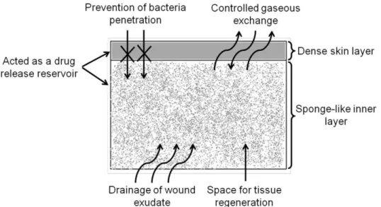
wound healing process. ... 11 Figure 1.4. Schematic representation of the different methods used to develop asymmetric
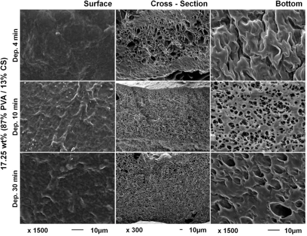
membranes. ... 14 Figure 1.5. Hypothetical ternary phase diagram for the system polymer/solvent/non-solvent. .. 19 Figure 2.1. Ideal characteristics of an asymmetrical membrane to be used as wound dressing 45 Figure 2.2. Scanning electron micrographs of 17.25 wt% (87% PVA / 13% CS) membranes. .. 46 Figure 2.3. Water uptake and contact angle analysis of PVA/CS membranes. ... 48 Figure 2.4. Evaluation of the structure stability of PVA/CS membranes at different pHs over 21
days ... 51 Figure 2.5. Young’s modulus analysis of PVA/CS membranes at dry and wet states and over 21
days at different pHs ... 52 Figure 2.6. Evaluation of the antimicrobial activity of native and PVA/CS membranes grafted
with OMetOx-DDA through disk diffusion tecnhique and rezasurin assay ... 54 Figure 2.7. Scanning electron micrographs of S. aureus in contact with native and PVA/CS
membranes grafted with OMetOx-DDA... 54 Figure 2.8. Evaluation of PVA/CS membranes solvent uptake ability and its influence on
ibuprofen release.. ... 55 Figure 2.9. Microscopic photographs of human fibroblast cells after being seeded in the
presence of the membranes during 24 and 72h ... 56 Figure 2.10. Cellular activities measured by the resazurin assay after 24 and 72h. ... 57 Figure 3.1. Scanning electron micrographs of PVA/CS membranes containing the different drug
delivery systems ... 71 Figure 3.2. Swelling behavior of PVA/CS membranes containing the different drug delivery
systems at different pHs. ... 72 Figure 3.3. Evaluation of the structural stability of PVA/CS membranes containing the different
drug delivery systems at different pHs, over 21 days ... 76 Figure 3.4. Young’s modulus analysis of PVA/CS membranes containing the different carriers in
dry and wet state. ... 77 Figure 3.5. In vitro drug release studies of IBP loaded into the several drug delivery systems
and IBP release profile fitted through Korsmeyer-Peppas mathematical model. ... 79 Figure 3.6. Evaluation of cellular activity in contact with the different dressings with and without
IBP through an MTS assay after 1, 3 and 7 days. ... 81 Figure 3.7. Characterization of the wound healing process under in vivo conditions by using the
different developed membranes.. ... 83 Figure 3.8. Magnified images of H&E-stained histological sections of the test groups after 10
Figure 3.9. Representative images of Masson’s Trichrome analysis of stained explants of the
control and PVA/CS+β-CD_IBP groups.. ... 85
Figure 4.1. Schematic representation of the effect of a sustained IBP release on skin wound regeneration taking into account the healing phases. ... 92
Figure S2.1. Scanning electron micrographs of 17.25 wt% (77% PVA/23% CS) membranes.115 Figure S2.2. Scanning electron micrographs of 13 wt% (67% PVA/33% CS) membranes. .... 116
Figure S2.3. Scanning electron micrographs of 9 wt% (50% PVA/50% CS) membranes ... 116
Figure S3.1. Maximal tensile strain (%) analysis of PVA/CS membranes with the different carriers at dry and wet state. ... 117
Figure S3.2. Tensile strength (MPa) analysis of PVA/CS membranes with the different carriers at dry and wet state. ... 117
Figure S3.3. Korsmeyer-Peppas Model for mechanism of IBP release from the different drug delivery systems ... 118
Figure S3.4. ATR-FTIR spectra of pure (S)-IBP and PF microbeads with/without IBP ... 118
Figure S3.5. ATR-FTIR spectra of pure (S)-IBP and PK microbeads with/without IBP ... 119
Figure S3.6. ATR-FTIR spectra of pure (S)-IBP and β-CD with/without IBP... 119
Figure S3.7. ATR-FTIR spectra of PVA/CS+PK, PVA/CS+PF, PVA/CS+β-CD and PVA/CS membranes. ... 120
Figure S3.8. ATR-FTIR spectra of PVA/CS+PK, PVA/CS+PF, PVA/CS+β-CD membranes loaded with IBP and PVA/CS membranes without IBP ... 120
Figure S3.9. Microscopic photographs of human fibroblast cells after being seeded in the presence of the developed membranes with the different carriers during 1, 3 and 7 days .. 121
Figure S3.10. Microscopic photographs of human fibroblast cells after being seeded in the presence of the developed membranes with the different carriers loaded with IBP during 1, 3 and 7 days. ... 121
Figure S3.11. Characterization of the wound healing process under in vivo conditions. ... 122
xvii
Index of Tables
Table 1.1. Tissue-engineered skin constructs commercially available. ... 9
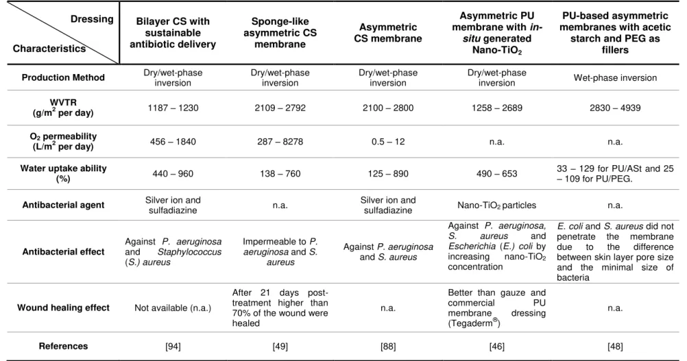
Table 1.2. Comparison between the different CS and PU asymmetric membranes developed in the last two decades... 12
Table 1.3. Summary of the production methods of asymmetric membranes: advantages and disadvantages. ... 23
Table 1.4. Summary of methods used during the development of wound dressings ... 29
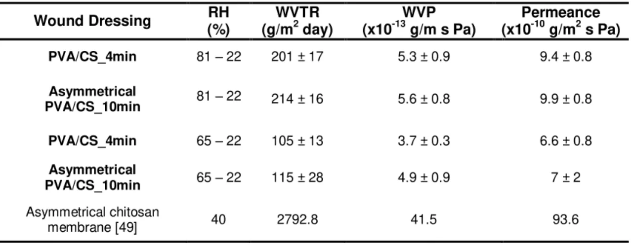
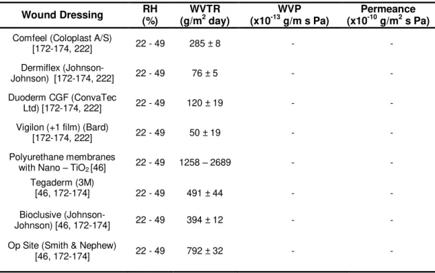
Table 2.1. Comparison of the water vapor transmission rate of different studied and commercially available wound dressings for burn treatment. ... 49
Table 2.2. Mechanical properties of various studied wound dressings. ... 52
Table 3.1. Average membranes’ pore diameter and porosity determined by mercury intrusion porosimetry. ... 73
Table 3.2. Water vapor permeation of the different developed membranes. ... 74
Table 3.3. Comparison of the oxygen permeability of the different developed membranes. ... 75
Table 3.4. Modeling of IBP release from carriers using the Korsmeyer-Peppas equation. ... 80
Table 4.1. Comparison between conventional and scCO2 – assisted phase inversion methods on asymmetrical membranes development ... 90
xix
Abbreviations
AgSD silver sulfadiazine AIBN 2,2-azo-isobutyronitrile
ATR-FTIR Attenuated Total Reflection-Fourier Transform Infrared spectroscopy
β-CDs β-cyclodextrins
BF3.OEt2 boron trifluoride diethyl etherate
CFCs chlorofluorcarbons CH3COOK potassium acetate
CO2 carbon dioxide
COX cyclooxygenase CS chitosan
DMEM-F12 Dulbecco’s modified Eagle’s medium DMF dimethylformamide
DMSO dimethylsulfoxide
E. coli Escherichiacoli
ECM extracellular matrix FBS fetal bovine serum GDMA glycerol dimethacrylate H&E hematoxylin & eosin HICs high-income countries IBP (S)-ibuprofen
IsoOx 2-isopropenyl-2-oxazoline K- negative control
K+ positive control
KGF keratinocyte growth factor LMICs low and middle-income countries MAFs micro-assembled fibers
MetOx 2-methyl-2-oxazoline MF micro-filtration miRNA microRNA
MSC mesenchymal stem cells
MTS 3-(4,5-dimethylthiazol-2-yl)-5-(3-carboxymethoxyphenyl)-2-(4-sulfophenyl)-2H- tetrazolium inner salt
NaOH-Na2CO3 sodium hydroxide-sodium carbonate
NF nano-filtration
NHDF normal human dermal fibroblasts adult (NH4)2SO4 ammonium sulfate
NSAID non-steroidal anti-inflammatory drug
O2 oxygen
P. aeruginosa Pseudomonas aeruginosa
PAN polyacrylonitrile
PBS phosphate buffered saline
pc critical pressure
PCL polycaprolactone pCO2 pressure of CO2
PEG polyethylene glycol PEO poly(ethylene oxide)
PF poly(1,3-glycerol dimethacrylate) microparticles with 30 wt% of fluorolink PFPE perfluoropolyether
PGDMA poly(1,3-glycerol dimethacrylate)
PK poly(1,3-glycerol dimethacrylate) microparticles with 10 wt% of krytox PLA poly(L-lactide)
PLLA poly(L-lactic acid)
PMO peptide-morpholino oligomer PS polystyrene
PU polyurethane PVA poly(vinyl alcohol) RH relative humidity
ROS reactive oxygen species
S. aureus Staphylococcus aureus
scCO2 supercritical carbon dioxide
SCF supercritical fluids
SEM scanning electron microscopy TBSA% total percent surface area injured
Tc critical temperature
THF tetrahydrofuran
TRIS tris(hydroxymethyl)aminomethane UF ultra-filtration
VOCs volatile organic solvents WHO World Health Organization WS wound size
C
HAPTER
1.
General Introduction
The major part of this chapter corresponds to the manuscript published
in:
2015, Journal of Membrane Science, 490:139-151.
Patrícia I. Morgado, Ana Aguiar-Ricardo and Ilídio J. Correia.
Asymmetric membranes as ideal wound dressings: an overview on production methods, structure, properties and performance relationship.
(http://www.sciencedirect.com/science/article/pii/S0376738815003956)
Reproduced with the authorization of the editor and subjected to the copyrights imposed.
Personal contribution:
C
HA
P
T
E
R
1
3
1. General Introduction
1.1.
Motivation
Skin is the biggest organ in vertebrates and occupies an area of about 2 m2, representing approximately one-tenth of the body mass [1, 2]. It has a complex three-layered structure (epidermis, dermis and hypodermis), which under normal physiological conditions, is intrinsically self-renewable [3]. This complex organ is the outermost barrier of the body that protects inner organs from microbial pathogens, mechanical and chemical insults, regulates the body temperature, gives support to blood vessels and nerves, and prevents dehydration [4]. Furthermore, it is also involved in the immune surveillance and sensory detection processes [5, 6].
As the largest external organ in the body, human skin is daily exposed to several toxic substances and pathogens, which makes it an easy target to be damaged. Indeed, skin wounds are a major social and financial burden and their healing process is extremely complex consisting of a cascade of biological responses with different cell types and growth factors involved. The loss of skin integrity can occur due to genetic disorders, acute trauma, chronic wounds (e.g. venous, diabetic and pressure ulcers) or even surgical interventions [7]. However,
thermal traumas like burns are one of the most common causes of major skin loss where different skin layers can be damaged and depending on the type of burn, the possibility of skin regeneration is unlikely [8]. Additionally, they can result in extensive and deep wounds that compromise immunity and body image, induce fluid losses and scarring, and ultimately significant disability or even patient death, causing not only physical but also emotional and mental consequences [5, 9].
Based on the World Health Organization (WHO) data over 300 000 deaths are annually attributable to fire-related burn injuries, with high incidence in low and middle-income countries (LMICs), where resources to treat and manage injuries are scarce or unavailable [10-12]. Fortunately, the mortality rate due to burns has been reduced over recent decades especially in high-income countries (HICs) where improved data gathering systems, new and more stringent legislation (e.g. use of smoke detectors, installation of sprinkle systems, development of safer
Despite there are several commercial available wound dressings, scientists continue to search for new or improve the currently available systems. Herein, our focus was to develop hydrogel-based asymmetrical dressings and drug delivery systems with highly controlled morphology toward the ideal wound dressing and able to mimic the native structure of skin, by taking advantage of the unique properties of supercritical fluid (SCF) technologies. We believe that our research could contribute for the continued reduction of mortality rate associated with burns. Furthermore, due to the low economic status of LMICs, the use of sustainable, low-cost and greener methods on the development of well-designed drug-loaded dressings could be extremely helpful for patients with low economic resources.
1.2.
Burn wound evaluation
According to the US Wound Healing Society, a wound can be described as a result of a
“disruption of normal anatomic structure and function” of the skin [16, 17]. The evaluation of skin
burned wounds is mainly done taking into account two aspects: the depth and the total percent surface area injured (TBSA %). Along with the extent of burn and patient’s age, burn depth is a
primary determinant of mortality following thermal injury. Burn depth is also the primary
determinant of the patient’s long term appearance and function [18, 19].
C
HA
P
T
E
R
1
5
Figure 1.1. Schematic representation of skin structure in relation to burn wound depth terminology (adapted from [20, 23, 24]).1.3.
Wound healing process
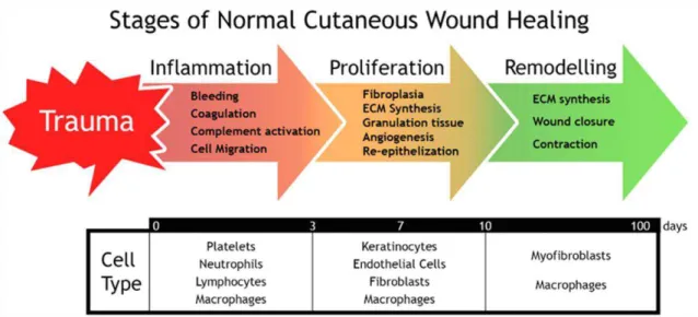
The repair of epidermal, superficial partial-thickness, deep partial-thickness and full-thickness wounds are one of the most dynamic, interactive and difficult processes that occur during human life [16, 25, 26]. It involves complex interactions between extracellular matrix (ECM) molecules, soluble mediators, various resident cells (fibroblasts and keratinocytes) and infiltrating leukocyte subtypes. They act together to reestablish the integrity of the damaged tissue and replace the lost one [16, 27, 28]. To achieve this goal, the wound healing comprises five overlapping stages: hemostasis, inflammation, migration, proliferation and maturation [16]. These can be summarized in three main phases, i.e., hemostasis and inflammation (typically
2-3 weeks after injury happened and lasts for a year or more [25, 28, 2-30]. Despite the importance of all the wound healing stages, the inflammatory phase is the most important one, since in burn wounds there is an expansion of the initial necrosis deeper into the tissue. After the initial injury, that leads to a progressive delay of wound re-epithelization and excessive formation of exudate, responsible for edema, it is imperative to use burn dressings to avoid progressive necrosis and exuberant inflammation [35]. Additionally, early wound closure decreases the severity of hypertrophic scarring, joint contractures and stiffness, and promotes quicker rehabilitation. Consequently, early healing is also a paramount for good aesthetic and functional recovery. For a suitable wound closure, the dressing used should present healing-based properties able to promote the restoration of the integrity of the damaged tissue as soon as possible [36].
Figure 1.2. Schematic representation of the three main stages of skin wound healing process: inflammation, proliferation and remodeling.
1.4.
Tissue-engineered skin constructs
Extensive skin loss is still a significant challenge to clinicians. Nowadays, the clinical “gold
standard” in full-thickness injuries treatment is autologous skin graft. However, healthy tissue
C
HA
P
T
E
R
1
7
wound healing. Furthermore, wound dressings can be used as temporary coverings, until an autograft is available, or remain in the wound during healing or even thereafter [37, 39-41].Depending on wound severity, epidermal, dermal and epidermal/dermal substitutes are available to clinicians (please see Table 1.1 for further details). Epidermal substitutes (e.g.
CellSpray®, Epicel®, Myskin®) seek to restore the epidermal layer of skin. In general, the available epidermal substitutes are expensive, difficult to handle due to their thin and fragile nature, are unable to treat third degree burn wounds and their production is time consuming [39]. Dermal substitutes (e.g. Alloderm®, Dermagraft®, Matriderm®) are biomatrices that fulfill all
the requirements of the dermal layer, being able to repair full-thickness skin defects, affecting both epidermis and dermis, and improve scar quality. These substitutes are also capable of preventing wound contraction, conferring mechanical support, and are available with different thickness and compositions. However, they cannot efficiently replace the dermal and epidermal layer and for some of them, further research is warranted to fully characterize side effects. Epidermal/dermal substitutes (e.g., Apligraf®, Integra®, OrCel®) are the most advanced skin
constructs available for clinic use. They contain keratinocytes and fibroblasts within their 3D matrix, gathering the potential to regenerate both the epidermal and dermal layers of skin. However, they have high production costs and some cases of immune rejection have been reported for this type of skin substitutes [41, 42].
Nowadays, advanced skin regeneration strategies combine biomaterials, cells, growth factors and modern biomanufacturing techniques to produce constructs that mimic skin anatomy and promote the regeneration of healthy and vascularized tissues. Despite the great advances attained in the area of skin tissue engineering, even the cutting-edge skin substitutes developed so far, do not incorporate many of the innate features of native skin, such as glands, dermal microvascularization, pilosity and other specialized cells that are responsible for the perception of heat, cold, pressure, vibration, pain and pigmentation. Moreover, the elasticity and strength of the native skin have not been attained until now [30, 43]. Recent studies in skin tissue engineering combined stem cells with gene recombination [44]. Due to their intrinsic characteristics, genetically modified stem cells can be used to produce and deliver cytokines and growth factors to the wound bed overcoming drawbacks such as physical inhibition and biological degradation of bioactive molecules that occur when they are administered topically.
In addition, other 3D matrices aimed at wound healing were also used as vehicles for nucleic acids. Kobsa et al., produced electrospun polymeric meshes, composed by poly(L-lactide)
(PLA) and polycaprolactone (PCL), that were loaded with plasmids encoding for keratinocyte growth factor (KGF). Such types of meshes allowed an improvement in the wound re-epithelization, keratinocyte proliferation and production of granulation tissue [45].
skin and suitable properties for a better wound healing process as it will be described in the next section. Regarding the available skin substitutes presented on Table 1.1, just the epidermal/dermal substitutes present an asymmetric geometry. The epidermal and dermal layers correspond to the dense skin and sponge-like inner layers of the asymmetric membranes, respectively. Usually the porous matrix of the epidermal/dermal substitutes is composed by collagen, hyaluronic acid, fibronectin or other ECM proteins. A bandage made of silicone is normally used to form the thin upper layer to protect the wound from moisture loss and infection. However, it is possible to produce epidermal/dermal substitutes with an integral structure, i.e., without the need to use a bandage to perform the dense skin layer. To improve
C
HA P T E R1
9
Table 1.1. Tissue-engineered skin constructs commercially available.Commercial Product Description Application References
Epidermal substitutes
Epicel® Cultured epidermal autograft (autologous keratinocytes grown in the presence of murine fibroblasts)
Full- and partial-thickness burns and
chronic ulcers treatment [50]
Epidex® Cultured epidermal autograft (autologous
outer root sheet hair follicle cells) Full- and partial-thickness burns and chronic ulcers treatment [51]
Laserskin® Sub-confluent autologous keratinocytes seeded on esterified laser-perforated hyaluronic acid matrix
Full- and partial-thickness burns and
chronic ulcers treatment [52]
BioSeed-S® Autologous oral mucosal cells on a fibrin
matrix Partial-thickness burns and chronic ulcers treatment [53, 54]
Myskin® Cultured epidermal autograft (autologous keratinocytes grown in the presence of irradiated murine fibroblasts)
Partial-thickness burns and chronic
ulcers treatment [55]
CellSpray® Pre-confluent autologous keratinocytes
delivered into a suspension for spray Partial-thickness burns and chronic ulcers treatment [56, 57]
Transcyte® Human fibroblast derived skin substitute composed by a nylon mesh coated with porcine dermal collagen and bonded to a silicone membrane
Full- and partial-thickness burns
[58]
Dermal substitutes
Dermagraft® Bioabsorbable polyglactin mesh scaffold seeded with human allogeneic nenonatal fibroblasts
Full-thickness diabetic foot ulcers
treatment [59]
Alloderm® Acellular allograft human dermis Full- and partial-thickness wounds
treatment [60]
EZ-Derm® Aldehyde-crosslinked porcine dermal
collagen Full- and partial-thickness wounds treatment [61, 62]
Cymetra® Micronized particulate acellular cadaveric
dermal matrix Wound filler in plastic surgery [63, 64]
Biobrane® Porcine collagen chemically bound to
silicone/nylon membrane Temporary thickness burns and wounds covering of partial- [65]
Hyalograft 3D® Esterified hyaluronic acid matrix seeded
with autologous fibroblasts Full- and partial-thickness wounds treatment [66]
Matriderm® Bovine dermal collagen type I, III, V and
elastin Full- or partial-thickness wounds treatment [67]
Epidermal/dermal substitutes
Integra® Thin silicone layer; cross-linked bovine tendon collagen type I and shark glycosaminoglycan (chondroitin-6-sulfate)
Full- or partial-thickness wounds
treatment [68]
OrCel® Human allogeneic neonatal keratinocytes on gel-coated non-porous side of sponge; bovine collagen sponge containing human allogeneic neonatal fibroblasts
Treat skin graft donor sites and
mitten-hand surgery for epidermolysis bullosa [69]
Apligraf® Human allogeneic neonatal keratinocytes; bovine collagen type I containing human allogeneic neonatal fibroblasts
Venous and diabetic foot ulcers
treatment [70]
TissueTech® Combination of Hyalograft 3D® and
1.5.
Asymmetric membranes as ideal wound dressings
The first asymmetric membrane was produced with cellulose acetate using the phase inversion method, in the late 1950s by Loeb and Sourirajan, and was used in reverse osmosis [72, 73]. Since then, asymmetric membranes found applications in almost every industrial field, namely: micro/nano/ultra-filtration (MF, NF, UF), dialysis, gas separation, per-evaporation, waste water treatment, and more recently as wound dressings for skin injuries treatment [74-87].
Researchers involved in regenerative medicine started by developing occlusive wound dressings like Opsite®, Omiderm® or Spandre®, which were impermeable and, consequently, did
not allow exudate absorption, resulting in a delayed healing process. Subsequently, macroporous constructs (e.g. Coldex® and Surfasoft®) appeared and allowed an effective
drainage of wound exudate. However, these dressings were unable to avoid penetration of microorganisms and wound dehydration. Later, researchers came to the conclusion that the combination of both systems (occlusive and macroporous structures) would be the ideal as it could prevent the bacteria penetration, and at the same time, allow the exudate absorption and gaseous exchange. Thus, dressings consisting of a macroporous sub-layer or a hydrogel linked to a dense or hydrophobic microporous top layer were developed. Lyofoam®, Epigard®, and
Duoderm® belong to such type of dressings. Nevertheless, they also presented some drawbacks: limited drainage capacity, exudate accumulation, and the need for frequent substitution which leads to an increased risk of wound infection. To overcome these handicaps, around 1990s, Hinrichs et al., based on the work developed by Loeb and Sourirajan, designed
for the first time an asymmetric membrane made of polyurethane (PU) [47]. The asymmetric PU membrane presented an interconnected microporous top layer (pore size < 0.7 µm), able to prevent rapid dehydration of the wound surface and bacterial penetration, as demonstrated through in vitro bacteriologic test using Pseudomonas (P.) aeruginosa. Furthermore, the
C
HA
P
T
E
R
1
11
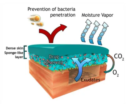
Figure 1.3. Schematic representation of the possible roles of an asymmetric membrane in the wound healing process.As previously stated, the first asymmetric membrane was made of PU, which is a fully synthetic biodegradable material with uniform hard segments composed of butanediol and 1,4-butanediisocyanate and soft segments of DL-lactide, ε-caprolactone and polyethylene glycol
(PEG). It has been used in wound healing due to its biocompatibility, mechanical (e.g. flexibility)
and hemostatic properties. The hydrophilic character of this material is responsible for attracting platelets and subsequently trigger the coagulation cascade [89]. In addition, asymmetrical membranes made of chitosan (CS) have also been produced. CS, a natural polymer that is obtained from the deacetylation of chitin, has been extensively used for wound dressing production, due to its intrinsic properties, e.g., antimicrobial activity, biocompatibility,
Dressing
Characteristics
Bilayer CS with sustainable antibiotic delivery Sponge-like asymmetric CS membrane Asymmetric CS membrane Asymmetric PU membrane with
in-situ generated Nano-TiO2
PU-based asymmetric membranes with acetic
starch and PEG as fillers
Production Method Dry/wet-phase inversion Dry/wet-phase inversion Dry/wet-phase inversion Dry/wet-phase inversion Wet-phase inversion
WVTR
(g/m2 per day) 1187 – 1230 2109 – 2792 2100 – 2800 1258 – 2689 2830 – 4939
O2 permeability
(L/m2 per day) 456 – 1840 287 – 8278 0.5 – 12 n.a. n.a.
Water uptake ability
(%) 440 – 960 138 – 760 125 – 890 490 – 653
33 – 129 for PU/ASt and 25
– 109 for PU/PEG.
Antibacterial agent Silver ion and sulfadiazine n.a. Silver ion and sulfadiazine Nano-TiO2 particles n.a.
Antibacterial effect
Against P. aeruginosa
and Staphylococcus
(S.) aureus
Impermeable to P.
aeruginosa and S.
aureus
Against P. aeruginosa
and S. aureus
Against P. aeruginosa,
S. aureus and
Escherichia (E.) coli by
increasing nano-TiO2
concentration
E. coli and S. aureus did not
penetrate the membrane due to the difference between skin layer pore size and the minimal size of bacteria
Wound healing effect Not available (n.a.)
After 21 days post-treatment higher than 70% of the wound were healed
n.a.
Better than gauze and
commercial PU
membrane dressing (Tegaderm®)
n.a.
References [94] [49] [88] [46] [48]
C
HA
P
T
E
R
1
13
Although PU and CS asymmetric membranes present several properties (antimicrobial activity, biocompatibility, hemostatic properties, gas and water permeation) that satisfy the common requirements of an ideal wound dressing, only 8 articles have been published in the last twenty years [74, 76, 78-82, 88]. This can be explained by the disadvantages of the most common methods used (wet- and dry/wet - phase inversion techniques) to prepare asymmetric membranes. These relatively simple methods usually require the use of toxic solvents which can only be removed by additional purification steps during the production process. In addition, the number of polymers used on skin wound regeneration that can be processed through the wet- and dry/wet - phase inversion techniques is limited by their solubility on the solvents used –always sodium hydroxide-sodium carbonate (NaOH-Na2CO3). This may be the reason why up
to 1991, only CS and PU asymmetric membranes were reported for wound healing, as shown on Table 1.2. More recently, other methods such as supercritical carbon dioxide (scCO2
)-induced phase inversion technique and electrospinning started being used for the production of asymmetric membranes proving to be feasible alternatives to the conventional methods used hitherto as it will be discussed on the next sections of this Chapter and thesis. In the first technique, it is important that scCO2 do not dissolve the polymers used but remove the solvents
where the polymers were dissolved while in the electrospinning technique the solvent used is the more suitable to dissolve the polymers. Thus, CS and PU polymers can also be processed with these two techniques being the most used solvents acidified solutions and organic solvents as dimethylformamide (DMF) and tetrahydrofuran (THF), respectively [95-100]. Furthermore, with these new methods other healing-based polymers for the production of asymmetric membranes have arisen as poly(vinyl alcohol) (PVA), polyacrylonitrile (PAN), polyethylene oxide (PEO), and polystyrene (PS) [101-103].
1.6.
Production methods of asymmetric membranes
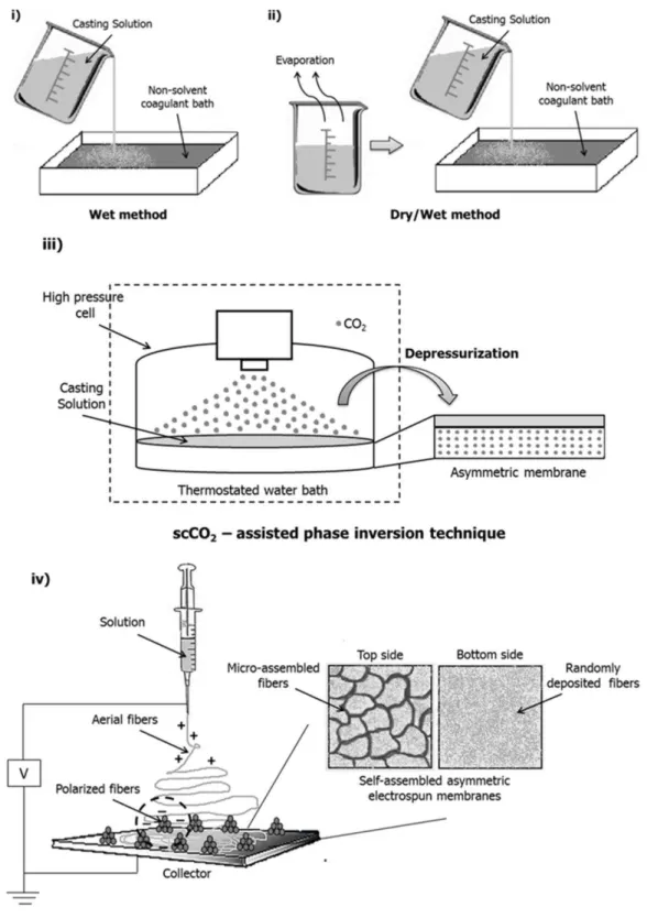
The phase separation process required to yield an asymmetric membrane can be induced by different techniques as shown in Figure 1.4: i) by directly immersing a polymer solution into a non-solvent bath, the wet method or immersion precipitation; ii) by evaporating a polymer solution for a time period and then immersing it in a non-solvent, the dry/wet method; iii) more recently, by using scCO2 to induce the phase separation, the scCO2-induced phase inversion
C
HA
P
T
E
R
1
15
1.6.1. Wet-phase inversion method
The wet-phase inversion method was the first technique developed to produce asymmetric membranes. This simple method involves the immersion of a casting polymer solution in a non-solvent coagulant bath, to promote membrane precipitation. The formation of a compact top cover with a porous sub-layer can be obtained through the delay of phase separation that takes place at the outmost interface region of the casting solution. When the membranes produced by wet-phase inversion, are to be used as wound dressings they present some limitations: the top
layer formed is quite thin (less than 1 μm) and may present defects which limit the capacity to
prevent excessive water vapor evaporation from the wound bed and compromise its barrier function against outside contaminants [46]. Lee et al [48] reported the use of wet-phase
inversion method to produce PU-based asymmetric membranes which were tested as wound dressings, as shown in Table 1.2. However, to obtain a more compact and dense top layer, the authors have combined polymer-filler hybridization, by using acetic starch and PEG, with the immersion precipitation phase inversion. The water vapor transmission rate (WVTR) and the water uptake ability are directly proportional to the filler content.
1.6.2. Dry/wet - phase inversion method
The dry/wet-phase inversion method was adopted to overcome the defects observed on the top layer of the membranes produced by the wet-phase inversion method. To do so, researchers started to per-evaporate the casting solution before its immersion into the coagulation bath. With this per-evaporation process, the non-solvent diffuses to the outermost region of the polymeric casting solution allowing the formation of an integral and dense top layer able to protect the wound against outside contamination. However, this method requires at least one volatile solvent for the membrane forming polymer. Usually, the casting solution is heated in an oven for dry phase separation at 50 ºC for 10-60 min. After that, the underlying polymer solution with formed skin layer is immersed into a coagulation bath for 24 h (usually containing NaOH-Na2CO3), inducing the wet-phase inversion by the diffusion of non-solvent into and solvent out
of the polymer solution. Afterwards, the solidified membrane is soaked in distilled water and finally frieze-dried [46, 88, 94, 104].
The dry/wet–phase inversion technique has been the most used method to produce asymmetric membranes composed of CS or PU. In some studies, the incorporation of antibacterial agents as silver sulfadiazine [88, 94] and nano-TiO2 particles [46], was reported (please refer to Table
sub-layer with a sponge-like structure also decreases with longer evaporation periods, since the polymer solution becomes more concentrated [66]. The type of non-solvent used on the coagulation bath also influences the asymmetry of the membranes. It is known that a polar non-solvent with low molar mass (e.g. methanol) tends to give a high overall porosity, and reduce
the membrane thickness, while a less polar non-solvent (e.g. butanol) allows the formation of a
very thick film and a more open porous sub-layer. This is due to the higher miscibility of polar non-solvents which promote an instantaneous demixing upon immersion of the casting solution in the coagulation bath. Furthermore, higher polymer concentrations reduce the porosity and finally induce the formation of a dense structure [105-107]. The possibility to form membranes with suitable porosity and thickness using this method allow for a faster and less painful wound healing process. Nevertheless, the dry/wet method often requires the use of additional post-treatments to remove any cytotoxic residues that might remain in the membranes.
1.6.3. Supercritical fluids and supercritical CO2 – assisted phase inversion
technique
After its discovery in 1922 by Hermann Staudinger, polymers have become part of our lives [108]. In view of their importance in numerous applications, increasing attention is being given, not only to the synthesis, but also to polymer processing. As stated before, the traditional methods normally use environmentally hazardous volatile organic solvents (VOCs) and chlorofluorcarbons (CFCs). Due to the huge increment of VOCs and CFCs emissions and generation of aqueous waste streams, there is a growing interest in the development of alternative technological processes with minimized environmental impact, such as reduction energy consumption, less toxic residues, better use of by-products and also better quality and safety of final products. SCF technology is one of such methods used as processing solvents or plasticizers [109, 110].
A fluid is termed supercritical when it is above the critical temperature (Tc) and pressure (pc) but
below the pressure needed for condensation [111]. In general terms, SCF show liquid-like densities with gas-like transport properties and solvent power for several applications that can be continuously adjusted by changes in pressure and temperature [112].
Several substances can be used as SCF as water, ethane, ethylene, propane, carbon dioxide (CO2), methanol and acetone [113]. However, in SCF technology, CO2 is the most common
used solvent for a variety of chemical and industrial processes. It is a clean and versatile solvent and a promising alternative to VOCs and CFCs. It is non-toxic, non-flammable, chemically inert and inexpensive. Though it is abundant in the atmosphere, a large amount is also available in high-purity as a by-product from many processes including the fermentation of biomass (NH3, H2 and ethanol production). Its supercritical conditions are easily achieved (Tc =
31.3 ºC and pc = 7.38 MPa) and it can be removed from a system by simple depressurization. In
addition, the use of scCO2 does not create a problem with respect to the greenhouse effect as it
C
HA P T E R1
17
scCO2 have found applications in a broad range of areas [117, 118] being the most popular thedecaffeination of tea [119] and coffee [120] and the extraction of flavours [121], spices [122], and essentials oils [123, 124] from plants. More recently, this unique solvent has found considerable commercial interest in applications as diverse as dry cleaning [125], metal degreasing [126], polymer modification and polymerization [127-129] and pharmaceutical processing including drug impregnation [130-132].
Nowadays, scCO2-assisted processes have been proposed in tissue engineering field
overcoming several limitations of the conventional biomaterials fabrication techniques (fiber bonding, solvent casting, particulate leaching, melt molding, gas foaming, freeze-drying, wet- and dry/wet-phase inversion) [133, 134]. Through the conventional techniques it is difficult to achieve: i) large porosity together with a control of the pore diameter, connectivity and mechanical resistance; ii) large porosity together with the nanometric surface that enhances cells adhesion and growth; iii) complex 3D symmetric or asymmetrical structures; iv) the removal of toxic solvents that are retained deep inside the structure [135]. By using SCF technologies as scCO2-assisted phase inversion technique, supercritical gel processing and
scCO2-assisted electrospinning, biomaterials with the aimed morphology can be obtained
thanks to the modulability of CO2 mass transfer properties, characteristic of dense gases and
the specific thermodynamic behavior of gas mixtures at high pressure. Additionally, an efficient solvent elimination can be obtained due to the high affinity of scCO2 with almost all the organic
solvents. Furthermore, short processing times are possible, taking advantage of the enhanced mass transfer rates [135].
When scCO2-induces the demixing of a polymeric solution forming a porous membrane the
method is often denominated as scCO2-assisted phase inversion method. Herein, CO2 acts as a
solvent, extracting the solvents used for preparing the casting solution, and as a non-solvent, for the polymers. The scCO2 solubilizes in the casting solution promoting the removal of solvent
from the polymer solution and, subsequently, the membrane formation through the polymer precipitation, as it is observed in Figure 1.4 (iii). The properties of the membranes can be modified by changing the concentration of the casting solution, the ratio of non-solvent/solvent, the pressure, the temperature and the depressurization rate. The relative affinity of a polymer and solvent can be assessed by comparing the solubility parameters [136]. It allows the production of porous matrices by the induction of nucleation and growth of bubbles inside the polymer stimulating the porous growth. Furthermore, it can allow the production of materials with high degree of swelling and transparent membranes in a wet state, which is useful to be used as wound dressings. It overcomes the several disadvantages of chemically crosslinking gels since it does not need the use of organic solvents that are difficult to be removed [137-139].
For instance, Temtem and co-workers [99] have explored this technique for the production of CS devices using moderate temperatures and three environmentally acceptable solvents (water, ethanol and CO2). They also have demonstrated that the scCO2-induced phase
inversion technique allowed a single-step strategy in the preparation of an implantable antibiotic system by co-dissolving gentamicin with CS and the solvent. These membranes were also biocompatible allowing the adhesion and proliferation of human mesenchymal stem cells
(MSC). The obtained results provided a starting point for the “green” design and production of
CS-based materials with potential applications in tissue engineering and regenerative medicine, as well as drug delivery. In a similar study [143] it was investigated the feasibility on the fabrication of porous crosslinked CS hydrogels in an aqueous phase using dense gas CO2 as
foaming agent. Glutaraldehyde and genipin were used as crosslinkers and CS hydrogels were obtained with a highly porous biocompatible skeleton with an average pore diameter of 30-40 µm. Furthermore, the produced hydrogels exhibited a comparable mechanical strength and swelling ratio compatible with its application for soft tissue (skin and cartilage) engineering regeneration.
The preparation of membranes using supercritical-fluid-assisted methods, implies the use of specialized and robust high-pressure apparatus as the one designed by Temtem et al. (2008)
[144]. The heart of the apparatus is a stainless steel high-pressure cell which can stand pressures up to 60 MPa. The cell includes a porous structure that supports a bed of Rasching rings inside to allow the homogeneous dispersion of CO2 through the casting solution as it is
schematically represented in Figure 1.4 (iii).
Taking advantage of the additional parameters that the scCO2-assisted phase inversion method
offers for controlling the morphological properties of the polymeric membrane, asymmetrical PVA/CS membranes were recently developed and their performance as skin wound dressing was evaluated, one the of the main achievements attained in this thesis project and well discussed on the following Chapters [103]. The concentration of the casting solution and the depressurization rate (fast or slow) were the key factors to obtain the desired membrane structure. Nevertheless, when all the parameters described before are well established, an asymmetrical membrane is easily and directly formed in just 4 h instead of the 24 h needed when using conventional methods. To the best of our knowledge it was the first time that asymmetric membranes for skin wound regeneration were developed using supercritical fluids. The technique can be also extended for the production of other polymeric membranes using different polymers and incorporating different bioactive agents on the top and bottom surfaces.
The events that lead to the membranes’ formation, i.e. thermodynamic interactions between
C
HA
P
T
E
R
1
19
represents each pure component (the polymer – PVA/CS, the solvent – acidified water, the non-solvent – scCO2 + ethanol), the axes represent the three pseudo-binary systems and any pointlocated inside the diagram represents a mixture of the three components. In the liquid + fluid region a primary (binodal curve) and a secondary envelope (spinodal curve) enclose demixing boundary. Both curves coincide at the critical point and the region between them corresponds to a metastable state. Two different mechanisms have to be considered as far as liquid-liquid (L-L) demixing is concerned: nucleation/growth and spinodal decomposition. The structures obtained and pore dimension are dependent on the path followed through the ternary diagram. The arrows numbered from 1 to 4, in Figure 1.5, report the different composition paths that can occur during membranes formation [99, 147]:
1 Increasing of polymer concentration since the outflow of the solvent from the solution is faster than the inflow of the non-solvent. The phase inversion does not occur and the polymer solidifies by gelation into a dense structure.
2 The ternary polymer solution becomes metastable. Cellular structure by nucleation and growth of droplets of the polymer-rich phase.
3 The ternary polymer solution becomes unstable. Bicontinuous structure due to spinodal phase separation.
4 Beads-like structure due to nucleation and growth of droplets of the polymer-poor phase.
Considering the formation of asymmetric membranes through scCO2-assisted phase inversion
technique the simultaneous presence of a dense top layer and porous (cellular) inner layer indicates that the competition between two different mass transport mechanisms occurs during membrane formation process. A hypothetical mechanism that may describe the asymmetry formation in the PVA/CS membranes is the following one: in a first step, the solvent (acidified water) outflows from the solution before the scCO2 can cause the phase separation leading to
polymer accumulation and the formation of the dense skin layer. Subsequently, the solvent outflow is stopped by the dense layer formation, and scCO2 with ethanol (co-solvent) diffuses
inside the remaining solution extraction the solvent and causing the L-L phase separation above the critical point leading to the cellular structure in the inner layer (path 2 schematically represented in Figure 1.5) [99, 147].
However, and as stated previously, the asymmetry and the inner morphology of the membranes can be easily changed taking into account the several process parameters. Matsuyama et al.
[148, 149] used scCO2-assisted phase inversion process to produce and analyze the effect of
several process parameters (temperature, pressure and polymer concentration) on the asymmetry of PS membranes [148] and the influence of a kind of solvents used in the formation of asymmetrical cellulose acetate membranes [149]. They observed that as the pressure increased from 8 to 16 MPa (maintaining the temperature (35 ºC) and casting solution concentration (20 wt%)), both the pore size (in the upper and bottom layers of the membrane) and the membrane thickness increased. The increase of CO2 incorporation leads to the
decrease of the solution viscosity due to polymer concentration decrease, resulting in higher membrane porosity. As the polymer concentration increased, the overall membrane thickness increased because of the higher amount of polymer. Finally, as the temperature increased, the asymmetry becomes more pronounced. As general, the pores are more isolated at the top surface because the polymer concentration becomes higher due to the evaporation of solvent before CO2 introduction and the dissolution of the solvent in the scCO2 phase. More recently,
Reverchon et al. [147] have performed a similar study by testing the versatility of the scCO2
-assisted phase inversion process on the formation of PVA/dimethylsulfoxide (DMSO) asymmetrical membranes. Polymer concentration, temperature, pressure and the affinity between scCO2 and solvent continued to be the key parameters to obtain an asymmetrical
structure. Regarding the affinity between scCO2 and solvent, they observed that when acetone
(with higher affinity to scCO2 than DMSO) is added to the procedure, the skin layer thickness
decreased since the phase-separation process is faster, limiting the outflow of the solvent. In addition to the processing conditions previously described, in our work it was found that the depressurization rate is also a key parameter to obtain the PVA/CS membranes with the desired structure, in addition to casting solution concentration [103]. As already observed by Temtem et al. [136] higher rates of depressurization produce larger pores, i.e. in a slow
depressurization the CO2 diffuses slowly out of the polymer phase (not affecting the polymer
structure), while for a fast depressurization a large amount of CO2, dissolved in the polymer,
![Figure 1.1. Schematic representation of skin structure in relation to burn wound depth terminology (adapted from [20, 23, 24] ).](https://thumb-eu.123doks.com/thumbv2/123dok_br/16479633.732353/25.892.172.696.91.408/figure-schematic-representation-structure-relation-wound-terminology-adapted.webp)
