w w w . r b h h . o r g
Hematology, Transfusion and Cell Therapy
Original article
Novel mutations associated with pyruvate kinase
deficiency in Brazil
Maria Carolina Costa Melo Svidnicki
a, Andrey Santos
b,
Jhonathan Angel Araujo Fernandez
c, Ana Paula Hitomi Yokoyama
a,
Isis Quezado Magalhães
d, Vitoria Regia Pereira Pinheiro
e, Silvia Regina Brandalise
f,
Paulo Augusto Achucarro Silveira
g, Fernando Ferreira Costa
a,b,
Sara Teresinha Olalla Saad
a,b,∗aCentro de Hematologia e Hemoterapia da Universidade Estadual de Campinas (HEMOCENTRO/UNICAMP), Campinas, SP, Brazil bDepartamento de Medicina Interna da Universidade Estadual de Campinas (UNICAMP), Campinas, SP, Brazil
cCentro de Biologia Molecular e Engenharia Genética da Universidade Estadual de Campinas (CBMEG/UNICAMP), Campinas, SP, Brazil dHospital da Crianc¸a de Brasília (HCB), Brasília, DF, Brazil
eCentro Integrado de Pesquisas Onco-Hematológicas na Infância da Universidade Estadual de Campinas (CIPOI/UNICAMP), Campinas, SP, Brazil
fCentro Infantil Bondrini, Campinas, SP, Brazil
gHospital das Clínicas da Faculdade de Medicina da Universidade de São Paulo (HCFMUSP), São Paulo, SP, Brazil
a r t i c l e
i n f o
Article history:
Received 15 February 2017 Accepted 2 August 2017
Available online 26 November 2017
Keywords:
Red cell disorder Pyruvate kinase Mutation
Hemolytic anemia
PKLRgene
a b s t r a c t
Background:Pyruvate kinase deficiency is a hereditary disease that affects the glycolytic pathway of the red blood cell, causing nonspherocytic hemolytic anemia. The disease is transmitted as an autosomal recessive trait and shows a marked variability in clinical expression. This study reports on the molecular characterization of ten Brazilian pyruvate kinase-deficient patients and the genotype–phenotype correlations.
Method:Sanger sequencing andin silicoanalysis were carried out to identify and characterize the genetic mutations. A non-affected group of Brazilian individuals were also screened for the most commonly reported variants (c.1456C>T and c.1529G>A).
Results:Ten different variants were identified in thePKLRgene, of which three are reported here for the first time: p.Leu61Gln, p.Ala137Val and p.Ala428Thr. All the three missense variants involve conserved amino acids, providing a rationale for the observed enzyme deficiency. The allelic frequency of c.1456C>T was 0.1% and the 1529G>A variant was not found.
Conclusion: This is the first comprehensive report on molecular characterization of pyruvate kinase deficiency from South America. The results allowed us to correlate the severity of the clinical phenotype with the identified variants.
© 2017 Associac¸ ˜ao Brasileira de Hematologia, Hemoterapia e Terapia Celular. Published by Elsevier Editora Ltda. This is an open access article under the CC BY-NC-ND license (http://creativecommons.org/licenses/by-nc-nd/4.0/).
∗ Corresponding author at: Centro de Hematologia e Hemoterapia da Universidade Estadual de Campinas (HEMOCENTRO/UNICAMP), Rua
Carlos Chagas, 480, 13083-878 Campinas, SP, Brazil. E-mail address:sara@unicamp.br(S.T. Saad).
https://doi.org/10.1016/j.bjhh.2017.08.007
Introduction
Pyruvate kinase (PK) deficiency is the most common enzy-matic defect of the glycolytic pathway, causing hereditary nonspherocytic hemolytic anemia. The prevalence of PK defi-ciency has been estimated to be 1:20 000 in the general white population.1 The disease is caused by mutations in thePKLRgene, which are transmitted as an autosomal reces-sive trait with affected individuals being either homozygotes or compound heterozygotes. The most commonly reported mutations are missense variants, including c.1529G>A in the United States and Northern/Central Europe, c.1456C>T in Southern Europe, and c.1468C>T in Asia.2–7
The severity of anemia is quite variable, ranging from mild or fully compensated forms to life-threatening neonatal anemia that requires continuous transfusions. Other clini-cal features include jaundice, splenomegaly and gallstones in some patients.8
This study reports on the molecular analysis and the clin-ical description of PK deficient patients of Brazilian origin.
Methods
Patients
This study involved ten unrelated patients with PK deficiency originating from Southern Brazil. Patients were either diag-nosed at the study center or diagdiag-nosed elsewhere and referred to this center for confirmation of the diagnosis and/or to estab-lish the molecular basis of their PK deficiency. Other causes of hemolytic anemia were ruled out in all patients. Appropri-ate informed consent was obtained either directly, when the patients were over 12 years of age, or from their parents or guardians. The diagnosis was based on clinical history, hema-tological data, and demonstration of reduced PK activity in red blood cells.
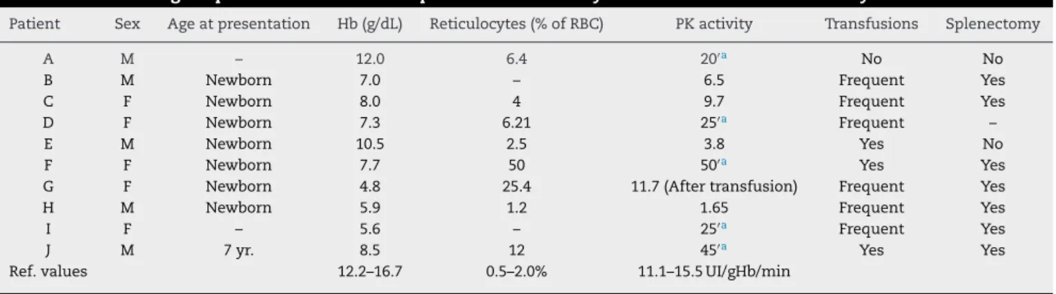
Eight patients (A–H) displayed a severe phenotype hemoglobin levels lower than 8 g/dL and/or transfusion dependence and/or splenectomy. Two patients (I and J) dis-played a moderate-to-mild phenotype defined by hemoglobin
greater than 8 g/dL, fewer than five transfusions and no splenectomy (Table 1).
Five hundred healthy blood donors from the Blood Center of the Universidade Estadual de Campinas were also stud-ied to analyze the frequency of the c.1456C>T and c.1529G>A variants in a sample Brazilian population.
DNA and structural analysis
Venous blood was used for hematological and DNA analy-sis. Hematological parameters including red blood cell indices were investigated and a reticulocyte count was performed. PK enzyme activity and other biochemical determinations were carried out according to standard methods.9
Total genomic DNA was isolated from peripheral blood leukocytes by the standard salting-out method. Individual exons of thePKLRgene were amplified by polymerase chain reaction (PCR) and DNA sequence analysis was performed using the Big Dye Terminator Cycle Sequencing Kit (Applied Biosystems, Foster City, CA) on an ABI 3500xL Genetic Ana-lyzer (Applied Biosystems) with the same primers used in the PCR reactions. The Chromas Lite 2.0 (Technelysium Pty Ltd) and CLC Sequence Viewer v.6.8.1 free software (CLC bio) were used to analyze and compare sequences with the ref-erence PKLRsequence. Structural analyses were performed using the PDB ID: 2VGB – chain A as a template.10The native and mutant models were constructed by the SWISS MODEL web-served program.11 Internal contacts were evaluated by STING Millennium12and 3D protein structures were generated using PyMOL (the PyMOL Molecular Graphics System, Version 1.8 Schrödinger, LLC).
To study the frequency of the c.1529G>A and c.1456C>T variants, exon 10 of thePKLRgene was amplified by PCR. The c.1529G>A variant was screened using the StyI enzyme, which only digests mutated fragments, producing fragments of 144 and 112 base pairs (bp). The c.1456C>T variant was screened using the BsmAI enzyme that only cleaves the DNA of peo-ple without these mutations resulting in fragments of 220 and 36 bp. The reactions were incubated at 37◦C for 16 h
accord-ing to the manufacturer’s recommendations and the products were subjected to agarose gel electrophoresis.
Table 1 – Hematological parameters of Brazilian patients with hemolytic anemia due to PK deficiency.
Patient Sex Age at presentation Hb (g/dL) Reticulocytes (% of RBC) PK activity Transfusions Splenectomy
A M – 12.0 6.4 20′a No No
B M Newborn 7.0 – 6.5 Frequent Yes
C F Newborn 8.0 4 9.7 Frequent Yes
D F Newborn 7.3 6.21 25′a Frequent –
E M Newborn 10.5 2.5 3.8 Yes No
F F Newborn 7.7 50 50′a Yes Yes
G F Newborn 4.8 25.4 11.7 (After transfusion) Frequent Yes
H M Newborn 5.9 1.2 1.65 Frequent Yes
I F – 5.6 – 25′a Frequent Yes
J M 7 yr. 8.5 12 45′a Yes Yes
Ref. values 12.2–16.7 0.5–2.0% 11.1–15.5 UI/gHb/min
a Fluorescence of NADPH (Ref. value≤15′).
Table 2 – Molecular data of 10 PK-deficient patients.
Patient Mutation Effect on protein Variant classificationb MAF ExAC (%)
A c.[182T>A]; [1282G>A] p.[Leu61Gln]; [Ala428Thr]
Uncertain significance; Uncertain significance
Absent; absent
B c.[410C>T]; [410C>T] p.[Ala137Val]; [Ala137Val]
Uncertain significance Absent
C c.[1529G>A]; [1529G>A] p.[Arg510Gln]; [Arg510Gln]
Pathogenic 0.04
D c.[1456C>T]; [993C>A] p.[Arg486Trp];
[Asp331Glu]
Likely pathogenic; uncertain significance
0.28; 0.03
E c.[1456C>T]; [−72A>G] p.[Arg486Trp]; Transcriptional mutation
Likely pathogenic; likely pathogenic
0.28; not provided
F c.[1511G>T]; [−72A>G] p.[Arg504Leu]; Transcriptional mutation
Pathogenic; likely pathogenic
0.0008/not provided
G c.[994G>A]; [994G>A] p.[Gly332Ser];
[Gly332Ser]
Pathogenic 0.006
H c.[721G>T]; [721G>T] p.[Glu241a]; [Glu241a] Pathogenic 0.003
I c.[1511G>T]; [1511G>T] p.[Arg504Leu]; [Arg504Leu]
Pathogenic 0.0008
J c.[1456C>T]; [?] p.[Arg486Trp]; [?] Likely pathogenic 0.28
a Stop codon.
b ACMG Guidelines (2015).22
?: the second mutation was not identified. Variants reported in bold are novel. ExAC: Exome Aggregation Consortium.27
Results
Ten different variants were identified among the ten PK-deficient patients, nine of which are in the coding region (eight missense and one nonsense variants) and one is in the promoter region (Table 2). Seven of these variants had
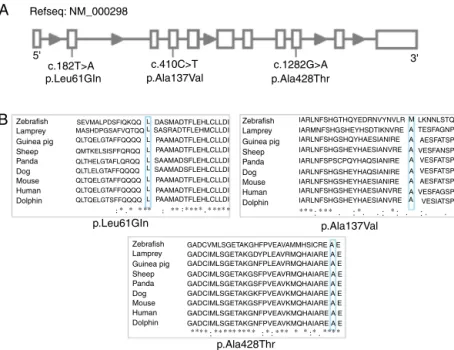
been previously reported: c.721G>T,7 993C>A,13 c.994G>A,14 c.1456C>T,7 c.1511G>T,15 c.1529G>A,7 c.-72A>G.16 The other three are described here for the first time: c.182T>A (p.Leu61Gln), c.1282G>A (p. Ala428Thr) and c.410C>T (p.Ala137Val) (Figure 1A).
All the new missense variants described appear to be damaging/disease causing according to computational
predic-A
B
Refseq: NM_000298
5' 3'
c.182T>A c.410C>T c.1282G>A p.Leu61GIn p.Leu61GIn p.Ala137Val p.Ala137Val p.Ala428Thr p.Ala428Thr
Zebrafish SEVMALPDSFIQKQQ DASMADTFLEHLCLLDI
SAAMADSFLEHLCLLDI SAAMADSFLEHLCLLDI SASRADTFLEHMCLLDI PAAMADTFLEHLCLLDI PAAMADTFLEHLCLLDI MASHDPGSAFVQTQQ QLTQELGTAFFQQQQ QMTKELSISFFQRQQ QLTHELGTAFLQRQQ QLTLELGTAFFQQQQ QLTQELGTAFFQQQQ QLTQELGTSFFQQQQ IARLNFSHGTHQYEDRNVYNVLR IARMNFSHGSHEYHSDTIKNVRE IARLNFSHGSHQYHAESIANIRE IARLNFSHGSHEYHAESIANVRE IARLNFSPSCPQYHAQSIANIRE IARLNFSHGSHEYHAQSIANIRE IARLNFSHGSHEYHAESIANIRE IARLNFSHGSHEYHAESIANVRE IARLNFSHGSHEYHAESIANVRE Lamprey Guinea pig Sheep Panda Dog Mouse Human Dolphin Zebrafish Lamprey Guinea pig Sheep Panda Dog Mouse Human Dolphin Zebrafish Lamprey Guinea pig Sheep Panda Dog Mouse Human Dolphin ∗ ∗ ∗ ∗∗ ∗ ∗∗ ∗ ∗∗ ∗ ∗∗ ∗∗ ∗ ∗ ∗ ∗ ∗∗ ∗ ∗ ∗ ∗∗∗ ∗ ∗∗ ∗ ∗∗ ∗ ∗ ∗∗∗ ∗ ∗ ∗ ∗ ∗ ∗ ∗ ∗ ∗ M LKNNLSTQ TESFAGNP AESFATSP VESFANSP VESFATSP VESFATSP VESFAGSP VESIATSP AESFATSP A A A A A A A A
GADCVMLSGETAKGHFPVEAVAMMHSICRE A E GADCIMLSGETAKGDYPLEAVRMQHAIARE A E GADCIMLSGETAKGNFPLEAVRMQHAIARE A E GADCIMLSGETAKGSFPVEAVRMQHAIARE A E GADCIMLSGETAKGSFPVEAVKMQHAIARE A E GADCIMLSGETAKGKFPVEAVKMQHAIARE A E GADCIMLSGETAKGSFPVEAVKMQHAIARE A E GADCIMLSGETAKGNFPVEAVKMQHAIARE A E GADCIMLSGETAKGNFPVEAVKMQHAIARE A E QLTQELGTAFFQQQQ PAAMADTFLEHLCLLDI PAAMADTFLEHLCLLDI PAAMADTFLEHLCLLDI L L L L L L L L L
tions: Polyphen-2,17 Sift18 and Mutation Taster.19 The three new variants were not detected in the DNA of the 80 normal controls studied.
The c.182T>A (p.Leu61Gln) and c.1282G>A (p.Ala428Thr) variants were identified in a heterozygous state in Patient A, a 21-year-old male. He had a mild phenotype, without the need of transfusions or splenectomy (Table 1). The patient presented with altered blood cell counts and splenomegaly. He has macro and microcytic hemolytic anemia, with the presence of target cells and poikilocytosis [Hemoglobin (Hb): 12.0 g/dL; mean corpuscular volume: 91.7 fl; mean corpuscular hemoglobin: 30.3 pg; mean corpuscular hemoglobin concen-tration: 33.1 g/dL; red blood cell distribution width: 13.6%; reticulocytes: 6.4%; ferritin: 525.1 ng/mL; haptoglobin less than 7.9 ng/dL and unconjugated hyperbilirubinemia]. The patient also presented with the sickle cell trait (Hb S: 40%).
The c.410C>T (p.Ala137Val) variant was identified in homozygosis in Patient B, a 14-year-old male. The patient pre-sented with a severe phenotype requiring transfusions since the first month of life. His parents are first-degree cousins; they lost their first child shortly after birth due to severe anemia.
Structural analyses of the new variants were obtained. The wild-type residue of the three variants is conserved among several species (Figure 1B). Neither the mutant residues nor another residue type with similar proper-ties was observed in these positions in other homologous sequences. The three new variants are in regions reported in UniProt to form ␣-helixes. The variants did not induce
significant conformational changes in the overall protein con-formation and, therefore, their description of the mutant structures is restricted here mainly to the sites affected by the variants.
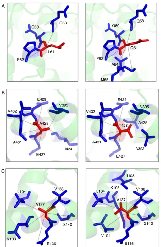
The p.Leu61Arg variant results in a change in the pro-tein conformation at the N domain interface. The residue is located on the surface of the protein. The hydrophobicity of the wild type and mutant residue differ and could cause loss of hydrophobic interactions with other molecules. The 3D rep-resentation of the normal and mutant amino acids shows that the mutant residue creates contacts with two additional amino acids of the protein (Figure 2A).
The p.Ala428Thr variant results in a change in the protein conformation at the A domain interface, the most highly con-served region. The mutant residue probably lost contact with one amino acid and gained contact with two other residues of the protein (Figure 2B).
The p.Ala137Val wild type probably lost contact with one amino acid and gained contact with three other residues of the protein (Figure 2C).
Discussion
The clinical picture of the patients was heterogeneous, ran-ging from mild chronic hemolytic anemia to severe anemia presenting at birth and requiring multiple transfusions. Anal-ysis of the correlation between molecular defect and disease severity in large series of patients has shown that the hetero-geneity depends mainly on the type and position of the variant affecting thePKLRgene.20,7,21,13
Studies in PK-deficient patients found that c.1529G>A was the most common variant in Central/Northern Europe and in the United States.6,13,14,22 However, in Southern/Western European patients the most common variant appears to be c.1456C>T.21,5,2 In this sample of PK deficient patients, the c.1456C>T and c.1511G>T variants had similar frequencies (15%).
The frequency of c.1456C>T in this sample is in accor-dance with that expected in Latino populations. In 500 normal individuals of southeastern Brazil screened in this study, the c.1456C>T variant was identified in 0.1% of the alleles and the c.1529G>A variant was not found. According to the Exome Aggregation Consortium (http://exac.broadinstitute.org/), the frequency of these alleles in Latino populations are 0.17% and 0%, respectively. In the general population, the frequencies are 0.28% and 0.04%, respectively.
The new variants (c.182T>A and c.1282G>A) were iden-tified in heterozygous in Patient A, who displayed a mild clinical phenotype. The c.182T>A (p.Leu61Gln) variant is in the N domain of the protein and could impair hydrophobic interactions with other molecules. Our prediction shows that the mutant residue creates additional contacts with other amino acids, and may perturb the structure of the protein. The c.1282G>A (p. Ala428Thr) variant is in the A domain and is likely to perturb the hydrophobic interior of the protein, forc-ing the surroundforc-ing side chains to rearrange thereby affectforc-ing protein stability and/or function.
The c.410C>T (p.Ala137Val) variant, located at the A domain interface of the protein, was identified in homozygosis. Sev-eral studies have demonstrated that variants in the A domain, especially in the homozygous form, are associated with severe hemolytic anemia,2,10,21,23such as in the case of Patient B in this study. The mutant residue is bigger and probably lost con-tact with one amino acid and gained concon-tacts with additional residues.
In the present study, the c.1529G>A variant was identified in the homozygous state in Patient C, a female child, who presented with a severe clinical phenotype. This variant was previously reported in homozygosis and was also associated with a severe clinical presentation.3
Analysis of thePKRLgene in Patient D showed the presence of biallelic variants (c.993C>A and c.1456C>T). This patient presented with a severe clinical phenotype requiring frequent transfusions and a hemoglobin level less than 8 g/dL. These two variants were also identified previously in compound het-erozygosity in an Italian patient with a mild phenotype.24The variability of clinical expression may be explained by individ-ual differences in metabolic or proteolytic activity, or even the ability to compensate for the enzyme deficiency by overex-pressing isozymes or using alternative pathways.
This study also reports two patients heterozygous for an A>G substitution at nucleotide−72 relative to the initiation
A
B
C
Q58
Q60
Q61
A64 P62
M65
E429
V395 V432
A431
E427
A392
T428 A425
Q58
Q60
L61
P62
I424 E427
A431
A428 E429
V395
V432
L104 V138
A137
S140
E136
N133 V101
E136
S140 V138 I108
K105 L104
V137
previous findings that also describe a mild phenotype in the presence of these two changes in heterozygosis.16Patient F, on the other hand, presented with the c.-72 A>G in compound heterozygosity with the c.1511G>T variant and expresses a severe phenotype.
Patient G presented with a severe phenotype including an extremely low hemoglobin level and frequent transfu-sions. The molecular analysis revealed homozygosity for the c.994G>A variant. Previous studies also show that this vari-ant in homozygosis leads to a severe anemia with the need of regular transfusions.21,14
The nonsense variant, c.721G>T, was found in Patient H in homozygosis. This variant causes the truncation of the protein in the Glu241 amino acid and thus explains the severity of the phenotype.
The c.1511G>T variant causes extreme instability of the protein26with severe anemia in PK-deficient patients homozy-gous for this variant.15This variant was found in Patient I, who presented with a severe clinical phenotype.
Patient J carried only one variant in heterozygosis, giving a genotype that could not explain the phenotype. One hypoth-esis is that the anemia was the result of an association with another red blood cell disease. Further studies are required to clarify the effect of such associations on the phenotype.
This is the first comprehensive report on the molecular characterization of PK deficiency in South America. Ten cases were characterized and three new variants were identified. The etiology was elucidated in nine patients. Collectively, the results contribute to a better understanding of the genotype-to-phenotype correlation in PK deficiency.
Conflicts of interest
The authors declare no conflicts of interest
Acknowledgements
The authors are grateful to all the members of the staff of our laboratory, and the patients and families participating in this study for their close cooperation and important contributions.
r e f e r e n c e s
1. Beutler E, Gelbart T. Estimating the prevalence of pyruvate kinase deficiency from the gene frequency in the general white population. Blood. 2000;95:3585–8.
2. Pissard S, Max-Audit I, Skopinski L, Vasson A, Vivien P, Bimet C, et al. Pyruvate kinase deficiency in France: a 3-year study reveals 27 new mutations. Br J Haematol. 2006;133:683–9. 3. Warang P, Kedar P, Ghosh K, Colah R. Molecular and clinical
heterogeneity in pyruvate kinase deficiency in India. Blood Cells Mol Dis. 2013;51:133–7.
4. Grace RF, Zanella A, Neufeld EJ, Morton DH, Eber S, Yaish H, et al. Erythrocyte pyruvate kinase deficiency: 2015 status report. Am J Hematol. 2015;90:825–30.
5. Zarza R, Alvarez R, Pujades A, Nomdedeu B, Carrera A, Estella J, et al. Molecular characterization of the PK-LR gene in pyruvate kinase deficient Spanish patients. Red Cell Pathology Group of the Spanish Society of Haematology (AEHH). Br J Haematol. 1998;103:377–82.
6. Lenzner C, Nürnberg P, Jacobasch G, Gerth C, Thiele BJ. Molecular analysis of 29 pyruvate kinase-deficient patients from central Europe with hereditary hemolytic anemia. Blood. 1997;89:1793–9.
7. Baronciani L, Beutler E. Analysis of pyruvate
kinase-deficiency mutations that produce nonspherocytic hemolytic anemia. Proc Natl Acad Sci U S A. 1993;90:4324–7. 8. Zanella A, Fermo E, Bianchi P, Valentini G. Red cell pyruvate
kinase deficiency: molecular and clinical aspects. Br J Haematol. 2005;130:11–25.
9. Beutler E, Beutler E. Red cell metabolism: a manual of biochemical methods. 3rd ed. Grune & Stratton; 1984. Orlando, FL.
10. Valentini G, Chiarelli LR, Fortin R, Dolzan M, Galizzi A, Abraham DJ, et al. Structure and function of human erythrocyte pyruvate kinase: molecular basis of nonspherocytic hemolytic anemia. J Biol Chem. 2002;277:23807–14.
11. Biasini M, Bienert S, Waterhouse A, Arnold K, Studer G, Schmidt T, et al. SWISS-MODEL: modelling protein tertiary and quaternary structure using evolutionary information. Nucleic Acids Res. 2014;42:W252–8.
12. Oliveira SRM, Almeida GV, Souza KRR, Rodrigues DN, Kuser-Falcão PR, Yamagishi MEB, et al. Sting RDB: a relational database of structural parameters for protein analysis with support for data warehousing and data mining. Genet Mol Res. 2007;6:911–22.
13. Baronciani L, Beutler E. Molecular study of pyruvate kinase deficient patients with hereditary nonspherocytic hemolytic anemia. J Clin Invest. 1995;95:1702–9.
14. Lenzner C, Nürnberg P, Thiele BJ, Reis A, Brabec V, Sakalova A, et al. Mutations in the pyruvate kinase L gene in patients with hereditary hemolytic anemia. Blood. 1994;83:2817–22. 15. Demina A, Varughese KI, Barbot J, Forman L, Beutler E. Six
previously undescribed pyruvate kinase mutations causing enzyme deficiency. Blood. 1998;92:647–52.
16. Manco L, Ribeiro ML, Maximo V, Almeida H, Costa A, Freitas O, et al. A new PKLR gene mutation in the R-type promoter region affects the gene transcription causing pyruvate kinase deficiency. Br J Haematol. 2000;110:993–7.
17. Adzhubei IA, Schmidt S, Peshkin L, Ramensky VE, Gerasimova A, Bork P, et al. A method and server for predicting damaging missense mutations. Nat Methods. 2010;7:248–9.
18. Ng PC, Henikoff S. SIFT: predicting amino acid changes that affect protein function. Nucleic Acids Res. 2003;31:3812–4. 19. Schwarz JM, Cooper DN, Schuelke M, Seelow M.
MutationTaster2: mutation prediction for the deep-sequencing age. Nat Methods. 2014;11(4):361–2. 20. Zanella A, Bianchi P. Red cell pyruvate kinase deficiency: from
genetics to clinical manifestations. Best Pract Res Clin Haematol. 2000;13:57–81.
21. Zanella A, Bianchi P, Baronciani L, Zappa M, Bredi E, Vercellati C, et al. Molecular characterization of PK-LR gene in pyruvate kinase-deficient Italian patients. Blood. 1997;89:3847–52. 22. Rouger H, Valentin C, Craescu CT, Galactéros F, Cohen-Solal
M. Five unknown mutations in the LR pyruvate kinase gene associated with severe hereditary nonspherocytic haemolytic anaemia in France. Br J Haematol. 1996:825–30,
http://dx.doi.org/10.1046/j.1365-2141.1996.405941.x. 23. Mouna J, Nadia H, Leila C, Miniar K, Fethi M, Imen D, et al.
Phenotypic and molecular genetic analysis of Pyruvate Kinase deficiency in a Tunisian family. Egypt J Med Hum Genet. 2015,http://dx.doi.org/10.1016/j.ejmhg.2015.09.001. 24. Zanella A, Fermo E, Bianchi P, Chiarelli LR, Valentini G.
Pyruvate kinase deficiency: the genotype–phenotype association. Blood Rev. 2007;21:217–31.
25. Max-Audit I, Eleouet J-F, Roméo P-H. Transcriptional
26. Fermo E, Bianchi P, Chiarelli LR, Cotton F, Vercellati C, Writzl K, et al. Red cell pyruvate kinase deficiency: 17 new mutations of the PK-LR gene. Br J Haematol. 2005;129:
839–46.
27. Lek M, Karczewski KJ, Minikel EV, Samocha KE, Banks E, Fennell T, et al. Analysis of protein-coding genetic variation in 60,706 humans. Nature. 2016;536:285–91. Available from: