CLINICAL SCIENCE
Phenotypic and immunohistochemical
characterization of sarcoglycanopathies
Ana F. B. Ferreira,I,IIMary S. Carvalho,IMaria Bernadete D. Resende,IAlda Wakamatsu,IIIUmbertina Conti Reed,ISuely Kazue Nagahashi MarieI
IDepartment of Neurology – Laboratory of Investigation in Neurology – LIM15, Faculty of Medicine, University of Sa˜o Paulo, Sa˜o Paulo/SP, Brazil. IIDepartment of Physiology and Biophysics, Institute of Biomedical Sciences, University of Sa˜o Paulo, Sa˜o Paulo, SP, Brazil.IIILaboratory of Investigation in Surgical Pathology – LIM14, Faculty of Medicine, University of Sa˜o Paulo, Sa˜o Paulo, SP, Brazil.
INTRODUCTION:Limb-girdle muscular dystrophy presents with heterogeneous clinical and molecular features. The primary characteristic of this disorder is proximal muscular weakness with variable age of onset, speed of progression, and intensity of symptoms. Sarcoglycanopathies, which are a subgroup of the limb-girdle muscular dystrophies, are caused by mutations in sarcoglycan genes. Mutations in these genes cause secondary deficiencies in other proteins, due to the instability of the dystrophin-glycoprotein complex. Therefore, determining the etiology of a given sarcoglycanopathy requires costly and occasionally inaccessible molecular methods.
OBJECTIVE: The aim of this study was to identify phenotypic differences among limb-girdle muscular dystrophy patients who were grouped according to the immunohistochemical phenotypes for the four sarcoglycans.
METHODS: To identify phenotypic differences among patients with different types of sarcoglycanopathies, a questionnaire was used and the muscle strength and range of motion of nine joints in 45 patients recruited from the Department of Neurology – HC-FMUSP (Clinics Hospital of the Faculty of Medicine of the University of Sa˜o Paulo) were evaluated. The findings obtained from these analyses were compared with the results of the immunohistochemical findings.
RESULTS: The patients were divided into the following groups based on the immunohistochemical findings: a -sarcoglycanopathies (16 patients),b-sarcoglycanopathies (1 patient),c-sarcoglycanopathies (5 patients), and non-sarcoglycanopathies (23 patients). The muscle strength analysis revealed significant differences for both upper and lower limb muscles, particularly the shoulder and hip muscles, as expected. No pattern of joint contractures was found among the four groups analyzed, even within the same family. However, a high frequency of tiptoe gait was observed in patients witha-sarcoglycanopathies, while calf pseudo-hypertrophy was most common in patients with non-sarcoglycanopathies. Thea-sarcoglycanopathy patients presented with more severe muscle weakness than did
c-sarcoglycanopathy patients.
CONCLUSION:The clinical differences observed in this study, which were associated with the immunohistochemical findings, may help to prioritize the mutational investigation of sarcoglycan genes.
KEYWORDS: Sarcoglycanopathies; Muscle weakness; Joint contractures; Tiptoe gait; Calf pseudo-hypertrophy.
Ferreira AFB, Carvalho MS, Resende MBD, Wakamatsu A, Reed UC, Marie SKN. Phenotypic and immunohistochemical characterization of sarcoglycanopathies. Clinics. 2011;66(10):1713-1719.
Received for publication onJune 13, 2011;First review completed onJune 22, 2011;Accepted for publication onJune 22, 2011 E-mail: [email protected]
Tel.: 55 11 3091-7242
INTRODUCTION
Muscular dystrophy is a necrotic degenerative/regenera-tive process of the muscles and results in progressive muscle weakness and wasting. The mechanisms by which various molecular defects result in muscular dystrophy are not yet fully understood.1
Limb-girdle muscular dystrophies (LGMD) are clearly distinct from other muscular disorders, such as dystrophino-pathies, myotonic disorders, or facioscapulohumeral dystro-phies.2The clinical course of LGMD is characterized by normal intelligence and great variability in muscle weakness and wasting, ranging from mild to severe forms. LGMD may show an early onset in the first decade of life with rapid disease progression or a later onset with slower disease progression.1,3 Hyperlordosis,4 scapular winging (escapula alata),5,6 tendon
contractures,7 and a tiptoe gait pattern4 with a wide-based stance7are associated with this disease. Serum creatine kinase
(CK) levels are elevated in most cases, and patients may develop cardiomyopathies and/or respiratory insufficiencies.6 Copyrightß2011CLINICS– This is an Open Access article distributed under
the terms of the Creative Commons Attribution Non-Commercial License (http:// creativecommons.org/licenses/by-nc/3.0/) which permits unrestricted non-commercial use, distribution, and reproduction in any medium, provided the original work is properly cited.
Due to the heterogeneity of LGMD and the lack of diagnostic specificity, estimates of the prevalence of all forms of LGMD range from 1/14,500 to 1/123,000.2 Autosomal dominant LGMD is relatively rare (5 loci described),8but cases of autosomal recessive LGMD have been characterized worldwide and are a heterogeneous group of disorders that lead to progressive muscle wasting and weakness. Current evidence suggests the involvement of at least 14 distinct loci in autosomal recessive LGMD (http://www.musclegenetable.org).9
LGMD2C – F are autosomal recessive LGMDs, also known as sarcoglycanopathies (SGP), which are caused by mutations in the genes encoding the c-, a-, b-, and d-sarcoglycan proteins (SG), respectively.10-13 The SGs are glycosylated proteins with single transmembrane domains,14and correct assembly of the sarcoglycan complex is required for the maintenance of the sarcolemma.15,16 Together with sarco-span, dystrophin, dystroglycans, syntrophins, anda -dystro-brevin, the SGs constitute the dystrophin-glycoprotein complex (DGC).17 The DGC acts as a link between the
muscle cell cytoskeleton and the extracellular matrix, providing mechanical support for the plasma membrane during myofiber contraction.14,18The function of the SGs is
not fully understood, but they appear to play both mechan-ical and non-mechanmechan-ical roles that mediate interactions among the extracellular matrix, the sarcolemma and the cytoskeleton.19,20 A primary mutation in any one of the sarcoglycan genes (a,b,cord) can lead to the total or partial loss of that sarcoglycan, secondary deficiencies of the other sarcoglycans and the occasional reduction of dystrophin labeling in muscle tissue.21 Inter- and intra-familial hetero-geneity is frequent.22Mutations in the gene fora-sarcoglycan are the most common sarcoglycan mutations, whereas mutations ind-sarcoglycan are the rarest.23
The phenotypes of the sarcoglycanopathies overlap with the dystrophinopathies with the important distinction that the learning disability specifically associated with Duchenne’s is not present, and scapular winging is more frequent in SGPs.6,9,22,24
Some studies have shown distinct patterns of sarcoglycan expression and labeling in various SGPs,1,14,16,24 but we believe that further studies are required to obtain more conclusive information because the characteristics of each population vary greatly.
OBJECTIVES
The purpose of this study was to identify phenotypic differences among LGMD patients who were grouped according to the immunohistochemical findings for the four sarcoglycans.
MATERIALS AND METHODS
Patients. Forty-five patients (from 40 families) with clinical diagnoses of LGMD from the Department of Neurology – HC-FMUSP were studied. The clinical diagnosis was based on the presence of limb-girdle muscle weakness, muscle retractions, myopathic alterations determined by electromyography (EMG) and altered levels of serum creatine kinase (CK). In all of the patients included in this study, the diagnosis of dystrophinopathy was previously excluded by mutational analysis using the multiple polymerase chain reaction (PCR) method and protein analysis by immunohistochemistry and western blotting.
This study was approved by the Institutional Review Board Ethics Committee on Human Experimentation of the Clinics Hospital of the University of Sa˜o Paulo, which follows the guidelines of the Helsinki Declaration of 1975, and all subjects in this study signed an informed consent form.
Clinical Evaluation. The following clinical features were evaluated: age of onset, consanguinity, family history, maximum motor ability (ambulant, ambulant with aid, or confined to a wheel-chair), calf pseudo-hypertrophy, tiptoe gait pattern (Achilles tendon shortening), muscle strength, and the range of motion (ROM) of nine joints (to identify tendon contractures). The serum CK levels and the presence of cardiomyopathies, as determined by electrocardiography and echodoppler, were also evaluated.
Muscle strength was determined according to the Medical Research Council (MRC) scale, which ranges from 0 to 5. On this scale, grade 5 is considered normal strength; grade 4 signifies that the patient can complete resisted movements but does not have normal strength; grade 3 represents the ability to complete movements against gravity only; in grade 2 there is movement but not against gravity; grade 1 represents contraction without movement; and grade 0 denotes an absence of muscle contraction. The MRC scale was applied to evaluate the muscle groups involved in essential movements because all of the patients included in the present study were in a chronic stage of the disease, which posed difficulties in assessing individual muscle strength. The muscles involved in the following movements were evaluated: flexion and extension of the shoulders, elbows, wrists, fingers, thumbs, hips, knees, ankles, and toes; specific movements of each joint, such as shoulder and hip adduction, abduction, internal rotation and external rotation, shoulder elevation, forearm pronation and supina-tion, wrist ulnar deviation and radial deviasupina-tion, finger interosseous, thumb opponency, and ankle inversion and eversion.
To determine the presence of contractures, the patients were given a score of complete or incomplete ROM for each joint, and each individual joint and the percentage of compromised joints were considered. The joints that were evaluated for contractures were also evaluated for muscle strength.
Muscle Biopsy. Muscle biopsies were obtained from the brachial biceps of all patients using local anesthesia in the operating room of HC-FMUSP. The samples were frozen in liquid nitrogen, and 6-mm thick sequential slices were collected on a cryostat. Routine histological and his-tochemical staining was conducted as described in detail by Dubowitz (1985).23
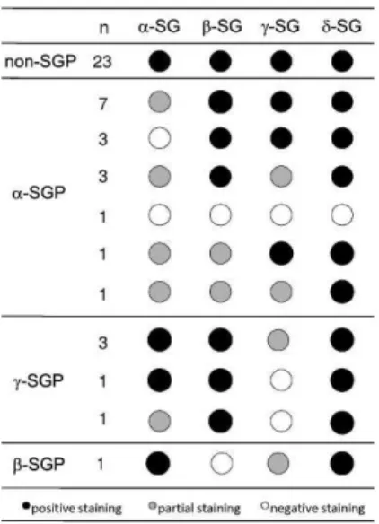
incomplete staining of most fibers) or negative staining (absence of staining of cell membrane). The samples with the lowest or absent immunoreactivity were classified as SG deficient, as shown in Figure 1. Examples of immunostained muscle biopsy samples from patients included in this series are presented in Figure 2.
Statistical Analyses. The data are expressed as the mean
¡ SEM. The Kruskal-Wallis test was used to identify
differences among the groups in the non-parametric clinical data. Pearson’s chi-square test was also employed to test for associations between components of the clinical data. A one-way analysis of variance (ANOVA) with Tukey’s post hoc test (Unequal N HSD) was applied after using Tukey’s test to record outliers and verify the differences, the power of these differences and the size effects on muscle strength.
RESULTS
The patients were divided into the following groups according to the immunohistochemical staining patterns:a -sarcoglycanopathy (a-SGP) (n = 16),c-sarcoglycanopathy (c -SGP) (n = 5) and non-SGP. The patients were classified as non-SGP when the staining for all four sarcoglycans was positive (n = 23). Only one patient was diagnosed as having ab-sarcoglycanopathy, and this patient was excluded so as not to bias the results. The immunohistochemical character-istics of each patient are shown in Figure 1, and the data collected from the clinical evaluations of the patients included in this study are shown in Table 1.
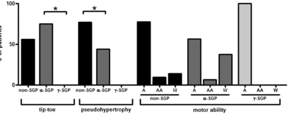
Differences in percentage of patients presenting with a tiptoe gait pattern and calf pseudo-hypertrophy were detected among the groups. The Kruskal-Wallis test revealed a significant difference in the percentage of patients present-ing with a tiptoe gait pattern between thea-SGP andc-SGP groups (p= 0.03) (Figure 3) and in the percentage of patients with calf pseudo-hypertrophy between thea-SGP and non-SGP groups (p= 0.02) (Figure 3). The maximum motor ability of the groups tended to vary. While 37.5% of the patients with ana-SG deficiency were confined to a wheelchair, only 12%
of the patients with non-SGP and none of thec-SG patients were wheelchair bound (Figure 3).
In general, the patients presented with symmetrical alterations in muscle strength and contractures with some exceptions due to falls or injuries affecting one side more than the other. Muscle weakness was more pronounced in the proximal than in the distal muscles as expected,7and the flexor muscles of these patients were found to be more affected than the extensor muscles. The one-way ANOVA analysis for the comparison of muscle strength per seg-ment showed significant differences among the groups (F(26.22) = 4.4; p= 0.0004). This analysis was highly signifi-cant (a= 0.99), and the partial eta squared (g2) value was
equal to 0.84, indicating that 84% of the differences found were due to the diagnosis. The post hoc test (Tukey -Unequal N HSD) revealed some significant differences in the muscle strengths of the upper and lower limbs among the groups as depicted in Figure 4.
The joints with decreased ROM, however, showed no characteristic contracture patterns among the three groups analyzed, even after comparing patients within the same family. Furthermore, no correlations between joint contrac-tures and muscle strength were found.
DISCUSSION
The present immunohistochemical study in a cohort of adult LGMD patients allowed us to classify 35% of cases asa -SGP, 12% as c-SGP, 2% asb-SGP and 51% as non-SGP (in which no deficiency in any of the four SG proteins was found). All four SGPs appear to be prevalent in Brazil; however, d-SGP is the least common form of autosomal recessive LGMD in Brazil26 and is equally rare world-wide.27,28Accordingly,d-SGP was not detected in the present
cohort; the proportion ofa-SGP andc-SGP in our cohort was similar to the proportion found in a previous study.29
Phenotypic differences were identified among thea-SGP,
c-SGP and non-SGP groups. A tiptoe gait pattern was more frequent in thea-SGP group than in the other groups. The high frequency of this pattern may be due to the marked presence of contractures in thea-SGP subtype,7including
contractures of the Achilles tendon, which lead to the tiptoe gait pattern. Also, patients classified as a-SGP in the present cohort presented with more severe muscle weak-ness and were more frequently confined to a wheelchair than the other patients. In primarya-SGP,a-SG is the most severely reduced protein, although there are also deficien-cies of the other three SGs.24,28-32However, the spectrum of protein deficiency may vary from total absence to partial reduction of this protein with normal staining for the other SGs.32,33
In the present study, patients classified as c-SGP had mild phenotype with remarkably preserved muscle strength; none of them was confined to a wheelchair or presented with a tiptoe gait pattern or calf pseudo-hypertrophy. These findings corroborate previous reports showing that c-SGP has a mild phenotype despite the complete absence ofc-SG and a decrease in the other three SGs.21,28,33-35However, in contrast to the present findings, some studies have reported that the early loss of ambulation, calf hypertrophy, contractures of the Achilles tendon, lumbar lordosis, scapular winging, and weak dorsal thigh and neck muscles are common clinical features ofc-SGP.36
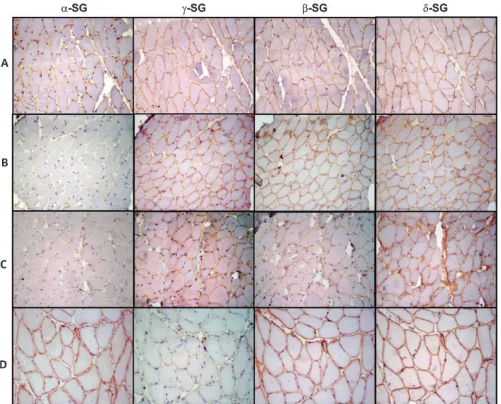
Figure 2 -Immunohistochemical preparations for alpha, gamma, beta and delta sarcoglycans (a-,c-,b- andd-SG, respectively). (A) A representative non-SGP case showing positive reactions for all four SGs; (B) a representative case ofa-SGP showing no expression ofa -SG and positive reactions for the remaining -SGs; (C) a representative case classified asa-SGP showing patchy expression of all four SGs with lower expression ofa-SG; and (D) a representative case ofc-SGP showing a negative reaction forc-SG and positive staining for the other three SGs. 200x magnification. SG: sarcoglycan; SGP: sarcoglycanopathy.
Table 1 -Demographic and clinical data collected from the patients.
Groups
non-SGP a-SGP c-SGP
n 23 16 5
Predominant sex M (54.2%) M (62.5%) F (60%)
Age (mean¡SD) 32.9 (¡13.9) 33.8 (¡16) 43.4 (¡17.1)
Age of Onset (mean¡SD) 18.3 (¡11.1) 17.3 (¡13) 29.3 (¡20.1)
Consanguinity 25% 37.5% 80%
Family history 58.3% 56.3% 60%
Cardiomyopathy 26% 25% 40%
CK nl – 50x 2 – 36x nl – 17x
Tiptoe gait pattern 54.6% 75% 0%
Maximum motor ability A (77.3%) A (56.3%) A (100%)
Calf pseudo-hypertrophy 77.3% 43.8% 0%
% of joints with affected ROM 43.1% (¡13.5) 55% (¡31.3) 37% (¡23.5)
Similar to a-SGP, studies on c-SGP have described a heterogeneous phenotype. Nonetheless, the clinical varia-tions from mild to severe disease have been correlated with the residual amount ofc-SG.33,37Although the parameters governing the phenotype/genotype correlation remain unclear, a linear association has been described between the degree of protein deficiency and the onset of symp-toms; a total absence of SGs is associated with an earlier mean age at disease onset than is observed with partial deficiencies.22
In the present cohort of patients, the patient presenting with a deficiency in all four SGs was classified as having
a-SGP, as previously reported by others.24,28-32 However, this lack of all four SGs has also been described in a
case confirmed to be c-SGP by molecular testing.29 Nevertheless, the results and statistical analyses were not affected by the classification of this case as eithera-SGP or
c-SGP.
The non-SGP group, which showed no immunohisto-chemical alterations in any of the four SGs, may include patients harboring either missense mutations in an SG gene (with no alterations in protein expression) or forms of LGMD other than SG deficiency. Therefore, patients with different etiologies were included in this group, which made the analysis difficult. However, the positive expres-sion of all four SGs excludes the presence of null mutations in the SG genes in this group; this type of mutation leads to a more predictable, uniform and severe phenotype with Figure 3 -Histograms illustrating the clinical findings. First, the percentage of patients in each group presenting with a tiptoe gait pattern are shown. The Kruskal-Wallis test showed a significant difference between thea-SGP andc-SGP groups (p= 0.03). Second, the percentage of patients in each group with calf pseudo-hypertrophy is shown. The Kruskal-Wallis test showed a significant difference between thea-SGP andc-SGP groups (p= 0.02). Third, the maximum motor ability of the groups is shown. 37.5% of the patients with ana-SG deficiency were confined to a wheelchair, while only 12% of the patients with ac-SG deficiency and none of the non-SGP patients were wheelchair bound. non-SGP: positive staining for all sarcoglycans;a-SGP:a-sarcoglycanopathy;c-SGP:c -sarcoglycano-pathy; A: ambulant; AA: ambulant with aid; W: confined to a wheelchair.
decreased expression levels of the protein encoded by the affected gene.22 The higher frequency of calf pseudo-hypertrophy observed in this group, however, might point to some other form of LGMD, such as calpainopathy, in which calf pseudo-hypertrophy is common.7
Molecular testing is fundamental for the establishment of the final diagnosis of LGMD and will certainly be mandatory when a gene-based treatment becomes available. However, considering the continuous increase in LGMD types caused by alterations in different proteins of the muscular system, it is important to find clinical parameters and accessible laboratory tools to prioritize the molecular characterization of LGMD. Efforts have been made to improve the differential diagnosis by comparing mutations to immunohistochemical findings from SGP patients29or by comparing general muscle strength to other laboratory data obtained from LGMD patients.38 Nonetheless, detailed muscular evaluations must be correlated with laboratory findings to identify clinical markers specific to each situation.
To this end, the present results show that a systematic clinical evaluation, together with immunohistochemical staining of muscle samples, enabled the characterization of LGMD patients. Applying this strategy to a larger number of patients may further refine these tools and help to determine the cost/benefit ratio of the molecular diagnosis.
ACKNOWLEDGMENTS
This study was supported by CNPq (Brazil). The authors would like to thank Eliene Dutra Campos, Caroline Alencar and Thais Freire for their technical assistance.
REFERENCES
1. ushby KM. The limb-girdle muscular dystrophies-multiple genes, multiple mechanisms. Hum Mol Genet. 1999;8:1875-82, doi: 10.1093/ hmg/8.10.1875.
2. Khadilkar SV, Singh RK. Limb girdle muscular dystrophies in India. Neurol India. 2008;56:281-8, doi: 10.4103/0028-3886.43446.
3. Zatz M, Vainzof M, Passos-Bueno MR. Limb-girdle muscular dystrophy: one gene with different phenotypes, one phenotype with different genes. Curr Opin Neurol. 2000;13:511-7, doi: 10.1097/00019052-200010000-00002.
4. Vermeer S, Verrips A, Willemsen MA, ter Laak HJ, Ginjaar IB, Hamel BC. Novel mutations in three patients with LGMD2C with phenotypic differences. Pediatr Neurol. 2004;30:291-4, doi: 10.1016/j.pediatrneurol. 2003.11.006.
5. Baumeister SK, Todorovic S, Milic-Rasic V, Dekomien G, Lochmuller H, Walter MC. Eosinophilic myositis as presenting symptom in gamma-sarcoglycanopathy. Neuromuscul Disord. 2009;19:167-71, doi: 10.1016/j. nmd.2008.11.010.
6. Bushby K. Diagnosis and management of the limb girdle muscular dystrophies. Pract Neurol. 2009;9:314-23, doi: 10.1136/jnnp.2009.193938. 7. Bushby KM. Making sense of the limb-girdle muscular dystrophies.
Brain. 1999;122:1403-20, doi: 10.1093/brain/122.8.1403.
8. Speer MC, Vance JM, Grubber JM, Lennon Graham F, Stajich JM, Viles KD, et al. Identification of a new autosomal dominant limb-girdle muscular dystrophy locus on chromosome 7. Am J Hum Genet. 1999;64:556-62, doi: 10.1086/302252.
9. Klinge L, Dekomien G, Aboumousa A, Charlton R, Epplen JT, Barresi R, et al. Sarcoglycanopathies: can muscle immunoanalysis predict the genotype? Neuromuscul Disord. 2008;18:934-41, doi: 10.1016/j.nmd. 2008.08.003.
10. Bonnemann CG, Modi R, Noguchi S, Mizuno Y, Yoshida M, Gussoni E, et al. Beta-sarcoglycan (A3b) mutations cause autosomal recessive muscular dystrophy with loss of the sarcoglycan complex. Nat Genet. 1995;11:266-73, doi: 10.1038/ng1195-266.
11. Nigro V, de Sa Moreira E, Piluso G, Vainzof M, Belsito A, Politano L, et al. Autosomal recessive limb-girdle muscular dystrophy, LGMD2F, is caused by a mutation in the delta-sarcoglycan gene. Nat Genet. 1996;14:195-8, doi: 10.1038/ng1096-195.
12. Noguchi S, McNally EM, Ben Othmane K, Hagiwara Y, Mizuno Y, Yoshida M, et al. Mutations in the dystrophin-associated protein
gamma-sarcoglycan in chromosome 13 muscular dystrophy. Science. 1995;270:819-22, doi: 10.1126/science.270.5237.819.
13. Roberds SL, Leturcq F, Allamand V, Piccolo F, Jeanpierre M, Anderson RD, et al. Missense mutations in the adhalin gene linked to autosomal recessive muscular dystrophy. Cell. 1994;78:625-33, doi: 10.1016/0092-8674(94)90527-4.
14. Ozawa E, Noguchi S, Mizuno Y, Hagiwara Y, Yoshida M. From dystrophinopathy to sarcoglycanopathy: evolution of a concept of muscular dystrophy. Muscle Nerve. 1998;21:421-38, doi: 10.1002/ (SICI)1097-4598(199804)21:4,421::AID-MUS1.3.0.CO;2-B.
15. Hack AA, Lam MY, Cordier L, Shoturma DI, Ly CT, Hadhazy MA, et al. Differential requirement for individual sarcoglycans and dystrophin in the assembly and function of the dystrophin-glycoprotein complex. J Cell Sci. 2000;113:2535-44.
16. Holt KH, Campbell KP. Assembly of the sarcoglycan complex. Insights for muscular dystrophy. J Biol Chem. 1998;273:34667-70.
17. Rando TA. The dystrophin-glycoprotein complex, cellular signaling, and the regulation of cell survival in the muscular dystrophies. Muscle Nerve. 2001;24:1575-94, doi: 10.1002/mus.1192.
18. Ibraghimov-Beskrovnaya O, Ervasti JM, Leveille CJ, Slaughter CA, Sernett SW, Campbell KP. Primary structure of dystrophin-associated glycoproteins linking dystrophin to the extracellular matrix. Nature. 1992;355:696-702, doi: 10.1038/355696a0.
19. Hack AA, Cordier L, Shoturma DI, Lam MY, Sweeney HL, McNally EM. Muscle degeneration without mechanical injury in sarcoglycan defi-ciency. Proc Natl Acad Sci U S A. 1999;96:10723-28, doi: 10.1073/pnas.96. 19.10723.
20. Ozawa E, Mizuno Y, Hagiwara Y, Sasaoka T, Yoshida M. Molecular and cell biology of the sarcoglycan complex. Muscle Nerve. 2005;32:563-76, doi: 10.1002/mus.20349.
21. Vainzof M, Passos-Bueno MR, Canovas M, Moreira ES, Pavanello RC, Marie SK, et al. The sarcoglycan complex in the six autosomal recessive limb-girdle muscular dystrophies. Hum Mol Genet. 1996;5:1963-9, doi: 10.1093/hmg/5.12.1963.
22. Guglieri M, Magri F, D’Angelo MG, Prelle A, Morandi L, Rodolico C, et al. Clinical, molecular, and protein correlations in a large sample of genetically diagnosed Italian limb girdle muscular dystrophy patients. Hum Mutat. 2008;29:258-66, doi: 10.1002/humu.20642.
23. Dubowitz V, Sewry C. Muscle biopsy: a practical approach. 3rd ed. Philadelphia: Saunders Elsevier; 2007.
24. Moore SA, Shilling CJ, Westra S, Wall C, Wicklund MP, Stolle C, et al. Limb-girdle muscular dystrophy in the United States. J Neuropathol Exp Neurol. 2006;65:995-1003, doi: 10.1097/01.jnen.0000235854.77716.6c. 25. Ferreira LG, Marie SK, Liu EC, Resende MB, Carvalho MS, Scaff M, et al.
Dystrophin-glycoproteins associated in congenital muscular dystrophy: immunohistochemical analysis of 59 Brazilian cases. Arq Neuropsiquiatr. 2005;63:791-800, doi: 10.1590/S0004-282X2005000500014.
26. Zatz M, de Paula F, Starling A, Vainzof M. The 10 autosomal recessive limb-girdle muscular dystrophies. Neuromuscul Disord. 2003;13:532-44, doi: 10.1016/S0960-8966(03)00100-7.
27. Duggan DJ, Gorospe JR, Fanin M, Hoffman EP, Angelini C. Mutations in the sarcoglycan genes in patients with myopathy. N Engl J Med. 1997;336:618-24, doi: 10.1056/NEJM199702273360904.
28. Passos-Bueno MR, Vainzof M, Moreira ES, Zatz M. Seven autosomal recessive limb-girdle muscular dystrophies in the Brazilian population: from LGMD2A to LGMD2G. Am J Med Genet. 1999;82:392-8, doi: 10. 1002/(SICI)1096-8628(19990219)82:5,392::AID-AJMG7.3.0.CO;2-0. 29. Gouveia TLF, Paim JFO, Pavanello RC, Zatz M, Vainzof M.
Sarcoglycanopathies: A Multiplex Molecular Analysis for the Most Common Mutations. Diagn Mol Pathol. 2006;15:95-100, doi: 10.1097/ 00019606-200606000-00006.
30. Draviam R, Billington L, Senchak A, Hoffman EP, Watkins SC. Confocal analysis of the dystrophin protein complex in muscular dystrophy. Muscle Nerve. 2001;24:262-72, doi: 10.1002/1097-4598(200102)24:2, 262::AID-MUS120.3.0.CO;2-3.
31. Duggan DJ, Fanin M, Pegoraro E, Angelini C, Hoffman EP. alpha-Sarcoglycan (adhalin) deficiency: complete deficiency patients are 5% of childhood-onset dystrophin-normal muscular dystrophy and most partial deficiency patients do not have gene mutations. J Neurol Sci. 1996;140:30-9, doi: 10.1016/0022-510X(96)00028-7.
32. Vainzof M, Moreira ES, Canovas M, Anderson LV, Pavanello RC, Passos-Bueno MR, et al. Partial alpha-sarcoglycan deficiency with retention of the dystrophin-glycoprotein complex in a LGMD2D family. Muscle Nerve. 2000;23:984-8, doi: 10.1002/(SICI)1097-4598(200006)23:6, 984::AID-MUS24.3.0.CO;2-#.
33. Moreira ES, Vainzof M, Suzuki OT, Pavanello RC, Zatz M, Passos-Bueno MR. Genotype-phenotype correlations in 35 Brazilian families with sarcoglycanopathies including the description of three novel mutations. J Med Genet. 2003;40:E12, doi: 10.1136/jmg.40.2.e12", -1,"xxx/2.e12.
35. Vainzof M, Passos-Bueno MR, Pavanello RC, Marie SK, Oliveira AS, Zatz M. Sarcoglycanopathies are responsible for 68% of severe auto-somal recessive limb-girdle muscular dystrophy in the Brazilian population. J Neurol Sci. 1999;164:44-9, doi: 10.1016/S0022-510X(99) 00040-4.
36. Norwood F, de Visser M, Eymard B, Lochmuller H, Bushby K. EFNS guideline on diagnosis and management of limb girdle muscular
dystrophies. Eur J Neurol. 2007;14:1305-12, doi: 10.1111/j.1468-1331.2007. 01979.x.
37. Angelini C, Fanin M, Freda MP, Duggan DJ, Siciliano G, Hoffman EP. The clinical spectrum of sarcoglycanopathies. Neurology 1999;52:176-9. 38. Comerlato EA, Scola RH, Werneck LC. Limb-girdle muscular dystrophy: