&
Universidad de Zaragoza
CANCER THERANOSTICS:
MULTIFUNCTIONAL GOLD NANOPARTICLES
FOR DIAGNOSTICS AND THERAPY
João Diogo Osório de Castro Conde
Doctorate in Biology
Specialty in Biotechnology
CANCER THERANOSTICS:
MULTIFUNCTIONAL GOLD NANOPARTICLES
FOR DIAGNOSTICS AND THERAPY
João Diogo Osório de Castro Conde
Thesis supervised by
Prof. Pedro V. Baptista
Prof. Jesus M. de la Fuente
Doctorate in Biology
Specialty in Biotechnology
All the statements expressed in this document are the sole responsibility of its author.
Todas as afirmações presentes no seguinte documento são da exclusiva responsabilidade do seu autor.
COPYRIGHT
Autorizo os direitos de copyright da presente tese de doutoramento, denominada “Cancer Theranostics: multifunctional gold nanoparticles for diagnostics and therapy”.
A Faculdade de Ciências e Tecnologia e a Universidade Nova de Lisboa têm o direito, perpétuo e sem limites geográficos, de arquivar e publicar esta dissertação através de exemplares impressos reproduzidos em papel ou de forma digital, ou por qualquer outro meio conhecido ou que venha a ser inventado, e de a divulgar através de repositórios científicos e de admitir a sua cópia e distribuição com objectivos educacionais ou de investigação, não comerciais, desde que seja dado crédito ao autor e editor.
Para as minhas princesas,
Catarina e Ritinha
Todo o meu Amor!
i
Acknowledgements ... ii
Preface ... iv
Publications published during the PhD Grant ... v
Sumário ... viii
Abstract ... x
Symbols and notations ... xii
Introduction .….………... 1
1. General considerations on Gold Nanoparticles……….... 2
1.1.Gold Nanoparticle synthesis ……….6
1.2.Stabilization against aggregation ………. 6
1.3.Biological applications ….………8
2. NanoDiagnostics ………... 10
2.1.Nanocarriers in Cancer diagnosis ……….. 12
3. NanoTherapy ……… 13
3.1.Tumour targeting ………... 14
3.2.Gene therapy ……….. 15
4. NanoToxicity ……… 17
References ………19
5. Review articles and book chapter……….... 24
6. General objectives………129
Gold Nanoparticles as Nanosensors for Diagnostic …... 131
Overview ... 132
Articles ... 133
Gold nanoparticles as Nanovectors for Therapy ... 169
Overview ... 170
Articles ... 173
Gold nanoparticles for Theranostics …………... 266
Overview ... 267
Articles ... 269
ii
“Fortune favours the audacious” – Desiderius Erasmus
This project has required the input from many people to acquire some of the “amazing” results we could achieved. Although the following words will never be enough to express all my appreciation to them, I would like here to present my admiration for all their support during this long passage. Hence, I would like to thanks:
My supervisor, Professor Pedro V. Baptista for giving me the opportunity to be a member of his working group. I would like to thanks for his never ending, dedicated and thoughtful scientific and personal input during my work. I owe him immensely. For his help, guidance, proof reading and scientific contributions with all publications, oral presentations and of course this doctoral thesis.
My co-supervisor, Professor Jesus M. de la Fuente, I am especially thankful for his guidance and magnanimity that allowed me to work independently. I would like to thanks for his support. I always enjoyed his commitment to achieve and foster a great working experience at his chair.
Professor João C. Lima for the numerous enthusiastic and successful scientific discussions. You are a great scientist. I want to be like you, someday!
Vanesa Sanz for all the things that I learned from you... and all the funny moments in the lab.
My Thesis Committee members, Prof. Jorge Gaspar (Faculdade de Ciências Médicas) and Prof. Ricardo Franco (REQUIMTE) for taking some time in evaluating my progress.
I would like to thanks Fundação para a Ciência e Tecnologia, for the financial support (SFRH/BD/62957/2009) provided to conduct this thesis and for the projects where I participate: Sensitive and selective detection of DNA/RNA based on functionalised gold nanoparticles - application to pathogen detection; mutation detection and RNA quantification - PTDC/BIO/66514/2006 ; NANOLIGHT - Nanosystems for delivery of caged compounds -
iii
multifunctional gold nanoconjugates towards cancer therapy - PTDC/BBB-NAN/1812/2012.
The members of the lab 315 for the great time in the lab. I believe I have every reason to convey my appreciation to all of you for the cooperation I have received from each and every one of you individually.
A special thanks to a colleague and friend, Leticia Giestas. I will always remember the talks on our joys and sorrows about the lab. Your support and judgment always kept me “in line”. Thank you for everything.
A todos os meus Amigos por estarem sempre lá…. Porque sempre me guiaram, porque sempre me apoiaram, porque sempre me fizeram sorrir…
Um agradecimento muito especial aos meus Pais, porque sempre acreditaram que seria possível… porque sempre acreditaram em mim. Pelo apoio incondicional nos melhores e nos piores momentos, especialmente quando achamos que não somos capazes. Muito obrigado. Sem vocês nada teria sido possível.
Por ultimo, não tenho palavras para descrever a gratidão e orgulho que tenho pela minha mulher, Catarina. Muito obrigado por todo o amor e carinho. Por todo o apoio e compreensão mesmo quando roubava um pouco do nosso tempo. Obrigado por fazeres de mim um homem melhor… uma pessoa melhor. E um obrigado do tamanho do mundo à nossa princesinha linda, Ritinha, que veio unir-nos ainda mais. Amo-vos muito….
iv
This Thesis compiles the work done during my doctoral studies that I started after invitation by Prof. Pedro V. Baptista for a PhD position at the Research Centre for Human Molecular Genetics, Dept. of Life Sciences at Science and Technology Faculty, New University of Lisbon. In 2010, I was awarded with a PhD Grant funded by the Portuguese Foundation for Science and Technology under the topic “Cancer Theranostics − Multifunctional gold nanoparticles for diagnostics and therapy” (part of the European project NanoScieE+ - NANOTRUCK). Prof. Pedro V. Baptista is an Associate Professor of Molecular Genetics & Nanomedicine at Dept. of Life Sciences, Science and Technology Faculty, New University of Lisbon.
From June 2010 to March 2011, the fellow carried out a part of the PhD Project at the Biofunctional Nanoparticles and Surfaces Group, IUI of Nanoscience of Aragon (INA), Zaragoza, Spain, under supervision of Prof. J.M. de la Fuente. Prof. J.M. de la Fuente established a multidisciplinary Nanotherapy and Nanodiagnostics Group at IUI of Nanoscience of Aragon, University of Zaragoza (Spain) after receiving an ERC Starting Grant.
The NANOTRUCK project intends to develop an innovative kind of multifunctional gold nanoparticles loaded with fluorescent, tumoral markers, cell penetrating peptides and RNAi complementary to the proto-oncogene c-Myc for cancer diagnosis and treatment. The fellow worked on the synthesis and functionalization of the multifunctional gold nanoparticles and on the in vitro studies for the silencing of c-Myc gene in human cells. From this work, he has broadened his experience in techniques in the molecular genetics field such as the cloning of constructs for applications in gene therapy and techniques related with the small interfering RNA approach for gene silencing. This project implies the collaboration with different groups in Europe with expertise in different subfields: Centre for Cell Engineering (University of Glasgow, UK), Instituto di Cibernetica “E. Caianiello” (Pozzuoli, Italy), Helmholtz Zentrum München (München, Germany), Science and Technology Faculty, New University of Lisbon (Lisbon, Portugal) and IUI of Nanoscience of Aragon at the University of Zaragoza (Zaragoza, Spain). Thanks to these collaborations, the interaction between nanoparticles and biological systems, ranging from in vitro cultured human cells to in vivo animal models (primitive Hydra and complex vertebrate mouse) were studied. From this work, the fellow acquired experience in the diagnostics and therapy fields, as can be seen from his publications on the development of nanosystems for the combined diagnostics/therapeutics (theranostics)
v
delivery and silencing systems of relevance for therapy strategies, with particular focus on enabling the development of safer, more efficient, and specific platforms to human disease. During the PhD the fellow has acquired experience in molecular biology techniques useful for the biomedical applications of nanoparticles (Human Cell culture, Microbiology, Confocal Microscopy, Electrophoresis, Real-Time PCR, etc), and skills useful in the development of applications of nanoparticles in gene therapy (plasmid DNA cloning, Antisense and RNA interference methodologies, work with reporter vectors such as EGFP and luciferase gene expression) and different techniques for nanoparticle characterization (Atomic Force Microscopy, Scanning Electron Microscopy, Transmission Electron Microscopy, Light-Scattering microscopy, Fluorescence, UV-VIS molecular absorption, etc). With a strong background in nanodiagnostics, nanotherapy and nanotoxicology fields, as can be seen from publications on the development of nanosystems (with ssDNA/ssRNA, siRNA, antibodies, peptides, tumoral markers, polymers and fluorescent dyes) for the combined diagnostics/therapeutics (theranostics) systems. These platforms involves DNA/RNA detection with strong impact in genetic diagnostics and in gene delivery/silencing and tumour targeting systems, with particular focus on enabling the development of safer, more efficient, and specific methods to human disease.
Overall, my doctoral studies have resulted in the following peer-reviewed publications with more than 200 citations:
1. “Gold Nanoparticle-mediated oncogene knockdown in human cancer cells: From cell proliferation to cell-cycle and apoptosis.” H.W. Child, Y. Hernandez, João Conde, P.V. Baptista, J.M. de la Fuente, C.C. Berry. (Submitted)
2. “Multifunctional Gold Nanocarriers for Cancer Theranostics – From bench to bedside and back again?” João Conde*, F. Tian, P.V. Baptista and J.M. de la Fuente (Accepted, Invited Book Chapter) (* Corresponding-author).
3. “In vivo tumour targeting via nanoparticle-mediated therapeutic siRNA coupled to inflammatory response in lung cancer mouse models.” João Conde*, F. Tian*, Y. Hernández, C. Bao, D. Cui, K.P. Janssene, M.R. Ibarra, P.V. Baptista, T. Stoëger and J.M. de la Fuente. (* Co-first authors). Biomaterials (2013), Vol. 34, pp. 7744–7753. Research article (IF=7.604)
vi
4. “A Gold-nanobeacon system for Gene therapy: evaluation of Genotoxicity, Cell toxicity and Proteome profiling analysis” João Conde, M. Larguinho, A. Cordeiro, L.R. Raposo, P.M. Costa, S. Santos, M.S. Diniz, A.R. Fernandes and P.V. Baptista. Nanotoxicology (2013). Research article (IF=7.844)
5. “Gold-nanobeacons for simultaneous gene specific silencing and intracellular tracking of the silencing events.” João Conde, J. Rosa, J.M. de la Fuente and P.V. Baptista. Biomaterials (2013), Vol. 34, pp. 2516–2523. Research article (IF=7.604)
6. “Design of Multifunctional Gold Nanoparticles for In vitro and In vivo Gene Silencing.” João Conde*, A. Ambrosone*, V. Sanz, Y. Hernández, V. Marchesano, F. Tian, H. Child, C.C. Berry, M.R. Ibarra, P.V. Baptista, C. Tortiglione and J.M. de la Fuente. (* Co-first authors). ACS Nano (2012), Vol. 6, pp. 8316–8324. Research article (IF=12.062)
7. “RNA Quantification Using Noble Metal Nanoprobes: Simultaneous Identification of Several Different mRNA Targets Using Colour Multiplexing and Application to Cancer Diagnostics.” João Conde, G. Doria, J.M. de la Fuente and P.V. Baptista. Nanoparticles in Biology and Medicine: Methods and Protocols Series. Methods in Molecular Biology (2012), Vol. 906, pp. 71-87. Humana Press, Springer Protocols.
8. “Effect of PEG biofunctional spacers and TAT peptide on dsRNA loading on Gold Nanoparticles.” V. Sanz, João Conde, Y. Hernández, P.V. Baptista, M.R. Ibarra and J.M. de la Fuente. Journal of Nanoparticle Research (2012), Vol. 14, pp. 1-9. Research article (IF=3.287)
9. “Modification of Plasmid DNA Topology by Histone-Mimetic Gold Nanoparticles.” João Conde, P.V. Baptista, Y. Hernández, V. Sanz and J.M. de la Fuente. Nanomedicine (Lond). (2012), Vol. 7, pp. 1657-1666. Research article (IF=5.260)
10. “Gold-Nanobeacons for Real-Time Monitoring of RNA Synthesis.” J. Rosa*, João Conde*, J.M. de la Fuente, J.C. Lima and P.V. Baptista. (* Co-first authors). Biosensors & Bioelectronics (2012), Vol. 36, pp. 161-167. Research article (IF=5.437)
vii
Veigas, L. Giestas, C. Almeida, M. Assunção, J. Rosa and P.V. Baptista. Sensors (Basel) (2012), Vol. 12, pp. 1657-1687. Review article (IF=1.953)
12. “Noble Metal Nanoparticles Applications in Cancer.” João Conde, G. Doria and P.V. Baptista. Journal of Drug Delivery (2012), Vol. 2012, pp. 1-12. Review article 13. “Nanophotonics for Molecular Diagnostics and Therapy Applications.” João Conde,
J. Rosa, J.C. Lima and P.V. Baptista. International Journal of Photoenergy (2012), Vol. 2012, pp. 1-11. Review article (IF=2.663)
14. “Alloy Metal Nanoparticles for Multicolour Cancer Diagnostics.” P.V. Baptista, G. Doria, João Conde. Proceedings of SPIE (2011), Vol. 7909, 79090K-1, Colloidal Quantum Dots/Nanocrystals for Biomedical Applications VI. Proceedings article 15. “In vitro Transcription and Translation Inhibition via DNA functionalized
Gold-Nanoparticles.” João Conde, J.M. de la Fuente and P.V. Baptista. Nanotechnology (2010), Vol. 21, 505101. Research article (IF=3.842)
16. “RNA Quantification using Gold Nanoprobes - application to Cancer Diagnostics.” João Conde, J.M. de la Fuente and P.V. Baptista. Journal of Nanobiotechnology (2010), Vol. 8, pp. 1-8. Research article IF=not yet determined by ISI (unofficial=5.09)
The three chapters in this Thesis (II-IV) are based on the inclusion of the respective article(s), preceded by a small overview explaining their context and declarations of authorship.
viii
O uso de nanopartículas de ouro tem ganho terreno em diagnóstico molecular, devido às suas propriedades físico-químicas únicas, apresentando inúmeras vantagens, tais como aumento da sensibilidade e especificidade, e do potencial para caracterizar sistemas “single molecule”. Devido à sua versatilidade, bem como fácil síntese e funcionalização, nanopartículas de ouro multifuncionais têm, também, sido propostas como sistemas ideais de “delivery” para terapia (nanovectores). Ser capaz de produzir tais sistemas significa o início de uma nova era na área de teragnóstico (diagnóstico e terapia), impulsionada pela criação de veículos fantásticos à nanoescala.
De facto, a Nanotecnologia tem a capacidade de explorar novos sistemas para teragnóstico em cancro, através do desenvolvimento de métodos de diagnóstico, tais como colorimetria e imunoensaios, e em abordagens terapêuticas por meio de terapia génica, “delivery” de fármacos e/ou no “targeting” de tumores.
As características únicas das nanopartículas à nanoescala, tais como a elevada relação de superfície/volume ou as propriedades ópticas dependentes de tamanho e forma, que são radicalmente diferentes das dos seus materiais à macroescala, constituem as principais vantagens para a criação de sistemas utilizados em ensaios clínicos.
Este projecto de doutoramento pretende optimizar a síntese e funcionalização de nanopartículas de ouro para a detecção de transcritos de oncogenes (c-Myc e BCR-ABL), podendo ser utilizado para a avaliação do perfil de expressão em células cancerígenas e, simultaneamente, o desenvolvimento de uma plataforma inovadora de nanopartículas de ouro multifuncionais, funcionalizadas com marcadores tumorais, péptidos de internalização celular, marcadores fluorescentes e siRNAs capazes de silenciar determinados genes, de forma a avaliar o nível de expressão e determinar a eficiência de silenciamento. Este trabalho é parte de uma colaboração entre o Centro de Investigação em Genética Molecular Humana, Faculdade de Ciências e Tecnologia, Universidade Nova de Lisboa, Portugal e o Instituto de Nanociencia de Aragón, Espanha no âmbito de um projecto europeu [NanoScieE+ - NanoTruck].
Por forma a atingir este objetivo, desenvolveram-se estratégias eficazes de conjugação para combinar, de uma forma altamente controlada, biomoléculas à superfície de nanopartículas de ouro com funções específicas, tais como: oligos ssDNA para detectar sequências específicas e para a quantificação de RNA mensageiro; Polímeros biofuncionais: poli(etilenoglicol) (PEG) utilizados para aumentar a solubilidade e biocompatibilidade, conferindo também uma
ix
internalização celular (por exemplo, péptido TAT): usados para superar de forma mais eficiente a barreira lipofílica das membranas celulares, por forma a alcançar mais facilmente o citoplasma das células alvo; amónios quaternários: para introduzir cargas positivas na superfície das nanopartículas de ouro (para fazer ligar/adsorver os RNAs); e siRNA complementar a um proto-oncogene, c-Myc, implicado no crescimento celular, proliferação, perda de diferenciação e morte celular programada.
A fim de estabelecer que estas nanopartículas são alternativas viáveis para os métodos disponíveis, estes sistemas inovadores foram extensivamente caracterizados na sua funcionalização química, nível de internalização celular, toxicidade celular e inflamação, e o “knockdown” da expressão da proteína MYC em várias linhagens celulares de cancro e em modelos in vivo.
Palavras-chave: Nanomedicina, Terapia, Diagnóstico, Teragnóstico, Cancro, Nanopartículas de ouro.
x
The use of gold nanoparticles (AuNPs) has been gaining momentum in molecular diagnostics due to their unique physico-chemical properties these systems present huge advantages, such as increased sensitivity, reduced cost and potential for single-molecule characterisation. Because of their versatility and easy of functionalisation, multifunctional AuNPs have also been proposed as optimal delivery systems for therapy (nanovectors). Being able to produce such systems would mean the dawn of a new age in theranostics (diagnostics and therapy) driven by nanotechnology vehicles.
Nanotechnology can be exploit for cancer theranostics via the development of diagnostics systems such as colorimetric and imunoassays, and in therapy approaches through gene therapy, drug delivery and tumour targeting systems.
The unique characteristics of nanoparticles in the nanometre range, such as high surface-to-volume ratio or shape/size-dependent optical properties, are drastically different from those of their bulk materials and hold pledge in the clinical field for disease therapeutics
This PhD project intends to optimise a gold-nanoparticle based technique for the detection of oncogenes’ transcripts (c-Myc and BCR-ABL) that can be used for the evaluation of the expression profile in cancer cells, while simultaneously developing an innovative platform of multifunctional gold nanoparticles (tumour markers, cell penetrating peptides, fluorescent dyes) loaded with siRNA capable of silencing the selected proto-oncogenes, which can be used to evaluate the level of expression and determine the efficiency of silencing. This work is a part of an ongoing collaboration between Research Centre for Human Molecular Genetics, Faculdade de Ciências e Tecnologia, Universidade Nova de Lisboa, Portugal and Biofunctional Nanoparticles and Surfaces Group, Instituto de Nanociencia de Aragón, Spain within a European project [NanoScieE+- NANOTRUCK].
In order to achieve this goal we developed effective conjugation strategies to combine, in a highly controlled way, biomolecules to the surface of AuNPs with specific functions such as: ssDNA oligos to detect specific sequences and for mRNA quantification; Biofunctional spacers: Poly(ethylene glycol) (PEG) spacers used to increase solubility and biocompatibility and confer chemical functionality; Cell penetrating peptides: to overcome the lipophilic barrier of the cellular membranes and deliver molecules into cells using TAT peptide to achieve cytoplasm and nucleus; Quaternary ammonium: to introduce stable positively charged in gold nanoparticles surface; and RNA interference: siRNA complementary to a master
xi
of differentiation, and cell death.
In order to establish that they are viable alternatives to the available methods, these innovative nanoparticles were extensively characterized on their chemical functionalization, ease of uptake, cellular toxicity and inflammation, and knockdown of MYC protein expression in several cancer cell lines and in in vivo models.
xii
AuNPs - gold nanoparticles
cDNA - complementary DNA (DNA synthesized from mRNA template) CML - chronic myelogenous leukemia
CPPs - Cell penetrating peptides DEPC - Diethylpyrocarbonate DLS - Dynamic Light Scattering DNA - Deoxyribonucleic Acid
dNTPs - Deoxyribonucleotide Triphosphate dsDNA - double-stranded DNA
DTNB - 5,5’-dithio-bis(2-nitrobenzoic) acid DTT - Dithiothreitol
EDC - 1-Ethyl-3-(3-dimethylaminopropyl)carbodiimide EGFP - Enhanced Green Fluorescent Protein
FRET - Förster’s or fluorescence resonance energy transfer GST - Glutathione
LSPR - localized surface plasmon resonance MDR - multiple-drug resistance
mRNA - Messenger RNA miRNA - micro RNA
MTT - 3-(4,5-dimethylthiazol-2-yl)-2,5-diphenyltetrazoliumbromide NIR - near-infrared
NPs - nanoparticles
NHS - N-hydroxysuccinimide NTP - Ribonucleotide Triphosphate PBS - Phosphate Buffer Saline PCR - Polymerase Chain Reaction PEG - Poly(ethylene glycol) Prot - Protamine
RGD Arginine-Glycine-Aspartic Acid (peptide) RNA - Ribonucleic Acid
RNAi - RNA interference
xiii
SDS - Sodium Dodecyl Sulfate SEM - Scanning electron microscopy
SERS - Surface-Enhanced Raman Scattering siRNA - Small interfering RNA
ssDNA - Single-stranded DNA ssRNA - single-stranded RNA
TAT - Transactivator of transcription (peptide) TEM - Transmission electron microscopy
Gold has been regarded as precious for as long as humans have existed, and has been associated with gods, kings and immortality. Already synthesized of gold at the times of Faraday, they generated ever-increasing interest since the ability to synthesize those particles in a controlled fashion way and their application in diverse nanosystems have come up in the last few decades [20;21]. Today, nanotechnology is enabling gold to help address critical global problems from cancer treatment to climate change. Although nanotechnology is thought of as a new branch of science that has only emerged over the past decade, gold nanoparticles (AuNPs) have been used, even though unintentionally, for several thousand years [1] (see Figure 1).
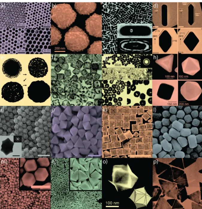
Gold nanoparticles have gained increasing interest due to their special features, such as extraordinary optical and electronic properties, high stability and biological compatibility, controllable morphology and size dispersion and shapes (see Figure 2), and easy surface functionalization.
Figure 1. (a) Golden burial mask of Egyptian Pharaoh Tutankhamun (King Tut) of the 18th Dynasty (ca. 1323 BC). (b) A gold medal presented at the Games of the II Olympiad (Paris, France; 1900). While bulk gold is highly un-reactive and predominantly reflects light, nanoscale gold can be highly reactive, exhibiting pharmacologic properties and the ability to absorb, transfer, and convert light energy into heat. The mask in (a), discovered in 1922 by Howard Cater, consists of solid gold with inlaid glass and stone (21 cm high and ca. 11 kg). Prior to the 1900 Olympics in (b), athletes received only silver and copper medals which easily oxidize. The winged goddess Nike is shown on the front in (b); a victorious athlete holding a laurel branch is shown on the back with The Acropolis in the background [2].
Figure 2. Gold nanoparticles of various size and shape with potential applications in biomedicine.
Small (a) and large (b) nanospheres, (c) nanorods, (d) sharpened nanorods, (e) nanoshells, (f) nanocages/frames, (g) hollow nanospheres, (h) tetrahedra/octahedra/ cubes/icosahedra, (i) rhombic dodecahedra, (j) octahedra, (k) concave nanocubes, (l) tetrahexahedra, (m) rhombic dodecahedra, (n) obtuse triangular bipyramids, (o) trisoctahedra, and (p) nanoprisms [2].
The main characteristics include the electrical, chemical, and optical properties. The optical properties of AuNPs are significant because its adsorption and emission of the wavelength are within the visible range of light and because of their size- and shape-dependent properties [3]. Of particular interest is the light extinction process in the UV-visible range, which occurs when an electromagnetic wave passes through a metal particle exciting its electronic or
respective frequency of the incident wave, and therefore disperse secondary radiation in all directions. This collective oscillation of the free conduction electrons is called localized surface plasmon resonance (LSPR). Surface plasmons are collective charge oscillations that occur at the interface between conductors and dielectrics (Figure 3). The oscillation frequency is usually in the visible region giving rise to the strong surface plasmon resonance absorption [25-28]. They can take various forms, ranging from freely propagating electron density waves along metal surfaces to localized electron oscillations on metal nanoparticles (NPs) [4;5]. When light passes through a metal nanoparticle induces dipole moments that oscillate at the respective frequency of the incident wave, consequently dispersing secondary radiation in all directions. Light on NP induces the conduction electrons to oscillate collectively with a resonant frequency that depends on the nanoparticles’ size, shape, composition, inter-particle distance and environment (dielectric properties) [30-32;58]. As a result of these SPR modes, the nanoparticles absorb and scatter light so intensely that single NPs are easily observed by eye using dark-field (optical scattering) microscopy.
Figure 3. Localized surface plasmon resonance. Schematic representation of how the interaction of
the electromagnetic waves with the metal NPs surface electrons generates a surface plasmon resonance [4].
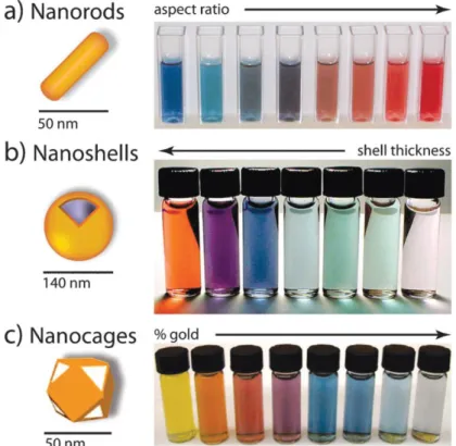
Size provides important control over many of the physical and chemical properties, including luminescence, conductivity, and catalytic activity. AuNPs give rise to both absorption and scattering whose proportions depend on the AuNP size, shell thickness, aspect ratio and percentage of gold [6] (see Figure 4). AuNPs with a diameter smaller than 20 nm essentially show absorption, but when the size increase to 80 nm also increases the ratio of scattering to absorption. As the size of the AuNP increases, light can no longer polarize the nanoparticles
and broadening of the surface plasmon band. Small AuNPs like the ones with 13 nm of diameter absorb green light, which corresponds to a strong absorption band at 520 nm in the visible light spectrum. However, solutions of AuNPs appear red in colour. For smaller AuNPs (i.e. 5 nm diameter), surface electrons are oscillated by the incoming light in a dipole mode, but the SPR it is very sensitive to the composition, size, shape, inter-particle distance and environment (dielectric properties) of the AuNPs [30-32]. For example, for citrate stabilised particles, the addition of NaCl shields the surface charge and leads to a concomitant decrease in the inter-particle distance and eventual particle aggregation [7]. For instance, 5 nm AuNPs are orange-red, but they turn blue-purple upon aggregation (network formation) to larger AuNPs.
Figure 4. Gold nanoparticles commonly applied in biomedical applications. (a) Gold nanorods, (b)
silica–gold core–shell nanoparticles, and (c) gold nanocages. The intense colour of these nanoparticles arises from the collective excitation of their conduction electrons, or surface plasmon resonance modes, which results in photon absorption at wavelengths which varies with (a) aspect ratio, (b) shell thickness, and/or (c) galvanic displacement by gold.
This also explains the corresponding surface plasmon band shifts (red shift), which results in colour changes (red-to-purple) that are observed during the aggregation of small AuNPs. When AuNPs aggregate, their surface plasmons combine (interparticle plasmon coupling),
interparticle plasmon coupling is rather complex and dependent on many factors, such as aggregate morphology and nanoparticles density [31;32;34].
1.1. GOLD NANOPARTICLE SYNTHESIS
Gold nanoparticles, also known as colloidal gold, is a suspension of sub-micrometre-sized gold metal particles in a fluid and can be obtained in sizes between 3 and 200 nm in diameter. As a colloidal particle is much larger than its constituent atoms, its typical shape mainly results from its surface energy, whose minimization decreases the surface area and favours the formation of spherical objects [8].
The most common methods for the synthesis of spherical gold colloidal particles involve the chemical reduction of Au ions.
Since the pioneering work of Turkevich [9] and the modification by G. Frens [10], the most widely studied reductant is sodium citrate [37;38]. The method of citrate reduction of Au(III) to Au(0) in water is still used nowadays to subsequently replace the citrate ligand of these AuNPs by appropriate ligands of biological interest.
In the citrate reduction method, sodium citrate reduces the gold cations in hydrogen tetrachloroaurate (HAuCl4). As the gold metal forms, anions coat the outside of the particles preventing them from forming larger particles. The small gold nanoparticles that are formed stay in solution because their coatings are negatively charged and repel each other, preventing aggregation. By other words, the particles are stabilized by citrate ions bound to the surface of the nanoparticles, resulting in negatively charged particles that repel each other by electrostatic repulsion [11].
Recent modifications of the Turkevitch method have allowed better size distribution and size control within the 9–120 nm range [32;39;40]. It is now well known that, by varying the citrate/Au ratio, colloidal particles with diameters ranging from 10 to 150 nm may be generated.
The synthesis of gold nanoparticles by reducing agents such as sodium borohydride [12], ascorbic acid in presence of cetyltrimethylammonium bromide (CTAB) [13], sugars (glucose, fructose and sucrose) [14] have also been reported.
1.2. STABILIZATION AGAINST AGGREGATION
In typical synthesis, AuNPs are produced by reduction of gold salts. Usually a stabilizing agent is also added to prevent the particles from aggregating. Because thiol groups bind to
agents which bind to the surface of the AuNPs by formation of Au-sulfur bonds [15].
Although AuNPs can be stabilized by a large variety of stabilizers (ligands, surfactants, polymers, dendrimers, biomolecules, etc.), [16] the most robust AuNPs were disclosed by Giersig and Mulvaney to be stabilized by thiolates using the strong Au–S bond between the soft acid Au and the soft thiolate base [17].
After synthesis, the stabilizing agents surrounding the AuNPs can be replaced by other molecules by ligand exchange reactions [18]. In addition, ligands can also be linked to the shell of stabilizing agents. One of the most common applications is the linkage of amino groups in biological molecules with carboxyl groups at the free ends of the stabilizing agents [19].
Functionalization of AuNPs makes it possible to adjust the surface properties and attach different kinds of molecules to the particles. This has encouraged the development of new forms and modifications of nanoparticles for biomedical, biological and treatment/diagnostic applications, as well as the use of these nanoparticles in self assembly paradigms using biomolecules such as DNA/RNA, oligonucleotides (i.e. siRNA, ssDNA), peptides and antibodies, fluorescent dyes, polymers, drugs, tumoral markers, PEGs, various enzymes and other proteins, that are easily attached to the nanoparticles’s surface (Figure 5).
comprise nucleic acids such as RNA and DNA used for gene silencing approaches and in colorimetric assays, respectively. Aptamers and anticancer drug molecules are also used for delivery to the target tissue. Carbohydrates may be useful as sensitive colorimetric probes. PEG is used to improve solubility and decrease immunogenicity. Responsive nanocarriers can also trigger reaction upon external stimuli through the functionality of valuable tumor markers, peptides, carbohydrates, polymers and antibodies that can be used to improve nanocarrier circulation, effectiveness and selectivity. Multifunctional systems can also carry fluorescent dyes that are used as reporter molecules tethered to the particle surface and employed as tracking and/or contrast agents.
1.3. BIOLOGICAL APPLICATIONS
Nanoparticle-based delivery systems in Theranostics (Diagnostics & Therapy) provide better penetration of therapeutic and diagnostic substances within the body at a reduced risk in comparison to conventional therapies [49;50].
Limitations in medical practice are closely associated with the fact that diagnostics, therapy and therapy guidance are three discrete and isolated stages. In order to overcome some of the sensitivity and specificity of current medicines, theranostics unites the three above stages in one single process, supporting early-stage diagnosis and treatment [51-53]. Nowadays, there is an ever-increasing need to enhance the capability of theranostics procedures where nanoparticle-based sensors may provide for the simultaneous detection of several gene-associated conditions and nanodevices with the ability to monitor real-time drug action. The unique characteristics of nanoparticles in the nanometre range, such as high surface-to-volume ratio or size-dependent optical and magnetic properties, are drastically different from those of their bulk materials and hold pledge in the clinical field for disease therapeutics [54;55].
In spite of these advantages, nanoparticles do have limitations. For example, their small size and large surface area can lead to particle-particle aggregation and may result in limited loading of functional components and burst release.
In fact, only nanoparticles with the appropriate size (and surface chemistry) are not immediately recognized by our immune system and show increased circulation times. The size plays an important role to avoid clearance. Hydrophilic nanoparticles with an effective size in the range of 10 to 100 nm are small enough to slow down activation of the mononuclear phagocyte system and are big enough to avoid renal filtration [20].
However, nanoparticles with unique and broad-based optical properties, ease of synthesis and facile surface chemistry and functionalization, and appropriate size scale are generating much
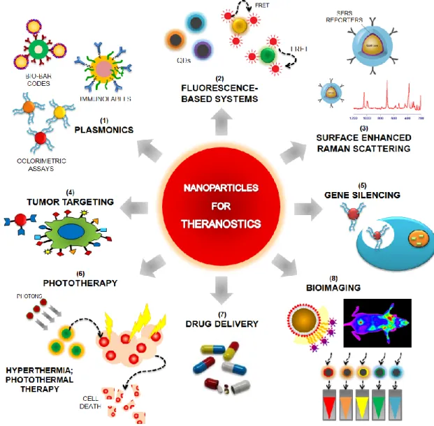
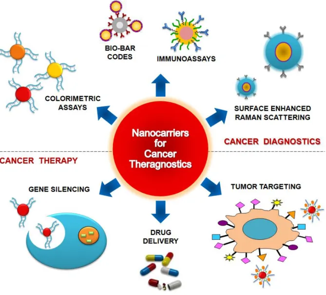
nanoparticles have been used so far are labelling, delivering, heating, sensing and detection [21] (Figure 6).
Figure 6. Nanoparticles for Theranostics. Nanoparticles-based strategies can be used for biosensing
using plasmonic nanosensors, such as metal nanoparticles functionalized with nucleic acid strand for colorimetric assays; Biobar-codes for protein detection or intense labels for immunoassays (1). Some nanoparticle systems can also be used for sensing by exploring a typical FRET system (2) or can be surrounded with Raman reporters (3) in order to provide in vivo detection (8), tumour targeting (4) and drug delivery (7). In fact, NPs symbolize an important class of materials with unique features suitable for biomedical imaging applications (8) such as increased sensitivity in detection and high quantum yields for fluorescence. Alternatively, NPs can survey near/far-field enhancing qualities that hold promise for a bounty of novel applications in optics and photonics. Engineered NPs can also act as phototherapeutic agents that can be attached to specific targets for selective gene silencing (5) or damage to cancer cells (6).
Nanodiagnostics can be defined as the use of nano-sized materials, devices or systems for diagnostics purposes. It is a burgeoning field as more and improved techniques are becoming available for clinical diagnostics with increased sensitivity at lower costs [3-6].
The use of the colloidal gold colour change upon aggregation is the best characterized example for diagnostic systems using AuNPs. In fact, AuNPs functionalized with ssDNA capable of specifically hybridizing to a complementary target for the detection of specific nucleic acid sequences in biological samples have been extensively used [3;5;61;63-68;70;71].
Other approaches are the use of AuNPs as a core/seed that can be tailored with a wide variety of surface functionalities to provide highly selective nanoprobes for diagnosis [22]; the utilization of Surface Plasmon resonance (SPR) scattering imaging or SPR absorption spectroscopy generated from antibody conjugated AuNPs in molecular biosensor techniques for the diagnosis and investigation of oral epithelial living cancer cells in vivo and in vitro [211]; the use of multifunctional AuNPs which incorporate both cytosolic delivery and targeting moieties on the same particle functioning as intracellular sensors to monitoring actin rearrangement in live fibroblasts [23]; and the employment of AuNPs in electrochemical based methods that can be coupled with metal deposition for signal enhancement [24].
Consequently, the use of thiol-linked ssDNA-modified gold nanoparticles (herein designated Au-nanoprobes) for the colorimetric detection of gene targets represents an inexpensive and easy to perform alternative to fluorescence or radioactivity-based assays [25]. In 1996, Mirkin et al. [26] described the use of a cross-linking method that relies on the detection of single-stranded oligonucleotide targets using two different Au-nanoprobes, each of them functionalized with a DNA-oligonucleotide complementary to one half of the given target [60;71].
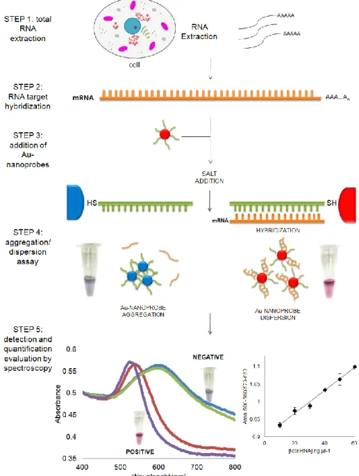
On the other hand, in 2005 Baptista et al. introduced a non-cross linking method where thiol-linked DNA-gold nanoparticles were used in a novel colorimetric method to detect the presence of specific mRNA from a total RNA extract of yeast cells [27]. The method consists in visual and/or spectrophotometric comparison of solutions before and after salt induced Au-nanoprobe aggregation (see Figure 7) – the presence of a complementary target prevents aggregation and the solution remains red and has a strong absorbance at ±520 nm; non-complementary/mismatched targets do not prevent Au-nanoprobe aggregation, resulting in a visible change of colour from red to blue and therefore a shift in the surface plasmon absorbance to 600-650 nm. This method has been successfully applied to detect eukaryotic
fully complementary from mismatched sequences, with a single base mismatch i.e. to detect common mutations within the β-globin gene [28]; and in a fast and straightforward assay for Mycobacterium tuberculosis DNA detection in clinical samples [6;62].
Figure 7. RNA detection using DNA-gold nanoparticles. Detection and quantification of the mRNA
using DNA thiol-modified gold nanoparticles. The assay is based on the increased stability of the nanoprobes upon hybridization with the complementary target in solution, while non-hybridized Au-nanoprobes easily aggregate once the solution’s ionic strength is increased. This method can be successfully applied to detect eukaryotic gene expression without retro-transcription or PCR amplification steps. Positive: sample in the presence of complementary target (solution remains red); Negative: sample in the presence of non-complementary target (solution turns blue).
advance molecular diagnosis assay that utilizes gold nanoparticles derivatized with thiol modified oligonucleotides is the most simple, rapid and with a great efficiency nanodiagnostic system.
NP-based biosensors provide a new horizon for novel functions with a variety of applications in clinical diagnostics and biological research. Gold NPs have already proven to be one of the most important groups of nanomaterials for biosensing approaches. Highly sensitive and specific biosensors based on AuNPs have open up the possibility of creating new diagnostic platforms for disease markers, biological and infectious agents in the early-stage detection of disease and threats, especially in cancer. [143;213]
2.1. NANOCARRIERS IN CANCER DIAGNOSIS
Cancer is the one of first leading causes of mortality in the modern world, with more than 10 million new cases every year [29]. However, advances in diagnosis and treating this disease that kills millions of people each year worldwide, have not been as effective as for other chronic diseases, and only for some types of cancer there are effective methods of detection [30]. Thus, the main challenge is to find new and more effective diagnostic agents for the monitorization of predictive cell molecular changes that are involved in tumour development. The key to the efficient and ultimately triumphant treatment of cancer is early and accurate diagnosis [31].
It’s here that nanotechnology enters the fray in the technological leap of controlling materials at nanoscale by offering a “big revolution” in new medical and healthcare diagnostic systems [20]. In fact, nanotechnology combined with biology and medicine is the most advanced technology both from an academic point-of-view and for commercial applications, producing major advances in cancer diagnostics and bioengineering [22;217].
Nanotechnology can be exploit for cancer theranostics via the development of diagnostics systems such as colorimetric and imunoassays, and in therapy approaches through gene therapy, drug delivery and tumour targeting systems [32].
The unique characteristics of nanoparticles in the nanometre range, such as high surface-to-volume ratio or size-dependent optical properties, are drastically different from those of their bulk materials and hold pledge in the clinical field for disease therapeutics [54;55].
Molecular nanodiagnostics applied to cancer may provide rapid and sensitive detection of cancer related molecular alterations, which would enable early detection even when those alterations occur only in a small percentage of cells. When referring to cancer therapy,
and decrease undesirable distribution to healthy organs and tissues. Multifunctional gold nanocarriers may potentiate the development of individualized cancer therapy based on the individual’s biological information within the tumour (biomolecular profiling). Gold nanocarriers can be modified with multiple cell-targeting and membrane translocating peptides, loaded with DNA/RNA and used as nanovectors [12;13].
A great effort has been applied to the detection of microorganisms and/or virus using gold SPR biosensors but very few were used for the detection of chronic diseases, such as asthma, Alzheimer's disease, diabetes, epilepsy, heart disease and specially cancer. Actually, molecular nanodiagnostics applied to cancer may provide rapid and sensitive detection of cancer related molecular alterations, which would enable early detection even when those alterations occur only in a small percentage of cells.
The high mortality rate in cancer is commonly attributed to the difficulties in detecting the disease at an early treatable stage. Therefore new synthesis, fabrication, and characterization methods are needed for developing highly advanced AuNPs capable of use in sensitive and multiple detection methods with negligible toxicity and high sensitivity. In the future, it might be possible to apply all AuNPs properties together and evolve new chemistry for synthesis of smart materials for diagnostic applications and clinical trials.
3. NANOTHERAPY
In medical term a therapeutic effect is a consequence of a medical treatment of any kind, the results of which are judged to be desirable and beneficial. Conventional therapy methods, for example in cancer, involve the employment of anticancer agents that do not greatly differentiate between cancerous and normal cells. Efficient in vivo targeting to heterogeneous population of cancer cells and tissue still requires better selectivity and non-cytotoxicity to surrounding normal cells. This fact leads to systemic toxicity, adverse effects and severe side effects [33].
In another way, universally targeting cells within a tumour is not always feasible because some drugs cannot diffuse efficiently and the random nature of the approach makes it difficult to control the process and may induce multiple-drug resistance (MDR), a situation where chemotherapy treatments fail patients owing to resistance of cancer cells towards one or more drugs [34]. Consequently, nanotechnology could offer a less invasive alternative, enhancing the life expectancy and quality of life of the patient [35].
by synergistic combinations of several multicomponent targeting strategies. Currently, it is imperative to develop technology for targeting and delivery of multiple therapeutic agents, and for the simultaneous capability of avoiding biological and biophysical barriers. For example, nanoparticles can extravasate into the tumour stroma through the fenestrations of the angiogenic vasculature, demonstrating targeting by enhanced permeation and retention. These particles are able to carry multiple antibodies, which further target them to epitopes on cancer cells, and direct antitumor action, leading to cell death. Irradiation might be use to activate the nanoparticles and set up the release of their cytotoxic action [36].
3.1. TUMOR TARGETING
It is expected that the greatest gains in therapeutic selectivity will be achieved by synergistic combinations of several multicomponent targeting strategies, i.e. capable of simultaneously target and deliver multiple therapeutic agents, while avoiding the organism’s biological and biophysiscal barriers. NPs targeting strategies to cancerous tissues have focused on passive and active targeting. In passive targeting, because numerous tumours present defective vasculature and poor lymphatic drainage due to the rapid growth of solid tumours, noble metal NPs can extravasate into the tumour stroma through the fenestrations of the angiogenic vasculature, demonstrating targeting by enhanced permeation and retention, thus accumulation at the tumour site [12;19;109]. Additionally, functionalization of the NP’s surface with hydrophilic molecules, such as PEG, can also greatly increase their solubility, help evading macrophage-mediated uptake and, thus, avoid removal from the systemic circulation and protect their carriers from enzymatic degradation when used in vivo [16]. For active targeting, NPs can be easily functionalized with a wide variety of biological moieties, such as antibodies, peptides and/or DNA/RNA to specifically target extracellular and intracellular receptors or pathways [16]. The use of NPs functionalized with multiple peptides or antibodies, such as monoclonal antibodies, have been described to successfully target specific cell surface proteins or receptors on cancer cells and further direct their antitumor action, leading to tumour cell death with minimal damage to collateral healthy cells [110-113]. In nucleic-acid functionalized NPs, DNA and RNA macromolecules can be used to simultaneously target specific sequences and exert their genetic-based therapy [114;115]. To help tracking nanoparticles in vivo and enhance the imaging properties of such moieties, leading to more efficient control of their therapeutic properties, they can also be
reporters.
3.2. GENE THERAPY
We are in the dawn of a new age in gene therapy driven by nanotechnology vehicles. Although there are technical challenges associated with the therapeutic application of nanoparticles, the integration of therapy with diagnostic profiling has accelerated the pace of discovery of new nanotechnology methods. The development of a safe, efficient, specific and nonpathogenic vehicle for gene delivery is highly attractive [17;18].
Gene therapy is receiving increasing attention and, in particular, small-interference RNA (siRNA) shows significant potential in new molecular approaches to down-regulate specific gene expression in cancerous cells. In fact, this non-viral-vector-mediated delivery of therapeutic siRNAs is highly desirable and constitutes an important challenge to gene therapy [14-16].
In fact, antisense DNA [119;120] and RNA interference (RNAi) via the use of small-interfering RNA [121-124] have emerged as powerful and useful tools to block gene function and for sequence-specific posttranscriptional gene silencing, playing an important role in downregulation of specific gene expression in cancer cells.
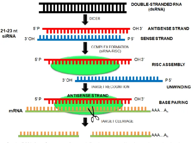
siRNAs are 21-23 nucleotide double strand nucleic acid molecules (dsRNA), with symmetric 2-3 nt 3’ overhangs and 5’-phosphate and 3’-hydroxyl groups, that mediate the cleavage of complementary mRNA sequences and thus regulate gene expression [121;129;347;348]. The RNA silencing pathway (Figure 8) begins with long dsRNA precursors that are processed to siRNA duplexes by the RNase-III-like enzyme Dicer. These short dsRNAs are subsequently unwound and assembled into an effector complex, RNA Induced Silencing Complex (RISC), which can direct RNA cleavage, mediate translational repression or induce chromatin modification. The antisense strand then binds to its complementary/target mRNA. The strand antisense to the targeted mRNA is often referred to as the guide strand, and its base-paired sense strand is known as the passenger strand, which is destroyed upon incorporation of the guide strand into RISC. The catalytic RISC recognizes mRNAs containing perfect or near-perfect complementary sequence to the guide siRNA and cleaves the mRNAs at a site precisely 10 nucleotides from the 5’-end of the guide strand. Finally, mRNA degradation is achieved by endo- and exonucleases, resulting in knockdown of the expression of the corresponding genes [349-351].
Figure 8. The RNA interference pathway, defined as a gene silencing pathway that is triggered by
double-stranded RNA (dsRNA). Gene silencing can be the result of nucleolytic degradation of the mRNA, or by translational suppression.
siRNAs can be transfected into mammalian cells by a variety of methods [125-128] that influence the strength and duration of the silencing response, which in turn is affected by the amount of siRNA that is delivered and on the potential of each siRNA to suppress its target. Thus, one drawback of using naked siRNAs is that they show extremely short half-lives, weak protection against action by RNases, poor chemical stability, and common dissociation from vector [37]. In fact, the major obstacle to clinical application is the uncertainty about how to deliver therapeutic RNAs (e.g., miRNA and/or siRNA) with maximal therapeutic impact. Nanotechnology offers an unprecedented opportunity to overcome these problems, as nanoscale devices, due to their small size, can readily interact with biomolecules on both the surface of cells and inside of cells for longer periods of time [38]. AuNPs have shown potential as intracellular delivery vehicles for antisense oligonucleotides [39] and for therapeutic siRNA by providing protection against RNAses and ease of functionalization for selective targeting [114;115]. For example, Mirkin and coworkers showed that AuNPs attached to single-stranded oligodeoxynucleotides can be used for gene therapy, providing a highly efficient gene regulator in terms of high loading of the antisense DNA with no toxicity
conjugates are readily taken up by cells and that the particle bound siRNA could effectively regulate genes in the context of RNA interference [40]. AuNPs modified with the hydrophilic PEG polymer, siRNAs and then coated with poly(β-aminoester)s have been shown to facilitate high levels of in vitro siRNA delivery and gene silencing in human cells [41]. Also, Braun et al. developed an Au-nanoshell functionalized with TAT-lipid layer for transfection and selective release of siRNA [42], where the TAT-lipid coating was used to efficiently mediate the cellular uptake of the nanoconjugates and the siRNA release was dependent on near-infrared (NIR) laser pulses. The authors demonstrated that this NIR strategy for siRNA release was proficient and time dependent. Several other studies using engineered NPs modified with siRNA have demonstrated a cytoplasmic delivery system of siRNA and efficient gene silencing using AuNPs [114;131;133;134]. However, almost all nanoconjugates using siRNA have exclusively been tested in cell cultures targeting only reporter genes. Further research into the fundamental mechanisms of gene therapy in vivo using nanodevices could unveil new dimensions of nanoparticle-mediated gene silencing that will have profound implications for understanding gene regulation, and which could also affect the development of functional genomics and therapeutic applications.
4. NANOTOXICITY
The AuNPs have a proclivity in vivo and in vitro to bioaccumulate within various types of cells with a special affinity for macrophage-type cells (both histiocytes and blood phagocytic cells), and reticuloendothelial cells throughout the body). They also produce varying degrees of bioaccumulation in such tissues as lymph nodes, bone marrow, spleen, adrenals, liver and kidneys [297-299].
Research shows that nanoparticles can stimulate and/or suppress the immune responses, and that their compatibility with the immune system is largely determined by their surface chemistry. In fact, is well known the influence of size, solubility and surface modification on the biocompatibility of nanoparticles and their use in biological applications [43].
AuNPs are generally considered to be benign. However, the size similarity of AuNPs to biological matters could provide “camouflage” to cellular barriers, leading to undesired cellular entry which might be detrimental to normal cellular function [44].
Pan and colleagues recently conducted a systematic investigation of the size-dependent cytotoxicity of AuNPs against four cell lines [45]. They found that AuNPs 1 to 2 nm in size displayed cell-type dependent cyotoxicity with high micromolar IC50s. In contrast, AuNPs 15
AuNPs. These results seemed to confirm size dependent toxicity of AuNPs [277;303-306], an inference that has been somewhat ambivalent.
Now the most imperative question rises up. Are the gold nanoparticles cytotoxic or biocompatible? And how can the gold nanoparticles be design to avoid these effects?
There does not seem to have a simple answer. Even though there is not any general mechanism for making nanoparticles universally ‘non-toxic’ to all living cells and all organisms, there are important findings that can be applied for increasing nanoparticle biocompatibility and reducing cytotoxic interactions in vivo and in vitro.
Using the lowest nanoparticle dose to get the desired response for the shortest period of time, in general, seems to promote biocompatibility as well as coating a nanoparticle if the outer coating completely covers the nanoparticle reactive surface (a non-continuous covering, the presence of cracks, roughness or interruptions could lead to complement or antibody attachment, or dissolution of the coating by cell digestion), and cannot be removed and utilized by the living cell [46].
It is essential to test nanoparticle/biological interactions experimentally and modify the nanoparticles for best biocompatibility with the cell in order to eliminate membrane lipid peroxidation; reduce the generation of reactive oxygen species; prevent acute and chronic release of inflammatory factors (and ‘complement’ activation); guard against alterations in genetic cellular function; and reduce the possibility of nanoparticles becoming ‘stuck’ during filtration or passage through pores and fenestrations [308] due to size, inflexibility of the nanoparticle core, or protein adsorption and agglomeration [46].
When interpreting nanoparticle interactions with biological cells and organisms, it is important to remember that living systems may appear normal and be capable of growth and function, but they may be genetically altered in subtle ways following nanoparticle exposure, which can produce serious consequences at some time in the distant future. Conversely, other cells that seem to be damaged may, in time, recover from nanoparticle exposure and function normally in the absence of the nanoparticles [46].
In conclusion, the only weapon that we have to insure that these new materials are well designed and safely used is to question and test each new nanoparticle to make sure that it has been designed for safety (with maximum biocompatibility) during handling, use and disposal.
These concepts were extensively reviewed in the Review Articles A, B and C and Book Chapter A at the end of this Introduction section.
1. Salata O. Applications of nanoparticles in biology and medicine. J Nanobiotechnology 2004; 2(1):3.
2. Dreaden EC, Alkilany AM, Huang XH, Murphy CJ, El-Sayed MA. The golden age: gold nanoparticles for biomedicine. Chemical Society Reviews 2012; 41(7):2740-2779.
3. El-Sayed MA. Some interesting properties of metals confined in time and nanometer space of different shapes. Acc Chem Res 2001; 34(4):257-264.
4. Kreibig U, Vollmer M. Optical Properties of Metal Clusters. Springer; 1995.
5. Barnes WL, Dereux A, Ebbesen TW. Surface plasmon subwavelength optics. Nature 2003; 424(6950):824-830.
6. Cao YC, Jin R, Mirkin CA. Nanoparticles with Raman spectroscopic fingerprints for DNA and RNA detection. Science 2002; 297(5586):1536-1540.
7. Storhoff JJ, Lazarides AA, Mucic RC, Mirkin CA, Letsinger RL, Schatz GC. What Controls the Optical Properties od DNA-Linked Gold nanoparticle assemblies? J Am Chem Soc 2000; 122:4640-4650.
8. Daniel MC, Astruc D. Gold nanoparticles: assembly, supramolecular chemistry, quantum-size-related properties, and applications toward biology, catalysis, and nanotechnology. Chem Rev 2004; 104(1):293-346.
9. Turkevich J, Garton G, Stevenson PC. The color of colloidal gold. Journal of Colloidal Science 1954; 9:26-35.
10. Frens G. Controlled nucleation for the regulation of the particle size in monodisperse gold suspensions. Nature (London), Phys Sci 1973; 241:20-22.
11. Wilcoxon JP, Abrams BL. Synthesis, structure and properties of metal nanoclusters. Chem Soc Rev 2006; 35(11):1162-1194.
by borohydride reduction. Chemical Engineering Journal 2008; 135:S104-S109. 13. Cao C, Park S, Sim SJ. Seedless synthesis of octahedral gold nanoparticles in
condensed surfactant phase. J Colloid Interface Sci 2008; 322(1):152-157.
14. Panigrahi S, Kundu S, Ghosh SK, Nath S, Pal T. General method of synthesis for metal nanoparticles. Journal of Nanoparticle Research 2004; 6(4):411-414.
15. Templeton AC, Wuelfing WP, Murray RW. Monolayer-protected cluster molecules. Acc Chem Res 2000; 33(1):27-36.
16. Sperling RA, Parak WJ. Surface modification, functionalization and bioconjugation of colloidal inorganic nanoparticles. Philos Transact A Math Phys Eng Sci 2010; 368(1915):1333-1383.
17. Giersig M, Mulvaney P. Preparation of ordered colloid monolayers by electrophoretic deposition. Langmuir 1993; 9(12):3408-3413.
18. Pellegrino T, Kudera S, Liedl T, Munoz JA, Manna L, Parak WJ. On the development of colloidal nanoparticles towards multifunctional structures and their possible use for biological applications. Small 2005; 1(1):48-63.
19. Sperling RA, Pellegrino T, Li JK, Chang WH, Parak WJ. Electrophoretic Separation of Nanoparticles with a Discrete Number of Functional Groups. Adv Funct Mater 2006; 16:943-948.
20. Gil PR, Parak WJ. Composite nanoparticles take aim at cancer. ACS Nano 2008; 2(11):2200-2205.
21. Sperling RA, Rivera GP, Zhang F, Zanella M, Parak WJ. Biological applications of gold nanoparticles. Chem Soc Rev 2008; 37(9):1896-1908.
22. You CC, Miranda OR, Gider B, Ghosh PS, Kim IB, Erdogan B et al. Detection and identification of proteins using nanoparticle-fluorescent polymer 'chemical nose' sensors. Nat Nanotechnol 2007; 2(5):318-323.
imaging intracellular biomarkers in live cells. Nano Lett 2007; 7(5):1338-1343.
24. Castaneda MT, Merkoci A, Pumera M, Alegret S. Electrochemical genosensors for biomedical applications based on gold nanoparticles. Biosens Bioelectron 2007; 22(9-10):1961-1967.
25. Storhoff JJ, Lucas AD, Garimella V, Bao YP, Muller UR. Homogeneous detection of unamplified genomic DNA sequences based on colorimetric scatter of gold nanoparticle probes. Nat Biotechnol 2004; 22(7):883-887.
26. Mirkin CA, Letsinger RL, Mucic RC, Storhoff JJ. A DNA-based method for rationally assembling nanoparticles into macroscopic materials. Nature 1996; 382(6592):607-609.
27. Baptista P, Doria G, Henriques D, Pereira E, Franco R. Colorimetric detection of eukaryotic gene expression with DNA-derivatized gold nanoparticles. J Biotechnol 2005; 119(2):111-117.
28. Doria G, Franco R, Baptista P. Nanodiagnostics: fast colorimetric method for single nucleotide polymorphism/mutation detection. IET Nanobiotechnol 2007; 1(4):53-57. 29. Siegel R, Naishadham D, Jemal A. Cancer statistics, 2012. CA Cancer J Clin 2012;
62(1):10-29.
30. Hanahan D, Weinberg RA. The hallmarks of cancer. Cell 2000; 100(1):57-70.
31. Etzioni R, Urban N, Ramsey S, McIntosh M, Schwartz S, Reid B et al. The case for early detection. Nat Rev Cancer 2003; 3(4):243-252.
32. Conde J, Doria G, Baptista P. Noble metal nanoparticles applications in cancer. J Drug Deliv 2012; 2012:751075.
33. Liu Y, Miyoshi H, Nakamura M. Nanomedicine for drug delivery and imaging: a promising avenue for cancer therapy and diagnosis using targeted functional nanoparticles. Int J Cancer 2007; 120(12):2527-2537.
emerging platform for cancer therapy. Nat Nanotechnol 2007; 2(12):751-760.
35. Cuenca AG, Jiang H, Hochwald SN, Delano M, Cance WG, Grobmyer SR. Emerging implications of nanotechnology on cancer diagnostics and therapeutics. Cancer 2006; 107(3):459-466.
36. Ferrari M. Cancer nanotechnology: opportunities and challenges. Nat Rev Cancer 2005; 5(3):161-171.
37. Hannon GJ, Rossi JJ. Unlocking the potential of the human genome with RNA interference. Nature 2004; 431(7006):371-378.
38. Baptista P. Cancer Nanotechnology - Prospects for Cancer Diagnostics and Therapy. Current Cancer Therapy Reviews 2009.
39. Rosi NL, Giljohann DA, Thaxton CS, Lytton-Jean AK, Han MS, Mirkin CA. Oligonucleotide-modified gold nanoparticles for intracellular gene regulation. Science 2006; 312(5776):1027-1030.
40. Giljohann DA, Seferos DS, Prigodich AE, Patel PC, Mirkin CA. Gene regulation with polyvalent siRNA-nanoparticle conjugates. J Am Chem Soc 2009; 131(6):2072-2073. 41. Lee JS, Green JJ, Love KT, Sunshine J, Langer R, Anderson DG. Gold,
poly(beta-amino ester) nanoparticles for small interfering RNA delivery. Nano Lett 2009; 9(6):2402-2406.
42. Braun GB, Pallaoro A, Wu G, Missirlis D, Zasadzinski JA, Tirrell M et al. Laser-Activated Gene Silencing via Gold Nanoshell-siRNA Conjugates. ACS Nano 2009. 43. Dobrovolskaia MA, McNeil SE. Immunological properties of engineered
nanomaterials. Nat Nanotechnol 2007; 2(8):469-478.
44. Connor EE, Mwamuka J, Gole A, Murphy CJ, Wyatt MD. Gold nanoparticles are taken up by human cells but do not cause acute cytotoxicity. Small 2005; 1(3):325-327.
cytotoxicity of gold nanoparticles. Small 2007; 3(11):1941-1949.
46. Bellucci S. Nanoparticles and Nanodevices in Biological Applications. Lecture Notes in Nanoscale Science and Technology ed. Springer; 2009.
In order to complement this introduction, we present three review articles and one book chapter on the application of noble metal nanoparticles in cancer diagnostics and therapy.
Review Article A
“Noble Metal Nanoparticles Applications in Cancer.” João Conde, G. Doria and P.V. Baptista. Journal of Drug Delivery (2012), Vol. 2012, pp. 1-12. Review article
Book Chapter A
“Multifunctional Gold Nanocarriers for Cancer Theranostics – From bench to bedside and back again?” João Conde, F. Tian, P.V. Baptista and J.M. de la Fuente (Submitted, Invited Book Chapter)
Review Article B
“Noble Metal Nanoparticles for Biosensing Applications.” G. Doria, João Conde, B. Veigas, L. Giestas, C. Almeida, M. Assunção, J. Rosa and P.V. Baptista. Sensors (Basel) (2012), Vol. 12, pp. 1657-1687. Review article (IF=1.953)
Review Article C
“Nanophotonics for Molecular Diagnostics and Therapy Applications.” João Conde, J. Rosa, J.C. Lima and P.V. Baptista. International Journal of Photoenergy (2012), Vol. 2012, pp. 1-11. Review article (IF=2.663)
Declaration of authorship
I, João Diogo Osório de Castro Conde, declare that the manuscripts preparation and writing was carried out by me, Prof. Pedro V. Baptista, Prof. Jesus M. de la Fuente, and all the associated co-authors.
I, Pedro V. Baptista, as supervisor of João Conde hereby acknowledge and confirm that the information above is correct.
![Figure 3. Localized surface plasmon resonance. Schematic representation of how the interaction of the electromagnetic waves with the metal NPs surface electrons generates a surface plasmon resonance [4]](https://thumb-eu.123doks.com/thumbv2/123dok_br/19188977.949037/23.892.164.745.596.847/localized-resonance-schematic-representation-interaction-electromagnetic-electrons-generates.webp)