Universidade de Lisboa
Faculdade de Ciências
Departamento de Biologia Animal
Characterization of the toxic potential of
nanomaterials using in vitro cell models
Mariana Salvado Pinhão
Dissertação
Mestrado em Biologia Humana e Ambiente
Universidade de Lisboa
Faculdade de Ciências
Departamento de Biologia Animal
Characterization of the toxic potential of
nanomaterials using in vitro cell models
Mariana Salvado Pinhão
Professora Doutora Teresa Rebelo (orientadora interna)
Departamento de Biologia Animal da Faculdade de Ciências da Universidade de Lisboa Doutora Maria João Silva (orientadora externa)
Departamento de Genética do Instituto Nacional de Saúde Doutor Ricardo Jorge, I.P.
Dissertação
Mestrado em Biologia Humana e Ambiente
Resumo
Os nanomateriais são estruturas com uma ou mais dimensões inferiores a 100 nanómetros. Devido à sua pequena dimensão, as nanopartículas apresentam atributos únicos, tais como a sua elevada área superficial relativamente à sua massa, reactividade ou força tênsil. Estas características influenciam grandemente algumas das propriedades dos nanomateriais, como a sua hidrofobicidade, carga ou toxicidade.
As propriedades das nanopartículas tornam-nas também muito úteis para o Homem, sendo aplicadas em medicina, farmácia, electrónica, cosmética, vestuário e biotecnologia, entre outras. O aumento de produção e utilização de nanomateriais tem vindo a aumentar também a possibilidade de exposição humana a este tipo de partículas, levando a preocupações relativas ao risco de toxicidade aguda ou crónica. A exposição humana pode ocorrer por diversas vias, sendo as mais relevantes a via inalatória, ingestão ou contacto com a pele. Dependendo do material e do órgão-alvo, a exposição a nanomateriais pode conduzir a diferentes consequências biológicas: a nível dos órgãos, os nanomateriais podem levar a inflamação ou a supressão do sistema imunitário e, a nível celular e molecular, a perturbações na estrutura e integridade do genoma, assim como a interacções com moléculas biológicas e inibição da actividade proteica, entre outras consequências.
Um dos nanomateriais mais utilizados são os nanotubos de carbono. Estes são constituídos por grafite cilíndrica disposta numa única camada (designados nanotubos de carbono de parede simples) ou em várias (nanotubos de carbono de parede múltipla). Os nanotubos de carbono apresentam propriedades como resistência e condutividade que os tornam muito úteis em aplicações como aparelhos electrónicos, vestuário ou biomedicina; cada vez mais, portanto, se torna provável a exposição ocupacional ou ambiental a este material. A semelhança estrutural destas partículas com fibras de amianto conduziu a questões relativas à sua segurança, pelo que já foram elaborados diversos estudos relativos aos seus efeitos biológicos. Alguns trabalhos sugerem que os nanotubos de carbono têm a capacidade de produzir toxicidade associada a lesões físicas, à produção de danos oxidativos por interacção com mecanismos celulares, ou a morte celular. Outros trabalhos defendem que estas partículas não causam toxicidade relevante.
O projecto de dimensão europeia “NANoREG” surgiu da necessidade de ser desenvolvida legislação e regulamentação apoiadas em conhecimento científico e adequadas à produção e ao uso actual de nanomateriais.
Este trabalho teve como objectivos principais a determinação do potencial cito- e genotóxico de um conjunto de nanotubos de carbono de parede múltipla (designados NM-400 a NM-403), e a consequente tentativa de associar este potencial às características físico-químicas dos nanomateriais. Com este
objectivo, a exposição por via inalatória foi analisada, pelo uso de duas linhas celulares in vitro provenientes de tecidos do tracto respiratório: epitélio pulmonar (células A549) e epitélio brônquico (células BEAS-2B).
A citotoxicidade dos nanotubos de carbono foi analisada com base em três parâmetros. Em primeiro lugar, as células foram contadas após a exposição aos nanomateriais utilizando o corante azul de tripanao para excluir as células inviáveis; a contagem foi realizada 3 e 24 horas após a exposição das células aos nanotubos. Os resultados deste ensaio apontam para a ausência de citotoxicidade após a exposição mais curta, e dados inconsistentes após a mais longa. Em segundo lugar, foi realizado o ensaio clonogénico, que se baseia na capacidade das células de se dividirem após a exposição ao agente em estudo. Este ensaio só foi realizado nas células A549 pois as BEAS-2B não permitem a formação de colónias. Os resultados apontam para uma citotoxicidade após a exposição a todos os nanomateriais, cuja intensidade se relaciona directamente com o tamanho das partículas, assim como ao seu diâmetro e área de superfície. Em terceiro lugar, foram calculados dois índices de viabilidade no ensaio dos Micronúcleos, cujo objectivo é avaliar se as células se dividiram durante a exposição aos nanomateriais em comparação com o controlo, e cujos resultados apresentam incoerências em relação aos outros já referidos. Estes dados podem ser justificados pelas diferenças existentes entre os ensaios, como o tempo de exposição ou a densidade celular.
Os efeitos genotóxicos dos nanomateriais foram avaliados com recurso aos ensaios do cometa e dos micronúcleos. O primeiro detecta lesões pequenas e reversíveis nas cadeias de DNA, ao passo que o segundo detecta efeitos irreversíveis ao nível cromossómico, tais como quebras ou perdas de cromossomas. Os resultados do ensaio do cometa sugerem que nenhum dos nanomateriais testados é genotóxico, uma vez que em ambas as linhas celulares e em ambos os tempos de exposição, os resultados são negativos. O ensaio dos micronúcleos, por outro lado, aponta para existência de genotoxicidade de dois dos nanomateriais (NM-401 e NM-402) nas células A549, mas não em células BEAS-2B.
Uma possível explicação para estes dados aparentemente contraditórios pode residir na hipótese de estes nanotubos de carbono serem compostos com efeitos aneugénicos, mas não clastogénicos: o ensaio dos micronúcleos permite a detecção de ambos os mecanismos de acção, ao passo que o ensaio do cometa só revela a quebra de cadeias de DNA. Outra justificação para os resultados é a possível influência da perda de viabilidade das células analisadas. Com base nos dados do ensaio clonogénico, estas partículas apresentam elevada citotoxicidade, pelo que os resultados dos ensaios de genotoxicidade, em particular do Ensaio do Cometa, poderão ser afectados por estes efeitos.
O meio de cultura usado para expor as células aos nanomateriais também é um parâmetro muito relevante na sua toxicidade. Neste trabalho, foram usados meios de cultura com proteínas, que podem ser adsorvidas pelas partículas e formar uma “corona” em seu redor; este processo pode alterar propriedades importantes dos nanomateriais, entre os quais o seu potencial efeito biológico. Também o método usado para conseguir uma dispersão homogénea de nanomateriais pode conduzir a diferenças nos resultados dos
ensaios de toxicidade. Neste estudo, foram observados alguns problemas relativos à perda de homogeneidade das dispersões de nanotubos de carbono, o que pode ter conduzido a que as células fossem expostas a massas de partículas de grandes dimensões conjuntamente com partículas individualizadas. O período durante o qual as células são expostas ao nanomaterial é também um aspecto essencial na produção de efeitos tóxicos.
Resumindo, este projecto forneceu informações relativas à toxicidade dos nanotubos de carbono que, complementadas pelas conclusões dos restantes parceiros do projecto europeu, poderão contribuir significativamente para a avaliação de risco e criação de legislação relativamente à utilização de nanomateriais. Na linha celular BEAS-2B, nenhum destes nanomateriais parece produzir efeitos tóxicos, quer a nível de célula, quer a nível de genoma, nas condições experimentais utilizadas. Nas células A549, por outro lado, os três nanomateriais testados parecem ser acentuadamente citotóxicos, e dois deles (NM-401 e NM-402) são também genotóxicos.
Em relação a perspectivas futuras, pode-se concluir que nem todos os ensaios de toxicidade existentes actualmente são adequados à análise de nanopartículas, pelo que novas metodologias devem ser desenvolvidas e complementadas por ensaios in vivo. Todos os estudos envolvendo nanomateriais deverão também descrever as características físico-químicas dos materiais usados, de forma a se poderem comparar os resultados com os de outros trabalhos.
Abstract
Carbon nanotubes are strong and flexible fibers that have a broad range of applications, such as in electronic devices, mechanical industry and medical procedures, among others. However, the use of these materials can also have consequences to human life and to the environment, as they may cause tissue inflammation, asthma or cancer. The general objective of this project was to provide some scientific evidence about the risks related to the use of carbon nanotubes, so that relevant legislation could be elaborated. More specifically, the study aimed at characterizing the cyto and genotoxicity of several carbon nanotubes. Cell counting coupled to the Trypan Blue Exclusion and the Clonogenic assays were performed to assess the cytotoxicity of a series of multi-walled carbon nanotubes (NM-400 to NM-403) in human lung (A549) and bronchial (BEAS-2B) epithelial cell lines, and the Comet and the Micronucleus assays were employed to characterize their genotoxicity.
The results pointed to a substantial difference in the nanomaterials toxicity in the two cell lines. In the BEAS-2B cells, no cytotoxicity or genotoxicity was produced by any of the nanotubes. In A549 cells, on the other hand, significant cytotoxicity was produced by three of the carbon nanotubes (401 to NM-403); the dose-response pattern appeared to be associated to the particle’s length, diameter and surface area. It is suggested that this may be related to physical aggression or to damage to the membranes of the cells by the particles. In terms of genotoxicity, the two longer nanotubes (NM-401 and NM-402) caused significant concentration-related chromosomal damage; a direct correlation between the aspect ratio of the particles was significant association to its genotoxicity. This may be related to interference of the particles in common biological processes, or to the generation of oxidative stress.
In the future, a standardized protocol should be used and a comprehensive list of characteristics should be provided in every study regarding nanomaterials, since parameters such as dispersion quality, medium protein content and properties of the particle are very influential in the toxicity of nanomaterials.
Acknowledgements
First of all, I want to show my gratitude for Doctor Maria João Silva, for allowing me to work at her laboratory and collaborate in this great project, and for helping me in every step of the way. Thank you for always defending by best interests, for giving me a lot of freedom in my work and for always being available to patiently discuss my confusing results.
I also want to thank Professor Teresa Rebelo, for all the support in the elaboration of this work, for the availability and for the good mood.
To the director of the National Institute of Health Doutor Ricardo Jorge, I.P., and to the coordinator of the Department of Genetics Doctor Glória Isidro, I am grateful for the opportunity to perform this study in the institute; to the Coordinator of the Research and Development Unit Doctor João Lavinha, I am also thankful for the opportunity to do this work in the institute, and for always having a moment to chat and to discuss important life decisions.
I need to express my gratitude for all the people who walked this road with me, because without them, this work would not exist. To Doctor Henriqueta Louro, for her constant availability to discuss my work and results, for her precious help with the assays and the writing, for patiently explaining how to perform the statistical analysis (twice), and for the permanent positive attitude. To Miguel Pinto, for teaching me most of what I know about laboratory work and management, for pushing me to do the best I can and for insisting that I work harder and harder every day. To Ana Tavares, for always giving me the opportunity to find the right answer by myself and for having patience when it took longer than it should have. To Sílvia José, for always having so much patience and joy when explaining things. To Doctor José Manuel Furtado, for helping us deal with technical problems and stubborn equipment.
I must also thank my lab partners and all the colleagues in the Genetics Department, for being a part of my life in this last year, and for always having a helping hand or a kind word.
Lastly, to my family and friends, for their help and support, and to my boyfriend, for always being right by my side. Thank you all for believing in me and for always encouraging me.
Table of Contents
Resumo ... i
Abstract ... iv
Acknowledgements ... v
Table of Contents ... vi
List of Figures ... viii
List of Tables ... x
List of Abbreviations ... xi
Introduction ... 1
1. Nanomaterials’ properties and biological interactions ... 1
2. Characterization of the Genotoxic Effects of the Nanomaterials ... 4
3. Multi-walled Carbon Nanotubes and Genotoxicity ... 7
4. The NANoREG Project ... 9
Objectives ... 11
Material and Methods ... 12
1. Cell Culture ... 12
2. Nanomaterials ... 14
a. Nanomaterial Sample Preparation ... 16
b. Scanning Electron Microscopy ... 16
3. Viability ... 17
a. Trypan Blue Exclusion Assay/Cell Counting Method ... 17
b. Clonogenic Assay ... 18
c. Proliferation and Replication indexes ... 18
4. Genotoxicity ... 19
a. Comet Assay... 19
b. Cytokinesis-blocked Micronucleus Assay ... 21
i. BEAS-2B cell line ... 23
ii. A549 cell line... 23
iii. Analysis ... 24
5. Statistical Analysis ... 25
Results ... 26
2. Tests for the Validation of the Methods – Ethyl Methanesulfonate ... 31 a. Viability ... 31 b. Genotoxicity... 32 3. Cytotoxicity of the MWCNT ... 36 a. Cell counting ... 36 b. Clonogenic Assay ... 38
c. Cytokinesis-blocked proliferation and replication indexes ... 39
4. Genotoxicity of the MWCNT ... 41
a. Comet Assay ... 41
b. Micronucleus Assay ... 44
Discussion ... 46
1. Qualitative Analysis of the Dispersion of the tested Nanomaterials... 47
2. Tests for the Validation of the Methods – Ethyl Methanesulfonate ... 49
3. Viability ... 52
4. Genotoxicity ... 58
Conclusions ... 65
References ... 66
Annexes ... a Annex A. Tables of the Results of the Tests for the Validation of the Methods – EMS ... a Annex B. Tables of the Results of the Viability Assays - MWCNT ... c Annex C. Tables of the Results of the Genotoxicity Assays – MWCNT ... f
List of Figures
Figure 1. Graphic representation of carbon nanomaterials. ... 7
Figure 2. Comparison of the structures of an agglomerate of carbon nanotubes and of a microtubule. ... 9
Figure 3. BEAS-2B cells in BEGM culture medium ... 13
Figure 4. A549 cells in DMEM culture medium ... 14
Figure 5. Examples of nucleoids obtained using Comet Assay ... 20
Figure 6. Examples of chromosomal damage in A549 cells ... 22
Figure 7. Scintillation vials containing the studied CNT ... 27
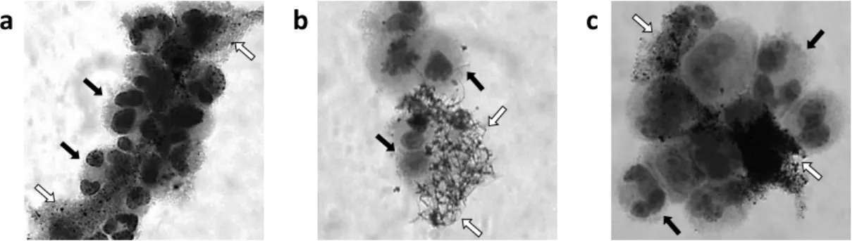
Figure 8. The three studied nanomaterials over or attached to Giemsa-stained BEAS-2B cells ... 28
Figure 9. NM-401 in different dispersion states ... 28
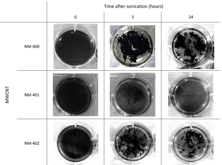
Figure 10. The three MWCNT in suspension in BEGM culture medium, immediately after cell exposure, three hours later and twenty-four hours after treatment ... 29
Figure 11. NM-402 suspension in BEGM culture medium, 15 minutes after the treatment (approximately 45 minutes after dispersion) ... 30
Figure 12. Lateral perspective of a plate well, with NM-402 in BEGM culture medium ... 30
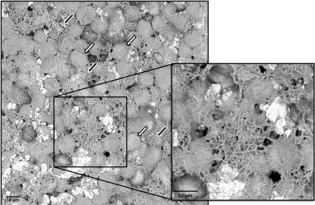
Figure 13. Scanning Electron Microscopy image of A549 cells treated with NM-401 ... 31
Figure 14. Results of the cell counting following EMS exposure. ... 32
Figure 15. Results of the unmodified Comet Assay with EMS on BEAS-2B cells ... 32
Figure 16. Results of modified Comet Assay with EMS on BEAS-2B cells ... 33
Figure 17. Oxidative damage caused by EMS on BEAS-2B cells. ... 34
Figure 18. Results of unmodified Comet Assay with EMS on A549 cells. ... 34
Figure 19. Results of modified Comet Assay with EMS on A549 cells ... 35
Figure 20. Oxidative damage caused by EMS on A549 cells ... 36
Figure 21. Comparison of the results of oxidative damage caused by EMS on BEAS-2B and A549 cell lines. 36 Figure 22. Results of the cell counting on BEAS-2B and A549 cells exposed to MWCNT. ... 37
Figure 23. Clonogenic assay results ... 38
Figure 25. Results of the CBPI and RI of BEAS-2B cells exposed to MWCNTs ... 40
Figure 26. Results of the CBPI and RI of A549 cells exposed to MWCNTs ... 40
Figure 27. Results of the Comet Assay in BEAS-2B cells exposed to MWCNT... 42
Figure 28. Results of the Comet Assay in A549 cells exposed to MWCNT ... 43
Figure 29. Results of the Micronucleus Assay in BEAS-2B cells exposed to MWCNTs ... 44
Figure 30. Results of the Micronucleus Assay in A549 cells exposed to MWCNTs ... 45
Figure 31. Association between the Clonogenic assay results and some of the tested MWCNT’s characteristics ... 57
Figure 32. Association between the maximum number of micronucleated binucleated A549 cells (MNBNCmax) following exposure to the tested MWCNT and the aspect ratio of the particles. ... 63
List of Tables
Table 1. Biomedical applications of some nanomaterials ... 2
Table 2. General uses of some of the most common nanomaterials ... 3
Table 3. General characteristics of the studied nanoparticles ... 15
Table 4. Results obtained in previous genotoxicity assays using the four mentioned nanomaterials ... 16
Table 5. Conclusions of the regression analysis performed on the results of the clonogenic assay on A549 cells, after 8-10 days exposure to each nanomaterial. ... 39
Table 6. Description of the symbols used in the summary tables presented in this chapter. ... 46
Table 7. Summary of the cytotoxicity results ... 53
List of Abbreviations
A549 – human epithelial lung adenocarcinoma cell lineATCC – American Type Culture Collection
BEAS-2B - human epithelial bronchial cell line
BEBM – Bronchial epithelial basal medium
BEGM – Bronchial epithelial growth medium
BSA – Bovine serum albumin
CBPI – Cytokinesis-blocked proliferation index
CNT – Carbon nanotube(s)
DAPI - 4',6-diamidino-2-phenylindole
DMEM – Dulbecco's Modified Eagle Medium
DMSO – Dimethyl Sulfoxide
DNA – Deoxyribonucleic acid
EDTA - Ethylenediamine Tetraacetic Acid
EMS – Ethyl Methanesulfonate
FBS – Fetal bovine serum
FBSi – Heat-inactivated fetal bovine serum
FP7 – Framework program 7
FPG – Formamidopyrimidine DNA Glycosylase
HEPES – 4-(2-hydroxyethyl)-1-piperazineethanesulfonic Acid
HPLC – High-Performance Liquid Chromatography
IC50 – Half maximal inhibitory concentration
INSA – Instituto Nacional de Saúde Doutor Ricardo Jorge (National Institute of Health, Portugal)
LDH – Lactate Dehydrogenase
MMC – Mitomycin C
MNBNC – Micronucleated binucleated cell(s)
MTT – 3-(4,5-dimethylthiazol-2-yl)-2,5-diphenyltetrazolium bromide
NM-400 to NM-403 – nanomaterial 400 to 403, as designated by Joint Research Centre
PBS – Phosphate Buffered Saline
RI – Replication index
ROS – Reactive oxygen species
RPMI – Roswell Park Memorial Institute
SD – Standard deviation
SEM – Scanning electron microscopy
SSC – saline-sodium citrate buffer
SWCNT – Single-walled carbon nanotube(s)
Introduction
1. Nanomaterials’ properties and biological interactions
Nanomaterials are natural or manufactured structures with unique properties, the most relevant being their size: at least one of their dimensions is inferior to 100 nanometers. Other characteristics – such as shape, surface area, chemical and biological reactivity, magnetism, conductivity, strength, resistance or charge – differ greatly from those of the same material in other dimensions [1-10].
These characteristics, however, have a great influence over some of the properties of the nanoparticles. Since most of the nanomaterials are hydrophobic and/or charged, they are easily attracted to one another and have a tendency to form aggregates or agglomerates (particles are held together by strong chemical bonds of by weak van der Walls forces, respectively [11]). This dynamic behavior alters the size of the particles and, consequently, may cause a variation of the other properties (particularly surface area and biological reactivity) [12-14].
Another important aspect to consider is that impurities that adhere to the exterior of the particle (resulting from air or water contaminants or residual materials left behind during the synthetic process) can change its properties, altering the charge or reactivity of the nanomaterial [6, 13].
In addition, when a biological system is exposed to nanomaterials, the particles have to travel through complex pathways inside the organism, subjecting them to very diverse environments, with specific biochemical conditions (protein composition, pH, enzymatic activity, metal composition, etc.) [12]. The small size and large aspect ratio of the particles cause them to easily interact with biological molecules in those environments, such as proteins, nucleic acid or lipids – depending on the type of particle and biomolecule, their characteristics, the agglomeration/aggregation state, the pH of the medium, etc. Consequently, some nanoparticles (such as metal oxides and carbon nanotubes) have the capacity to adsorb proteins, creating a “protein corona” – which may happen instantly when the nanoparticles contact with the biological medium. This corona may alter some of the particles’ properties, such as size, reactivity, charge or ability to aggregate/agglomerate or to bind to other proteins, as well as the preferred route inside the cells and the organism; besides, throughout the life cycle of the particle, the corona proteins may detach and contaminate the surrounding environment, possibly causing negative effects (which may, erroneously, be attributed to the nanoparticles) [1, 6, 12, 13]. Conversely, the formation of a protein corona around the nanoparticles may affect the proteins’ properties, such as secondary structure, flexibility, chemical composition and thermodynamic stability; these characteristics also influence the strength of the nanoparticle-protein association and the rate of binding and unbinding [12]. The adsorption of proteins to the nanoparticles may affect other events downstream, such as interactions between proteins, cellular signaling, DNA transcription and loss of enzyme activity – ultimately, it may lead to cellular
damage, apoptosis or abnormal immune response [12]. Gold nanoparticles and zinc oxide, for example, have been reported to cause conformational changes in bovine serum albumin, which does not happen with carbon C60 fullerenes; titanium dioxide reduces polymerization of tubulin, influencing cellular structure [12].
However, since the protein corona may, in fact, enhance the uptake of nanoparticles into the cells, and some nanoparticles have the ability to cross largely impermeable membranes such as the blood-brain barrier, nanotechnology represents a new possibility in the field of biomedical applications: the efficiency of the transport into the brain may be drastically increased by binding drugs and specifically targeted proteins to these nanoparticles, possibly enabling the treatment of neurological diseases [12, 15].
All of these properties have made nanoparticles very useful in several fields of human life [2, 3, 6], and the number of uses for nanotechnology keeps increasing. The most prominent field is medicine – nanoparticles are used in bioimaging, drug delivery, cancer therapy, fluorescent labels, and diagnostic agents, among others [16] (described in greater detail in Table 1).
Nanomaterials are also broadly used in other industries: pharmaceutical, automotive, aircraft, electronics, optics, ceramic, glass, paints, cosmetics, clothing, biochemical and environmental engineering, biotechnology, food, construction, etc. [10, 14, 17, 18] A short list of some of the main uses of nanomaterials in consumer products can be found in Table 2.
Table 1. Biomedical applications of some nanomaterials [15].
Nanomaterial Uses
Metallic nanoparticles
Iron oxide nanoparticles; manganese oxide nanoparticles; crystals of gadolinium oxide;
metal nanoshells; quantum dots
Tumor targeting, analysis and therapy. Contrast agents in optical imaging. Treatment for gliomas.
Quantum dots can also be used as a dye in fluorescence-based bioanalytical techniques
Carbon Nanotubes
May have a large variety of applications depending on size.
Molecular therapy or immunotherapy: direct delivery of antigens to antigen-presenting cells or
microglia in the central nervous system
Inorganic Nanoparticles
Ceramic nanoparticles; organosilicates; transition metal oxides; metalloids; metal
sulfides
Drug carriers; protect the drug molecules and keep them from being denatured or degraded by
the organism
Dendrimers
Highly branched macromolecules, with exterior end groups that can be functionalized
by the attachment of specific molecules
Drug carriers, by encapsulation of drugs. Large variety of groups can be attached to the exterior
of the nanoparticles, allowing for different properties and, consequently, different
Table 2. General uses of some of the most common nanomaterials.
Nanomaterial Uses Reference
Metallic nanoparticles
TiO2, Cr2O3,
Mn2O3, Fe2O3,
NiO, CuO, ZnO, ZrO2
Biochemical catalysts, electronic devices, water purification, cancer diagnostics and therapy, drug delivery, food additives,
artificial dyes, cosmetics, medical and dental implants, sunscreens, biosensors, bioimaging (as contrast agents).
[19-28]
Silver nanoparticles Textiles, cosmetics, health care, antibiotic agent in bandages
and medical material. [29-31]
Carbon Nanotubes
Drug carriers, rubber tires, pigments, electronics, catalysts, biosensors, photonics, tissue engineering, batteries,
composites.
[30, 32-34]
The number of individuals working in all of these industries who are, therefore, potentially exposed to the nanomaterials is very high and constantly increasing, which is raising many questions concerning the safety of the nanomaterials. Also, a large number of industries using nanotechnology means a large number of products with nanoparticles imbedded in them, which may increase the environmental exposure of the general population too [5, 8, 35, 36].
During the nanomaterial lifecycle, from the synthesis to the disposal of the nanomaterials, there are many occasions in which environment contamination is also possible and, in some cases, even likely. Accidentally or deliberately, nanoparticles may reach the soil or courses of water, where they can deposit or react, possibly altering their properties, or be transported far from where they were originally produced or discharged. Agglomerates/aggregates will likely be formed, as well as bonds between nano- and metallic particles naturally present in the environment [14, 17].
There are several routes of human or animal exposure to the nanoparticles, such as inhalation of airborne particles (more likely and frequent in both occupational and environmental exposure), absorption of nanoparticles through the skin, ingestion of the materials included in food products, inoculation with pure or processed nanoparticles (relevant in medical applications) and through physical contact of the nanomaterials with cuts in the skin [1, 3, 7, 37]. It is, therefore, necessary to study the effect the different nanoparticles have in the human organism through each of these routes, since each of them may affect different organs or vital processes. However, it is possible that the nanoparticles not only have an effect on the more exposed organ (such as skin, lung or stomach), but also on the others: nanomaterials may be transported to different locations throughout the body, travelling through the blood circulation or the lymph system [1, 37]. They may then deposit in sensitive organs, such as bone marrow, lymph nodes, spleen, heart and central nervous system [1, 7, 10]. The distribution of the nanoparticles inside the organism, as well as the interactions with the cells depend greatly on the characteristics of the particles (size, shape, surface area, coating, agglomeration/aggregation, etc.) and the cell types in question [1, 4, 6, 32, 36].
The size of the nanoparticles is the most influential feature in terms of biological reactivity and toxicity, due to the increase in the ratio between surface area and mass of the particle. Their size also allows the particles to disperse throughout the entire organism and to penetrate several barriers and membranes, reacting with biomolecules and interfering with biological processes. Additionally, the increase in surface area leads to an increase in the release of free radicals and metal ions from the nanoparticle, which may interact with the cells and molecules. Other characteristics of the surface of the nanoparticles, such as charge or coating, may also influence greatly the biological properties of the material, particularly those relating to toxicity. Due to those changes to the particles, the binding of other molecules (or nanoparticles) may be enabled of inhibited, altering the way these particles are internalized, processed or eliminated from the organism. Agglomerated particles, for example, may lose the ability to enter the cells or the organelles due to the increase in size, reacting differently to biological systems than the non-agglomerated particles. It is not possible to extrapolate toxicity data from larger particles with the same chemical composition, since nanomaterials may have different properties, causing alterations in the uptake, distribution, metabolism and elimination of the particles [35].
Once inside the organism, the nanoparticles able to penetrate cell membranes mostly do it through endocytosis (phagocytosis or pinocytosis)[1] or electrostatic attraction, and may stay free in the cytosol or within phagosomes [12] – unlike most xenobiotics, which are degraded in lysosomes and exocytosed. Consequently, nanoparticles may react to cell organelles or enter the nucleus, interfering in the normal cell processes and damaging essential molecules, such as DNA and proteins [34]. Some particles may even be able to cross the nuclear membrane and enter the nucleus by diffusion across the membrane or by transport through nuclear pore complexes [35]. Besides, some nanoparticles are made from non-biodegradable materials, which give them the capacity to stay in biologic tissues for years after the original exposure [4, 6, 35, 36].
At the organ and tissue level, it is thought that nanoparticle exposure may result in inflammation, oxidative stress [6, 35, 38, 39], stroke, myocardial infarction, alterations in the permeability of the blood-brain barrier [37] and suppression of the immune system response.
2. Characterization of the Genotoxic Effects of the Nanomaterials
A genotoxic event is one in which a chemical molecule triggers a reaction which will, after a period of time, lead to a permanent change in the genome of the cell. Examples of genotoxic events are direct damage to the DNA molecule, interference with processes such as mitosis and DNA replication or repair and disruption of the normal function of enzymes and proteins. Depending on the type of damage and its extent, these events may be deadly to the cells, or may be repaired quickly (through base or nucleotide
excision repair or mismatch repair, for example). To the organism, these occurrences may result in reproductive defects, developmental abnormalities, genetic diseases and carcinogenesis [40, 41].
At the cellular level, nanoparticles may cause genotoxicity and, consequently, possibly lead to carcinogenesis. This genotoxicity may be caused by the direct interaction of the nanoparticles with the DNA molecules, or by damage from by-products of the nanoparticle biotransformation (such as reactive oxygen species or ions released from the particles). The first, called direct genotoxicity, disrupts processes such as replication and transcription, and affects the structure of the DNA molecule; at the chromosome level, nanomaterials may cause breaks and losses (clastogenesis and aneugenesis, respectively), both mechanically and by chemically binding to the molecules [35]. The latter is known as indirect genotoxicity. The nanomaterials interact with proteins and the mitotic structures, disturbing the processes of replication, transcription, repair and cell division. Protein activity may be inhibited or altered, and the DNA molecules may be affected by by-products of nanoparticle processing – such as reactive oxygen species (ROS), transition metals (such as Fe2+, Ag+, Cu+, Mn2+, Cr5+ and Ni2+) and antioxidants – or remains of cell components – such as mitochondria (which, when damaged, can also produce ROS), inflammatory cells, ions and transcription factors. These by-products, particularly ROS, may attack the DNA molecules, and cause purine- and pyrimidine-derived oxidized base lesions (producing 8-OxoGuanine, for example, which is the most frequent product of purine oxidation, and is highly mutagenic) and DNA strand breaks [13, 35, 39]. Besides genotoxicity, nanoparticles can cause damage in other biomolecules and cell processes, such as methylation and phosphorylation; modification of proteins may lead to the silencing of genes and to changes in gene expression, altering the production and metabolism of proteins and the normal survival of the cell [35].
An extensive review of several assays that can be used to test nanomaterials for their geno- and cytotoxicity, as well as other useful data, can be found in Singh et al. [42]. As a concise review of this information, and focusing solely on the genotoxicity testing, the following list summarizes the main assays used to analyze the genotoxic effects of nanomaterials: the alkaline comet assay (or single cell gel electrophoresis assay), which detects the single- or double-strand breaks in DNA and, with modifications, other endpoints, such as oxidative damage; the cytokinesis-blocked micronucleus assay, which identifies chromosome instabilities, such as loss or fragmentation of chromosomes, as well as apoptotic or necrotic events, or even cytotoxicity; Ames test, which uses Salmonella bacteria to determine the capacity of a certain chemical to cause reversion of mutations; chromosome aberrations test, where the cell cycle is arrested at metaphase and the chromosomes are observed for structural or numeric alterations; detection of DNA adducts, such as 8-hydroxydeoxyguanosine, through HPLC or mass spectrometry based techniques [42].
Besides from these tests, other endpoints that do not involve genotoxic events include analysis of protein expression, phosphorylation and activity, testing cell viability and proliferation (with MTT assay,
Neutral Red assay, trypan blue staining of unviable cells, or analysis of the proliferation and replication indexes provided by the micronucleus assay), measuring glutathione production, analyzing cytokine activity, analyzing histological aspects, etc. [42]
The properties of the particles are key factors in their distribution, transport and toxicity; the coating and impurities, for example, may influence the particles’ tendency to bind to one molecule and not to another, creating the possibility that a toxicity result may be attributable to the coating, rather than to the nanoparticle itself [6, 13]. Another example is the protein corona: it can increase the particles' mobility and their capacity to penetrate organs otherwise inaccessible, as well as affect their biological activity [6, 13]; besides, the transformation and degradation of the proteins attached to the nanoparticles (as they travel throughout the organism and possibly deposit in one or more organs) may affect these parameters even further, causing changes in the toxicity of the nanomaterials over time [13].
It is not clear whether any of these (or other) properties is singularly responsible for the possible toxicity of the nanomaterials, or if it is due to the combination of several [6, 35]. The majority of the authors have obtained conflicting results concerning the toxicological impact of nanomaterials; this is most likely caused by differences in the physicochemical properties of the studied nanomaterials (such as type, size, composition, shape, stability, coating, surface area, electrical charge, etc.) [6, 10, 32, 35], as well as to other variables inherent to the assays used and the characteristics of the exposure (dispersion method, exposure time, concentration, culture medium, etc.) [6, 32, 35]. The solvents used for the preparation of the nanoparticles solutions or dispersions, for example, may differ in pH, temperature or dissolved molecules (such as proteins or metals), altering the results obtained in the assays. The dispersion protocol – procedure intended to break apart the nanoparticle agglomerates or aggregates in order to obtain as many single particles as possible – is a large influence on the outcome of the assays, since it alters the size of the particles. The type of assay also influences greatly the obtained results regarding the toxicity of the nanomaterials – in vitro methods can mainly detect the primary genotoxicity, and in vivo assays may be able to detect secondary genotoxicity too, due to the influence of the immune system of the tested animals. Concerning the in vitro assays, the toxicity of the nanoparticles depends on cell line, since different cell types have distinct susceptibility, metabolic activity, DNA repair capacity and particle internalization properties, among other unique characteristics. The assessment of nanoparticle genotoxicity should be performed using several assays and endpoints, due to the fact that there may be more than one mechanism leading to the DNA and cellular damage [35].
3. Multi-walled Carbon Nanotubes and Genotoxicity
Carbon nanotubes (CNT) are nanoparticles with a unique structure and very attractive physical, chemical and electrical properties, such as high tensile strength and conductivity. They may be separated in three groups, depending on the structure of the material: if they have only one flat graphite layer, they are known as graphene; if they have only one graphite layer, but it is cylindrical, they are Single-Walled Carbon Nanotubes (SWCNT); if, on the other hand, they have several layers of cylindrical graphite, they are Multi-Walled Carbon Nanotubes (MWCNT) [39, 43] (see Figure 1).
Figure 1. Graphic representation of carbon nanomaterials. (A) Graphene sheet; (B) Single-walled carbon nanotube; (C) Multi-walled carbon nanotube. [44]
CNT have a broad range of applications, such as in electronics, electrical appliances, batteries, clothing, biotechnology and composites [32, 34, 39, 43]; in biomedicine alone, CNT are used for diagnostics and therapeutics, as well as for recognition of antibodies, sequencing of nucleic acids, as biocatalysts of biological reactions, as components in regenerative surgery (in the central nervous system and in orthopedic interventions) and as drug delivery vectors [43].
However, CNT are hydrophobic and, therefore, difficult to solubilize in biological fluids; as a result, several different protocols have been proposed to disperse these particles [45-48]. In these procedures, CNT are often modified by binding them to chemical and/or biological molecules (such as proteins, polymers or surfactants), enhancing their solubility and bioavailability. These modifications are controversial, though, since they may alter properties of the nanoparticles other than their solubility, and may increase their toxicity [40, 43].
Due to CNT’s small dimensions and low density, exposure through the respiratory route is likely, especially in occupational settings; if the particles have been modified as stated above, a reaction between the CNT and the biological molecules may happen, leading to a response. Besides, these particles are degraded slowly, and therefore can stay in the organism or in the environment for a long period of time after initial exposure [49].
These facts have raised some concerns regarding the possible toxicity of the CNT. Some authors have proposed a toxicity profile identical to that of asbestos, based on the similarity of shape between the two materials: the particles may penetrate the intrapleural space and cause inflammation and tissue fibrosis
[40, 49, 50]. Other authors suggest that the damage induced by the CNT to the DNA of the cells is related to direct mechanical injury (thus, associated to the physical characteristics of the nanotubes), and not to the production of ROS and oxidative damage [35]. There have been reports with discrepant results, some displaying genotoxicity and some not, possibly due to differences in the experimental methods used or in the characteristics of the particles (for example, contaminants or residues in the particles, such as metals) [32, 35, 40, 49].
Several in vivo studies reported that exposure to CNT cause granulomatous inflammation, pulmonary fibrosis, increased release of lactate dehydrogenase, cell hypertrophy and hyperplasticity, nuclear abnormalities in macrophages or disruption of the mitotic spindle, among other negative effects. Genotoxicity is a frequently observed consequence of CNT exposure, as well as increased proliferation of epithelial cells, which is a common feature of pulmonary carcinogenesis [40, 49].
In vitro studies have shown evidence of increased release of lactate dehydrogenase, and depletion of glutathione and superoxide dismutase – enzymes that protect the cells from oxidative damage. These effects indicate production of ROS; ROS may be formed due to the direct effect of the CNT inside the cell, or due to the internalization of the nanotubes by the mitochondria and subsequent mitochondrial dysfunction. This induces tissue inflammation, activation of cellular signaling pathways, DNA damage, chromosomal aberrations, abnormalities in the cell growth and ultimately, cell death; all these processes may lead to lung injury and carcinogenesis [39, 40, 49].
There are authors who propose yet another consequence of CNT exposure: nanotubes may be able to interact with microtubules [49]. Microtubules are dynamic polymers, formed by subunits of alpha and beta tubulin bound by non-covalent hydrogen bonds; when cell division occurs, the microtubules assemble at the centrosome and form the mitotic spindle, pulling the chromosomes to opposites sides of the cell. Throughout this process, the length of the spindle varies due to the activity of cellular motors (such as kinesin and dynein), which assures the correct segregation of the duplicated chromosomes into the two daughter cells. The CNT may disturb this process, inhibiting the activity of the cellular motors and disrupting the centrosomes; it may even occur the incorporation of nanotubes in the mitotic spindle (the two structures have approximately the same diameter; see Figure 2), preventing it from varying in size or shape, and not allowing the separation of the daughter cells. This theory offers an explanation for the number of abnormalities in number and size of chromosomes observed in cells exposed to CNT (micronuclei or multinucleated cells, for example) and, ultimately for the possible link to cancer [49].
Figure 2. Comparison of the structures of an agglomerate of carbon nanotubes and of a microtubule. The straight lines on the agglomerate of CNT represent the individual fibers; the small spheres in the microtubule represent the
alpha and beta tubulin. Adapted from [49].
4. The NANoREG Project
Considering the importance of nanotechnology in the current society and the effort made by laboratories world-wide to understand its toxic potential, it is essential to make the connection between the scientific body and the regulators and legislators, and to start making informed decisions concerning the safety of nanoparticles. With this purpose, the European Commission approved a project called NANoREG - A common European approach to the regulatory testing of nanomaterials (FP7/2007-2013, grant agreement 310584; http://www.nanoreg.eu/), which is inserted in the Seventh Framework Program (FP7) [51].
The main objective of NANoREG is to provide solutions for already existing problems concerning nanomaterials, providing regulators and scientists with the means to perform risk assessment, involving toxicity testing and exposure measurements. In the long term, the project aims to develop or adapt new protocols for testing the particles, as well as to establish a better collaboration between science, industry and regulators, in order to more efficiently manage the risk inherent to the utilization of nanotechnology [51].
A large number of European countries are contributing to NANoREG, such as Netherlands, Belgium, Germany, Denmark, France, Austria, the United Kingdom, Switzerland, Spain, Ireland, Sweden, Norway, Italy, Finland and Portugal. However, this project also intends to establish connections and associations with other countries, such as the USA, Canada, Australia, Japan and Russia, in order to globalize the standards to nanotoxicology testing and legislation [51].
In Portugal, the NANoREG project is represented by PToNANO, which is a consortium of four entities: the Institute for Welding and Quality, the National Institute of Health Doutor Ricardo Jorge, I.P (INSA), the Portuguese Institute for Quality, I.P and the Portuguese General Directorate of Health [51].
INSA, being the state health laboratory and having the mission of contributing to international knowledge and guidelines in public health, is involved in performing research concerning the toxicological
aspects of the nanoparticles, in the development of new analytical methods, and in the answering of questions related to the materials’ safety. This data will be provided to the authorities and to the industry, in order to better develop legislation and solutions regarding occupational and environmental exposure to nanomaterials [51].
Objectives
The general objective of this project was to contribute to the assessment of the influence of the physicochemical properties (such as length, thickness and surface area) of a set of well characterized multi-walled carbon nanotubes on their cyto- and genotoxic potential, in order to understand the characteristic that is the most relevant to its toxicity.
The specific objectives of this project were: i) to test the genotoxic potential of three carbon nanotubes in human respiratory tract cells using in vitro methodologies (comet and micronucleus assays); ii) to test the cytotoxicity of these three nanomaterials (cell counting method and the clonogenic assay); iii) to try to disclose relationships between a certain physicochemical property and the toxicity of the carbon nanotubes.
Material and Methods
1. Cell Culture
There are many advantages in using in vitro cell models in toxicity testing of any chemical instead of in vivo systems, such as the reduction of the number of animals, monetary costs and time, as well as the existence of numerous well-characterized and available cell models. Besides, there is also the possibility to increase the complexity of the in vitro system, by including different cell lines in the study, in order to analyze different endpoints; by adding chemical agents (such as antioxidants, cell pathways inhibitors, or growth factors), it is also possible to alter several biochemical parameters and, therefore, study other mechanisms [52].
However, there are also disadvantages in the use of in vitro models, the most prominent being the limited number of different cell lines that can be combined in only one in vitro system; these systems, consequently, cannot adequately mimic the processes that happen in vivo, where several cell types interact, and the different tissues and organs may influence the cellular and molecular mechanisms. Another aspect to have in mind when using in vitro cell models is that the target organ for the exposure must be already known, since the toxicity testing will be focused on a single tissue; in addition, in in vitro systems, evidently, the influence of the immune system on the cellular and molecular mechanisms is inexistent [52].
The route of exposure and the potential target organ of a given particle or molecule are the main factors that define the in vitro cell types used in a toxicity study. However, this decision may be problematic, as different cell lines can produce different assay results, due to the fact that the molecular metabolism and toxicity response of cells from different tissues can change greatly. In addition, the characteristics of the cells when growing in culture may influence their susceptibility to the chemicals or particles, as their metabolism may be altered due to changes of medium or to cell density (the concentration of proteins or serum may cause morphological changes in the cells, and the achievement of confluence may inhibit the growth of some cell lines) [52].
The Bronchial Epithelium Cell Basal Medium (BEBM), as well as the growth supplements (BEGM SingleQuots) is from Lonza/Clonetics (Basel, Switzerland). The Hyclone Fetal Bovine Serum (FBS) is from Thermo Scientific (Waltham, MA, USA). All the other reagents used in cell culture – phosphate buffer saline (PBS 1X; without calcium, magnesium or phenol red), trypsin-EDTA 0.05% (1X), RPMI-1640 Medium (1X) + GlutaMAX (RPMI-1640), the heat-inactivated Fetal Bovine Serum (FBSi), Dulbecco’s Modified Eagle Medium (DMEM; with 1 g/L glucose, L-glutamine and pyruvate), HEPES buffer solution (1M), amphotericin B (Fungizone; 250 µg/mL), Penicillin/Streptomycin mix (Pen/Strep; with 10000 units/mL of penicillin and 10000 µg/mL of streptomycin) and Trypan Blue Stain (0,4%) – are from Gibco (Scotland, UK).
Considering the aim of this project, the chosen cell lines were the BEAS-2B and the A549, both isolated from human respiratory tract tissues.
The BEAS-2B cell line was obtained from the American Type Culture Collection (ATCC No. CRL-9609; see Figure 3); it was isolated from healthy human bronchial epithelium, and was obtained from the autopsy of non-cancerous individuals. Later, the cells were infected with an adenovirus 12-SV40 virus hybrid (Ad12SV40), and then cloned. These cells display adherence properties, and are suitable hosts for transfection processes [53].
Figure 3. BEAS-2B cells in BEGM culture medium.
Due to their main purpose – being a protective barrier to the bronchial tissue – these cells produce and release important immunological molecules, such as lipid mediators, inflammatory enzymes and cytokines [54]. For this reason, and since this cell line retains the ability to differentiate when exposed to serum, it is often used to analyze chemical and biological agents, testing for potential to induce or affect events of differentiation and/or carcinogenesis in pulmonary tissue [53].
The growth medium used for the BEAS-2B cell cultures was the serum-free Bronchial Epithelium Growth Medium (BEGM), which consisted of 500 mL of BEBM and 6 mL of BEGM SingleQuots: 2 mL of bovine pituitary extract, 0.5 mL of hydrocortisone, 0.5 mL of human epidermal growth factor, 0.5 mL of epinephrine, 0.5 mL of transferrin, 0.5 mL of insulin, 0.5 mL of retinoic acid, 0.5 mL of triiodothyronine and 0.5 mL of gentamycin/amphotericin B. The cells were maintained in culture flasks in an incubator at 37 ºC, in 5% CO2.
When the cells reached a confluence of 80%, a subculture was performed: the cells were washed with preheated sterile PBS and incubated for 5-7 minutes with trypsin-EDTA in an incubator at 37ºC. When the cells were detached from the flask, inactivation medium (RPMI 1640 with 10% FBSi) was added. Fifty µl of the cell suspension were added to equal volume of Trypan Blue solution, counted in a Neubauer chamber, and the cell density was determined. The volume corresponding to 1x10⁶ cells was centrifuged for 5 minutes at 800 rpm, the cell suspension was ressuspended in BEGM medium and then transferred to a new culture flask and incubated.
The A549 cell line was obtained from the American Type Culture Collection (ATCC No. CCL-185; see Figure 4); it was isolated from a lung carcinoma of a 58 year old Caucasian male. These cells display adherence properties, and are suitable transfection hosts [53].
Figure 4. A549 cells in DMEM culture medium.
These cells possess important molecules involved in the detoxification of the cells, such as P450 cytocrome. For this reason, the A549 cell line is important in the study of metabolic pathways, as well as the mechanisms involved in drug delivery and processing at the pulmonary epithelium [55].
The growth medium used for the cell cultures in the first 2/3 passages after defrosting was DMEM, with 1% Pen/Strep, 2.5% HEPES buffer and 10% heat-inactivated Hyclone FBS. Later, when the cells stabilized in their normal pattern of growth, this medium was supplemented with 1% fungizone. The cells were maintained in culture flasks in an incubator, at 37 ºC, in 5% CO2.
When the cells reached 80% confluence, a subculture was performed: the cells were washed with preheated trypsin-EDTA, and incubated for 4 minutes in an incubator at 37ºC with trypsin-EDTA. When the cells were detached from the flask, culture medium was added to the cell suspension to inactivate the trypsin-EDTA; the suspension was then divided to new culture flasks, depending on the growth rate of the cells before the trypsinization process, and incubated in the same conditions as before.
2. Nanomaterials
In this project, three different MWCNT were studied: NM-400, NM-401 and NM-402 from the Joint Research Centre Repository (Institute for Health and Consumer Protection, European Commission, Ispra, Italy). Previously, a different project with similar goals as this one (“NANOGENOTOX – Safety Evaluation of Manufactured Nanomaterials by Characterization of their Potential Genotoxic Hazard”; Grant agreement 2009 2101) tested NM-402 and NM403 (another MWCNT with comparable features to the three mentioned above) in human lymphocytes and in vitro models of human respiratory tract tissues cells (BEAS-2B and A549 cell lines). The characteristics of NM-403 were added to this section and the conclusions
obtained in the mentioned work were added to the analysis of the results, in order to compare them to those obtained in this project.
All of these nanomaterials have applications in the energy industry and are commonly used as structural composites. In terms of impurities, a small percent has been found in these nanomaterials, particularly aluminum, iron and zinc [56, 57]. Their properties differ in several aspects, especially in size; the most relevant characteristics may be consulted in Table 3.
Table 3. General characteristics of the studied nanoparticles [32]. * – [57] Nanomaterial Average Length ± SD
(nm) Average Diameter/Thickness ± SD (nm) Surface Area (m2/g) Aspect Ratio ± SD NM-400 726.3 ± 1.8 10.8 ± 1.3 280 67.3 ± 1.8 NM-401 3366.4 ± 1.9 62.8 ± 1.4 300 53.6 ± 2.0 NM-402 1141.3 ± 2.0 10.7 ± 1.3 250 107.1 ± 1.9 NM-403 394.3 ± 1.6 11.1 ± 1.5 135* 35.6 ± 1.8
NM-402 and NM-403 have already been tested for their genotoxicity in the A549 cell line and in human lymphocytes. The results were ambiguous, pointing to toxicity in some cases and not in others: in A549 cells, a decrease in cell viability was noted in both nanomaterials, but a genotoxic effect (coupled with a dose-response relationship) was only proved in NM-402 and in one of the performed genotoxicity assays (Micronucleus assay); in human lymphocytes, on the other hand, no cytotoxicity was observed in either case, but both nanomaterials have shown genotoxic effects (without a dose-response association). NM-403 was also tested in BEAS-2B, and the results proved that this nanomaterial is not cytotoxic or genotoxic in this cell line.
In that project, it was not possible to make any association between the dimensions of the nanomaterials and their toxicity; consequently, other explanations for those results were proposed, such as surface properties or impurities in the solution [32]. This last point is in agreement with the findings of other authors, who suggested that metal traces may be responsible for the biological effects nanomaterial solutions have in cells [58].
The other two nanomaterials (NM-400 and NM-401) have been tested only in human lymphocytes, showing negative results for genotoxicity [32]. This data is summed in Table 4.
Table 4. Results obtained in previous genotoxicity assays using the four mentioned nanomaterials.
- negative results, + positive results, +/- positive results is some assays, and negative in others.
Nanomaterial Lymphocytes A549 BEAS-2B
Cytotoxicity Genotoxicity Cytotoxicity Genotoxicity Cytotoxicity Genotoxicity
NM-400 - -
NM-401 - -
NM-402 - + + +/-
NM-403 - + + - - -
a. Nanomaterial Sample Preparation
For the dispersion of the MWCNT, an adaptation of the protocol used in the project NANOGENOTOX was used [59]. The nanomaterials were weighed in a precision scale, inside a glass scintillation vial. Then, the powder was prewetted with 96% ethanol (0.5% of the volume of the final solution), and diluted in sterile 0.05% w/v Bovine Serum Albumin (BSA; 99.5% of the volume of the final solution; from Sigma-Aldrich, St. Louis, MO, USA), in order to obtain a stock solution with the final concentration of 2.56 mg/mL (1.28 mg/cm2).
The concentration of BSA to be used was determined by viscosity, size and dispersibility analysis of the dispersion [56]. Since previous studies indicated that none of the tested nanomaterials presented significant cytotoxicity, the stock nanomaterial concentration was chosen based on the dispersibility of the particles (the maximum amount of nanomaterial that could disperse homogenously in BSA water 0.05%) [32].
The scintillation vial with the nanomaterial dispersion was placed in a container with ice to avoid overheating, and was sonicatedfor 16 minutes at 400 W and 10% amplitude using a Branson Sonifier S-450D and a 13 mm probe (Branson Ultrasonics Corporation, Danbury, USA). All the treatment solutions were prepared by successive dilutions of the nanomaterial stock dispersion in sterile 0.05% BSA-water, and then by diluting the resulting solutions in culture medium in order to obtain the different concentrations used: 16, 32, 64 and 128 µg/cm2. The highest concentration was chosen based on the percentage of the batch dispersion (10%) that could be added to a cell culture without interfering with the normal cellular activity and proliferation capacity [32, 59].
b. Scanning Electron Microscopy
Several different samples were prepared with the objective of being observed on a scanning electron microscope (SEM). A549 cells were plated on a 24-well plate at a density of 2x10⁵ cells/well and allowed to
grow for approximately 24 hours, at 37 ºC, in 5% CO2. Then, the cells were treated with NM-401 in only the
highest concentration tested – 128 µg/cm2 – for 18-20 hours, and incubated as before. After this exposure period, the cells were washed with PBS, detached from the plate with trypsin-EDTA and centrifuged for 5 minutes at 800 rpm. The samples were put on custom-made pins and allowed to dry before being inserted on the microscope and observed.
3. Viability
Cell proliferation and viability are important parameters in the assessment of any particle’s toxicity. Cytotoxicity, or the ability to reduce the number of viable cells, is a common trait of a lot of different particles, including nanomaterials; for that reason, cytotoxicity and viability assays are usually the first tests performed when analyzing a given chemical or particle [60].
The Giemsa and the methanol are from Merck (Darmstadt, Germany). Mitomycin C is from Sigma-Aldrich (St. Louis, MO, USA). The Gurr’s phosphate buffer is from VWR (Radnor, PA, USA).
a. Trypan Blue Exclusion Assay/Cell Counting Method
Trypan blue is a dye that, when added to a cell suspension, enter the cells which membranes have been compromised (or, generally, the non-viable cells) and make them blue, leaving the viable (not damaged) cells in their original color, undyed. Using a Neubauer chamber, it is then possible to count the number of viable and non-viable cells, allowing the comparison with a control sample, and the assessment of the cytotoxicity of the tested particle [52, 61]. Even though this assay is simple and quickly performed, it requires manual counting of cells, being prone to errors.
After each exposure to the nanomaterials (3 or 24 hours) or to the ethyl methanesulfonate (1 or 3 hours), an assessment of the cell viability was performed, using Trypan Blue to exclude non-viable cells. A small volume of the cell suspension was diluted 1:1 in Trypan Blue dye, placed in a Neubauer chamber, and counted. The result was then doubled, to compensate the previous dilution with the dye and the cell concentration times 104 (cells/ml) was obtained. In general, few or no unviable cells were detected using these exposure times. Thus, the concentration of viable cells in the exposed wells was then compared to the concentration of viable cells in the negative control well, and the percentage of viability was determined, relatively to the control.
b. Clonogenic Assay
Two of the most common features of cell death are the loss of reproductive integrity and the inability to proliferate. These are the endpoints of the clonogenic assay. It stands on the principle that a cell that retains the capacity to divide is, therefore, able to form a colony; by comparing the number of cells initially plated with the number of colonies formed after an incubation period, the toxicity of the agent can be calculated [62, 63].
This assay could not be performed in BEAS-2B cells, since the reduced number of cells necessary for the formation of individual colonies does not allow these cells to grow.
The A549 cells were plated in a very low density – approximately 250 cells per plate well, in a 6-well plate – and allowed to attach for 20 hours; then, the cells were exposed to the nanomaterials, in the same concentrations as stated above. The attachment period was shorter than the doubling time of the cells – 22 hours – guaranteeing that the cells were attached but not divided at the time of the treatment with the nanomaterials. Mitomycin C (MMC) was used as positive control. The plates were then incubated for 8 days, at 37 ºC, in 5% CO2.
After the growth period, the cells were washed twice with PBS and fixed with absolute cold methanol for 10 minutes. After a drying period, the colonies were stained with Giemsa (10%) for 10 minutes, washed twice with Gurr’s phosphate buffer and allowed to dry. The colonies were counted, and several parameters were calculated in order to compare the nanomaterial-treated cultures to the ones of the control sample, using the following equations [63]:
(equation 1)
(equation 2)
(equation 3)
From the equations obtained in the regression analysis performed on these results, when possible, the half maximal inhibitory concentration (IC50) was also calculated.
c. Proliferation and Replication indexes
The viability of the cells exposed to the carbon nanotubes was also explored with the cytokinesis-blocked proliferation index and the replication index, calculated in the analysis of the results of the Micronucleus assay, as is explained below.
4. Genotoxicity
As mentioned above, genotoxicity refers to the capacity of an agent to cause damage to DNA, either directly or indirectly; this can ultimately lead to cell death or, alternatively, to cancer. Some of the most common aggressions to the genome of cells are breaks in the DNA sequence, and structural or numeric alterations to the chromosomes, which can cause alterations in gene expression. Mutations in some of the genes essential for the maintenance of the good functioning of the DNA processes can lead to carcinogenesis: oncogenes and tumor suppressor genes, for example, are critical in the preservation of the genome health, and an agent that causes mutations in them may be associated to an increased risk of cancer. Indeed, the accumulation of genomic errors and, consequently, the acquisition of genomic instability are known events in the beginning of cancer development [64].
The Ethyl Methanesulfonate (EMS), the low melting point agarose, the dimethyl sulfoxide (DMSO), the Triton-X100, the HEPES, the Trizma-base and the ethidium bromide, as well as the cytochalasin-B and the acridine orange are all from Sigma-Aldrich (St. Louis, MO, USA). The normal melting point agarose is from Amersham Biosciences (Uppsala, Sweden), the Na2EDTA.2H2O is from Calbiochem (Darmstadt,
Germany), and the Tris-HCl is from Invitrogen (Carlsbad, CA, USA). The NaCl, KCl, acid EDTA, NaOH, Entellan, as well as KH2PO4 and Na2HPO4 are from Merck (Darmstadt, Germany). The Saline-Sodium Citrate
Buffer (SSC) is from Gibco (Scotland, UK).
a. Comet Assay
The Comet Assay, or Single Cell Gel Electrophoresis Assay, is a technique that allows the detection of DNA damage in individual cells. The cells, after being trapped on a microscope slide in an agarose gel and lysed (to remove all cellular components except for the nucleus), are subjected to an electrophoresis under alkaline conditions (pH>13). DNA fragments, resulting from single or double strand breaks, being smaller than the molecule itself, have the capacity to migrate more rapidly in the gel towards the anode. After staining the slide with a DNA-binding fluorescent dye (e.g. ethidium bromide) and analyzing the results on a fluorescence microscope, it is possible to identify a distinct comet shape, the head being the largely undamaged DNA molecule, and the tail the trail of broken DNA fragments (presented in Figure 5). Therefore, the length of the comet tail is directly related to the extent of the damage to the DNA [65-69].