Braz. J. of Develop., Curitiba, v. 6, n. 7, p. 42856-42866, jul. 2020. ISSN 2525-8761
Remodeling of gingival contour in the rehabilitation with fixed partial dentures
– case report
Remodelação do contorno gengival em reabilitação com prótese parcial fixa –
relato de caso
DOI:10.34117/bjdv6n7-053
Recebimento dos originais: 03/06/2020 Aceitação para publicação: 02/07/2020
Gabrielle Caroline Rodrigues
Graduate Student of School of Dentistry of the University of Marilia (UNIMAR). Institutions: University of Marilia (UNIMAR)
Address: Avenue Hygino Muzzy Filho, 1001, Marília, SP, 17525-902, Brazil. E-mail: gabrielle.rd2010@hotmail.com
Eliana de Souza Bastos Mazuqueli Pereira Professor of the University of Marilia (UNIMAR).
Doctoral Student of School of Dentistry, São Leopoldo Mandic – Campinas (SLMANDIC). Institutions: University of Marilia (UNIMAR) and São Leopoldo Mandic Institute and Research
Center – Campinas (SLMANDIC).
Address: Avenue Hygino Muzzy Filho, 1001, Marília, SP, 17525-902, Brazil. Address: R. Dr. José Rocha Junqueira, 13, Campinas, SP, 13045-755, Brazil.
E-mails: elianabastosmsn@hotmail.com / elianabastos@unimar.br * Correspondence: elianabastosmsn@hotmail.com
Rachel Gomes Eleutério
Professor of the University of Marilia (UNIMAR).
Doctoral Student of School of Dentistry, São Leopoldo Mandic – Campinas (SLMANDIC). Institutions: University of Marilia (UNIMAR) and São Leopoldo Mandic Institute and Research
Center – Campinas (SLMANDIC).
Address: Avenue Hygino Muzzy Filho, 1001, Marília, SP, 17525-902, Brazil. Address: R. Dr. José Rocha Junqueira, 13, Campinas, SP, 13045-755, Brazil.
E-mail: rachel.ge@hotmail.com
Fernando Accetturi
Dental Surgeonat Private Dental Clinic, Fernando Accetturi Implants and Aesthetic Dentistry Address: R. José Joaquim de Oliveira, 161 – Jardim Acapulco, Marília, SP, 17525-170, Brazil
E-mail: dr@fernandoaccetturi.com.br
Juliana Trindade Clemente-Napimoga
Professor of School of Medicine and Dentistry, São Leopoldo Mandic – Campinas (SLMANDIC). Institution: São Leopoldo Mandic Institute and Research Center, School of Medicine and Dentistry,
São Leopoldo Mandic – Campinas (SLMANDIC).
Address: R. Dr. José Rocha Junqueira, 13, Campinas, SP, 13045-755, Brazil. E-mail: juliana.napimoga@slmandic.edu.br
Braz. J. of Develop., Curitiba, v. 6, n. 7, p. 42856-42866, jul. 2020. ISSN 2525-8761 Daniela Vieira Buchaim
PhD by Bauru School of Dentistry, University of São Paulo (USP). Professor of the Postgraduate Program in Structural and Functional Interactions in Rehabilitation, University of Marilia
(UNIMAR) and in Medical School, University Center of Adamantina (UniFAI).
Institutions: University of Marilia (UNIMAR) and University Center of Adamantina (UniFAI). Address: Avenue Hygino Muzzy Filho, 1001, Marília, SP, 17525-902, Brazil.
Address: Nove de Julho Street, 730 – Centro, Adamantina, SP, 17800-000, Brazil. E-mail: danibuchaim@usp.br
Rogério Leone Buchaim
Associate Professor, Bauru School of Dentistry, University of São Paulo. Coordinator of the Postgraduate Program in Structural and Functional Interactions in Rehabilitation, University of
Marilia (UNIMAR).
Institutions: Bauru School of Dentistry, University of São Paulo (USP) and University of Marilia (UNIMAR).
Address: Alameda Dr. Octávio Pinheiro Brisolla, 9-75, Vila Universitaria – Bauru, SP, 17012-901, Brazil
Address: Avenue Hygino Muzzy Filho, 1001, Marília, SP, 17525-902, Brazil. E-mail: rogerio@fob.usp.br
ABSTRACT
One of the most challenging aspects in rehabilitations is the correct manipulation of gingival tissues, aiming to achieve a natural contour, with interdental papillae and emergence profile, compatible with a natural tooth. The rehabilitation is impaired when there is loss of the interproximal papilla. To solve this problem, it is possible to use the gingival conditioning technique, which comprises relining the provisional crown with acrylic resin, applying gradual pressure, leading to formation of a gingival papilla. This paper presents a case of esthetic and functional recovery in a fixed partial denture by remodeling of the gingival contour by the gradual pressure technique, by gradual relining of the provisional crown. It is concluded that this gingival conditioning technique is simple, easy to accomplish and very effective for esthetic-functional reestablishment in rehabilitations with fixed partial dentures, provided the patient maintains correct hygiene and plaque control.
Keywords: gingiva; denture partial fixed; dental esthetics; emergency profile. RESUMO
Um dos pontos mais desafiadores nas reabilitações é a correta manipulação dos tecidos gengivais, buscando obter contorno natural, com papilas interdentais e perfil de emergência, compatível com um dente natural. Quando há perda de papila interproximal, a reabilitação se torna dificultada. Para solucionar esse problema, podemos utilizar a técnica do condicionamento gengival que consiste no reembasamento da coroa provisória com resina acrílica, por meio de uma pressão gradual, levando à formação de papila gengival. Esse trabalho tem como objetivo apresentar, por meio de um caso clínico, o alcance estético e funcional através de remodelação do contorno gengival pela técnica da pressão gradual por meio do reembasamento gradativo da coroa provisória. Conclui-se que, essa técnica de condicionamento gengival é simples, fácil de ser executada e bastante eficiente no restabelecimento estético-funcional em reabilitações com próteses parciais fixas, desde que o paciente mantenha uma correta higienização e controle de placa.
Braz. J. of Develop., Curitiba, v. 6, n. 7, p. 42856-42866, jul. 2020. ISSN 2525-8761 1 INTRODUÇÃO
Currently, dentistry is constantly searching for esthetic, functional and biological excellence in all segments, for patients who are increasingly differentiated and often have great expectations concerning the final esthetic outcome of their treatment. In prosthetic rehabilitation, the harmony of gingival contour is fundamental; its achievement is based on biological, functional and esthetic concepts of a rehabilitative approach, keeping the integrity of periodontal tissues adjacent to the preparation for fixed denture, to achieve the required gingival architecture (QUESADA et al., 2014).
It is known that tooth loss causes resorption of adjacent bone with consequent loss of gingival volume and interdental papillae (GROISMAN & HARARI, 2001). This poses a great difficulty in relation to gingival esthetics, especially in cases of anterior region with unfavorable smile line. In this context, gingival conditioning is one of the most satisfactory and simplest clinical approaches to optimize the gingival esthetics in implant-supported and fixed partial dentures (JOLY et al., 2010; WITTNEBEN et al., 2013; FURZE et al., 2016). Gingival conditioning would be the directing of interdental or inter-implant gingival tissue and reestablishment of the gingival concave arch, improving the gingival and dental harmony (OLIVEIRA et al., 2002; MORTON et al., 2014; FUENTEALBA & JOFRÉ, 2015).
To achieve adequate gingival conditioning, some factors as facial symmetry, smile line, lip support, emergence profile, types of prosthetic components, and especially the quality, thickness and height of the alveolar ridge should be previously analyzed (ASKARY, 2004; CHO et al., 2010). Thus, it is possible to achieve an emergence profile of the prosthetic element and pontic similar to that of natural teeth, hiding the cervical crown line and eliminating the so-called “black holes” (OLIVEIRA et al., 2002; KIM et al., 2009; JOLY et al., 2010; RAIGRODSKI et al., 2014; LEVINE et al., 2017). The provisional denture stage is one of the most important and essential steps for the success of definitive rehabilitation (HAMMOND et al., 2009; STRUPP, 2010). Some techniques as gradual pressure, scarification and electrosurgery are applied to achieve the ideal gingival conditioning (FRANCISCHONE & VASCONCELOS, 1998).
The scarification technique is indicated for areas with more than one pontic and consists of sculpting the gingival tissue with pear-shaped diamond burs at high speed under constant irrigation, creating interdental papillae, a regular concave arch and the concavities that will receive the pontics (NEALE & CHEE, 1994). This generates a wound area on which the provisional restoration is placed, which is relined and polished to avoid any pressure on the sculpted area, only juxtaposing, serving as protection and a healing guide to complement the scarification. After 12 days the tissue will be repaired, and thus the sculpture of gingival architecture will be done (SANSEVERINO, 1998). The provisional restoration should not exert pressure on the gingiva, which will present a wound area.
Braz. J. of Develop., Curitiba, v. 6, n. 7, p. 42856-42866, jul. 2020. ISSN 2525-8761 The technique allows good esthetics of large areas and the goal is quickly achieved, but the presence of blood impairs visualization of the operative field and the wound area complicates the hygiene (MAINIERI & RIVALDO, 1991; LINDHE, 2018).
Electrosurgery is similar to the scarification technique, yet it is performed with an electric scalpel, which generates an unpleasant smell for many patients and presents slower healing, leading to tissue necrosis and bone resorption; for these reasons, this technique is scarcely performed (OLIVEIRA et al., 2002; PEGORARO, 1998). Thus, one of the effective options to establish the gingival architecture in fixed dentures is the gradual pressure technique, presenting excellent esthetic results without the need for invasive procedures. Even though this technique requires a longer time, it involves simple materials and instruments, thus presenting optimal cost-benefit relationship (PEGORARO, 1998; OLIVEIRA et al., 2002; QUESADA et al., 2014; LINDHE, 2018).
Gradual pressure is best indicated for areas requiring single crowns for teeth or implants and fixed partial dentures with one or two pontics, for better accuracy (WITTNEBEN et al., 2013; FURZE et al., 2016). The gingival tissue should be thick enough to allow conditioning by the gradual pressure (hyperpressure) technique. By gradual pressure conditioning, adequate gingival contour is achieved by adding a small amount of self-curing acrylic resin in temporary restorations, causing slight ischemia on the tissue to be conditioned, avoiding impairment of tissue vascularization (JACQUES et al., 2009). Convex pontics are preferable to replace missing teeth because they create the illusion of a pontic emerging from the gingival tissue (OLIVEIRA et al., 2002; AZER, 2010). The esthetic success focuses mainly on the pontic shape, which plays a key role in creating an emergence profile that mimics the cervical contour of the natural tooth (LIU, 2004).
Therefore, this case report presents the esthetic and functional improvement by gingival conditioning by the gradual pressure technique, achieved by relining the provisional crown in a ceramometal fixed partial denture, obtaining a gingival concave arch harmonious with the teeth.
2 CASE PRESENTATION
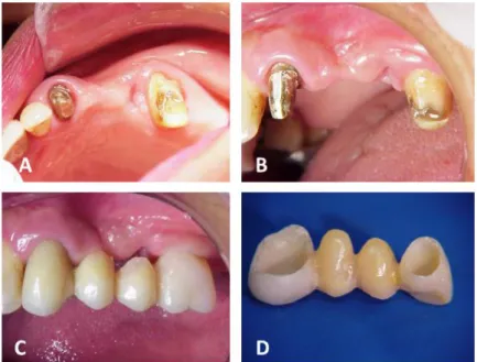
A female 48-year-old patient attended the dental clinic for definitive prosthetic rehabilitation of the upper left quadrant, exhibiting deep dissatisfaction with the esthetics and wish to achieve a better oral condition. Clinical and radiographic examination evidenced absence of teeth 24 and 25; full extracoronal preparations in teeth 23 and 26 and provisional crowns over these teeth (Figure 1A). The gingival architecture was also altered at the prosthetic space, concerning the esthetics and function (Figure 1B).
Thus, the proposed treatment included reestablishment of the lost gingival architecture, to optimize the esthetic and functional outcome of definitive rehabilitative treatment with fixed a more
Braz. J. of Develop., Curitiba, v. 6, n. 7, p. 42856-42866, jul. 2020. ISSN 2525-8761 biocompatible fixed partial denture. The patient presented favorable residual ridge for reconstruction of the gingival concave arch by soft tissue conditioning.
The technique selected to improve the gingival architecture was gingival conditioning by gradual pressure of provisional crowns. They were relined by adding self-curing acrylic resin at the cervical region of pontics, three times in a period of 30 days (Figures 1C and 1D).
Figure 1 – (A) Initial clinical case; (B) Initial condition of gingival tissue; (C) Initial provisional crowns, evidencing the need of relining the pontics at the cervical region for gingival modeling; (D) Addition of acrylic resin at the cervical region of pontics, making them convex, smooth and polished.
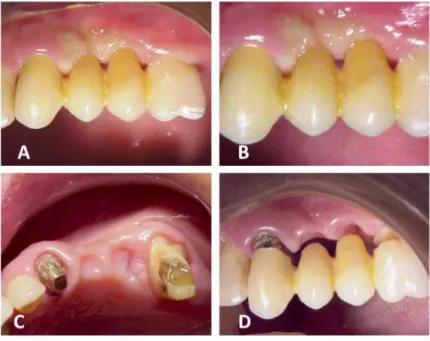
The patient received oral hygiene instructions to avoid plaque accumulation at the region. Using controlled pressure, the convex polished pontics and strict plaque control by the patient, the gingival epithelium only presented thinning, reducing the connective tissue crests, without presence of tissue inflammation. At each addition of acrylic resin at the cervical region, there was gingival ischemia, which was normalized after 2 to 3 minutes (Figures 2A and 2B). Thus, a favorable gingival condition was achieved by the provisional crowns, respecting the biological space, allowing correct emergence profile, besides serving as a model for the future full crowns of the definitive fixed partial denture.
The gingival conditioning by gradual pressure of the provisional crowns enhanced the shape of the residual ridge, providing adequate space for the emergence profile of pontics, which would be placed at this site in a ridge with concave arch and interdental papillae (Figures 2C and 2D).
Braz. J. of Develop., Curitiba, v. 6, n. 7, p. 42856-42866, jul. 2020. ISSN 2525-8761 Figure 2 – (A-B) Gradual pressure of provisional crowns on the gingival tissue; (C) Architecture o gingival tissue during conditioning with gradual pressure (occlusal view); (D) Relationship of provisional crowns (teeth 24 and 25) with the gingival tissue after conditioning with gradual pressure (buccal view).
The stage of gingival remodeling using the provisional crowns contributed to achieve adequate harmony between the soft tissues and the emergence profile of the future definitive denture (Figures 3A and 3B).
Figure 3 – (A) Final condition of gingival tissue after conditioning by gradual pressure (buccal view); (B) Final condition of gingival tissue after conditioning by gradual pressure, evidencing the formation of interdental papillae; (C) Fitting and adjustment of metallic framework of the fixed partial denture; (D) Interocclusal registry with self-curing acrylic resin with good dimensional accuracy.
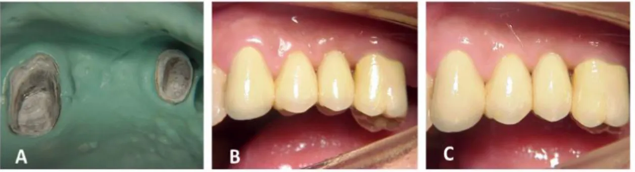
Braz. J. of Develop., Curitiba, v. 6, n. 7, p. 42856-42866, jul. 2020. ISSN 2525-8761 After achievement of the desired gingival contour, functional impression of the maxillary arch was obtained using putty and light condensation silicone, by the double manipulation technique, using a gingival retraction cord on the prosthetic preparations on teeth 23 and 26, for correct gingival retraction. After fabrication of the metallic framework of the fixed partial denture, this was placed in the mouth for fitting and adjustment of cervical adaptation (Figure 3C). Using chemically cured acrylic resin with good dimensional accuracy, the interocclusal recording was obtained (Figure 3D) and the metallic framework was removed from the mouth by impression with irreversible hydrocolloid using stock trays, allowing remounting of the dental cast (Figure 4A).
Figure 4 – (A) Removal of metallic framework from an alginate impression; (B) Definitive cementation of fixed partial denture; (C) Final case.
After application of feldspathic ceramics on the metallic copings and proper esthetic and functional adjustments in the patient’s mouth, definitive cementation was performed using zinc phosphate cement (Figures 4B and 4C). The patient was instructed about the denture hygiene and maintenance. After treatment finalization, the patient reported great satisfaction about the esthetic and functional aspects of rehabilitation.
3 DISCUSSION
The search for esthetic excellence faces great challenges, including the reestablishment of interdental papillae and proper filling of interdental space. To reestablish the ideal conditions of interdental papilla, regular concave arch and buccal prominences, the soft tissues can be properly manipulated (MORTON et al., 2014). This manipulation includes prosthetic gingival conditioning procedures that allow soft tissue conditioning using provisional dentures, aiming at formation of a gingival concave arch and consequently interdental papillae. The interdental papilla should be regular, occluding the interdental spaces in a V-shape with vertex coinciding with the proximal contact point (CONCEIÇÃO, 2007; FURZE et al., 2016).
Braz. J. of Develop., Curitiba, v. 6, n. 7, p. 42856-42866, jul. 2020. ISSN 2525-8761 The white esthetics plays a key role in the success of rehabilitative treatment, especially in anterior areas; this begins by the appropriate use of provisional restorations that allow preparation of gingival architecture and adequate emergence profile (BUSER et al., 2011). When the aim is to rehabilitate esthetic and visible areas, the ovoid-shaped pontic with convex cervical shape allows to recreate a natural emergence profile to meet the esthetic requirement and to allow a good oral hygiene by the patient (ORSINI et al., 2006; KIM et al., 2009; VASCONCELLOS et al., 2010; WITTNEBEN et al., 2013; PEREIRA et al., 2015). The emergence profile of a fixed partial denture pontic avoids formation of the so-called “black holes” and food accumulation in the reestablished oral environment. The functional and esthetic success of implant-supported and conventional dentures depends not only on the denture quality, but also on the positioning and stability of soft tissues. Thus, it is necessary to establish an integrated planning, which may require periodontal procedures (FRIZZERA et al., 2012), as in the present case. Gingival remodeling by provisional dentures is a simple and satisfactory clinical option to achieve better esthetics in rehabilitations with implanted-supported and conventional dentures, in which the emergence profile of the prosthetic element and pontic is similar to natural teeth, hiding the cervical line of crowns and eliminating the empty interpapillary space (KIM et al., 2009; JOLY et al., 2010). This is simple to perform and has great importance for the final esthetic result when performed following certain criteria as proper polishing, adaptation and hygiene. This technique requires a longer clinical time compared to other techniques, yet it is less invasive and more controlled. It does not remove gingiva; rather, it only remodels it. This technique consists of working on the gingival wall of a pontic by light and constant compression by adding successive layers of acrylic resin in areas where a marked contour is desired, until there is gingival tissue repair conditioned to the denture shape at the site (TARNOW et al., 1992; GASTALDO et al., 2004). Several studies report that the main criteria to achieve only epithelial thinning of the soft tissue without inflammation are the application of moderate pontic pressure to the soft tissue, strict biofilm control by the patient, and adequate pontic polishing (KIM et al., 2009; DREY & FREITAS, 2013; PEREIRA et al., 2015).
Besides esthetics, the goal of provisional dentures is to lead the soft tissue to the best cervical contour, as demonstrated in the technique of gingival conditioning by gradual pressure of provisional crowns. Since this is a non-surgical procedure and is considered minimally invasive because it does not remove soft tissue, gradual pressure is considered a safer technique, eliminating the risk of postoperative complications and anatomically and functionally restoring the components of the stomatognathic system (BUCHAIM et al., 2014; PEREIRA et al., 2019).
Braz. J. of Develop., Curitiba, v. 6, n. 7, p. 42856-42866, jul. 2020. ISSN 2525-8761 4 CONCLUSION
The search for an outcome combining satisfactory function and esthetics has become an increasing demand in the rehabilitation with conventional and implant-supported fixed dentures. Adequate soft tissue manipulation, besides accurate prosthetic work, are fundamental in esthetic areas. This report demonstrated the achievement of adequate gingival architecture in esthetic and functional aspects, by applying gradual and controlled pressure on the provisional crowns, which allows gingival conditioning by cervical increase of pontics, thus providing gingival and dental harmony in prosthetic rehabilitation.
CONFLICTS OF INTEREST
The authors declare that there is no conflict of interest regarding the publication of this paper.
ACKNOWLEDGMENTS
The authors thank Gisele da Silva Dalben, expert in scientific English, for manuscript editing.
REFERENCES
ASKARY, A. S. Cirurgia estética reconstrutiva na implantodontia. São Paulo: Santos, 2004.
BUCHAIM, R. L.; ANDREO, J. C.; RODRIGUES, A. C.; GONÇALVES, J. B. O.; DARÉ, L. R.; ROSA JÚNIOR, G. M. et al. Multidisciplinary Approach in the Teaching of Dental Sculpture and Anatomy. Int. J. Morphol., v. 32, n. 2, p. 399-403, 2014.
BUSER, D.; WITTNEBEN, J.; BORNSTEIN, M. M.; GRUTTER, L.; CHAPPUIS, V.; BELSER, U. C. Stability of contour augmentation and esthetic outcomes of implant-supported single crowns in the esthetic zone: 3-year results of a prospective study with early implant placement postextraction. Journal of Periodontology, v. 82, n. 3, p. 342-349, 2011.
CHO, H. L.; LEE, J. K.; UM, H. S.; CHANG, B. S. Esthetic evaluation of maxillary single- tooth implants in the esthetic zone. Journal of Periodontal & Implant Science., v. 40, n. 4, p. 188-193, 2010.
CONCEIÇÃO, E. N. Aplicações clínicas dos sistemas cerâmicos em dentes anteriores. In: CONCEIÇÃO, E. N. et al. Restaurações estéticas: compósitos, cerâmicas e implantes, Porto Alegre: Ed. Artmed. 2007, pp. 250-283.
DREY, S. E. & FREITAS, F. F. A. Técnica de condicionamento gengival em reabilitação protética: relato de caso clínico. RFO UPF, v. 18, n. 3, p. 1-10, 2013.
Braz. J. of Develop., Curitiba, v. 6, n. 7, p. 42856-42866, jul. 2020. ISSN 2525-8761 FRANCISCHONE, C. E. & VASCONCELOS, L. W. Otimização estética das próteses unitárias sobre implantes. In: FRANCISCHONE, C. E.; VASCONCELOS, L. W. Próteses Unitárias e Osseointegração. São Paulo: Artes Médicas. 1998, pp. 79-103.
FRIZZERA, F.; FONTANARI, L. A.; TONETTO, M. R.; KABBACH, W.; OTTONI, J.; MASIOLI, M. A.; JUNIOR, E. M. Escultura gengival: abordagem cirúrgica em alterações gengivais estéticas. Clínica Int J Braz Dent, v. 8, n. 4, p. 388-400, 2012.
FUENTEALBA, R. & JOFRÉ, J. Esthetic failure in implant dentistry. Dental Clinics., v. 59, n. 1, p. 227-246, 2015.
FURZE, D.; BYRNE, A.; ALAM, S.; WITTNEBEN, J. G. Esthetic utcome of implant supported crowns with and without peri‐mplant conditioning using provisional fixed prosthesis: a randomized controlled clinical trial. Clinical Implant Dentistry and Related Research., v. 18, n. 6, p. 1153-1162, 2016.
GASTALDO, J. F.; CURY, P. R.; SENDYK, W. R. Effect of the vertical and horizontal distances between adjacent implants and between a tooth and an implant on the incidence of interproximal papilla. Journal of Periodontology, v. 75, n. 9, p. 1242-1246, 2004.
GROISMAN, M.; HARARI, N. D. Técnicas de cirurgia plástica periodontal visando a estética em implantes orais. In: DINATO, J. C.; POLITO, W. D. Implantes osseointegrados: cirurgia e prótese. São Paulo: Artes Médicas, 2001, pp. 243-260.
HAMMOND, B. D.; COOPER, J. R.; LAZARCHILK, D. A. Predictable repair of provisional restorations. Journal of Esthetic and Restorative Dentistry, v. 21, n. 1, p. 19-24, 2009.
JACQUES, L. B.; COELHO, A. B.; HOLLWEG, H.; CONTI, P. C. Tissue sculpturing: an alternative method for improving esthetics of anterior fixed prosthodontics. Journal of Prosthetic Dentistry, v. 81, n. 5, p. 630-633, 2009.
JOLY, J. C.; CARVALHO, P. F. M.; SILVA, R. C. Reconstrução tecidual estética, técnicas de manipulação tecidual. São Paulo: Artes Médicas, 2010.
KIM, T. H.; CASCIONE, D.; KNEZEVIC, A. Simulated tissue using a unique pontic design: a clinical report. Journal of Prosthetic Dentistry, v. 102, n. 4, p. 205-210, 2009.
LEVINE, R. A.; GANELES, J.; GONZAGA, L.; KAN, J. Y.; RANDEL, H.; EVANS, C. D.; CHEN, S. T. 10 keys for successful esthetic-zone single immediate implants. Compendium of Continuing Education in Dentistry., v. 38, n. 4, p. 248-260, 2017.
LINDHE, J. Tratado de periodontia clínica e implantologia oral. Rio de Janeiro: Guanabara Koogan, 2018.
LIU, C. L. S. Use of a modified ovate pontic in areas of ridges defects: a report of two cases. Journal of Esthetic and Restorative Dentistry, v. 16, n. 5, p. 273-283, 2004.
MAINIERI, E. T.; RIVALDO, E. G. Conduta eletrocirúrgica nos tecidos moles aplicada às restaurações protéticas. Odont. Mod., v. 18, n. 4, p. 16-24, 1991.
Braz. J. of Develop., Curitiba, v. 6, n. 7, p. 42856-42866, jul. 2020. ISSN 2525-8761 MORTON, D.; CHEN, S. T.; MARTIN, W. C.; LEVINE, R. A.; BUSER, D. Consensus statements and recommended clinical procedures regarding optimizing esthetic outcomes in implant dentistry. The International Journal of Oral & Maxillofacial Implants., v. 29, p. 216-220, 2014.
NEALE, D. & CHEE, W. W. L. Development of implant soft tissue emergence profile: A technique. Journal of Prosthetic Dentistry, v. 71, n. 4, p. 364-368, 1994.
OLIVEIRA, J. A.; RIBEIRO, E. D. P.; CONTI, P. C. R.; VALLE, A. L.; PEGORARO, L. F. Condicionamento gengival: Estética em tecidos moles. Revista da Faculdade de Odontologia de Bauru, v. 10, n. 2, p. 99-104, 2002.
ORSINI, G.; MURMURA, G.; ARTESE, L.; PIATTELLI, A.; PICCIRILLI, M.; CAPUTI, S. Tissue healing under provisional restorations with ovate pontics: a pilot human histological study. Journal of Prosthetic Dentistry, v. 96, n. 4, p. 252-257, 2006.
PEGORARO, L. F. Coroas provisórias. In: PEGORARO, L. F.; VALLE, A. L.; ARAÚJO, C. R. P.; BONFANTE, G.; CONTI, P. C. R.; BONACHELA, W. Prótese Fixa. São Paulo: Artes Médicas, 1998.
PEREIRA, J. R.; GHIZONI, J. S.; OLIVEIRA, M. T.; PAMATO, S. Transferring conditioned partially edentulous ridge form to a master cast. Journal of Prosthodontics, v. 25, n. 7, p. 595-598, 2015.
PEREIRA, E. S. B. M.; ACCETTURI, F.; ELEUTÉRIO, R. G.; BUCHAIM, D. V.; BUCHAIM, R. L.; CLEMENTE-NAPIMOGA, J. T. Reverse Cast Metallic Core Based on the Original Prosthetic Crown. Case Rep Dent. 2019 Jun 23;2019:6936573. doi: 10.1155/2019/6936573. eCollection 2019. PubMed PMID: 31341682; PubMed Central PMCID: PMC6612380.
QUESADA, G. A. T.; RIZZARDI, M.; FRANCISCATTO, L. J.; ARRAIS, F. R. Condicionamento gengival visando o perfil de emergência em prótese sobre implante. Saúde, v. 40, n. 2, p. 9-18, 2014.
RAIGRODSKI, A. J.; SCHWEDHELM, E. R.; CHEN, Y. A simplified technique for recording an implant-supported ovate pontic site in the esthetic zone. Journal of Prosthetic Dentistry, v. 111, n. 2, p. 154-158, 2014.
SANSEVERINO, C. A. M. Manipulação do tecido gengival para um melhor resultado protético. Rev. APCD, v. 52, n. 3, p. 203-204, 1998.
STRUPP, W. C. Provisional materials. Comp. Cont. Educ. Dent., v. 31, n. 2, p. 167-174, 2010.
TARNOW, D. P.; MAGNER, A. W.; FLETCHER, P. The effect of the distance from the contact point to the crest of bone presence or absence of the interproximal dental papilla. Journal of Periodontology, v. 63, n. 12, p. 995-996, 1992.
VASCONCELLOS, D. K.; VOLPATO, C. A. M.; ZANI, I. Z.; BOTTINO, M. A. Impression technique for ovate pontics. Journal of Prosthetic Dentistry, v. 105, n. 1, p. 59-61, 2010.
WITTNEBEN, J. G.; BUSER, D.; BELSER, U. C.; BRÄGGER, U. Peri-implant soft tissue conditioning with provisional restorations in the esthetic zone: the dynamic compression technique. Int J Periodontics Restorative Dent., v. 33, n. 4, p. 447-55, 2013.