Non-neoplastic proliferative gingival
processes in patients undergoing
orthodontic treatment
Irineu Gregnanin Pedron*, Estevam Rubens Utumi**, Ângelo Rafael Calábria Tancredi***, Flávio Eduardo Guillin Perez****, Gilberto Marcucci*****
Introduction:Orthodontic appliances render oral hygiene difficult and may contribute to the development of gingival lesions such as non-neoplastic proliferative gingival processes. These lesions, depending on such factors as development time, histopathological components and oral conditions may be reversible in some cases, through oral hygiene advice and basic periodontal therapy. In most cases, however, surgical treatment is required. Objectives:The purpose of this paper is to report the case of a patient using fixed orthodontic appliance who presented with two distinct gingival lesions diagnosed as pyogenic granuloma and inflamma-tory gingival hyperplasia. The clinical and histopathological features, incidence and frequency, treatment modalities and prevention of both lesions were discussed, highlighting the impor-tance of submitting the material collected from the lesions to histopathological examination given the possibility of different diagnostic hypotheses. Surgical excision was performed on both lesions. The upper arch lesion, diagnosed as pyogenic granuloma, relapsed, which led us to provide basic periodontal therapy and repeat the surgical procedures. Results:The lesion in the lower arch, diagnosed as gingival hyperplasia, was surgically removed and followed up clinically, whereas the patient was instructed to perform proper oral hygiene.
Abstract
Keywords: Pyogenic granuloma. Gingival hyperplasia. Periodontal diseases. Orthodontics. Gingiva.
* Specialist in Periodontics and MSc in Dental Sciences (Area of concentration: General Dentistry), School of Dentistry, University of São Paulo. Lieuten-ant, Brazilian Air Force Dentist - Brazilian Air Force Hospital of São Paulo (HASP).
** Specialist in Oromaxillofacial Surgery and Traumatology. MSc in Dental Sciences (Area of concentration General Dentistry), School of Dentistry, Univer-sity of São Paulo. Lieutenant, Brazilian Air Force Dentist - Brazilian Air Force Hospital of São Paulo (HASP).
*** Specialist in Stomatology. MSc in Oral Diagnosis (subarea: Semiology), School of Dentistry, University of São Paulo. **** Professor and PhD, Discipline of General Dentistry, School of Dentistry, University of São Paulo.
***** Head Professor, Discipline of Oral Diagnosis (subarea: Semiology), School of Dentistry, University of São Paulo.
IntROductIOn
The effects of fixed and removable orthodon-tic appliances on the periodontium have been widely investigated. Orthodontic appliances usually hinder proper oral hygiene, contribut-ing to the development of gcontribut-ingival inflammation,
Gingival hyperplastic and inflammatory re-sponses during orthodontic treatment are com-mon and can lead to complications that require periodontal therapy.1,5,15
Pyogenic granuloma and inflammatory gingi-val hyperplasia are tissue growths of inflammatory origin with varying degrees of granular and fibrous inflammatory tissues, possibly caused by low in-tensity chronic irritation.2,9,11,12
Both occur frequently in clinical dentistry and are clinically characterized by exophytic, sessile or pedunculated tissue growths, of pale pink to ery-thematous color, which may manifest ulceration and spontaneous bleeding.3,6,7,11,12,17-20 They are most prevalent in the female gender during the second decade of life, possibly due to vascular ef-fects exerted by feminine hormones.9 Treatment often consists of surgical excision associated with the removal of local irritating factors.2-7 However, recognition and identification of these factors are not always possible and the recurrence rate of le-sions is relatively high.12,17
The purpose of this study was to report the case of a patient using fixed orthodontic appli-ance who presented with two distinct lesions— pyogenic granuloma and inflammatory gingival hyperplasia. The development of these conditions
is related to chronic low intensity trauma. We en-courage orthodontists and dentists to routinely submit any material collected from the lesions to histopathological examination after surgery.
cAse RepORt
Black female patient, aged 20 years, after 6 months of orthodontic treatment presented to our private clinic complaining of gingival changes.
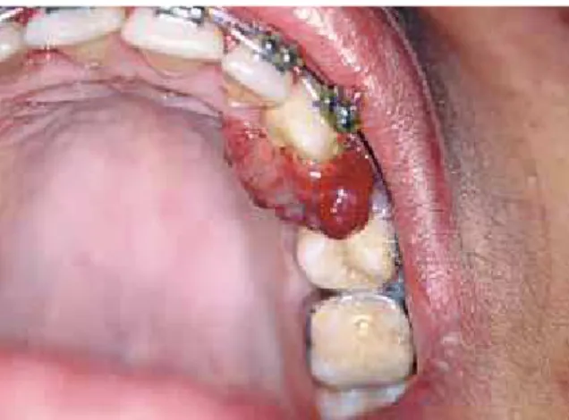
On examination, a tumor-like lesion was ob-served, of erythematous color, irregularly shaped, with a smooth surface and pedunculated base, lo-cated in an edentulous region between teeth 23 and 25, under occlusion trauma. The condition had been developing for a week, starting with a node in the aforesaid region. The diagnostic potheses were pyogenic granuloma, gingival hy-perplasia and peripheral giant cell lesion (Fig 1). Tooth 24 had been extracted 4 months earlier with no history of postoperative complication. The other lesion was observed between teeth 41 and 42. It was characterized by moderate gingi-val enlargement, pale pink in color, sessile base, smooth surface extending from the papilla to the brackets. The hypothetical diagnosis was inflam-matory gingival hyperplasia (Fig 2). Radiographs of the lesions yielded no significant findings.
FIGURE 1 - Erythematous tumor mass with heavy bleeding to the touch, resembling pyogenic granuloma.
Under local anesthesia, excisional biopsy of the lesion was performed in tooth 24 region by incising it by the pedicle, from which a wedge was removed as a safety measure and tissue was removed from the edentulous area. The region was sutured and surgical cement added and maintained for 7 days, aided by the orthodontic appliance. The removed part was fixed in 10% formalin and submitted for labo-ratory analysis. Histopathological examination showed fragments of mucosa lined by para-keratinized stratified squamous epithelium exhibiting areas of spongiosis and acanthosis, and an ulceration area covered with fibrin-haemorrhagic exudate and bacterial colonies. In the lamina propria—permeating the dense connective tissue—we observed the prolifera-tion of endothelial cells delimiting sometimes congested vascular spaces. There was exuber-ant hemorrhagic exudate and intense mono-nuclear and polymorphomono-nuclear inflammatory infiltration in the ulceration areas. The histo-pathological diagnosis was pyogenic granuloma (Fig 3). In the same consultation excisional bi-opsy of the lesion in the region of teeth 41 and 42 was performed, and the removed piece was
also prepared and treated histologically. Light microscopy disclosed fragments of mucosa lined by parakeratinized stratified squamous epithelium, showing acanthosis, exocytosis and hyperparakeratosis. In surface areas there were fibrin-hemorrhagic exudate and bacterial colonies. In the lamina propria we observed intense deposition of collagen fibers forming a dense stroma that sustained intense chronic inflammatory infiltrate. Finally, there were also numerous vascular spaces and areas of hemor-rhagic exudate. The histopathological diagnosis was inflammatory gingival hyperplasia (Fig 4).
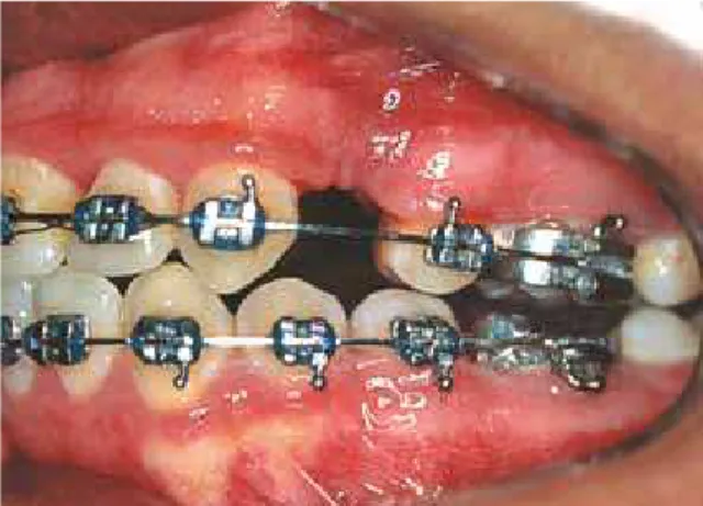
After a postoperative period of 7 days the surgical cement and remaining sutures were removed. The patient was evaluated after 20 days, showing satisfactory repair of the region between teeth 41 and 42 (Fig 5). However, there was recurrence of pyogenic granuloma. Periodontal treatment was then performed and once again excision, submitting the lesion to the same laboratory, which confirmed the diagnosis of pyogenic granuloma. Repair was satisfactory with no signs of relapse (Fig 6). After four years of treatment, the patient is still being monitored and exhibits no signs of recurrence.
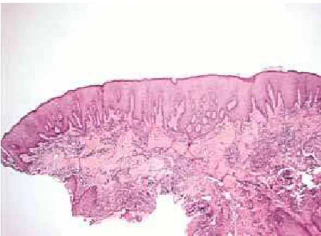
FIGURE 3 - Histological section of pyogenic granuloma (original color: HE; smaller magnification).
dIscussIOn
Among the most frequent gingival prolifera-tive processes are inflammatory gingival hyper-plasia and pyogenic granuloma. Peripheral fi-broma, peripheral giant cell lesions and gingival hyperplasia are also part of this group, although not as common.4
In order to facilitate lesion exposure the dis-cussion was divided into topics.
etiopathogenesis
Etiopathogenesis of both lesions is usually related to chronic low intensity trauma, pro-ducing in most cases gingival inflammation and infection (periodontal diseases) caused by dif-ficulty in removing biofilm in patients wearing an orthodontic appliance, which translate into traumatic injuries and hormonal factors.1,4,6,9 The physical set-up (brackets and bands that could invade the periodontium’s biological space) and mechanical set-up (forces delivered by orthodontic and / or orthopedic movement), associated with biofilm, were reported as hy-potheses to explain the etiopathogenesis of gin-gival hyperplasia.1,9 And so was trauma during placement of the orthodontic appliance, which
causes pressure areas that result in epithelium thickening, connective tissue proliferation and an increased amount of tissue. The possible allergic process triggered by the acrylic resin monomer placed on the base of the removable orthodontic appliances, when associated with the presence of fungi (Candida albicans) may also cause a slight increase in plaque and gin-gival indices. The possibility of an unusual host response against the local irritant (biofilm), ex-acerbated by the patients’ hormonal changes (puberty and menstruation) was also reported.1
Local irritants such as excessive restorations and neonatal teeth combined with poor oral hy-giene, plaque and dental calculus were also con-sidered in the etiopathogenesis.2,3,6,7,11,12,17,18,19 Hormonal changes such as menarche, use of oral contraceptives and pregnancy were also re-ported. During gestation, lesions usually arise in the 2nd or 3rd quarters, but tend to regress there-after.2,6,11,13,19
Increased levels of progesterone and estrogen produce dilatation and proliferation of gingival microvasculature and destruction of mast cells, which result in an increased release of vasoac-tive substances in the adjacent tissue, inducing FIGURE 5 - Postsurgical view after 20 days showing satisfactory repair in
the region between 41 and 42 teeth.
the formation of pyogenic granuloma.17 A de-crease in keratinization of the epithelium of the attached gingiva, rendering it more vulnerable to trauma and triggering a tendency towards growth of vascular tumors in the gingiva and alveolar mucosa has also been reported. The development of pyogenic granuloma depends on factors such as sufficient amount of tissue, degree of gingival inflammation, degree of vul-nerability to trauma, presence of teeth and den-tures, and level of oral hygiene. Low intensity tissue trauma could facilitate the invasion of nonspecific low virulence saprophytic microor-ganisms, causing a tissue response characterized by excessive proliferation of vascular-type con-nective tissue.3
clinical Features
Non-neoplastic proliferative processes are generally characterized by gingival tissue growth, either well defined, such as nodules, or diffuse, like tissue masses; fibrous or flac-cid texture (resilient); variable symptoms and ranging from pink to erythematous color; ses-sile or pedunculated base; usually bleeding to touch; loss of the “orange peel” look on the sur-face.1,2,4,5 Gingival growth stems from the in-terdental papilla and expands to the marginal gingiva.1,2,4,5,13 Although they have a predilec-tion for the gingiva, they can be found in extra-gingival regions with various clinical features that often mimic malignant lesions.10 Pyogenic granuloma, in particular, differs from inflamma-tory gingival hyperplasia because it is character-ized by well circumscribed papular, nodular or tumoral exophytic soft tissue; erythematous to brownish in color, depending on the maturity of the lesion; hemorrhagic aspect and a bleeding tendency; smooth or lobulated surface; soft and resilient texture when young, and more fibrous when mature due to obliteration of the capil-laries; rapid growth; may cause bone resorption. It may be covered with a pus-filled membrane
due to trauma, hence the name of the lesion, despite the absence of a relationship between suppuration and pathological entity. It varies in size from a few millimeters to a few centimeters and as it grows increasingly larger it can inter-fere with physiological activities in the oral cav-ity2,3,6-8,11,12,17-22
Both lesions may present with an ulcerated surface under occlusion trauma.1,2,4,5,13
There was no incidence of inflammatory gin-gival hyperplasia. Pyogenic granuloma is more common in the gingiva, in the anterior maxilla. It affects adolescents and young adults, with 60% incidence at ages 11-40 years and no race predilection. Women are two to four times more affected than men.2,3,6,7,11,12,17-20 Studies have confirmed the incidence of pyogenic gran-ulomas in young adults.19,21,22
Histopathological Features
The histological picture of inflammatory gin-gival hyperplasia is characterized by parakera-tinized stratified squamous epithelium issuing long, thin projections towards the connective tissue. The lamina propria is made up of dense, well cellularized and collagenized connective tissue permeated by an intense mononuclear in-flammatory infiltrate,1 as shown in Figure 4.
differential diagnosis
Among the lesions that make up the differ-ential diagnosis are peripheral ossifying fibroma, peripheral giant cell lesions and inflammatory gingival hyperplasia.2,4
Particularly in the case of pyogenic granu-loma, given its clinical aspects and marked vascularization, the differential diagnosis com-prises hemangioma, lymphoma, nevus flamme-us, Kaposi’s sarcoma, metastatic tumor, parulis, hemangioendothelioma, hemangiopericytoma, leiomyoma, cytomegalovirus infection and gin-gival lesions by bacilli.3,11,19
Hemangioma is an important differential di-agnosis since some smaller lesions may be indis-tinguishable9. Dyscopia tests are used in case of suspected vascular lesions. Inflammatory fibrous hyperplasia should also be considered as a dif-ferential diagnosis of pyogenic granuloma.
Given the breadth of the differential diag-nosis, a histopathological examination was sug-gested as a means to verify and clarify the diag-nosis of gingival lesions.7,8
treatment
Surgical excision has usually been the treat-ment of choice for both lesions.1-3,12,16-19. How-ever, some changes have been suggested, such as curettage,1,2,7 gingivectomy or gingivoplasty techniques.2,3,7 The latter is determined by the amount of attached gingiva.6 Barack et al1 cited the need for flap procedure (modified Widman technique) in the presence of periodontal pock-et with attachment loss. Other modalities have been recommended. Surgical removal using la-ser (CO2 or Nd:YAG) has been proposed.3,4,12,14 The advantages of laser use in these procedures are: Enhanced hemostasis with better visualiza-tion of the surgical field, less discomfort or pain, reducing the need for postoperative medication; satisfactory tissue healing, improved patient ac-ceptance, fewer anesthetics, and reduction of postoperative bacteremia in the surgical site.4
Cryosurgery was cited in the treatment of pyo-genic granuloma.6 Silverstein et al13 performed free gingival graft for root coverage and ke-ratinized gingiva loss resulting from surgical excision of pyogenic granuloma. The use of chlorhexidine mouthwashes pre and post-sur-gically have prevented potential post-surgical infection and inflammation.11,17 The removal of the base of the lesion in order to avoid recur-rence has been recommended.1,7,16 For cases of pyogenic granuloma, the clinical follow-up and supervision of oral hygiene during pregnancy is recommended if the lesion is small, asymptom-atic and not bleeding.17,18
The need for removal of causative factors through basic periodontal treatment (scraping sessions, coronoradicular smoothing and pol-ishing and oral hygiene advice) has been advo-cated.1-4,6,7,17,18 It is suggested that periodontal treatment be performed prior to surgery in view of a milder inflammatory process and surgery procedure, reducing heavy bleeding and de-creasing the chance of recurrence.
prognosis
It would be timely to make some consider-ations regarding the monitoring of gingival le-sions in orthodontic patients. Orthodontists should use appropriate orthodontic compo-nents that do not put the periodontium at risk. Periodontal changes should be diagnosed and treated as early as possible in order to control periodontal disease (periodontal treatment and reinforcement of basic oral hygiene).1
cOnclusIOns
In view of the foregoing, we may conclude that:
1. Pyogenic granuloma and inflammatory gingival hyperplasia usually exhibit typical clin-ical and histopathologclin-ical features.
2. Periodontal disease, usually present due to the difficulty in performing adequate oral hygiene because of the orthodontic appliance, must be treated before surgical removal of the proliferative processes so as to avoid heavy tran-soperative bleeding and postoperative
compli-cations, such as lesion recurrence itself.
3. Surgical excision is the most widely em-ployed technique today. Regardless of treatment modality, submitting the collected material to histopathological examination is not only en-lightening but a sine qua non measure to avoid the underestimation of these lesions and pos-sible errors in the final diagnosis since different diagnostic hypotheses are possible.
1. Barack D, Stafileno H, Sadowsky C. Periodontal complication during orthodontic therapy. Am J Orthod. 1985 Dec;88(6):461-5. 2. Binnie WH. Periodontal cysts and epulides. Periodontol 2000.
1999 Oct;21:16-32.
3. Campos V, Bittencourt LP, Maia LC, Andrade M, Mascarenhas A. Granuloma piogênico - descrição de dois casos clínicos. J Bras Odontoped Odontol Bebê. 2000;3(12):170-5.
4. Coleman GC, Flaitz CM, Vincent SD. Differential diagnosis of oral soft tissue lesions. Tex Dent J. 2002 Jun;119(6):484-8, 90-2, 494-503.
5. Convissar RA, Diamond LB, Fazekas CD. Laser treatment of orthodontically induced gingival hyperplasia. Gen Dent. 1996 Jan-Feb;44(1):47-51.
6. Falabella MEV, Falabella JM. Granuloma gravídico - caso clínico. Periodontia. 1994;3(2):167-70.
7. Graham RM. Pyogenic granuloma: an unusual presentation. Dent Update. 1996 Jul-Aug;23(6):240-1.
8. Halliday H, Gordon S, Bhola M. Case report: an unusually large epulis on the maxillary gingiva of a 24-year-old woman. Gen Dent. 2007 May-Jun;55(3):232-5.
9. Jafarzadeh H, Sanatkhani M, Mohtasham N. Oral pyogenic granuloma: a review. J Oral Sci. 2006;48(4):167-75.
10. Patil K, Mahima VG, Lahari K. Extragingival pyogenic granuloma. Indian J Dent Res. 2006;17(4):199-202.
11. Ramirez K, Bruce G, Carpenter W. Pyogenic granuloma: case report in a 9-year-old girl. Gen Dent. 2002 May-Jun;50(3):280-1. 12. Rivero ELC, Araújo LMA. Granuloma piogênico: uma análise
clínico-histopatológica de 147 casos bucais. Rev Fac Odontol Univ Passo Fundo. 1998;3(2):55-61.
ReFeRences
13. Romero M, Albi M, Bravo LA. Surgical solutions to periodontal complications of orthodontic therapy. J Clin Pediatr Dent. 2000 Spring;24(3):159-63.
14. Satpathy AK, Mohanty PK. Large pyogenic granuloma: a case report. J Indian Med Assoc. 2007 Feb;105(2):90-8.
15. Scaramella F, Quaranta M. Hypertrophic and/or hyperplastic gingivopathy during orthodontic therapy. Dent Cadmos. 1984 Feb;52(2):65-72.
16. Shenoy SS, Dinkar AD. Pyogenic granuloma associated with bone loss in an eight year old child: a case report. J Indian Soc Pedod Prev Dent. 2006 Dec;24(4):201-3.
17. Silva-Sousa YT, Coelho CM, Brentegani LG, Vieira ML, Oliveira ML. Clinical and histological evaluation of granuloma gravi -darum: case report. Braz Dent J. 2000;11(2):135-9. 18. Silverstein LH, Burton CH Jr, Garnick JJ, Singh BB. The late
development of oral pyogenic granuloma as a complication of pregnancy: a case report. Compend Contin Educ Dent. 1996 Feb;17(2):192-8; quiz 200.
19. Terezhalmy GT, Riley CK, Moore WS. Pyogenic granuloma (preg -nancy tumour). Quintessence Int. 2000;31(6):440-1.
20. Vélez LMA, Souza LB, Pinto LP. Granuloma piogênico. Análise dos componentes histológicos relacionados com a duração da lesão. Rev Gaúcha Odontol. 1992;40(1):52-6.
21. Zarei MR, Chamani G, Amanpoor S. Reactive hyperplasia of the oral cavity in Kerman province, Iran: a review of 172 cases. Br J Oral Maxillofac Surg. 2007 Jun;45(4):288-92.
22. Zhang W, Chen Y, An Z, Geng N, Bao D. Reactive gingival lesions: a retrospective study of 2,439 cases. Quintessence Int. 2007 Feb;38(2):103-10.
contact address
Irineu Gregnanin Pedron Rua Flores do Piaui, 347
CEP: 08.210-200 – São Paulo/SP, Brazil E-mail: igpedron@usp.br
Submitted: October 2008