Na,K-ATPase: a molecular target
for
Leptospira interrogans
endotoxin
1Laboratoire de Biologie Intégrée des Cellules Rénales,
CNRS URA 1859, Commissariat à l’Energie Atomique, Saclay, France 2Laboratório de Biologia Celular, Universidade do Estado do Rio de Janeiro,
20559-900 Rio de Janeiro, RJ, Brasil M. Younes-Ibrahim1,
B. Buffin-Meyer1, L. Cheval1, P. Burth2, M.V. Castro-Faria2, C. Barlet-Bas1, S. Marsy1 and A. Doucet1
Abstract
On the basis of our report that a glycolipoprotein fraction (GLP) extracted from Leptospira interrogans contains a potent inhibitor of renal Na,K-ATPase, we proposed that GLP-induced inhibition of Na,K-ATPase might be the primary cellular defect in the physiopa-thology of leptospirosis. The present study was designed to test this hypothesis by determining whether or not 1) GLP inhibits all the isoforms of Na,K-ATPase which are expressed in the tissues affected by leptospirosis, 2) Na,K-ATPase from leptospirosis-resistant spe-cies, such as the rat, is sensitive to GLP, 3) GLP inhibits Na,K-ATPase from intact cells, and 4) GLP inhibits ouabain-sensitive H,K-ATPase. The results indicate that in the rabbit, a leptospirosis-sensitive species, GLP inhibits with similar efficiency (apparent IC50: 120-220 µg protein GLP/ml) all isoforms of Na,K-ATPase known to be expressed in target tissues for the disease. Na,K-ATPase from rat kidney displays a sensitivity to GLP similar to that of the rabbit kidney enzyme (apparent IC50: 25-80 and 50-150 µg protein GLP/ml for rat and rabbit, respectively), indicating that resistance to the disease does not result from the resistance of Na,K-ATPase to GLP. GLP also reduces ouabain-sensitive rubidium uptake in rat thick ascending limbs (pmol mm-1 min-1 ± SEM; control: 23.8 ± 1.8; GLP, 88 µg protein/ml: 8.2 ± 0.9), demonstrating that it is active in intact cells. Finally, GLP had no demonstrable effect on renal H,K-ATPase activity, even on the oua-bain-sensitive form, indicating that the active principle of GLP is more specific for Na,K-ATPase than ouabain itself. Although the hypoth-esis remains to be demonstrated in vivo, the present findings are compatible with the putative role of GLP-induced inhibition of Na,K-ATPase as an initial mechanism in the physiopathology of leptospirosis.
Correspondence
A. Doucet
SBCe, Centre d’Etudes de Saclay Bâtiment 520
91191 Gif sur Yvette cedex France
Fax (33) 01-69083570
Research supported in part by the Centre National de la Recherche Scientifique to the Unités de Recherche Associées 219 and 1859, and the Commissariat à l’Energie Atomique. M. Younes-Ibrahim was the recipient of a scholarship from the PhD program of CNPq and Comité Français d’Evaluation de la Coopération Universitaire avec le Brésil.
Received February 23, 1996 Accepted November 7, 1996
Key words
•Na,K-ATPase •H,K-ATPase
Introduction
Although Leptospira interrogans has been incriminated in the origin of leptospiro-sis since the beginning of this century (1), the cellular disorders involved in the multiorganic dysfunctions during this infec-tion remain unknown. We have previously reported that a crude preparation of glycolip-oprotein endotoxin (GLP) extracted from
Leptospira interrogans is a potent inhibitor of Na,K-ATPase from rabbit kidney (2). Based on this observation, it was also pro-posed that GLP-induced inhibition of Na,K-ATPase might be the initial cellular defect responsible for the leptospirotic physiopa-thology in the different organs. Indeed, many of the clinical manifestations of this disease may be viewed as a consequence of a pri-mary defect in transmembrane transport of cations, the motor of which is Na,K-ATPase. The clinical manifestations include cardiac arrhythmia, hypovolemia, neurological dis-orders, myalgia, diarrhea, and disorders of kidney electrolyte handling. The present stud-y was designed to further elaborate this phstud-ys- phys-iopathological hypothesis.
First, we investigated whether or not GLP was able to inhibit Na,K-ATPase from sources other than the kidney. Although a direct interaction between the renal pump and the endotoxin was previously established (2), it remained to be determined whether GLP would equally inhibit the activity of all the isoforms of Na,K-ATPase, in particular the α2 and/or α3 isoforms which
predomi-nate in leptospirosis target tissues and cells other than kidney (heart, muscle, nervous system, etc.). For this purpose, the effect of GLP was compared on purified Na,K-ATPase from brain (which contains mainly the α2
and α3 isoforms, Ref. 3) and kidney.
Be-cause in the kidney itself the successive seg-ments constituting the nephrons express forms of Na,K-ATPase displaying different functional and pharmacological properties (4,5), the effect of GLP was also evaluated at
the level of the different segments of the nephron.
Second, because the rat is the main vec-tor of leptospirosis (6) and is also a species resistant to ouabain (7,8), another inhibitor of Na,K-ATPase, we determined whether the resistance of rats to leptospirosis might be related to the resistance of their Na,K-ATPase to the inhibitory action of GLP. For this purpose, the sensitivity of renal Na,K-ATPase to GLP was compared in rat and rabbit nephrons.
Third, since GLP is likely to interfere with Na,K-ATPase on the cytosolic side of the membrane (since it modulates the af-finity of Na,K-ATPase for sodium, Ref. 2), we addressed the question of whether or not it may alter Na,K-ATPase in intact, non-permeabilized cells, as expected if it were involved in the pathogenicity of Leptospira. For this purpose, the inhibitory action of GLP was investigated on the Na,K-pump activity determined by ouabain-sensitive ru-bidium intake into intact renal cells.
Finally, it is now well established that ouabain not only inhibits Na,K-ATPase but also some specific types of H,K-ATPase (9,10). We therefore determined whether GLP might be more specific for Na,K-ATPase than ouabain by evaluating its effect on oua-bain-sensitive H,K-ATPase activity.
Material and Methods
Preparation of leptospirotic glycolipoprotein
prepared at a concentration of 350 µg GLP protein/ml, which corresponds to approxi-mately 1 mg GLP/ml since the protein frac-tion of GLP accounts for about 35% of its whole mass (Adler B, Monash University, Clayton, Australia, personal communication). The stock solution was aliquoted and stored at -20oC. The protein content of GLP was
used as reference for concentrations.
Preparation of purified Na,K-ATPase from rabbit brain and kidney
Na,K-ATPase was purified from whole rabbit brain (except meninges) and kidney medulla according to the procedure of Jorgensen (14). After anesthesia, the organs were removed and homogenized in a Potter apparatus (5 strokes at 1,000 rpm) in homoge-nization buffer (10 ml per g of tissue) con-taining 250 mM sucrose and 30 mM histi-dine, pH 7.2. After 15 min of centrifugation at 6,000 g, the supernatant was centrifuged for 30 min at 48,000 g and the pellet was resuspended (5-6 mg protein/ml) in the ho-mogenization buffer. This preparation was then diluted to 1.35-1.40 mg protein/ml in a medium containing 3 mM Na2-ATP, 2 mM
EDTA, 50 mM imidazole and 0.58 mg/ml sodium dodecyl sulfate (SDS), pH 7.5, and incubated for 30 min at 20oC. After this
treatment with SDS, the preparation was layered onto a sucrose gradient (12.5 ml at 29.4%, 7.5 ml at 15% and 5 ml at 10% in 25 mM imidazole and 1 mM EDTA, pH 7.5) and centrifuged for 90 min at 60,000 rpm, and the pellet was resuspended in the imida-zole/EDTA solution (2 mg protein/ml), lyo-philized and stored at -20oC. Before use, the
enzyme was resuspended in 10 mM Tris (hydroxymethyl) aminomethane (Tris)-HCl. Purification was only partial since the spe-cific activity of the enzyme was 1.38 and 2.73 µmol mg-1 min-1 for brain and renal
Na,K-ATPase, respectively. Dose-inhibition curves of the enzyme preparations with oua-bain confirmed that they consisted of the α1
isoform for the kidney enzyme and the α2
and α3 isoforms for the brain Na,K-ATPase
(data not shown).
Tubule microdissection
Kidneys were obtained from male New Zealand rabbits (1.5-2.0 kg body weight) or male Wistar rats (180-200 g body weight) fed the usual laboratory diet with free access to tap water. Except for the slight variations dictated by the difference in kidney size, the procedure for kidney perfusion and mi-crodissection described below was similar for the two species.
After anesthesia with pentobarbital (50 mg/kgbody weight, ip for rats or iv for rab-bits), the left rabbit kidneys were excised and perfused in vitro via the renal artery (20 ml, 1.5 ml/min) while the rat kidneys were perfused in situ via the aorta (4 ml, 5 ml/min) with cold microdissection solution (see com-position below) containing 0.16% (w/v) col-lagenase (from Clostridium histolyticum, 0.452 U/mg, Boehringer Mannheim, Ger-many). After perfusion, the kidney was im-mediately sliced into small pyramids, which were incubated in aerated dissection solu-tion containing 0.08% (w/v) collagenase at either 35oC for 30 min or at 30oC for 20 min,
for rabbit and rat, respectively. Pyramids were then thoroughly rinsed in ice-cold dis-section solution and stored in the cold until use.
The composition of the microdissection solution varied as a function of the experi-mental protocol. For measurement of ATPase activities, the solution (A) contained 137
mM NaCl, 0.8 mM MgSO4, 0.33 mM
Na2HPO4, 0.44 mM NaH2PO4, 1 mM MgCl2,
10 mM Tris-HCl, 1 mM CaCl2 and 0.1% (w/v)
BSA, pH 7.4. For the measurement of 86Rb
uptake the solution (B) derived from Eagle’s minimal essential medium contained 120 mM NaCl, 5 mM RbCl, 1 mM MgSO4, 0.15
mM Na2HPO4, 0.2 mM NaH2PO4, 4 mM
mM lactate, about 4 mM essential and non-essential amino acids, 0.03 mM vitamins, 20 mM N-2-hydroxyethylpiperazine-N’-2-ethane-sulfonic acid (HEPES), 0.3% (w/v) dextran (Mr 40,000), and 0.1% BSA.
Single pieces of nephron were dissected in microdissection solution at 0-4oC under
stereomicroscopic observation and were identified by morphological and topogra-phical criteria as previously described (15). In the rabbit kidney, segments of proximal convoluted and straight tubules (PCT and PST) were dissected in the superficial cortex next to their attachment to the glomeruli and in the outer stripe of the outer medulla next to their attachment to the thin descending limb, respectively. Medullary (MTAL) and cortical portions (CTAL) of the thick ascen-ding limb of Henle’s loop were obtained from the inner stripe of the outer medulla and from the cortex, respectively. The initial bright portion of distal convoluted tubules (DCT) and the late portion, or connecting tubule (CNT), were dissected separately. Collecting ducts were dissected either from the cortex below the last branching point (cortical collecting duct, CCD) or from the inner stripe of the outer medulla (outer me-dullary collecting duct, OMCD). In the rat kidney, the study was restricted to the PCT, MTAL, CTAL, CCD and OMCD which were characterized by the same criteria as in the rabbit. The length (0.4-1.0 mm) of each seg-ment was determined by computerized im-age analysis.
ATPase assay
Pretreatment of the samples before meas-urement of ATPase activity varied as a func-tion of the sample. To increase the cell per-meability of nephron segments to reagents, samples were individually rinsed with dis-tilled water and, after sucking away the sur-rounding water, 0.2 µl of either GLP (at various concentrations) or distilled water was added. Samples were frozen on dry-ice
and, after thawing, they were preincubated for 5 min at 37oC. For purified enzymes, 0.2
µl of either distilled water or GLP (at various concentrations) was added to 0.2 µl of en-zyme (about 20 µg protein/ml). Then, samples were preincubated for 5 min at 37oC.
ATPase activities were measured as pre-viously described (10,16). Briefly, 1 µl of ATPase assay medium (see below) was added to each sample and the samples were incu-bated for 15 min at 37oC. The reaction was
then stopped by the addition of 5 µl of 5% (w/v) ice-cold trichloroacetic acid and the samples were transferred individually to 2 ml of a suspension of 10% (w/v) activated charcoal. After mixing and centrifugation, the radioactivity of 500 µl of supernatant, which contained Pi formed from ATP, was
determined by liquid scintillation. For tech-nical reasons, it was not possible to maintain the same concentration of GLP during ATPase measurement and preincubation. Thus, the concentrations of endotoxin indi-cated in the results and Figures correspond to those present during the preincubation. Those effectively present during the ATPase assay were only 1/6 (nephron segments) or 1/7 (purified enzymes) of it.
The distinction between the different ATPases was based on their cation-specific stimulation and on their sensitivity to specif-ic inhibitors. Thus, for each structure 10-14 samples were distributed randomly into two groups, one for measuring basal Mg-ATPase activity and the other for measuring stimu-lated ATPase activity. In addition, samples without nephron segments were treated in parallel in each experiment to determine the spontaneous breakdown of ATP.
For determination of Na,K-ATPase ac-tivity, stimulated ATPase was determined in an assay medium containing 50 mM NaCl, 5 mM KCl, 10 mM MgCl2, 1 mM ethylene
glycol-bis(ß-aminoethylether)-N,N,N’,N’ -tetraacetic acid (EGTA), 100 mM Tris-HCl, and 10 mM Na2-ATP as well as tracer
medium for determination of basal ATPase was similar except that KCl and NaCl were omitted and 1 mM ouabain was added. Na,K-ATPase activity was calculated for each struc-ture as the difference between the mean stimulated ATPase activity and the mean basal ATPase activity.
For determination of H,K-ATPase activ-ity, stimulated ATPase was measured in a medium containing 2.5 mM KCl, 10 mM MgCl2, 1 mM EGTA, 25 mM Tris-HCl, 5
mM Tris-ATP and tracer amounts of [γ32
P]-ATP, pH 7.4. Basal ATPase was determined in a similar medium except that KCl was omitted, and H,K-ATPase activity was calculated as the difference between the mean stimulated ATPase activity and the mean basal ATPase activity.
Because the measurement of tubular H,K-ATPase is based on the nominal absence of Na+, we determined the actual concentration
of this cation during the assay. For this pur-pose, nephron segments were treated as done in a usual ATPase assay (permeabilization, preincubation with GLP and incubation) ex-cept that [32P]-ATP was omitted. At the end
of the incubation, the concentration of Na+
was measured in ≈30-nl aliquots of the assay medium by flame microspectrophotometry, as described previously (17). The Na+
con-centration ranged from 50 to 70 µM, which, based on the activation curves of Na,K-ATPase by Na+ previously reported for
neph-ron segments (18), would account for Na,K-ATPase activities <1 pmol mm-1 h-1, i.e.,
100-fold lower than K-ATPase activities measured in this study (see Figure 5).
ATPase activities were reported either as a function of protein content (purified ATPase) or of tubular length (nephron seg-ments).
Measurement of 86Rb+ uptake
86Rb+ uptake was measured according to
a method previously developed in our labo-ratory (19) and modified slightly (20). Pools
of 10-12 segments of MTAL were trans-ferred with 0.7 µl microdissection solution B into the concavity of a sunken bacterio-logical slide. After addition of another 0.7 µl of solution B supplemented or not with 5 mM ouabain and/or GLP, segments of the nephron were preincubated for 10 min at 37oC to allow the regeneration of
transmem-brane ion gradients. Incubation was then initiated by adding 0.5 µl of prewarmed solution B supplemented with 86RbCl (100
nCi/sample, Amersham) and stopped after 30 sec by adding 20 µl of an ice-cold rinsing solution containing 150 mM choline chlo-ride, 1.2 mM MgSO4, 1.2 mM CaCl2, 2 mM
BaCl2 and 5 mM HEPES, pH 7.4, the
osmo-larity being adjusted to 500 mOsmol/kg. The segments of the nephron on each slide were then rapidly rinsed in three successive baths and individually transferred with 0.2 µl of rinsing solution to a small microscopic cover-slip. After photography for the determina-tion of its length, each sample was dropped into a counting vial containing 500 µl of 1% (w/v) deoxycholic acid, and its radioac-tivity was measured. In each experiment, the blank which was substracted from all data was determined by measuring the mean radioactivity of 8-12 replicate samples consisting of 0.2 µl of the last rinsing so-lution.
For all experimental conditions, Rb+
in-flux was measured on 6-8 replicate samples. Na,K-ATPase-dependent Rb+ influx was
taken as the difference between the means of Rb+ influx measured in the absence or
pre-sence of ouabain, and is reported as pmol of Rb+ transported per ml of tubule length per
min.
Statistical analysis
Results
Effect of GLP on purified Na,K-ATPase
Figure 1 shows that GLP completely in-hibited Na,K-ATPase purified from both rab-bit brain and rabrab-bit kidney in a dose-depend-ent manner. Half maximum inhibition was observed in the presence of GLP in the range of 120 to 220 µg protein/ml for brain and kidney enzyme, respectively. It is worth not-ing that complete inhibition of Na,K-ATPase activity occurred within a very narrow range
of variation of GLP concentration (50-400 µg protein/ml for the kidney enzyme), which suggests some cooperativity in the interac-tion between GLP and Na,K-ATPase.
Effect of GLP on tubular Na,K-ATPase
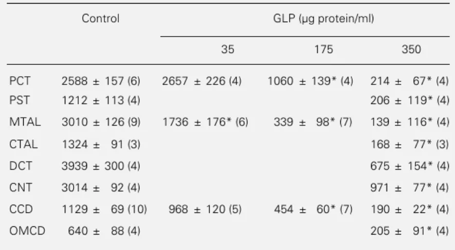
The data (Table 1 and Figure 2, top panel) show that GLP (350 µg protein/ml) almost abolished Na,K-ATPase activity in all seg-ments of the rabbit nephron, including those which express the α1 isoform of the catalytic
subunit of Na,K-ATPase (proximal tubule, thick ascending limb) and the α3-like form
(collecting duct) (4). This inhibition was dose dependent (Table 1 and Figure 2, bot-tom panel) and half-maximal inhibition was observed with GLP in the range of 50-150 µg protein/ml in the thick ascending limbs, proxi-mal tubules and collecting ducts.
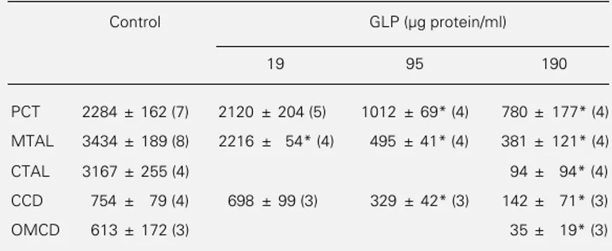
In the nephron of the rat, a species resist-ant to leptospirosis, Na,K-ATPase activity was also markedly reduced by GLP (190 µg protein/ml) in the proximal tubule, thick as-cending limb and collecting tubule (Table 2 and Figure 3, top panel). Rat kidney Na,K-ATPase appeared to be slightly more sensi-tive to GLP than the rabbit enzyme since half-maximal inhibition was observed in the range of 25-80 µg protein/ml for the thick ascending limbs, proximal tubules and collec-ting ducts (Table 2 and Figure 3, bottom panel).
Effect of GLP on Rb+ uptake
In order to determine whether GLP in-hibits Na,K-ATPase in intact cells, we evalu-ated its effect on the Na,K-pump activity, as determined by ouabain-sensitive Rb+ uptake
in intact rat nephron segments. The results in Figure 4 indicate that at a concentration of 88 µg protein/ml, GLP inhibited ouabain-sensitive Rb+ uptake by more than 65% in
the rat MTAL, i.e., to an almost similar extent as Na,K-ATPase activity (see Figure 3, bottom).
100
Na,K-ATPase activity (%)
50
0
Brain
Kdney
50 100 200 400 GLP (µg protein/ml)
Figure 1 - Inhibition of Na,K-ATPase purified from rabbit brain and kidney by GLP. Na,K-ATPase activity was measured in en-zyme purified from rabbit brain and rabbit kidney after preincu-bation for 5 min at 37oC in the
absence or presence of different concentrations of GLP. Data are reported as percent of the acti-vity determined in the absence of GLP (1.38 and 2.73 µmol mg-1
min-1 for brain and kidney
en-zymes, respectively), and are means ± SEM of 4 experiments.
Table 1 - Inhibition of Na,K-ATPase activity by GLP along the rabbit nephron.
Na,K-ATPase activity was determined in rabbit nephron segments preincubated for 5 min at 37oC in the absence or presence of 35, 175 or 350 µg GLP protein/ml. Data (in
pmol mm-1 h-1) are reported as means ± SEM for the number of animals given in
parentheses. *P<0.001 compared to control (analysis of variance).
Control GLP (µg protein/ml)
35 175 350
PCT 2588 ± 157 (6) 2657 ± 226 (4) 1060 ± 139* (4) 214 ± 67* (4)
PST 1212 ± 113 (4) 206 ± 119* (4)
MTAL 3010 ± 126 (9) 1736 ± 176* (6) 339 ± 98* (7) 139 ± 116* (4)
CTAL 1324 ± 91 (3) 168 ± 77* (3)
DCT 3939 ± 300 (4) 675 ± 154* (4)
CNT 3014 ± 92 (4) 971 ± 77* (4)
CCD 1129 ± 69 (10) 968 ± 120 (5) 454 ± 60* (7) 190 ± 22* (4)
Effect of GLP on ouabain-sensitive and -insensitive tubular H,K-ATPases
The effect of GLP was evaluated on both the ouabain-sensitive and the ouabain-in-sensitive H,K-ATPase activities present in the rat thick ascending limb and collecting duct, respectively (10). At a concentration which induces a maximal inhibition of Na,K-ATPase activity in these two nephron seg-ments (190 µg protein/ml, Figure 3), GLP had no effect on H,K-ATPase activities (Fig-ure 5).
Discussion
The present data confirm the previous observation (2) that a glycolipoprotein frac-tion (GLP) extracted from Leptospira inter-rogans inhibits the Na,K-ATPase α1
isoform from rabbit kidney, and extend this observation to other isoforms of Na,K-ATPase from rabbit and rat. These results also indicate that GLP has no effect on Na, K-ATPase-related proteins such as renal H,K-ATPases and finally demonstrate that GLP inhibits the transport activity of Na,K-ATPase.
Although the active principle of GLP responsible for the inhibition of Na,K-ATPase has not yet been purified or charac-terized, the present study was undertaken to further elucidate the putative role of Na,K-ATPase inhibition by GLP in the physio-pathological mechanism of leptospirosis. Indeed, several studies on animals (22-25) as well as humans (26-28) have demon-strated the presence of leptospirae and/or leptospira antigens in diseased tissues. The presence of GLP itself in these tissues was also detected immunologically during ex-perimental leptospirosis (23). However, be-cause leptospirosis is a pleiotropic disease which alters the function of many tissues and organs, we determined whether or not GLP inhibits Na,K-ATPase from these distinct tissues.
Indeed, it is now well established that different tissues express different isoforms of Na,K-ATPase which display distinct prop-erties, in particular towards inhibitors such as ouabain (reviewed in Ref. 3). The results in Figure 1 demonstrate that GLP inhibited not
Na,K-ATPase activity (pmol mm
-1 h
-1) 4000
3000
2000
1000
0
Rabbit
4 PCT
4 PST
4 MTAL
3 CTAL
4 DCT
4 CNT
4 OMCD N
123 123
12 12
123 123
123 123
123 123 123 123 123
12 12 12 12 12 12
123 123
123 123
4 CCD
* * * *
* *
* *
Na,K-ATPase activity (%)
100
80
60
40
20
0
PCT
MTAL
CCD
0 100 200 300 400
GLP (µg protein/ml)
*
* *
*
* * *
Figure 2 - Inhibition of Na,K-ATPase activity in the rabbit nephron by GLP. Top, Na,K-ATPase activity was measured in nephron segments dissected from rabbit kidneys and preincu-bated for 5 min at 37oC in the absence (
à) or presence ( 123 123
123) of 350 µg GLP protein/ml. Data are reported as means ± SEM for N animals. PCT and PST, Proximal convoluted and straight tubules; MTAL and CTAL, medullary and cortical thick ascending limb of Henles loop; DCT, distal convoluted tubule; CNT, connecting tubule; CCD and OMCD, cortical and outer medullary collecting duct. *P<0.025 compared to respective control (Student t-test). Bot-tom, Segments from rabbit PCT (!), MTAL(
·
), and CCD (à) were preincubated for 5 min at 37oC in the presence of different concentrations of GLP prior to the measurement ofonly the α1 isoform of the Na,K-ATPase
catalytic subunit which predominates in the rabbit kidney but also the α2 and α3 isoforms
present in the brain. In the rabbit kidney itself, the two different forms of Na,K-ATPase which were previously demonstrated (4,29) in the collecting duct (α3-like form)
and in more proximal nephron segments (α1
form) proved to be sensitive to GLP (Figure 2). The dose-inhibition curves depicted in Figures 1 and 2 deserve three additional comments. First, they suggest a positive co-operativity in the inhibition of Na,K-ATPase by GLP. Second, they suggest that there might be some small differences in the sensi-tivity of the different isoforms of Na,K-ATPase to GLP. The brain enzyme (α2 and
α3 isoforms) is 3-fold more sensitive than
the kidney enzyme (α1 isoform). However, it
is not known whether such differences in sensitivity have functional consequences. Third, they indicate the range of GLP con-centrations required for inhibition of Na,K-ATPase. Although the concentration of GLP achieved in infected tissues is not known, it is possible to estimate it on the basis of the following considerations. According to the procedure used in this study (see Material and Methods), approximately 1010 bacteria
were needed to obtain 350 µg of GLP pro-Table 2 - Inhibition of Na,K-ATPase activity by GLP along the rat nephron.
Na,K-ATPase activity was determined in rat nephron segments preincubated for 5 min at 37oC in the absence or presence of 19, 95 or 190 µg GLP protein/ml. Data (in pmol
mm-1 h-1) are reported as means ± SEM for the number of animals given in
parenthe-ses. *P<0.001 compared to control (analysis of variance).
Control GLP (µg protein/ml)
19 95 190
PCT 2284 ± 162 (7) 2120 ± 204 (5) 1012 ± 69* (4) 780 ± 177* (4)
MTAL 3434 ± 189 (8) 2216 ± 54* (4) 495 ± 41* (4) 381 ± 121* (4)
CTAL 3167 ± 255 (4) 94 ± 94* (4)
CCD 754 ± 79 (4) 698 ± 99 (3) 329 ± 42* (3) 142 ± 71* (3)
OMCD 613 ± 172 (3) 35 ± 19* (3)
Na,K-ATPase activity (pmol mm
-1 h -1)
4000
3000
2000
1000
0
Rat
4 PCT
4 PCT
4 MTAL
4 CTAL
4 CCD N
123 123 123 123 123 123
123 123 123 123
123 123
3 OMCD
* *
123 123 123
Na,K-ATPase activity (%)
100
80
60
40
20
0
MTAL
CCD
0 100 200
GLP (µg protein/ml)
*
* *
*
* * *
* *
PCT
Figure 3 - Inhibition of Na,K-ATPase activity in the rat nephron by GLP. Top, Na,K-ATPase activity was meas-ured in nephron segments dissected from rat kidneys and preincubated for 5 min at 37oC in the absence (à)
or presence (12
12) of 190 µg GLP protein/ml. Data are reported as means ± SEM for N animals. For abbrevia-tions, see the legend to Figure 2, top. *P<0.025 com-pared to control (Student t-test). Bottom, Segments from rat proximal convoluted tubule (à, PCT), medul-lary thick ascending limb (
·
, MTAL), and cortical col-lecting tubule (!, CCD) were preincubated for 5 min at 37oC in the presence of different concentrations ofGLP prior to the measurement of Na,K-ATPase activity. Data are reported as percent of the activity measured in the absence of GLP in the corresponding segments of nephron and are means ± SEM for 3-5 animals. Control activities (100%) were similar to those shown in the top of Figure 3. *P<0.001 compared to controls (analysis of variance).
tein. Assuming that the release of GLP from leptospirae occurs in vivo with a similar efficiency, and based on the concentration of bacteria detected in kidneys of infected animals (1010 bacteria per g of tissue, Ref.
30), the tissue concentration of GLP could be as high as 350 µg protein/ml, i.e., suffi-cient to fully inhibit renal Na,K-ATPase. This is a rather rough estimate of the pos-sible concentrations of GLP in infected tis-sues, but it should be stressed that partial inhibition of Na,K-ATPase promoted by lo-wer concentrations of GLP would be suffi-cient to induce pathological alterations.
The finding (2) that GLP increases the apparent affinity of Na,K-ATPase for so-dium (uncompetitive inhibition) suggested that it might interact with the sodium-bind-ing sites of Na,K-ATPase, i.e., on the cyto-solic surface of cell membranes. This raised the question of whether GLP might also be able to inhibit Na,K-ATPase in intact cells, as expected if such inhibition were to play a causal role in the physiopathology of lepto-spirosis. In fact, GLP inhibits to the same extent both Na,K-ATPase in permeabilized (ATPase activity, Figure 3) and in intact tubular cells (transport activity, Figure 4), which is consistent with a possible physio-pathological role.
That GLP inhibits Na,K-ATPase-medi-ated transport in rat thick ascending limb cells may appear at variance with the report of Magaldi et al. (31) who showed that MTAL from normal and leptospirotic guinea pigs display similar transport capacity when microperfused in vitro. In fact, two explana-tions may account for this apparent discrep-ancy. First, it is possible that the concentra-tion of GLP achieved in vivo at the level of MTAL from leptospirotic animals is too low to inhibit Na,K-ATPase. More likely, the interaction between Na,K-ATPase and GLP is too labile to persist under the in vitro
microperfusion conditions. In particular, GLP is likely to be washed out of the bath.
Interspecies comparison revealed that
Na,K-ATPase from rat and rabbit kidneys displays a similar sensitivity to inhibition by GLP (Figures 2 and 3), which may seem paradoxical since the rat is resistant to the disease (6). In fact, this finding suggests that the resistance of a given species to the dis-ease does not result from the resistance of its cells to GLP but rather from the amount of toxin released at the level of its tissues. The latter would vary with the degree of bacterial colonization of the tissues, itself depending on species-specific immunological res-ponses. Under these circumstances, the cel-lular sensitivity of a species naturally
resist-Figure 4 - Inhibition of ouabain-sensitive Rb+ uptake by GLP.
Ouabain-sensitive Rb+ uptake
was determined in rat MTAL preincubated for 10 min at 37oC
in the absence (à) or presence (
123
123) of GLP (88 µg protein/ml).
Data are reported as means ± SEM for 5 animals. *P<0.001 compared to control (Student t -test).
* 1234567890123456 1234567890123456 1234567890123456 1234567890123456 1234567890123456 1234567890123456 1234567890123456 1234567890123456 1234567890123456 1234567890123456 1234567890123456
Ouabain-sensitive Rb
+ uptake
(pmol mm
-1 min -1)
30
25
20
15
10
5
0
H,K-ATPase activity (pmol mm
-1 h -1)
300
200
100
0
MTAL
(Ouabain-sensitive H,K-ATPase)
OMCD
(Ouabain-insensitive H,K-ATPase) 200
150
100
0 50
Control GLP Control GLP
Figure 5 - Lack of effect of GLP on tubular H,K-ATPase activities. Ouabain-sensitive and -insensitive H,K-ATPase activities were determined in rat MTAL and OMCD, respectively. Nephron segments were preincubated for 5 min at 37oC in the absence (control) or
ant to leptospirosis is not inconsistent with the physiopathological potential of GLP. That im-munosuppression turns resistant-species sen-sitive to the multiorganic alterations of lepto-spirosis (32) corroborates this hypothesis.
Because rabbit tubular Na,K-ATPase (α1
form) is about 100-fold more sensitive to ouabain than its rat counterpart (5,29), the similar sensitivities of rat and rabbit renal Na,K-ATPase to GLP further demonstrate that GLP is not a ouabain-like substance. This conclusion is also supported by the fact that 1) in contrast to ouabain, GLP does not alter the apparent affinity of Na,K-ATPase
for potassium (2), and 2) GLP does not alter the activity of renal ouabain-sensitive H,K-ATPase (Figure 5).
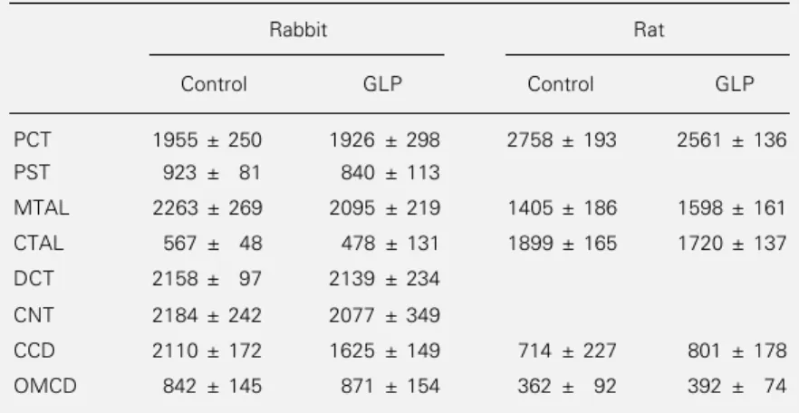
Taken together with the findings that GLP alters neither basal Mg-ATPase activity (Table 3) nor hormone-sensitive adenylate cyclase activity (2), this last result demon-strates the rather high specificity of GLP for Na,K-ATPase. In addition, it indicates that GLP, or better its active principle when it will become available, should be preferred to ouabain when attempts are made to spe-cifically inhibit Na,K-ATPase.
In conclusion, the present results support the hypothesis that GLP released during ly-sis of Leptospira interrogans is able to in-hibit Na,K-ATPase activity in epithelial as well as non-polarized cells. Cellular dys-functions induced by this inhibition in in-fected tissues might account for many symp-toms, in particular those associated with elec-trolytic disorders such as disturbances of renal electrolyte handling, cardiac arrhyth-mia or diarrhea. Depending on the intensity of Na,K-ATPase inhibition in colonized tis-sues, disorders may vary from slight me-tabolic dysfunction to organic failure. Fur-ther studies, in particular the demonstration that Na,K-ATPase activity is lower in tissues from diseased animals than in tissues from normal ones, will be needed to determine the role of GLP in the physiopathology of lepto-spirosis.
Table 3 - GLP has no effect on Mg-ATPase activity along the rabbit and the rat nephron.
Mg-ATPase activity was determined in rabbit and rat nephron segments preincubated for 5 min at 37oC in the absence or presence of 350 µg GLP protein/ml (rabbit) or 190
µg GLP protein/ml (rat). Data (in pmol mm-1 h-1) are reported as means ± SEM for the
same animals as in Figures 2 and 3. GLP had no significant effect on Mg-ATPase activity in any nephron segment (Student t-test).
Rabbit Rat
Control GLP Control GLP
PCT 1955 ± 250 1926 ± 298 2758 ± 193 2561 ± 136
PST 923 ± 81 840 ± 113
MTAL 2263 ± 269 2095 ± 219 1405 ± 186 1598 ± 161
CTAL 567 ± 48 478 ± 131 1899 ± 165 1720 ± 137
DCT 2158 ± 97 2139 ± 234
CNT 2184 ± 242 2077 ± 349
CCD 2110 ± 172 1625 ± 149 714 ± 227 801 ± 178
OMCD 842 ± 145 871 ± 154 362 ± 92 392 ± 74
References
1. Inada R, Ido Y, Hoki R, Kanero R & Ito H (1916). Etiology, mode of infection and specific therapy of Weils disease ( spiro-chaetosis icterohaemorrhagica). Journal of Experimental Medicine, 23: 377-402. 2. Younes-Ibrahim M, Burth P, Castro Faria
MV, Buffin-Meyer B, Marsy S, Barlet-Bas C, Cheval L & Doucet A (1995). Inhibition of Na,K-ATPase by an endotoxin extract-ed from Leptospira interrogans: a pos-sible mechanism for the physiopathology of leptospirosis. Comptes Rendus de lAcadémie des Sciences, Paris, 318: 619-625.
3. Sweadner KJ (1989). Isozymes of the Na+/
K+-ATPase. Biochimica et Biophysica
Acta, 988: 185-220.
4. Barlet-Bas C, Arystarkhova E, Cheval L, Marsy S, Sweadner K, Modyanov N & Doucet A (1993). Are there several isoforms of Na-K-ATPase α subunit in the rabbit kidney? Journal of Biological Chem-istry, 268: 11512-11515.
5. Féraille E, Barlet-Bas C, Cheval L, Rousselot M, Carranza ML, Dreher D, Arystarkhova E, Doucet A & Favre H (1995). Presence of two isoforms of Na-K-ATPase with different pharmacological and immunological properties in the rat kidney. Pflügers Archiv, 430: 205-212. 6. Minette HP (1983). Leptospirosis in
7. Tobin T & Brody TM (1972). Rates of dis-sociation of enzyme-ouabain complexes and K0.5 values in (Na++K+) adenosine
triphosphatase from different species. Biochemical Pharmacology, 21: 1553-1560.
8. Price EM & Lingrel JB (1988). Structure-function relationship in the Na,K-ATPase α subunit. Site-directed mutagenesis of glutamine-111 to arginine and asparagine-122 to aspartic acid generates a ouabain-resistant enzyme. Biochemistry, 27: 8400-8408.
9. Jaisser F, Horisberger JD, Geering K & Rossier B (1993). Mechanism of urinary K+ and H+ excretion: primary structure
and functional expression of a novel H,K-ATPase. Journal of Cell Biology, 123: 1421-1429.
10. Younes-Ibrahim M, Barlet-Bas C, Buffin-Meyer B, Cheval L, Rajerison R & Doucet A (1995). Ouabain-sensitive and -insensi-tive K-ATPases in rat nephron: effect of K depletion. American Journal of Physiolo-gy, 268: F1141-F1147.
11. Braun V & Wolff H (1970). The murein-lipoprotein linkage in the cell wall of Es-cherichia coli. European Journal of Bio-chemistry, 14: 387-391.
12. Vinh T, Adler B & Faine S (1986). Glycolip-oprotein cytotoxin from Leptospira inter-rogans serovar copenhageni. Journal of General Microbiology,132: 111-123. 13. Bradford M (1976). A rapid and sensitive
method for the quantitation of microgram quantities of protein utilizing the principle of protein-dye binding. Analytical Bio-chemistry,72: 248-254.
14. Jorgensen PL (1974). Purification and characterization of (Na+ plus K+)-ATPase.
III. Purification from the outer medulla of mammalian kidney after selective removal of membrane components by sodium dodecylsulphate. Biochimica et Biophy-sica Acta, 356: 36-52.
15. Morel F, Chabardès D & Imbert-Teboul M (1978). Methodology for enzymatic stud-ies of isolated tubular segments: adenyl-ate cyclase. In: Martinez-Maldonado M (Editor), Methods in Pharmacology. Ple-num Press, New York, 297-323.
16. Doucet A, Katz AI & Morel F (1979). De-termination of Na-K-ATPase activity in single segments of the mammalian neph-ron. American Journal of Physiology,237: F105-F113.
17. Sudo J & Morel F (1984). Na+ and K+ cell
concentrations in collagenase-treated rat kidney tubules incubated at various tem-peratures. American Journal of Physiolo-gy, 246: C407-C414.
18. Buffin-Meyer B, Marsy S, Barlet-Bas C, Cheval L, Younes-Ibrahim M, Rajerison R & Doucet A (1996). Regulation of renal Na,K-ATPase in rat thick ascending limb during K-depletion: Evidence for modula-tion of Na affinity. Journal of Physiology, 490: 623-632.
19. Cheval L & Doucet A (1990). Measure-ment of Na-K-ATPase-mediated rubidium influx in single segments of rat nephron. American Journal of Physiology, 259: F111-F121.
20. Féraille E, Marsy S, Cheval L, Barlet-Bas C, Khadouri C & Doucet A (1992). Sites of antinatriuretic action of insulin along the rat nephron. American Journal of Physiol-ogy, 263: F175-F179.
21. Dunnett CW (1955). A multiple compari-son procedure for comparing several treatments with a control. American Sta-tistical Association Journal, 50: 1096-1121.
22. De Brito T, Freymuller E, Hishino S & Penna DO (1966). Pathology in the kidney and liver of the experimental leptospirosis of the guinea-pig. A light and electron microscopy study. Virchows Archiv of Pa-thology and Anatomy, 341: 64-78. 23. Macedo Santos RT, Sakata EE, Yasuda
PH, Wakamatsu A, Kanamura CT, Candelori I, Pestana CB & Alves VAF (1989). Glicolipoproteína de Leptospira in-terrogans sorogrupo icterohaemorrhagi-ae: distribuição em fígado e rim de co-baias experimentalmente infectadas. Revista do Instituto de Medicina Tropical de São Paulo, 31: 235-241.
24. Miller DA, Wilson MA & Beran GW (1991). Survey to estimate prevalence of Lep-tospira interrogans infection in mature cattle in the United States. American Jour-nal of Veterinary Research, 52: 1761-1765.
25. Sitprija V, Papatanagul V, Mertowidjojo K, Boonpcknavig V & Boonpcknavig S (1980). Pathogenesis of renal disease in leptospirosis: Clinical and experimental studies. Kidney International, 17: 827-836. 26. Arean VM (1962). The pathologic anatomy and pathogenesis of fatal human leptospi-rosis (Weils disease). American Journal of Pathology, 40: 393-422.
27. De Brito T, Penna DO, Pereira VG & Hoshino S (1967). Kidney biopsies in hu-man leptospirosis: a biochemical and elec-tron microscopy study. Virchows Archiv of Pathology and Anatomy, 343: 124-135. 28. Ferreira Alves VA, Vianna MR, Yasuda PH & De Brito T (1987). Detection of leptospi-ral antigen in the human liver and kidney using an immunoperoxidase staining pro-cedure. Journal of Pathology, 151: 125-131.
29. Doucet A & Barlet C (1986). Evidence for differences in the sensitivity to ouabain of Na,K-ATPase along the nephrons of rabbit kidney. Journal of Biological Chemistry, 261: 993-995.
30. Rudland SV (1989). Leptospirosis. Do you consider the diagnosis? Journal of the Royal Navy Medical Service, 75: 146. 31. Magaldi AJ, Yasuda PN, Kudo LH, Seguro
AC & Rocha AS (1992). Renal involvement in leptospirosis: A pathophysiological study. Nephron, 62: 332-339.