online | memorias.ioc.fiocruz.br
Preliminary multiplex microarray IgG immunoassay
for the diagnosis of toxoplasmosis and rubella
Priscila T Baschirotto1,2, Marco A Krieger1,2, Leonardo Foti1,2/+
1Fundação Oswaldo Cruz-Fiocruz, Instituto Carlos Chagas, Curitiba, PR, Brasil 2Instituto de Biologia Molecular do Paraná, Curitiba, PR, Brasil
BACKGROUND During pregnancy, toxoplasmosis and rubella can cause serious damage to the mother and the foetus through vertical transmission. Early diagnosis enables implementation of health measures aimed at preventing vertical transmission and minimising damage caused by these diseases.
OBJECTIVE Here, we report the development of a multiplex assay for simultaneous detection of IgG antibodies produced during toxoplasmosis and rubella infection.
METHODS This assay is based on xMap technology. Initially, by singleplex assays, we evaluated the following antigens: one Toxoplasma gondii lysate; two antigenic extracts of T. gondii (TOX8131 and TOX8122); fragments of T. gondii antigens [SAG-1 (amino acids 45-198), 7 (24-100), 1 (57-149), ROP-4, and MIC-3 (234-306)]; two chimeric antigens composed of fragments of SAG-1, GRA-7, and P35 (CTOX and CTOXH); and fragments of Rubella virus antigens [E-1 (157-176, 213-239, 374-390), E-2 (31-105), and C (1-123)]. FINDINGS A multiplex assay to simultaneously diagnose toxoplasmosis and rubella was designed with the best-performing antigens in singleplex and multiplex assays, which included CTOXH, T. gondii lysate, TOX8131, E-1, and E-2. The multiplex assay showed 100% sensitivity and specificity for anti-T. gondii IgG detection and 95.6% sensitivity and 100% specificity for anti-R. virus IgG detection. MAIN CONCLUSIONS We found that, despite the difficulties related to developing multiplex systems, different types of antigens (extracts and recombinant proteins) can be used to develop high-performance diagnostic tests. The assay developed is suitable to screen for prior T. gondii and R. virus infections, because it is a rapid, high-throughput, low-cost alternative to the current standard diagnostic tools, which require multiple individual tests.
Key words: toxoplasmosis - rubella - diagnosis - multiplex - liquid microarray
doi: 10.1590/0074-02760160509
Financial support: CNPq, Instituto Nacional de Ciência e Tecnologia em Diagnósticos para a Saúde Pública, Fundação e Instituto Oswaldo Cruz - FIOCRUZ.
+ Corresponding author: [email protected] Received 23 November 2016
Accepted 17 March 2017
Toxoplasmosis and rubella are caused by Toxo-plasma gondii and Rubella virus, respectively, which have been reported worldwide. Primary infections in pregnant women may result in vertical transmission of the pathogens, which can cause congenital disease that significantly affects foetal development. The possible effects include structural and functional sequelae in several systems and organs, miscarriage, foetal deaths, stillbirths, premature births, and development of disease postnatally (Havelaar et al. 2007, Simons et al. 2016).
Since clinical manifestations of toxoplasmosis and rubella can be absent or nonspecific, serological screen-ing for toxoplasmosis and rubella in pregnant women is routinely performed worldwide (Montoya & Rosso 2005, Wandinger et al. 2011). An individual is considered sus-ceptible to infection when specific antibodies are absent from serum. In this case, measures to prevent infection by T. gondii or R. virus can be implemented to reduce cases of vertical transmission. Detection of specific IgG without IgM antibodies is the classical serological
pat-tern indicating past infection and/or vaccination in the case of rubella (Montoya & Rosso 2005, Vauloup-Fellous & Grangeot-Keros 2007, Montoya & Remington 2008). In most cases, pregnant women are considered immuno-logically protected, and new events are extremely rare (Banatvala & Brown 2004, Montoya & Rosso 2005). Primary infection during pregnancy is most commonly identified by the detection of IgM and/or IgG. However, because of variations in antibody production following infection, a definitive diagnosis is only possible after additional assessments such as confirmation of a sig-nificant increase in antibody titre or seroconversion and IgG avidity testing (Wandinger et al. 2011, Hotop et al. 2012). If acute toxoplasmosis and rubella are confirmed, pregnancy can be terminated based on maternal and/or foetal risks and the law(s) of the country (Banatvala & Brown 2004, Montoya & Remington 2008), or toxoplas-mosis therapy can be initiated to reduce foetal and neo-natal damage or prevent vertical transmission of T. gondii
for each marker investigated, and relatively low through-put because of time-consuming procedures. These limi-tations can be overcome by multiplex technologies that are amenable to automation. xMap technology involves multiplex testing using a liquid microarray. Beads serve as a solid support and contain differing proportions of red and near-infrared fluorophores that result in different colour codes. Each class of beads can be coupled to a spe-cific capture molecule and used in a multiplex format to detect multiple targets. The microspheres are analysed in a Luminex 100/200 System, which has a laser that excites the R-phycoerythrin conjugates and quantifies antigen-antibody interactions, and another laser that excites the fluorochromes in the microsphere to identify bead colour codes (Kellar & Iannone 2002, Elshal & McCoy 2006).
Most commercial tests incorporate lysates or ex-tracts of T. gondii or R. virus as specific antibody cap-ture molecules. However, there are technical limita-tions to their production, such as the need to maintain living parasites and difficulties in the standardisation of cultures (Schmidt et al. 1996, Pfrepper et al. 2005, Holec-Gasior 2013). For these reasons, there are several reports of evaluations of recombinant proteins by EIAs that were designed to replace extracts and lysates of T. gondii (Holec-Gasior 2013) and R. virus (Schmidt et al. 1996, Liu et al. 2013) used in current tests.
In this study, we developed a single bead-based immunoassay to detect IgG antibodies produced in re-sponse to T. gondii and/or R. virus infection. We showed that this preliminary multiplex platform can be used to develop more informative and rapid tests, and that, de-spite the difficulties associated with the development of multiplex systems, different types of recombinant pro-teins, lysates, and extracts can be exploited in multiplex format to produce high-performance diagnostic tests.
MATERIALS AND METHODS
Samples - To develop assays for the diagnosis of tox-oplasmosis, we used a quality control panel from the Central Laboratory of Paraná (PR-LACEN). This panel contains 92 anti-T. gondii IgG-positive serum samples and 30 negative serum samples. The samples were tested for T. gondii IgG by an ELFA assay (Vidas, BioMérieux). There is no clinical data associated with these samples. The presence of IgM and IgG anti-T. gondii antibodies, as well as IgG avidities, were determined by the ELFA assays VIDAS Toxo IgM, VIDAS Toxo IgG, and VIDAS Toxo IgG Avidity (BioMérieux), respectively. According to the manufacturer, high-avidity test results are obtained only from individuals who have been infected for at least four months. The panel of samples included those with variable IgG test results, including 42 samples that were weakly positive for IgG (up to 500 IU/mL), 42 moderate-ly positive samples (up to 3000 IU/mL), and 10 strong-ly positive samples (up to 10,000 IU/mL), with different patterns of avidity (low, medium, and high), and positive or negative results for anti-T. gondii IgM antibodies.
To develop assays for the detection of rubella, 23 serum or plasma samples classified as positive for an-ti-R. virus IgG antibodies and two samples negative for anti-R. virus IgG antibodies were used. These samples
were purchased from SeraCare Life Sciences (Catalogue No. PTR 201). According to the supplier, the samples were evaluated for the presence of anti-R. virus IgG and IgM by EIA (Abbott EIA-Rubella IgG, BioWhittaker EIA-Rubella IgG, Abbott EIA-Rubella IgM, and BioW-hittaker-Rubella IgM EIA), MEIA (Abbott IMX-Rubella IgG), and latex-agglutination assays (Becton Dickinson Rubella-Total Latex Agglutination, Murex Rubella-Total Latex Agglutination, and Seradyn Rubella-Total Latex Agglutination). There is no clinical information associ-ated with these samples. This performance panel includ-ed samples with variable IgG reactivities, IgM anti-T. gondii-positive and -negative samples, and three sam-ples from individuals who had recently been immunised.
Antigens - To detect anti-T. gondii antibodies, we pur-chased recombinant antigens from Prospec, Inc. (Rehov-ot, Israel), including SAG-1 [amino acids (aas) 45-198, Catalogue No. TOX-261], GRA-7 (aas 24-100, Catalogue No. 262), GRA-1 (aas 57-149, Catalogue No. TOX-263), ROP-4 (Catalogue No. TOX-266), MIC-3 (aas 234-307, Catalogue No. TOX-264), as well as two chimeric antigens from Fapon Biotech, Inc., that are referred to in this report as CTOX (Catalogue No. GECTOXI101) and CTOXH (Catalogue No. GEETOXC101). These chimeric antigens are composed of epitopes of P29+P30+P35 frag-ments from T. gondii and differ only in that horserad-ish peroxidase (HRP) is conjugated to the CTOXH an-tigen. Lysates from whole tachyzoites (RH strain) were obtained from Fitzgerald Industries (Catalogue No. 30-AT56). In addition, two extracts from tachyzoites (RH strain) were obtained from Meridian Life Sciences, Inc. (Catalogue Nos. 8122 and 8131) and are referred to in this report as TOX8122 and TOX8131, respectively.
Recombinant R. virus E-1 antigens (aas 157-176, 213-239, and 374-390, Catalogue No. RUB-291), E-2 (aas 31-105, Catalogue No. RUB-292), and core antigen C (aas 1-123, Catalogue No. RUB-293) were purchased from Prospec.
Coupling of beads to antigens - Briefly, 106
Bead-based immunoassay standard protocol - Se-rum samples were diluted 1:100 in assay buffer (PBS at pH 7.2, 1% BSA, 0.02% Tween 20, and 0.05% sodium azide), and 50 µL of the resulting mixture was added to each well. The microspheres were diluted to a concentra-tion of 50 beads/µL in assay buffer containing Escheri-chia coli extract at 0.2 mg/mL, and 50 µL was added to each well of a 96-well plate, resulting in a 1:200 sample dilution. In the singleplex assays, only coupled beads with a single colour code were diluted to 50 beads/µL and added to each well in a total of 2500 beads/well. In the multiplex assays, a single dilution of coupled micro-spheres containing different colour codes was used, each with a concentration of 50 beads/µL. Thereafter, each well received 2500 beads of each colour code.
Diluted serum (50 µL) and beads (50 µL) were mixed and incubated for 15 min in the dark. The beads were then washed twice with 100 µL of wash buffer (PBS pH 7.2, 1% BSA, 0.02% Tween 20, 0.05% sodium azide), 100 µL of 1:1000-diluted goat anti-human IgG conju-gated to R-phycoerythrin (Moss Substrates, MO, GTIG-001) was added, and the beads were incubated for 15 min in the dark. The beads were washed twice with 100 µL of wash buffer, and then reporter bead fluorescence and median fluorescence intensity (MFI) were determined using a Luminex 200 reader (Luminex Corp, TX). All incubations were performed at 37ºC on a microplate shaker (300 rpm), and the wash steps were performed with a Hydroflex plate washer with a magnetic plate sup-port (Tecan, NC). For background fluorescence meas-urements, 50 µL of blocking buffer was added to at least two wells per plate for incubation with beads.
MFIs and cutoff determinations - Each antigen and its associated MFI were identified with a Luminex 200 reader. We considered MFI values as valid when the number of microspheres reached a threshold of 100 beads per well. The MFI values were obtained by sub-tracting the MFI of each sample from the MFI obtained from the average of the background wells (no serum added). The cutoff value for each antigen was defined by the lowest MFI, which gave the highest sensitivity when the specificity reached 100%. Samples were then classi-fied as “positive” or “negative” according to the cutoff values defined for each specific antigen.
Statistical analysis - A performance evaluation of the tests was based on an analysis of receiver operat-ing characteristic (ROC) curves, the area under the ROC curve, scatter plots, specificity and sensitivity values, and respective 95% confidence intervals (CIs). Some assays showed similar sensitivity. In such cases, the se-lection of the best assay was based on a greater distance between the positive MFI and the cutoff value. The Pear-son correlation coefficient (r) was calculated from the test results, using the reference method. The ROC curve, area under the curve (AUC), and Pearson correlation co-efficient were calculated using the statistical program Medcalc version 12.1.4. Scatter plots of the results were constructed in GraphPad software version 6.0.
Evaluation of antigens in singleplex assays - In this initial stage of testing, each antigen was coupled to a sin-gle-code bead and tested in singleplex format. This step was performed to analyse the performance of individual antigens and adjust the buffer and antigen concentrations for optimal coupling. The buffers used were 2-(N -mor-pholino)ethanesulfonic acid (MES; 100 mM), PBS, and sodium bicarbonate (NaHCO3). The coupling conditions that showed the most satisfactory results were used for further optimisation in subsequent experiments. The an-tigens with the best performance were selected for use in the multiplex assays.
Evaluation of antigens in multiplex assays - The anti-gens selected were evaluated in multiplex format tests to verify their collective performance and, subsequently, to adjust the test conditions, if necessary. First, all antigens were separately coupled to beads with different colour codes for each antigen using the best concentration and buffer for coupling, as defined during singleplex assay optimisation. Each antigen and its associated MFI were identified with a Luminex 200 reader. Finally, we select-ed multiplex test conditions for those antigens that per-formed well when mixed together and that showed the greatest distance between the MFI of the positive sample and the cutoff value. To calculate the overall sensitivi-ty, samples were considered true positives if they gave a positive result with at least one antigen.
RESULTS
Antigen detection in singleplex assays - First, we evaluated the best buffer and antigen concentrations for bead coupling. Then, the antigens that resulted in the best singleplex assay sensitivity were selected for multiplex assays. Supplementary data, Table I and Sup-plementary data, Figure show sensitivity and specific-ity results, respectively, as well as scatter plots for all singleplex assays performed with T. gondii and R. virus
antigens evaluated under different coupling conditions. After evaluating different coupling conditions, we de-termined that the SAG-1, GRA-7, GRA-1, and ROP-4 an-tigens did not perform optimally, and they were not eval-uated further. In contrast, assays using T. gondii antigens MIC-3 (234-306), T. gondii lysate, TOX8122, TOX8131, CTOX, and CTOXH, when coupled optimally, resulted in ~100% specificity and sensitivities of 99% (95% CI: 94-100%), 100% (95% CI: 96-94-100%), 97% (95% CI: 91-99%), 100% (95% CI: 96-100%), 99% (95% CI: 94-100%), and 99% (95% CI: 94-100%), respectively. Among them, the assays using T. gondii antigens TOX8131, T. gondii lysate, CTOX, and CTOXH coupled under optimal conditions showed the greatest distance between positive MFIs and the cutoff values; therefore, these antigens were selected for the multiplex assay for toxoplasmosis diagnosis.
positive. Our antigen selection protocol proved robust, because the samples that gave a false negative result for one particular antigen were positive with at least one of the other antigens. Hence, the utilisation of all antigens yielded a more reliable diagnostic test because the false negative rate was minimised. For this reason, we decid-ed to evaluate antigens E-1, E-2, and C in multiplex as-says for rubella diagnosis.
Antigen evaluation in multiplex assays - Antigens previously selected and individually coupled to beads under optimised conditions were evaluated in multi-plex format tests to verify and optimise their collective performance related to overall sensitivity, which corre-sponds to proportion of positive results for true positive samples with at least one antigen.
Multiplex assay for detecting anti-T. gondii IgG (Toxoplex assay) - The sensitivity and specificity of the Toxoplex assay in detecting CTOX, CTOXH, T. gondii lysate, and TOX8131 are presented in Table I. Comparing Toxoplex and singleplex assay results (Fig. 1) revealed that samples characterised by very low IgG anti-T. gondii titers showed a 37% and 51% decrease in MFI for TOX8131 and T. gondii lysate, respectively, re-sulting in some false negative results. For this reason, tests performed with the antigenic extract TOX8131 and
T. gondii lysate showed lower sensitivity (96%, 95% CI: 89-99%; and 98%, 95% CI: 92-100%, respectively) when compared with the 100% sensitivity obtained when test-ing in stest-ingleplex format. However, CTOX and CTOXH, which showed 99% (95% CI: 94-100%) sensitivity, were particularly useful for detecting samples with low IgG levels. Thus, the combination of TOX8131, T. gondii ly-sate, and CTOXH was selected for the final multiplex assay, because all samples were positive for at least one of these antigens, resulting in a 100% overall sensitivity and specificity. CTOX and CTOXH antigens differ only by the presence of HRP, and CTOXH antigen was se-lected for multiplexing because of it showed the highest AUC among the respective assays, as shown by Table I.
Multiplex assay for detecting anti-R. virus IgG (Rubplex assay) - Antigens E-1, E-2, and C, coupled op-timally for singleplex assays, were evaluated in a mul-tiplex assay, referred to here as the Rubplex assay. The Rubplex assay showed an overall sensitivity of 82% and a specificity of 100%. Antigens E-1, E-2, and C showed 100% specificity and individual sensitivities of 53% (95% CI: 34.5-77%), 70% (95% CI: 47-87%), and 30% (95% CI: 13-53%), respectively.
The overall sensitivity of this multiplex assay was not satisfactory; thus, optimisation steps were necessary. Com-TABLE I
Results of sensitivity and specificity of the best singleplex, multiplex and TR multiplex assays with
Toxoplasma gondii and Rubella virus antigens coupled to beads under optimised conditions
Format Antigens Individual sensitivity (100% specificity; 95% CI)
AUC (95% CI)
Overall sensitivitya
(100% specificity)
Rubella IgG detection
Singleplex
E1 78% (56-92.5%) 0.826 (0.6659 - 0.9863)
-E2 78% (56-92.5%) 0.7826 (0.6140 - 0.9512)
C 57% (34.5-77%) 0.7174 (0.4527 - 0.9821)
Optimised Rubplex
E1 91% (72-99%) 0.9674 (0.8908 - 1)
100%
E2 96% (78-99%) 0.9783 (0.919 - 1)
C 35% (16-57%) 0.6304 (0.2135 - 1)
TR Multiplex
E1 70% (47-87%) 0.8370 (0.5824 - 1)
95.6%
E2 96% (78-100%) 0.9783 (0.9190 - 1)
Toxoplasmosis IgG detection
Singleplex
T. gondii lysate 100% (96-100%) 1.0000 (1 - 1)
-TOX8131 100% (96-100%) 1.0000 (1 - 1)
CTOXH 99% (94-100%) 0.9967 (0.9899 - 1)
CTOX 99% (94-100%) 0.9967 (0.9899 - 1)
Toxoplex
T. gondii lysate 98% (92-100%) 0.9985 (0.9953 - 1)
100%
TOX8131 96% (89-99%) 0.9981 (0.9943 - 1)
CTOXH 99% (94-100%) 0.9960 (0.9878 - 1)
CTOX 99% (94-100%) 0.9936 (0.9809 - 1)
TR Multiplex
T. gondii lysate 98% (92-100%) 0.9959 (0.9885 - 1)
100%
TOX8131 96% (88-99%) 0.9950 (0.9906 - 1)
CTOXH 100% (96-100%) 1.0000 (1 - 1)
paring the results of the Rubplex assay with those of the sin-gleplex assays, the observed decrease in sensitivity could have been related to an increased consumption of conju-gate, because the multiplex assay employed three times more microspheres than did the singleplex assays (7500 vs. 2500). In addition, the use of a higher concentration of se-rum might also have increased signal intensity. For these reasons, we evaluated additional serum dilutions and sec-ondary antibodies conjugated with R-phycoerythrin.
Assays were performed with samples diluted 1:200, with the phycoerythrin conjugate diluted from 1:500 to 1:100. Additional assays were performed with samples diluted 1:100, using the phycoerythrin conjugate diluted to 1:1000, 1:500, or 1:100.
As shown in Supplementary data, Table II, test con-ditions in which the serum and phycoerythrin conjugate were diluted to 1:100 showed the highest overall sensitivity and specificity (equivalent to 100%). Antigens E-1, E-2, and C showed 100% specificity and individual sensitivities of 91% (95% CI: 72-99%), 96% (95% CI: 78-99%), 35% (95% CI: 16-57%), respectively. The reduced sensitivity and MFI of positive samples revealed that the multiplex assay was still not optimised when compared to the single-plex assays, which was clearly a result of insufficient quan-tities of sample and phycoerythrin conjugates. Further op-timisation resulted in a general increase in the MFIs and better separation from the cutoff value, as shown in Figure. Antigen C underperformed and, thus, was not selected for use in the Toxo+Rub (TR) multiplex assay.
Multiplex assay for the detection of anti-T. gondii and anti-R. virus IgGs (TR multiplex assay) - The TR multiplex assay, including the antigens TOX8131, T. gon-dii lysate, CTOXH, E-1, and E-2, was performed using serum and secondary antibody dilutions of 1:100, which were ideal test conditions for the Rubplex assay. The overall sensitivity and specificity of each antigen in the TR multiplex assay compared to those of the singleplex, Toxoplex, and Rubplex assays are shown in Table I.
For toxoplasmosis diagnosis, the TR multiplex showed 100% overall sensitivity and specificity, because all true positive samples were detected by at least one of the T. gondii antigens selected for the tests. For rubella diagnosis, the TR multiplex showed 95.6% overall sensi-tivity and 100% specificity. Figure shows a comparison of the best singleplex and multiplex assays for each of the antigens tested, revealing the impact of multiplexing and changing the assay parameters on the MFI. The use of a lower dilution of both the serum and phycoerythrin conjugate reduced the sensitivity of detecting T. gondii
lysate and TOX8131 antigens by specific antibodies.
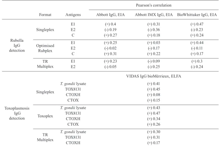
Table II shows the Pearson’s correlation coefficients for each antigen in the singleplex, Rubplex, and TR mul-tiplex formats with respect to results obtained from tests used to characterise the serological panel.
The ELFA method and liquid microarray assays us-ing T. gondii lysate and TOX8131 showed a moderate positive correlation between all test formats. In general, the chimeric antigens CTOX and CTOXH showed a less robust positive correlation.
Correlations between the singleplex and multiplex assay results based on EIAs and MEIAs using R. virus
antigens varied considerably. In general, the results of tests performed with E-1 correlated strongly among these methods, whereas those obtained with E-2 were poorly correlated.
DISCUSSION
Here, we developed a preliminary assay based on liq-uid microarray technology and evaluated 13 antigens for the detection of IgG anti-T. gondii or anti-R. virus. The performance of six of these antigens was sufficiently ro-TABLE II
Pearson’s correlation between enzyme-linked fluorescent assay (ELFA), enzyme immunoassay (EIA) and multiplex enzyme immunoassay (MEIA) assays, and the best singleplex, multiplex and TR multiplex assays
using Rubella virus and Toxoplasma gondii antigens coupled to beads under optimised conditions
Format Antigens
Pearson’s correlation
Abbott IgG, EIA Abbott IMX IgG, EIA BioWhittaker IgG, EIA
Rubella IgG detection
Singleplex
E1 (+) 0.4 (+) 0.31 (+) 0.47
E2 (-) 0.19 (-) 0.36 (-) 0.23
C (+) 0.27 (+) 0.18 (+) 0.24
Optimised Rubplex
E1 (+) 0.25 (+) 0.03 (+) 0.44
E2 (-) 0.02 (-) 0.17 (-) 0.11
C (+) 0.31 (+) 0.22 (+) 0.17
TR Multiplex
E1 (+) 0.23 (-) 0.09 (+) 0.3
E2 (-) 0.05 (-) 0.25 (-) 0.24
VIDAS IgG bioMérrieux, ELFA
Toxoplasmosis IgG detection
Singleplex
T. gondii lysate (+) 0.41
TOX8131 (+) 0.45
CTOXH (+) 0.08
CTOX (+) 0.15
Toxoplex
T. gondii lysate (+) 0.43
TOX8131 (+) 0.47
CTOXH (+) 0.34
CTOX (+) 0.26
TR Multiplex
T. gondii lysate (+) 0.30
TOX8131 (+) 0.31
CTOXH (+) 0.17
bust for their inclusion in a multiplex diagnostic test for rubella and toxoplasmosis. This multiplex assay showed 100% overall sensitivity and specificity for toxoplasmosis detection, and 95.6% overall sensitivity and 100% speci-ficity for rubella detection. In addition, we showed that E1, E2, and chimeric CTOXH (P29+P30+P35) recombi-nant proteins provided satisfactory results, so that their application in immunoassays may be further optimised.
The protein concentration and pH of the buffer used for sample dilution are critical factors that influence the qual-ity of bead coupling and, hence, the qualqual-ity of the assay. The bonds between the carboxyl groups of microspheres and free amino groups present in the antigens are mainly ionic, and are thus controlled by the pH of the buffer used. The buffer pH can cause conformational changes in the structure of the molecule, thereby altering the sites of in-teraction with specific antibodies. The optimal antigen concentration in a microsphere depends on the antibody titers in the blood, as well as the dilution of the plasma or serum used in the assay. In the current study, this opti-misation process increased the sensitivity of detection for E-1 (from 57% to 82%), E-2 (from 43% to 78%), C (from 9% to 57%), and MIC-3 (from 66% to 99%).
The performance of a test is affected by the choice of samples that are included in the test panel. The panel of samples used for the validation tests to diagnose ru-bella comprised only two negative samples. Obtaining negative samples is currently hampered by the extensive vaccination campaign being conducted against rubella in Brazil, which has almost eradicated the disease in the country. However, the panel of toxoplasmosis and ru-bella samples are representative of the various stages of infection, including seroconversion and acute, chronic, and late stage samples. Moreover, 40% of positive sam-ples in both panels showed low reactivity for specific IgG in gold standard assays, yielding unbiased sensitiv-ity results. Importantly, there were no discordant results. The assays developed in this work serve as proof of con-cept. Validation tests must be conducted, including tests with a number of representative samples from the popu-lation to be studied and based on the acceptable margin of error and other validation criteria such as the detec-tion limit, linearity, absolute recovery, reproducibility, repeatability, and analytical sensitivity.
The singleplex assays performed with TOX8131, TOX8122, and T. gondii lysate antigens and CTOXH (P29+P30+P35) showed sensitivities of 100%, 97%, 100%, and 99%, respectively, and a specificity of 100% in each case. This excellent performance was related to the fact that the antigenic extracts and chimeric antigen pro-vided a wider range of epitopes for binding anti-T. gon-dii antibodies compared to those of individual antigens. During infection, a wide variety of antigens are present, as they define exposure to the immune system and de-termine the avidity of IgG-specific antibodies, and stage of infection. These factors lead to noticeable variation in the intensity of humoral responses elicited against dif-ferent T. gondii antigens (Ferrandiz et al. 2004, Pfrepper et al. 2005, Holec et al. 2008, Holec-Gasior 2013). Thus, the increased variety of epitopes in the T. gondii extracts
and lysate antigen resulted in the best Pearson’s correla-tions by ELFA, which employs an antigenic extract as the capture molecule. Likewise, differences in performance between the assays using T. gondii extract and lysate were related to differences in antigenic composition.
In singleplex and all multiplex assays, the E-1 and E-2 antigens performed significantly better than the C antigen. Both vaccination and natural infection by R. vi-rus elicit a host immune response against the structural proteins E-1, E-2, and C (Chaye et al. 1992a, Vauloup-Fellous & Grangeot-Keros 2007). The mosaic E-1 protein employed in this study includes the amino acid fragment 214-240 containing the hemagglutinin epitope, fragments 214-233 and 219-233, which correspond to neutralising epitopes, and fragment 374-390, all of which are highly recognised in enzyme-linked immunosorbent assays (ELISAs) (Chaye et al. 1992b). The E-2 and C antigens were limited to fragments 31-105 and 1-123, which con-tain recognised epitopes (Wolinsky et al. 1991, 1993). An-tigen E-1 is more immunogenic in adults and children, vaccinated individuals and those with recent infections and who are currently infected, and children with con-genital rubella syndrome) (Chaye et al. 1992a, Nedeljkov-ic et al. 1999, Wilson et al. 2006). This finding explains why E-1 showed the best Pearson’s correlation between all assays, as E-1 was present in the R. virus extracts used in tests to characterise the serostatus of the sample panel.
The development of multiplex assays presents many challenges related to the use of different antigens and antibodies in a single assay, as well as the same assay parameters to detect different targets (de Jager et al. 2003, Elshal & McCoy 2006). Here, dilution of serum and secondary antibody to 1:100 was optimal for detect-ing R. virus antigens. However, the use of these dilutions and increased complexity of the multiplex system led to reduced performance with respect to TOX8131 and T. gondii lysate, compared to their performances in single-plex assays. Nevertheless, the multisingle-plex assay resulted in 100% detection of IgG anti-T. gondii in all positive samples. CTOXH showed 100% sensitivity in the TR multiplex assay, which indicates that including multiple antigens can replace antigenic extracts produced from
T. gondii cultures, as seen in toxoplasmosis diagnostics (Holec-Gasior 2013). Therefore, a multiplex assay can be developed including only this chimeric antigen for the detection of anti-T. gondii IgG antibodies.
read-ing), which corresponds to approximately 15 min for each molecule employed in antigen capture. Thus, the efficiency and throughput is increased when a greater number of targets is simultaneously detected, particu-larly when compared to a single ELISA assay that takes 5 h to perform. Reducing the required hands-on time en-ables operators to perform almost two full Luminex as-says in the same time needed to perform a single ELISA. Furthermore, the cost of reagents for this multiplex as-say is 60% lower than the costs for equivalent ELISA asas-says used to diagnose both diseases, excluding the costs related to laboratory operating times. Moreover, unlike ELISA as-says, the cost of our multiplex assay can be further reduced by including additional targets, as this requires only the cost of adding the microspheres and antigens and maintains the same amounts of other reagents and labor.
This multiplex assay for the detection of anti-T. gon-dii and anti-R. virus IgGs required the use of only 2 µL of serum or plasma. This is a significant improvement, particularly for use with pregnant women, because cur-rent prenatal screening for multiple infectious diseases requires large volumes of blood samples.
In conclusion, the multiplex assay, which includes R. virus recombinant antigens and T. gondii chimeric anti-gens, extract, and lysate, showed promising sensitivities and specificities. This multiplex assay for specific IgG detection may be useful for determining whether preg-nant woman have had prior contact with the pathogens that cause toxoplasmosis and rubella, as the analysis of anti-R. virus IgG antibodies in women already vaccinat-ed is useful for surveillance of immunity against R. vi-rus, which allows a public health risk assessment. How-ever, this assay is not sufficient for toxoplasmosis and rubella diagnosis. For this purpose, assays that specifi-cally detect IgM can be integrated or developed on sepa-rate platforms for IgG and IgM detection. This decision depends on the assay formats used, because there is the possibility of cross-reaction between test components. Avidity assays can be included as an additional step af-ter the first IgG fluorescence reading. It is important to understand that the specific IgG response may evolve differentially with different antigens, so results positive for one specific antigen and negative for others must be confirmed through an analysis of paired samples, which is already common. The test parameters were calculated based on a well-characterised set of sera, but the number of samples was limited, so the performance of the as-say must be further validated with a larger number of samples. The Luminex-based multiplex assay is rapid and can process large numbers of samples, which is clearly advantageous for screening programs in a public health setting. In this way, this multiplex assay shows the potential to be used as a platform for the inclusion of additional markers of prenatal infections for systematic screening of pregnant women.
ACKNOWLEDGEMENTS
To Irina Nastassja Riediger, for providing the panel of samples from the reference laboratory of the Central Labora-tory of Paraná (PR-LACEN).
AUTHORS’ CONTRIBUTION
PTB and LF - Contributed substantially to the conception or design of the work, the acquisition, analysis, and interpreta-tion of data, drafting and revision of the report for critically important intellectual content; PTB, LF and MAK - provided final approval of the version to be published and are account-able for all aspects of the work, ensuring the accuracy and in-tegrity of all parts of the study.
REFERENCES
Banatvala JE, Brown DW. Rubella. Lancet. 2004; 363(9415): 1127-37.
Binnicker MJ, Jespersen DJ, Harring JA. Multiplex detection of IgM and IgG class antibodies to Toxoplasma gondii, rubella virus, and cytomegalovirus using a novel multiplex flow immunoassay. Clin Vaccine Immunol. 2010; 17(11): 1734-8.
Binnicker MJ, Jespersen DJ, Rollins LO. Evaluation of the Rad Bio-Plex measles, mumps, rubella, and varicella-zoster virus IgG multi-plex bead immunoassay. Clin Vaccine Immunol. 2011; 18(9): 1524-6.
Chaye H, Chong P, Tripet B, Brush B, Gillam S. Localization of the virus neutralizing and hemagglutinin epitopes of E1 glycoprotein of rubella virus. Virology. 1992b; 189(2): 483-92.
Chaye HH, Mauracher CA, Tingle AJ, Gillam S. Cellular and humor-al immune responses to rubella virus structurhumor-al proteins E1, E2, and C. J Clin Microbiol. 1992a; 30(9): 2323-9.
de Jager W, te Velthuis H, Prakken BJ, Kuis W, Rijkers GT. Simul-taneous detection of 15 human cytokines in a single sample of stimulated peripheral blood mononuclear cells. Clin Diagn Lab Immunol. 2003; 10(1): 133-9.
Dhiman N, Jespersen DJ, Rollins LO, Harring JA, Beito EM, Bin-nicker MJ. Detection of IgG-class antibodies to measles, mumps, rubella, and varicella-zoster virus using a multiplex bead immu-noassay. Diagn Microbiol Infect Dis. 2010; 67(4): 346-9.
Dimech W, Panagiotopoulos L, Francis B, Laven N, Marler J, Dicke-son D, et al. Evaluation of eight anti-Rubella virus immunoglobu-lin G immunoassays that report results in international units per milliliter. J Clin Microbiol. 2008; 46(6): 1955-60.
Elshal MF, McCoy JP. Multiplex bead array assays: performance evaluation and comparison of sensitivity to ELISA. Methods. 2006; 38(4): 317-23.
Enders M, Bartelt U, Knotek F, Bunn K, Strobel S, Dietz K, et al. Performance of the elecsys rubella IgG assay in the diagnostic laboratory setting for assessment of immune status. Clin Vaccine Immunol. 2013; 20(3): 420-6.
Ferrandiz J, Mercier C, Wallon M, Picot S, Cesbron-Delauw MF, Pey-ron F. Limited value of assays using detection of immunoglobu-lin G antibodies to the two recombinant dense granule antigens, GRA1 and GRA6 Nt of Toxoplasma gondii, for distinguishing between acute and chronic infections in pregnant women. Clin Diagn Lab Immunol. 2004; 11(6): 1016-21.
Havelaar AH, Kemmeren JM, Kortbeek LM. Disease burden of con-genital toxoplasmosis. Clin Infect Dis. 2007; 44(11): 1467-74.
Holec L, Gasior A, Brillowska-Dabrowska A, Kur J. Toxoplasma gon-dii: enzyme-linked immunosorbent assay using different frag-ments of recombinant microneme protein 1 (MIC1) for detection of immunoglobulin G antibodies. Exp Parasitol. 2008; 119(1): 1-6.
Holec-Gasior L. Toxoplasma gondii recombinant antigens as tools for serodiagnosis of human toxoplasmosis: current status of studies. Clin Vaccine Immunol. 2013; 20(9): 1343-51.
Kellar KL, Iannone MA. Multiplexed microsphere-based flow cyto-metric assays. Exp Hematol. 2002; 30(11): 1227-37.
Liu Y, Yu F, Huang H, Han J. Development of recombinant antigen array for simultaneous detection of viral antibodies. PLoS ONE. 2013; 8(9): e73842.
Montoya JG, Remington JS. Management of Toxoplasma gondii in-fection during pregnancy. Clin Infect Dis. 2008; 47(4): 554-66.
Montoya JG, Rosso F. Diagnosis and management of toxoplasmosis. Clin Perinatol. 2005; 32(3): 705-26.
Murat J-B, L’Ollivier C, Hidalgo HF, Franck J, Pelloux H, Piarroux R. Evaluation of the new elecsys toxo IgG avidity assay for toxoplas-mosis and new insights into the interpretation of avidity results. Clin Vaccine Immunol. 2012; 19(11): 1838-43.
Nedeljkovic J, Jovanovic T, Mladjenovic S, Hedman K, Peitsaro N, Oker-Blom C. Immunoblot analysis of natural and vaccine-in-duced IgG responses to Rubella virus proteins expressed in insect cells. J Clin Virol. 1999; 14(2): 119-31.
Pfrepper KI, Enders G, Gohl M, Krczal D, Hlobil H, Wassenberg D, et al. Seroreactivity to and avidity for recombinant antigens in toxoplasmosis. Clin Diagn Lab Immunol. 2005; 12(8): 977-82.
Schmidt M, Lindqvist C, Salmi A, Oker-Blom C. Detection of rubella virus-specific immunoglobulin M antibodies with a baculovirus-expressed E1 protein. Clin Diagn Lab Immunol. 1996; 3(2): 216-8.
Simons EA, Reef SE, Cooper LZ, Zimmerman L, Thompson KM. Systematic review of the manifestations of congenital rubella syndrome in infants and characterization of disability-adjusted life years (DALYs). Risk Analysis. 2016; 36: 1332-56.
Vauloup-Fellous C, Grangeot-Keros L. Humoral immune response after primary rubella virus infection and after vaccination. Clin Vaccine Immunol. 2007; 14(5): 644-7.
Villard O, Breit L, Cimon B, Franck J, Fricker-Hidalgo H, Godineau N, et al. Comparison of four commercially available avidity tests for Toxoplasma gondii-specific IgG antibodies. Clin Vaccine Im-munol. 2013; 20(2): 197-204.
Wandinger KP, Saschenbrecker S, Steinhagen K, Scheper T, Meyer W, Bartelt U, et al. Diagnosis of recent primary rubella virus infections: significance of glycoprotein-based IgM serology, IgG avidity and immunoblot analysis. J Virol Methods. 2011; 174(1-2): 85-93.
Wilson KM, Di Camillo C, Doughty L, Dax EM. Humoral immune response to primary rubella virus infection. Clin Vaccine Immu-nol. 2006; 13(3): 380-6.
Wolinsky JS, McCarthy M, Allen-Cannady O, Moore WT, Jin R, Cao SN, et al. Monoclonal antibody-defined epitope map of expressed rubella virus protein domains. J Virol. 1991; 65(8): 3986-94.