RET
Variants and Haplotype Analysis in a Cohort of Czech
Patients with Hirschsprung Disease
Eliska Vaclavikova1*, Sarka Dvorakova1, Richard Skaba2, Lucie Pos2, Vlasta Sykorova1, Tereza Halkova1, Josef Vcelak1, Bela Bendlova1
1Department of Molecular Endocrinology, Institute of Endocrinology, Prague, Czech Republic,2Department of Paediatric Surgery, 2nd Faculty of Medicine, Charles University and Hospital Motol, Prague, Czech Republic
Abstract
Hirschsprung disease (HSCR) is a congenital aganglionosis of myenteric and submucosal plexuses in variable length of the intestine. This study investigated the influence and a possible modifying function ofRETproto-oncogene’s single nucleotide polymorphisms (SNPs) and haplotypes in the development and phenotype of the disease in Czech patients. Genotyping of 14 SNPs was performed using TaqMan Genotyping Assays and direct sequencing. The frequencies of SNPs and generated haplotypes were statistically evaluated using chi-square test and the association with the risk of HSCR was estimated by odds ratio. SNP analysis revealed significant differences in frequencies of 11 polymorphicRETvariants between 162 HSCR patients and 205 unaffected controls. Particularly variant alleles of rs1864410, rs2435357, rs2506004 (intron 1), rs1800858 (exon 2), rs1800861 (exon 13), and rs2565200 (intron 19) were strongly associated with increased risk of HSCR (p,0.00000) and were over-represented in males vs. females. Conversely, variant alleles of rs1800860, rs1799939 and rs1800863 (exons 7, 11, 15) had a protective role. The haploblock comprising variants in intron 1 and exon 2 was constructed. It represented a high risk of HSCR, however, the influence of other variants was also found after pruning from effect of this haploblock. Clustering patients according to genotype status in haploblock revealed a strong co-segregation with several SNPs and pointed out the differences between long and short form of HSCR. This study involved a large number of SNPs along the entireRETproto-oncogene with demonstration of their risk/protective role also in haplotype and diplotype analysis in the Czech population. The influence of some variant alleles on the aggressiveness of the disease and their role in gender manifestation differences was found. These data contribute to worldwide knowledge of the genetics of HSCR.
Citation:Vaclavikova E, Dvorakova S, Skaba R, Pos L, Sykorova V, et al. (2014)RET Variants and Haplotype Analysis in a Cohort of Czech Patients with Hirschsprung Disease. PLoS ONE 9(6): e98957. doi:10.1371/journal.pone.0098957
Editor:Xiaoping Miao, MOE Key Laboratory of Environment and Health, School of Public Health, Tongji Medical College, Huazhong University of Science and Technology, China
ReceivedMarch 3, 2014;AcceptedMay 8, 2014;PublishedJune 4, 2014
Copyright:ß2014 Vaclavikova et al. This is an open-access article distributed under the terms of the Creative Commons Attribution License, which permits unrestricted use, distribution, and reproduction in any medium, provided the original author and source are credited.
Funding:This work was supported by grant projects IGA MH CR NT13901-4, GAUK 411611 and MH CZ for conceptual development of research organisation 00023761 (Institute of Endocrinology, Prague, Czech Republic). The funders had no role in study design, data collection and analysis, decision to publish, or preparation of the manuscript.
Competing Interests:The authors have declared that no competing interests exist. * E-mail: [email protected]
Introduction
Hirschsprung disease (HSCR) is a congenital developmental malformation characterised by the absence of enteric ganglion cells of myenteric and submucosal plexuses in the intestine. The incidence of the disease is 1 per 5000 live births. Short-segment aganglionosis (80%) and long-segment aganglionosis (20%) are classified according to the length of the aganglionic segment. The short-segment form of HSCR (S-HSCR), comprising recto sigmoid HSCR and ultra-short segment HSCR, affects the distal portion, part of the anal canal and, in contrast to long-segment HSCR, occurs four times more often in males than females. The long-segment form of HSCR (L-HSCR) can present as total colonic aganglionosis (TCA) extending from the rectum up to the terminal ileum and, in rare cases, as nearly total bowel (NTBA) or total intestinal aganglionosis (TIA) comprising nearly total or the whole intestine. It occurs as an isolated disorder in 70% of cases, about 12% of cases have a chromosomal abnormality and 18% of cases have additional congenital anomalies [1].
Isolated HSCR appears as a sporadic (85%) or less commonly as a familial disorder (15%). To date, more than 14 genes and 5
susceptibility loci have been associated with the disease [1,2]. However, the inactivating germline mutations in theRET
proto-oncogene play the major role in the pathogenesis. The RET
(Rearranged during Transfection), a gene consisting of 21 exons and located on chromosome 10q11.2, encodes a transmembrane tyrosine kinase receptor. It is expressed in neural crest-derived cells and plays an important role in the development, proliferation and differentiation of neuroendocrine cells. The various alterations (missense mutations, deletions, insertions, frame shifts) have been found along the entire gene. Inactivating germlineRETmutations
are detected in about 50% of familial and 15–20% of sporadic HSCR cases [1].
Although the detection rate of germline RET mutations is
relatively low, linkage analysis has shown that nearly all familial HSCR patients are in linkage with theRETproto-oncogene [3].
Therefore, noncoding variants and single nucleotide polymor-phisms (SNPs) as well as some specific haplotypes of the RET
proto-oncogene have been revealed to be a potential low susceptibility loci and have a function of genetic modifying factors in HSCR pathogenesis. SeveralRET polymorphisms have been
investigated in the association with HSCR. Previous studies [3–5]
SNP Location Amino acid change Nucleotide change
HSCR (n = 162): Variant allele (%)
Controls (n = 205): Variant allele (%)
p-value
(forx2) OR (95% CI) p-value (for OR)
rs1864410 Intron 1 IVS1+2846G/T c.73+2846G/T 233 (71.9) 113 (27.6) 0.00000 6.73 (4.86–9.31) 0.00000
rs2435357 Intron 1 IVS1+9277C/T c.73+9277C/T 234 (72.2) 115 (28.0) 0.00000 6.67 (4.82–9.23) 0.00000
rs2506004 Intron 1 IVS1+9494C/A c.73+9494C/A 234 (72.2) 115 (28.0) 0.00000 6.67 (4.82–9.23) 0.00000
rs1800858 Exon 2 Ala45Ala c.135G/A 231 (71.3) 112 (27.3) 0.00000 6.61 (4.78–9.14) 0.00000
rs1800860 Exon 7 Ala432Ala c.1296G/A 82 (25.8) 139 (33.9) 0.01816 0.68 (0.49–0.94) 0.02255
rs1799939 Exon 11 Gly691Ser c.2071G/A 27 (8.3) 94 (22.9) 0.00000 0.31 (0.19–0.48) 0.00000
rs1800861 Exon 13 Leu769Leu c.2307T/G 141 (43.5) 89 (21.8) 0.00000 2.76 (2.00–3.81) 0.00000
rs111264957 Intron 13 IVS13-94C/T c.2393-94C/T 6 (1.9) 12 (2.9) 0.34978 0.63 (0.23–1.69) 0.48723
rs1800862 Exon 14 Ser836Ser c.2508C/T 8 (2.5) 12 (2.9) 0.70528 0.84 (0.34–2.08) 0.88083
rs2472737 Intron 14 IVS14-24G/A c.2608-24G/A 82 (25.3) 99 (24.1) 0.71678 1.06 (0.76–1.49) 0.78213
rs1800863 Exon 15 Ser904Ser c.2712C/G 28 (8.6) 94 (22.9) 0.00000 0.32 (0.20–0.50) 0.00000
rs2565200 Intron 19 IVS19-627C/T c.3188-627C/T 133 (41.0) 77 (18.8) 0.00000 3.01 (2.16–4.20) 0.00000
rs143948954 39UTR c.4391G/C 8 (2.5) 2 (0.5) 0.02149 5.16 (1.09–24.49) 0.04785
rs2435355 39UTR c.4461T/C 105 (32.6) 103 (25.1) 0.02579 1.44 (1.04–1.99) 0.03181
doi:10.1371/journal.pone.0098957.t001
RET
Variants
in
Czech
Hirschspru
ng
Patients
ONE
|
www.ploson
e.org
2
June
2014
|
Volume
9
|
Issue
6
|
described the variants in the coding regions - polymorphism in exon 2 (rs1800858, p.Ala45Ala, c.135G/A) was overrepresented in HSCR patients with respect to controls, as well as polymorphisms in exon 7 (rs1800860, p.Ala432Ala, c.1296G/A) and in exon 13 (rs1800861, p.Leu769Leu, c.2307T/G) are risk factors for HSCR. On the other hand, SNPs in exons 11 (rs1799939, p.Gly691Ser, c.2071G/A), 14 (rs1800862, p.Ser836Ser, c.2508C/T) and 15 (rs1800863, p.Ser904Ser, c.2712C/G) were underrepresented in HSCR cases, suggesting a protective role of these SNPs. However, results could differ depending on methods, cohorts and frequencies of SNPs.
Consequently, the study of SNPs has been focused on noncoding regions - 59untranslated region (UTR), intron variants and 39UTR. The investigation of the strong association of rs1800858 (p.Ala45Ala, c.135G/A) with HSCR was extended upstream of exon 2 to intron 1 and promoter. A haplotype spanning 27 kb of the 59UTR region to exon 2 was determined as HSCR-associated [2,6–8]. The investigation of 39UTR variants underrepresented in HSCR patients resulted in the suggestion of the protective haplotype in 39UTR [9–10].
In this large case-control study, we investigated the possible role of variants in the RETproto-oncogene in cohorts of 162 Czech
patients with HSCR and 205 unaffected control individuals and the role of these variants in gender manifestation differences and their influence on the aggressiveness of the disease. We carried out a screening of 14 polymorphisms throughout the RET
proto-oncogene to identify risk or protective SNPs and haplotypes associated with the HSCR phenotype from the Czech population.
Material and Methods
In this study, blood samples were obtained from 162 Czech patients with HSCR (121 males and 41 females). The patients were chosen for molecular genetic analysis prospectively (88 cases operated on from 2003 till now) as well as retrospectively (74 cases operated on between 1979 and 2003). According to the length of the aganglionic segment, the cohort consisted of 117 patients with short-segment HSCR (recto-sigmoid form; patients with ultra-short segment were not involved in the study), 41 patients with long-segment aganglionosis (including 20 patients with total colonic aganglionosis and 2 patients with nearly total bowel aganglionosis), and 4 patients with an unspecified form of HSCR. The control group included 205 Czech healthy individuals (95 males, 110 females).
Ethics statement
This study was approved by the Ethical Committee of the Institute of Endocrinology and University Hospital Motol, Prague. A signed informed consent for this study was obtained from each patient or legitimate representative who participated.
Genetic analysis
Genomic DNA was isolated from peripheral blood leukocytes using the QIAamp DNA Blood Kit (Qiagen, Germany) and the QuickGene 610 L machine (Fujifilm, Japan). Studied polymor-phisms are listed in Table 1. Five of SNPs were analysed during routine sequencing testing of HSCR patients for mutations in exons 10, 11, 13, 14, 15 and 16 in theRETproto-oncogene due to
the risk of medullary thyroid cancer. The standard conditions for amplification ofRETexons were provided previously [11] as well
as sequenase reaction preparation [12]. Direct sequencing was performed on the CEQ 8000 sequencing machine (Beckman Coulter) and analysed by CEQ 8000 sequencing software. Sequence traces were compared with RET reference sequence
(NG_007489.1). Genotyping of the other 9 SNPs in patients and all 14 SNPs in control samples were determined by real-time polymerase chain reaction (PCR) with TaqMan Genotyping Assays (Applied Biosystems) on LightCycler 480 Real-Time PCR System (Roche) with using non template control in each run.
Statistical analysis
The frequencies of studied SNPs were statistically evaluated and compared using the NCSS programme and chi-square test with establishing p-value. The results were considered as statistically significant if the p-value was less than 0.05. Association with risk of HSCR was estimated by odds ratio (OR) and their 95% confidence interval (CI). For generating haplotypes, the Haploview programme (version 4.1) was used and a haplotype block of polymorphisms which were in linkage disequilibrium was constructed using Gabriel’s methods. Consequently, haplotypes of the particular haploblock were generated in each individual patient and control and diplotypes were estimated using the PHASE programme (version 2.1).
Results
SNP analysis
Statistical evaluation of studied SNPs revealed significant differences in genotype and allele distribution of polymorphic variants between HSCR patients and normal controls (Table 1). No significant deviation from Hardy-Weinberg equilibrium was found in the control cohort. Studied cohorts significantly differed in 11 out of 14 investigated SNPs. In SNPs rs1864410, rs2435357, rs2506004, rs1800858 the variant allele dominated in HSCR patients (all p,0.00000) and their carriership was associated with more than 6.6-fold elevated risk for development of HSCR compared with the wild-type allele carriership. These 4 SNPs were in complete linkage disequilibrium and went together with almost the same genotype distribution in all cohorts. The variant allele was also over-represented in rs1800861 (p,0.00000, OR = 2.76, 95% CI = 2.00–3.81) and rs2565200 (p,0.00000, OR = 3.01, 95% CI = 2.16–4.20). In contrast, the variant allele was under-represented in two SNPs in linkage disequilibrium: rs1799939 (p, 0.00000, OR = 0.31, 95% CI = 0.19–0.48) and rs1800863 (p, 0.00000, OR = 0.32, 95% CI = 0.20–0.50). Significant differences in allele distribution were established in rs1800860, rs143948954 and rs2435355. Allele distributions of SNPs considering the phenotype of the disease (L- and S-HSCR) and gender were described in Table S1 and Table S2.
Three SNPs (rs111264957, rs1800862, rs143948954) were detected in very low frequencies of variant allele (2.9%, 2.9%, and 0.5%, respectively in controls).
Haplotype and diplotype analysis
Using the Haploview programme for generating haplotypes, two haplotype blocks were identified (Figure 1) comprising 10 out of 14 investigated polymorphisms that were in linkage disequilib-rium. The first haploblock was composed of 4 SNPs (rs1864410, rs2435357, rs2506004, rs1800858) that were very closely related together. The second haploblock consisting of 6 SNPs (rs1799939, rs1800861, rs2472737, rs1800863, rs2565200, rs2435355) was very heterogeneous and differed in distribution between particular cohorts. Therefore, these SNPs were rather evaluated separately. Moreover, 4 SNPs not included in any of two haploblocks due to low frequencies in cohorts or its localization in the gene were also investigated separately.
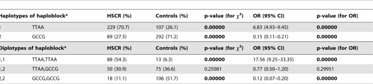
The two most frequent haplotypes of 59region haploblock were represented by TTAA and GCCG (Table 2). The presence of only RETVariants in Czech Hirschsprung Patients
two main haplotypes was caused by above-mentioned trend that in nearly all cases these 4 SNPs went together in wild type or variant allele mode. The distribution of haploblock-haplotypes signifi-cantly differed between HSCR patients and controls (p,0.00000). Although the most frequent haplotype in HSCR patients was TTAA (HSCR 70.7% vs. controls 26.1%), the most frequent in controls was haplotype GCCG (HSCR 27.5% vs. controls 71.2%). The haplotype TTAA had a 6.83-fold (95% CI = 4.93–9.45, p, 0.00000) elevated risk for development of HSCR than other haplotypes. The different distribution of estimated combinations of haplotypes in two alleles of each subject resulted from this converse representation of haplotypes. The diplotype TTAA,T-TAA had a 17,56-fold (95% CI = 9.25–33.35, p,0.00000) elevated risk for the development of HSCR than other diplotypes. If we consider the phenotype of the disease with haploblock status, variant allele frequencies in L-HSCR and S-HSCR were significantly different from controls, but did not vary between forms of HSCR (Table 3). The risk of HSCR was higher in the S-form than in L-S-form resulting from odds ratios (7.62 vs. 5.66 for
rs1864410). By comparing frequencies of SNPs in haploblock between male patients and male controls as well as female patients and female controls, similar significant results were obtained– contrary representation of the variant allele in controls vs. patients. In comparison between male and female patients, there were also statistically significant differences. The different allele distribution in haploblock (for rs1864410 variant allele in male 76.0% vs. female 59.8%) was related to the different risk for development of HSCR in males (OR = 8.20, 95% CI = 5.32–12.65) and females (OR = 3.96, 95% CI = 2.33–6.74).
Distribution of 39region SNPs with respect to 59region haploblock status
Other SNPs located in 39 RET region were examined
depending on what haplotype in 59 part of gene was carried. Given allelic distribution of TTAA and GCCG haplotypes (Table 4), frequencies of variant alleles of rs1800861 and 2565200 as well as rs1799939 and rs1800863 significantly differed Figure 1. Haplotype blocks generated by the Haploview Programme in cohorts of HSCR patients and control population.The scheme is shown with confidence bounds. LD values are reported in D9.
between cases and controls with the same risk/protective trends as were shown in previous SNP analysis (Table 1). In genotype distribution of these haplotypes, there was possible to observe the tendency of the increasing/decreasing variant allele frequencies depending on carriership of TTAA in two, one or no alleles (Table S3).
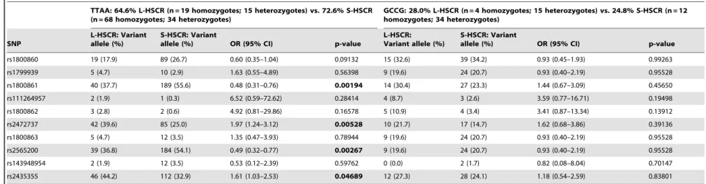
In carriers of TTAA haplotype, differences between L-HSCR and S-HSCR phenotype were intensified in variant alleles of rs1800861, rs2472737, rs2565200 and rs2435355, whereas in carriers of GCCG no significant differences between phenotypes were found (Table 5). In TTAA carriers, the variant alleles of rs1800861 and rs2565200 were over-represented in S-HSCR (55.6% and 54.1%, respectively) vs. L-HSCR (37.7% and 36.8%, respectively). The variant alleles of rs2472737 and rs2435355 were over-represented in L-HSCR (39.6% and 44.2%, respectively) vs. S-HSCR (25.0% and 32.9%, respectively). Genotype distribution of haploblock-haplotypes TTAA and GCCG describing differenc-es in 39 region SNPs frequencies between L- and S-HSCR is shown in Table S4. No differences between male and female patients and controls were observed considering the 59 region haplotype (Table S5).
Discussion
The genetics of HSCR is complex. It is believed that it can be inherited in a dominant or recessive trait, but probably it is polygenic with incomplete penetrance, genetic heterogeneity, variable expression of the disease and a large number of additive syndromes. The main impact of the RET proto-oncogene
mutation is a loss of function (haploinsufficiency). Almost all HSCR cases are linked to the RET locus despite no detected
mutation. Therefore, noncoding RET variants and SNPs must
play at least a modifying role in the remaining HSCR cases. We focused our study on SNPs along the entire RET
proto-oncogene and selected 14 promising SNPs in coding and intronic sequences and 39UTR. Allele frequencies in our tested normal population were similar with frequencies in previously reported in a European control population [13] and greatly varied from an Asian control population [2,10,14,15].
Our data show that mainly 8 SNPs are very important in the development of HSCR. We found a strong association of variant alleles of 4 investigated SNPs in intron 1 and exon 2 grouped in haploblock (rs1864410, rs2435357, rs2506004, rs1800858) with HSCR (p,0.00000). These alleles were the most frequent in our patient cohort and formed the haplotype TTAA with a very elevated risk (OR = 6.83). In the homozygous diplotype TTAA,T-TAA the risk was even more increased (OR = 17.56). These findings are consistent with other studies describing these SNPs as
belonging to HSCR-causing region covering 27 kb in total. It is a highly conserved region called MSC+9.7 (Multi species conserved) and starts 4 kb upstream of theRET transcription start site and
going along the way to the beginning of exon 2. This risk haplotype decreasesRET promoter activity, reduces the binding
affinity of TTF-1 (thyroid transcription factor 1), disrupts a binding site for transcription factor SOX10, decreases enhancer for RET expression and thus regulates RET expression which was confirmed by in vitro studies [7,8,16,17].
In previous studies, there was suggested the hypothesis about the different role of SNPs in two linkage disequilibrium regions. Besides the risk haplotype at the 59 end of the RET
proto-oncogene, a protective function of the 39 half of the gene was proposed. SNPs in this region encompassing rs1799939 up to 39UTR variants were reported as underrepresented in HSCR patients [3,9,10]. We cannot confirm this hypothesis because we detected several SNPs with increased risk in this region. The main contradiction with the hypothesis of the protective 39 half RET
gene haplotype is the high overrepresentation of rs2565200 located in intron 19 and rs2435355 located in 39UTR. SNP rs1800861 in exon 13 was also strongly associated with HSCR as previously described [3,15]. The risk effect of rs1800861 and rs2565200 in Czech patients was confirmed. Moreover, after pruning from the effect of 59 region haploblock, variant allele frequencies of rs1800861 and rs2565200 still significantly differed in comparison of patients and controls and were associated with the risk of HSCR. In rs2435355, a divergent tendency was observed in Czech patients that differed from a study of Chinese patients [10] which instead had a protective role. Perhaps the discrepancy in the theory of protective 39half gene role can be due to various studied cohorts and ethnic differences. We defined three main protective SNPs in our series located in exons 7 (rs1800860), 11 (rs1799939) and 15 (rs1800863). The protective role of rs1799939 and rs1800863 was described previously [3]. Consid-ering the 59region haploblock status in investigated carriers, only rs1799939 with rs1800863 variant alleles stayed protective. In contrast to our results, some studies detected rs1800862 variant allele underrepresented in HSCR with a protective role [18], and in the Chinese population the variant allele was even absent [14], which was correlated with increased HSCR incidence in the Chinese population. We identified this SNP in similar low frequencies in both our case and control cohorts, which was not in agreement with these findings. It seems that the other SNPs studied do not have any major role in the development of HSCR in our series. The low frequencies of variant allele were also identified in rs111264957 that was in linkage disequilibrium with rs1800862, and rs143948954, where a significantly higher representation of the variant allele in patients was noticed.
Table 2.Distribution of haplotypes and diplotypes of 59region haploblock in HSCR patients and controls.
Haplotypes of haploblock* HSCR (%) Controls (%) p-value (forx2) OR (95% CI) p-value (for OR)
1 TTAA 229 (70.7) 107 (26.1) 0.00000 6.83 (4.93–9.45) 0.00000
2 GCCG 89 (27.5) 292 (71.2) 0.00000 0.15 (0.11–0.21) 0.00000
Diplotypes of haploblock* HSCR (%) Controls (%) p-value (forx2) OR (95% CI) p-value (for OR)
1,1 TTAA,TTAA 88 (54.3) 13 (6.3) 0.00000 17.56 (9.25–33.35) 0.00000
1,2 TTAA,GCCG 50 (30.9) 75 (36.6) 0.25081 0.77 (0.50–1.20) 0.29951
2,2 GCCG,GCCG 18 (11.1) 106 (51.7) 0.00000 0.12 (0.07–0.20) 0.00000
* Haplotypes/diplotypes with occurrence,2% in both cohorts are not included. doi:10.1371/journal.pone.0098957.t002
RETVariants in Czech Hirschsprung Patients
HSCR patients.
L-HSCR (n = 41) S-HSCR (n = 117) L-HSCR vs. S-HSCR
SNP
Cases: Variant allele (%)
Controls: Variant allele (%)
p-value
(forx2) OR (95% CI)
p-value (for OR)
Cases: Variant allele (%)
Controls: Variant allele (%)
p-value
(forx2) OR (95% CI)
p-value (for OR)
p-value
(forx2) OR (95% CI)
p-value (for OR)
rs1864410 56 (68.3) 113 (27.6) 0.00000 5.66 (3.39–9.46) 0.00000 174 (74.4) 113 (27.6) 0.00000 7.62 (5.29–10.98)0.00000 0.28819 0.74 (0.43–1.29) 0.35865
rs2435357 57 (69.5) 115 (28.0) 0.00000 5.85 (3.49–9.81) 0.00000 174 (74.4) 115 (28.0) 0.00000 7.44 (5.17–10.71)0.00000 0.39437 0.79 (0.45–1.37) 0.47956
rs2506004 57 (69.5) 115 (28.0) 0.00000 5.85 (3.49–9.81) 0.00000 174 (74.4) 115 (28.0) 0.00000 7.44 (5.17–10.71)0.00000 0.39437 0.79 (0.45–1.37) 0.47956
rs1800858 55 (67.1) 112 (27.3) 0.00000 5.42 (3.26–9.02) 0.00000 173 (73.9) 112 (27.3) 0.00000 7.55 (5.24–10.86)0.00000 0.23315 0.72 (0.42–1.24) 0.29412
Male cases (n = 121) vs. male controls (n = 95) Female cases (n = 41) vs. female controls (n = 110) Male vs. female cases
SNP
Cases: Variant allele (%)
Controls: Variant allele (%)
p-value
(forx2) OR (95% CI)
p-value (for OR)
Cases: Variant allele (%)
Controls: Variant allele (%)
p-value
(forx2) OR (95% CI)
p-value (for OR)
p-value
(forx2) OR (95% CI)
p-value (for OR)
rs1864410 184 (76.0) 53 (27.9) 0.00000 8.20 (5.32–12.65)0.00000 49 (59.8) 60 (27.3) 0.00000 3.96 (2.33–6.74) 0.00000 0.00459 2.14 (1.26–3.63) 0.00710
rs2435357 185 (76.4) 54 (28.4) 0.00000 8.17 (5.30–12.60)0.00000 49 (59.8) 61 (27.7) 0.00000 3.87 (2.28–6.58) 0.00000 0.00354 2.19 (1.28–3.72) 0.00554
rs2506004 185 (76.4) 54 (28.4) 0.00000 8.17 (5.30–12.60)0.00000 49 (59.8) 61 (27.7) 0.00000 3.87 (2.28–6.58) 0.00000 0.00354 2.19 (1.28–3.72) 0.00554
rs1800858 181 (74.8) 53 (27.9) 0.00000 7.67 (4.99–11.79)0.00000 50 (61.0) 59 (26.8) 0.00000 4.26 (2.50–7.28) 0.00000 0.01683 1.90 (1.12–3.23) 0.02450
doi:10.1371/journal.pone.0098957.t003
RET
Variants
in
Czech
Hirschspru
ng
Patients
ONE
|
www.ploson
e.org
6
June
2014
|
Volume
9
|
Issue
6
|
Table 4.Allelic distribution of single nucleotide polymorphisms considering allelic distribution of haploblock-haplotypes TTAA and GCCG in HSCR patients and controls.
TTAA: 70.7% cases (88 homozygotes; 50 heterozygotes) vs. 26.1% controls (13 homozygotes; 75 heterozygotes)
GCCG: 27.5% cases (18 homozygotes; 50 heterozygotes) vs. 71.2% controls (106 homozygotes; 75 heterozygotes)
SNP
Cases: Variant allele (%)
Controls: Variant
allele (%) OR (95% CI) p-value
Cases: Variant allele (%)
Controls: Variant
allele (%) OR (95% CI) p-value
rs1800860 108 (24.2) 50 (24.8) 0.97 (0.66–1.43) 0.96110 54 (32.5) 216 (37.6) 0.80 (0.55–1.15) 0.26667
rs1799939 15 (3.3) 19 (9.4) 0.33 (0.16–0.66) 0.00229 35 (20.4) 161 (28.1) 0.66 (0.43–0.99) 0.05563
rs1800861 233 (51.6) 78 (38.6) 1.69 (1.21–2.37) 0.00292 45 (26.2) 86 (15.0) 2.01 (1.33–3.03) 0.00109
rs111264957 4 (0.9) 3 (1.5) 0.59 (0.13–2.67) 0.78107 8 (4.7) 19 (3.3) 1.42 (0.61–3.31) 0.55297
rs1800862 6 (1.3) 3 (1.5) 0.89 (0.22–3.60) 0.83892 10 (5.8) 19 (3.3) 1.80 (0.82–3.96) 0.20575
rs2472737 129 (28.5) 63 (31.2) 0.88 (0.61–1.26) 0.55238 27 (15.7) 119 (20.7) 0.71 (0.45–1.13) 0.17698
rs1800863 17 (3.8) 19 (9.4) 0.38 (0.19–0.74) 0.00616 35 (20.4) 161 (28.1) 0.66 (0.43–0.99) 0.05563
rs2565200 226 (50.0) 76 (37.6) 1.66 (1.18–2.33) 0.00439 36 (20.9) 64 (11.2) 2.11 (1.34–3.31) 0.00150
rs143948954 14 (3.1) 3 (1.5) 2.12 (0.60–7.46) 0.35174 2 (1.2) 1 (0.2) 6.74 (0.61–74.80) 0.26690
rs2435355 160 (35.6) 61 (30.2) 1.28 (0.89–1.82) 0.21242 40 (23.5) 129 (22.5) 1.06 (0.71–1.59) 0.85377
doi:10.1371/journal.pone.0098957.t004
Table 5.Allelic distribution of single nucleotide polymorphisms considering allelic distribution of haploblock-haplotypes TTAA and GCCG in patients with long-segment and short-segment form of HSCR.
TTAA: 64.6% L-HSCR (n = 19 homozygotes; 15 heterozygotes) vs. 72.6% S-HSCR (n = 68 homozygotes; 34 heterozygotes)
GCCG: 28.0% L-HSCR (n = 4 homozygotes; 15 heterozygotes) vs. 24.8% S-HSCR (n = 12 homozygotes; 34 heterozygotes)
SNP
L-HSCR: Variant allele (%)
S-HSCR: Variant
allele (%) OR (95% CI) p-value
L-HSCR: Variant allele (%)
S-HSCR: Variant
allele (%) OR (95% CI) p-value
rs1800860 19 (17.9) 89 (26.7) 0.60 (0.35–1.04) 0.09132 15 (32.6) 39 (34.2) 0.93 (0.45–1.93) 0.99263
rs1799939 5 (4.7) 10 (2.9) 1.63 (0.55–4.89) 0.56398 9 (19.6) 24 (20.7) 0.93 (0.40–2.19) 0.95528
rs1800861 40 (37.7) 189 (55.6) 0.48 (0.31–0.76) 0.00194 14 (30.4) 27 (23.3) 1.44 (0.67–3.09) 0.45650
rs111264957 2 (1.9) 1 (0.3) 6.52 (0.59–72.62) 0.28414 4 (8.7) 3 (2.6) 3.59 (0.77–16.71) 0.19498
rs1800862 3 (2.8) 2 (0.6) 4.92 (0.81–29.86) 0.16578 5 (10.9) 4 (3.4) 3.41 (0.87–13.34) 0.13912
rs2472737 42 (39.6) 85 (25.0) 1.97 (1.24–3.12) 0.00528 10 (21.7) 17 (14.7) 1.62 (0.68–3.86) 0.39136
rs1800863 5 (4.7) 12 (3.5) 1.35 (0.47–3.93) 0.78944 9 (19.6) 24 (20.7) 0.93 (0.40–2.19) 0.95528
rs2565200 39 (36.8) 184 (54.1) 0.49 (0.32–0.77) 0.00267 9 (19.6) 24 (20.7) 0.93 (0.40–2.19) 0.95528
rs143948954 2 (1.9) 12 (3.5) 0.53 (0.12–2.39) 0.59762 0 (0.0) 2 (1.7) 0.82 (0.08–8.04) 0.70147
rs2435355 46 (44.2) 112 (32.9) 1.61 (1.03–2.53) 0.04689 12 (27.3) 28 (24.1) 1.18 (0.54–2.59) 0.83801
doi:10.1371/journal.pone.0098957.t005
RET
Variants
in
Czech
Hirschspru
ng
Patients
PLOS
ONE
|
www.ploson
e.org
7
June
2014
|
Volume
9
|
Issue
6
|
However, due to very low frequency, this significance on the phenotype could not be definitively revealed.
In our study, we also focused on the evaluation of the possible influence of the variant alleles on aggressiveness of the disease (L-HSCR vs. S-(L-HSCR) and their role in gender manifestation differences. The risk of variant alleles in the 59region haploblock was highly elevated both in L-HSCR and S-HSCR patients in relation to controls. Interestingly, the frequency of these risk alleles was even 6% higher in the less aggressive S-HSCR form. Similar results were described in rs2435357 [8] and rs1800858 [5], whereas no difference was revealed between L-HSCR and S-HSCR by Lantieri et al [3]. However, in our cohort, the higher frequency of variant alleles in S-HSCR was likely caused by a higher representation of male patients in S-HSCR (78%) than in the L-HSCR cohort (71%). A gender effect was previously described in rs2435357 where the variant allele was present in 65% of males vs. 56% of females [19]. Our data documented this trend not only in SNPs of the haploblock (76% of males vs. 60% of females) but also in risk variant alleles of rs1800861 and rs2565200. The frequency of these two risk alleles was about 10% higher in males, but nonsignificant. In variants of the haploblock, the associated risk was much more profound (OR = 8 in males vs. OR = 4 in females). It is not clear if over-representation of rs1800861 and rs2565200 in S-HSCR patients and a significant difference related to L-HSCR was also influenced by sex differences between cohorts. The risk of L-HSCR was influenced especially by rs2472737 and rs2435355 where the variant allele elevated the risk of L-HSCR nearly two fold compared to S-HSCR. Both these findings–rs1800861 and rs2565200 variant allele over-representation in S-HSCR and rs2472737 and rs2435355 variant allele over-representation in L-HSCR–were even confirmed considering the 59region haploblock status, but only in TTAA carriers.
Conclusions
This study aimed to identify the risk and protective SNPs and haplotypes that could be associated with Hirschsprung disease. From our results, we propose several risk and several protective SNPs and haplotypes. The influence of some variant alleles on the
aggressiveness of the disease and differences in allele frequencies between males and females were confirmed. These data could contribute to world data about the genetics of HSCR because we studied a large collection of SNPs in theRETproto-oncogene. We
did not confirm the hypothesis about some protective SNPs in 39UTRRETregion. It seems that it would be more difficult and it
depends on the exact SNPs and their position and function in the gene. The molecular function background of all these SNPs needs to be elucidated and the connection between SNPs among one gene seems also to be important and interesting.
Supporting Information
Table S1 Allelic distribution of single nucleotide polymorphisms in patients with long-segment and short-segment form of HSCR. (DOC)
Table S2 Allelic distribution of single nucleotide polymorphisms in male and female HSCR patients.
(DOC)
Table S3 Allelic distribution of single nucleotide polymorphisms considering genotypic distribution of haploblock-haplotypes TTAA and GCCG in HSCR patients and controls.
(DOC)
Table S4 Allelic distribution of single nucleotide polymorphisms considering genotypic distribution haploblock-haplotypes TTAA and GCCG in patients with long-segment and short-segment form of HSCR.
(DOC)
Table S5 Allelic distribution of single nucleotide polymorphisms considering haplotype TTAA in male and female HSCR patients. (DOC)
Author Contributions
Conceived and designed the experiments: EV SD BB. Performed the experiments: EV SD VS TH. Analyzed the data: EV JV. Contributed reagents/materials/analysis tools: RS LP. Wrote the paper: EV SD BB. Responsible for diagnostics of patients, long-term follow-up, acquisition and interpretation clinical data and sample collection: RS LP.
References
1. Amiel J, Emison ES, Garcia-Barcelo M, Lantieri F, Burzynski G, et al. (2008) Hirschsprung Disease Consortium: Hirschsprung Disease Consortium. Hirsch-sprung disease, associated syndromes and genetics: a review. J Med Genet 45(1): 1–14.
2. Miao X, Leon TY, Ngan ES, So MT, Yuan ZW, et al. (2010) Reduced RET expression in gut tissue of individuals carrying risk alleles of Hirschsprung’s disease. Hum Mol Genet 19(8): 1461–7.
3. Lantieri F, Griseri P, Puppo F, Campus R, Martucciello G, et al. (2006) Haplotypes of the HumanRETProto-oncogene Associated with Hirschsprung
Disease in the Italian Population Derive from a Single Ancestral Combination of Alleles. Ann Hum Genet 70: 12–26.
4. Borrego S, Ruiz A, Saez ME, Gimm O, Gao X, et al. (2000) RET genotypes comprising specific haplotypes of polymorphic variants predispose to isolated Hirschsprung disease. J Med Genet 37(8): 572–578.
5. Fitze G, Cramer J, Ziegler A, Schierz M, Schreiber M, et al. (2002) Association between c135G/A genotype andRETproto-oncogene germline mutations and
phenotype of Hirschsprung’s disease. Lancet 359(9313): 1200–5.
6. Fernandez RM, Boru G, Pecina A, Jones K, Lopez-Alonso M, et al. (2005) AncestralREThaplotype associated with Hirschsprung’s disease shows linkage
disequilibrium breakpoint at -1249. J Med Genet 42(4): 322–7.
7. Garcia-Barcelo M, Ganster RW, Lui VC, Leon TY, So MT, et al. (2005)TTF-1
andRETpromoter SNPs: regulation ofRETtranscription in Hirschsprung’s
disease. Hum Mol Genet 14(2): 191–204.
8. Emison ES, Garcia-Barcelo M, Grice EA, Lantieri F, Amiel J, et al. (2010) Differential contributions of rare and common, coding and noncodingRET
mutations to multifactorial Hirschsprung disease liability. Am J Hum Genet 87(1): 60–74.
9. Griseri P, Lantieri F, Puppo F, Bachetti T, Di Duca M, et al. (2007) A common variant located in the 39UTR of theRETgene is associated with protection from
Hirschsprung disease. Hum Mutat 28(2): 168–76.
10. Pan ZW, Luo CF, Liu ZJ, Li JC (2012)RET39UTR polymorphisms and its
protective role in Hirschsprung disease in southeastern Chinese. J Pediatr Surg 47(9): 1699–705.
11. Dvorakova S, Dvorakova K, Malikova M, Skaba R, Vlcek P, et al. (2005) A novel Czech kindred with familial medullary thyroid carcinoma and Hirsch-sprung’s disease. J Pediatr Surg 40(6): e1–6.
12. Vaclavikova E, Kavalcova L, Skaba R, Dvorakova S, Macokova P, et al. (2012) Hirschsprung’s disease and medullary thyroid carcinoma: 15-year experience with molecular genetic screening of theRETproto-oncogene. Pediatr Surg Int
28(2): 123–8.
13. Lesueur F, Corbex M, McKay JD, Lima J, Soares P, et al. (2002) Specific haplotypes of theRETproto-oncogene are over-represented in patients with
sporadic papillary thyroid carcinoma. J Med Genet 39(4): 260–5.
14. Garcia-Barcelo MM, Sham MH, Lui VC, Chen BL, Song YQ, et al. (2003) Chinese patients with sporadic Hirschsprung’s disease are predominantly represented by a singleREThaplotype. J Med Genet 40(11): e122.
15. Tou J, Wang L, Liu L, Wang Y, Zhong R, et al. (2011) Genetic variants inRET
and risk of Hirschsprung’s disease in Southeastern Chinese: a haplotype-based analysis. BMC Med Genet 12: 32.
16. Burzynski GM, Nolte IM, Bronda A, Bos KK, Osinga J, et al. (2005) Identifying candidate Hirschsprung disease-associated RETvariants. Am J Hum Genet
76(5): 850–8.
17. Sribudiani Y, Metzger M, Osinga J, Rey A, Burns AJ, et al. (2011) Variants in
RET associated with Hirschsprung’s disease affect binding of transcription
18. Griseri P, Sancandi M, Patrone G, Bocciardi R, Hofstra R, et al. (2000) A single-nucleotide polymorphic variant of theRETproto-oncogene is underrepresented
in sporadic Hirschsprung disease. Eur J Hum Genet 8(9): 721–4.
19. Nunez-Torres R, Fernandez RM, Acosta MJ, Enguix-Riego Mdel V, Marba M, et al. (2011) Comprehensive analysis ofRETcommon and rare variants in a
series of Spanish Hirschsprung patients confirms a synergistic effect of both kinds of events. BMC Med Genet 12: 138.
RETVariants in Czech Hirschsprung Patients